Article

Bone Temperature Variation Using a 3D-Printed Surgical Guide with Internal Irrigation

1
Section of Dentistry, Department of Neurosciences, University of Padova, Via Giustiniani 2, 35128 Padova, Italy
2
Department of Oral and Maxillofacial Surgery and Oral Medicine, Faculty of Odontology, Malmö University, Carl Gustavs Väg 34, 20506 Malmö, Sweden
3
Independent Statistician, 36020 Solagna, Italy
4
Department of Management and Engineering, University of Padova, Stradella San Nicola 3, 36100 Vicenza, Italy
*
Author to whom correspondence should be addressed.
Appl. Sci. 2021, 11(6), 2588; https://doi.org/10.3390/app11062588
Submission received: 12 February 2021 / Revised: 7 March 2021 / Accepted: 10 March 2021 / Published: 14 March 2021
(This article belongs to the Special Issue New Materials and Technologies for Implant Dentistry)

Abstract

:
Bone overheating is a possible cause of implants early failure. When a surgical guide is used, the risk of heat injury is greater due to the reduced efficacy of the irrigation. The aim of this ex vivo study was to evaluate the effect of an additional built-in irrigation on bone temperature variation during implant osteotomy. Twelve bovine ribs were used. Cone beam computerized tomography (CBCT) was performed and a 3D-printed surgical guide with additional built-in irrigation tubes was produced for each rib. A total of 48 osteotomies were prepared, to compare the supplementary internal irrigation system (Group A) with external irrigation alone (Group B), no irrigation (Group C) and with free-hand surgery with external irrigation (Group D). Temperature was measured by three thermocouples placed at depths of 1.5, 7, and 12 mm. The largest temperature variation at each thermocouple showed median values of 3.0 °C, 1.9 °C, and 2.3 °C in Group 1; 2.3 °C, 1.7 °C, and 0.9 °C in Group 2; 3.2 °C, 1.6 °C, and 2.0 °C in Group 3; 2.0 °C, 2.0 °C, and 1.3 °C in Group 4, respectively. No differences were found among the four groups. In general, the highest temperature increase was observed with the use of the first drill (cortical perforator). Post-experimental CBCT revealed the presence of radiopaque material clogging the aperture of the internal irrigation channels. Additional internal irrigation was not found to significantly contribute to decrease bone temperature in this ex vivo setting.

1. Introduction

Overheating is a possible cause of implants failure during the early phases of healing, due to an excessive thermal stress to the bone [1,2,3].
Frictional heat is always generated during implant site preparation with cutting tools at high speed [4,5] and during implant insertion [6]. When bone is exposed to excessive heat, tissue necrosis can be triggered [7]. It has been demonstrated that the threshold temperature for bone tissue viability is 47 °C for 1 min and a linear relationship between time of exposure and the extent of the damaged area has been observed [8].
Heat causes a dehydration of the tissue, a denaturation of membrane proteins, a decrease in osteoblastic and osteoclastic activity, and interrupts the microcirculation contributing to cells necrosis [9,10]. Besides, necrotic tissue is more prone to bacterial infections [10]. Ultimately, a thermal injury induces an irreversible bone resorption [7], which can impede the osseointegration process. Several factors that influence overheating during implant site preparation have been identified, including the drill characteristics [9,11,12], the host bone [13], operator-related variables [14,15], and the surgical technique [16,17,18].
Computer-guided surgery allows accurate implant positioning and results in less postoperative discomfort [19]. However, there are some concerns regarding the risk of bone overheating during a guided surgery. Previous studies have found that a drilling procedure with the use of a surgical guide can cause a greater increase of temperature compared with a conventional drilling method [11,16,20].
As reported in previous studies, increased drill length and physical barriers, represented by the sleeve and the guide itself, could block the irrigation fluid flow to the drilling site, thus resulting in a deficient irrigation [11,21]. Moreover, the possible friction between drills and the sleeve may increase heat generation. For this reason, various strategies have been advocated, including a copious and continuous irrigation, a frequent intermittent drill movement, the renovation of the drill kit after a certain usage and procedures for an improved cooling [16,19]. Other surgical strategies are pilot-drill guided surgery, half-guided surgery with a free-handed implant insertion and an open-flap guided surgery [22,23].
In the recent years, the huge advancements in the three-dimensional (3D) printing technology have offered alternative techniques to the conventional stereolithography for the production of surgical guides [24,25]. In this context, a novel type of 3D-printed surgical guide characterized by a supplementary built-in internal irrigation has been proposed, aiming to provide an additional irrigation to the surgical field. This type of guide may be a viable option to decrease the risk of bone overheating, however, its efficacy has never been tested. It is hypothesized that, during a guided implant surgery, the use of this supplementary irrigation may reduce the bone temperature increase when compared with a conventional external irrigation.
The purpose of this ex vivo study was to investigate the effect of a supplementary built-in irrigation in terms of bone temperature variation during dental implant osteotomy. For this purpose, surgical guides provided with supplementary built-in irrigation tubes were produced for guided osteotomies in bovine ribs. Different irrigation methods were tested, and the bone temperature was recorded during the drilling procedures in three fixed positions.

2. Materials and Methods

2.1. Study Design

In this ex vivo experiment, a 3D-printed surgical guide provided with built-in irrigation tubes for supplementary internal irrigation was tested. Bone temperature was recorded during osteotomy preparation. The workflow of the study is presented in Figure S1. Twelve fresh bovine ribs were used to conduct the study. The specimens were acquired from a local butcher shop and stored at −18 °C. They all had the soft tissue removed before the initiation of the experiment.

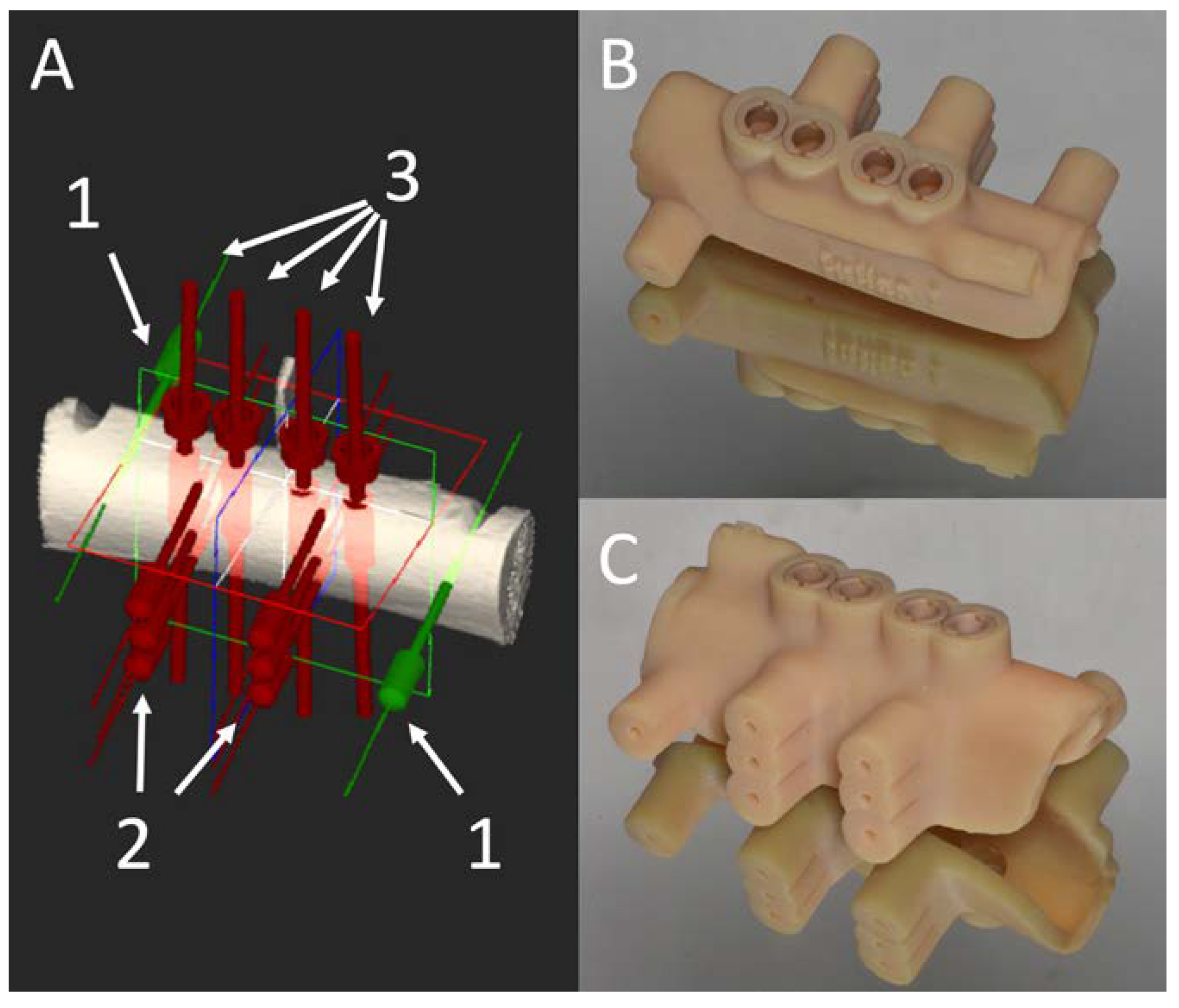
2.2. Surgical Guide Production

Each bone specimen was subjected to cone beam computed tomography (CBCT) (Orthophos XG 3D unit; Dentsply Sirona, Bensheim, Germany) with the following parameters: 85 kV, 4 mA, 14.4 s. From the CBCT data, the implant planning was carried out using a dedicated software (NaviMAX®, Biomax S.p.a., Vicenza, Italy). For each specimen, four 4.1 × 13 mm T3 Certain® Parallel Walled Non-Platform Switched Implants (Zimmer Biomet Dental, Palm Beach Gardens, FL, USA) were planned. The implants were intended to be perfectly parallel to each other, perpendicular to the bone crest with an inter-implant distance of 3 mm. The fixation pins were planned one on each lateral side of the rib. Drill holes of 1.8 mm in diameter for the insertion of the thermocouples were also designed laterally and perpendicularly to the osteotomies (Figure 1a).
An acrylic (MED620) 3D-printed surgical guide (NaviGUIDE®, Biomax S.p.a., Vicenza, Italy) was then produced for each rib, according to the individual planning using an Objet30 OrthoDesk (Stratasys, Ltd., Eden Prairie, MN, USA) printer. The axes resolution was 600 dpi, 600 dpi and 900 dpi for X-, Y- and Z-axes respectively.
The guide was provided with a built-in tube system for internal irrigation, with an aperture in the proximity of each polyetheretherketone (PEEK) sleeve (Figure 1b,c). The average diameter of the internal irrigation tube system was 2.2 mm (range 3.6 mm to 1 mm).

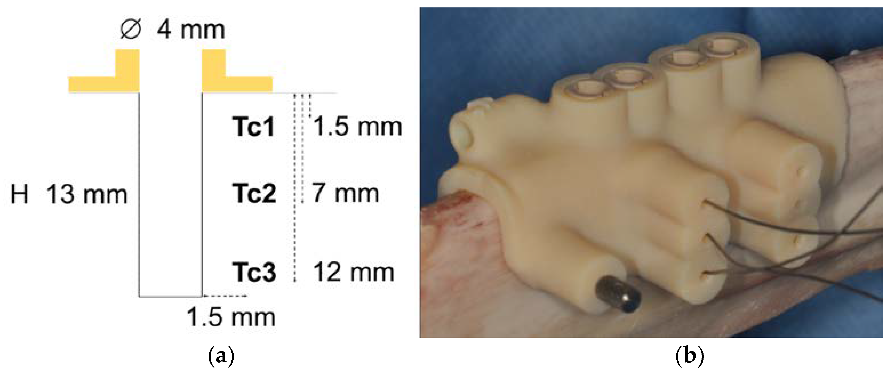
2.3. Temperature Measurement

Bone temperature was measured using type-K thermocouple (TC Instruments, Torino, Italy) with a 0.5 mm tip, coupled to a data logger thermometer (Pico Technology TC-08, Pico Technology, Cambridgshire, UK), which allowed constant, real-time temperature recording (PICOLOG06 software was used as interface). This device was able to record the temperature with an interval of 0.5 s and a resolution of 0.01 °C. The bone temperature was recorded at 1.5 mm from the nominal implant surface at three osteotomy depths: thermocouples (Tc) were inserted at the depths of 1.5 mm (Tc1), 7 mm (Tc2), and 12 mm (Tc3) from the bone crest (Figure 2).

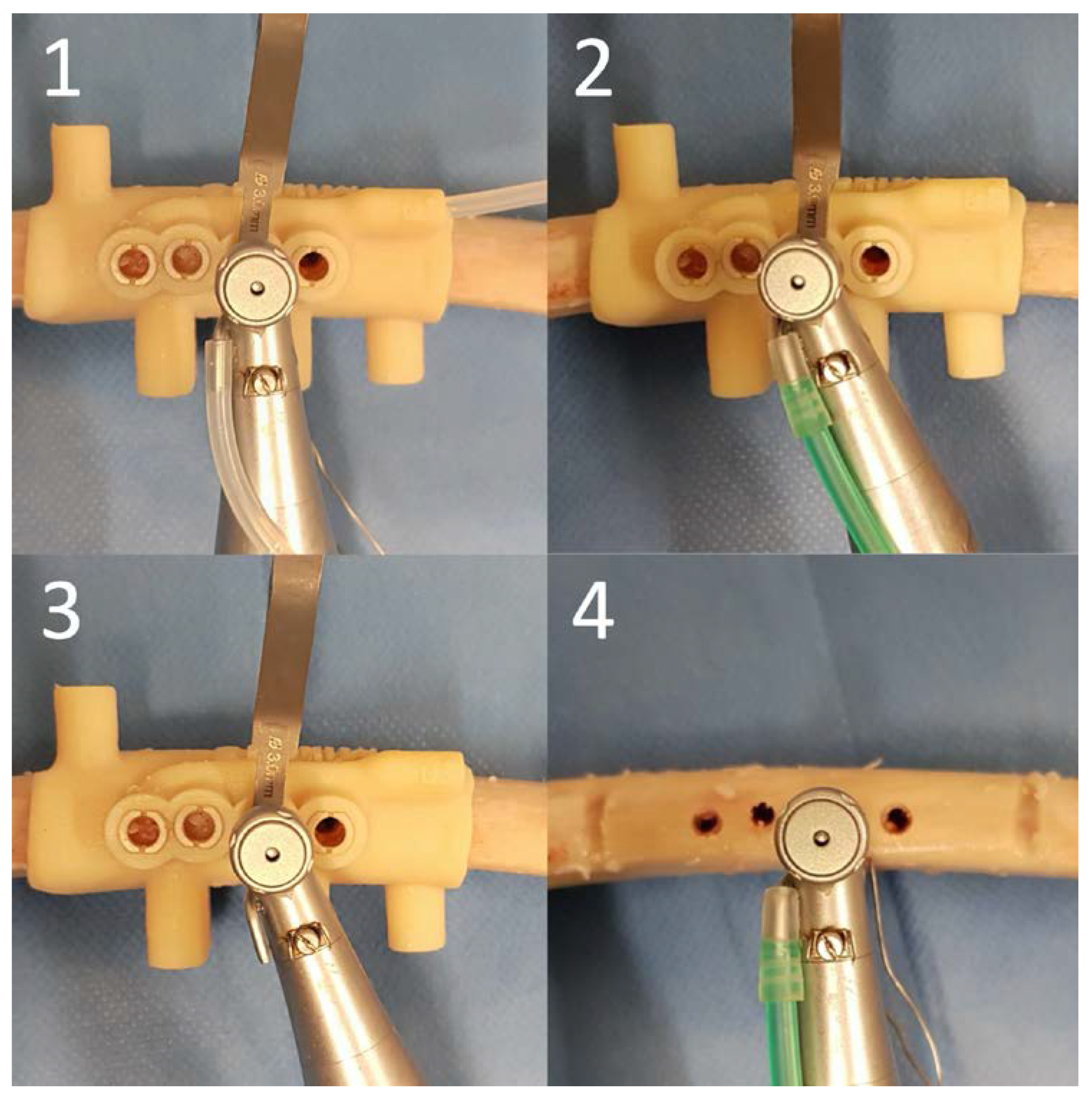
2.4. Surgical Protocol

The following conditions were tested: guided osteotomy using the surgical guide with external irrigation and an additional internal irrigation with the use of built-in tubes (Group 1); guided osteotomy using the surgical guide with external irrigation only (Group 2); guided osteotomy using the surgical guide without irrigation (Group 3); osteotomy finalized without the use of a surgical guide, with external irrigation (Group 4).
Four dental implant osteotomies—one for each group—were carried out in each of the 12 ribs, so that a total of 48 sites were included in this study (Figure 3). The position of the osteotomies in each rib was randomized using a computer-generated method.
Before the initiation of the experiment, the specimens were immersed in room-temperature (19 °C) water for 12 h. Each rib was clamped to a bench vise, the fixation pin holes were made and the surgical guide was then secured to the substrate. Lateral holes for the thermocouples were prepared with a 1.8 mm calibrated drill and their tips were then inserted.
Osteotomies were performed using a surgical motor (Implantmed, W&H, Burmoos, Austria) and a 20:1 reduction surgical contra angle handpiece (WS-75, W&H) at 1200 rpm according to a sequential drill preparation, as suggested by the manufacturer (Zimmer Biomet Dental). Regarding the irrigation modalities, a 0.9% saline solution at room temperature was used for irrigation using the pump of the surgical motor for groups 1, 2 and 4 with an emission flow of 90 ml/min. A Y-type irrigation tubing set was connected to the external and to the built-in tube system for internal irrigation in Group 1; irrigation tube was connected only to the external irrigation in Group 2 and Group 4; in Group 3 irrigation was disabled.
Certain® Tapered Navigator® System For Guided Surgery (Zimmer Biomet Dental) was utilized. The following drill sequence was used with a continuous movement: Cortical Perforator drill, 2.0 mm, 2.75 mm, 3.00 mm, and 3.25 mm twist drills. After each drilling, the osteotomy site was irrigated with room temperature saline solution with a syringe to remove bone chips and debris. Thirty seconds were waited between the finalization of one drilling step and the initiation of the following one. To secure a precise osteotomy in Group 4, an initial cortical engraving (approximately 1.5 mm deep) was carried out with the use of the surgical guide. According to the manufacturer, drill positioning handles were used. To prevent the effect of drill wear, four new drill sets were used, so that each drill was used for 12 osteotomies only.
The guides were removed after the completion of the experiment and subjected to a CBCT with the same parameters as above with the aim of qualitatively investigating the condition of the internal tube system.

2.5. Outcome Measures

The primary outcome measure was the largest temperature variation, calculated as the difference from the maximum temperature during the experiment and the starting temperature at Tc1.
The secondary outcome measures were the largest temperature variation at Tc2 and Tc3, and the average temperature increase with respect to starting temperature (calculated as average difference between each recorded temperature and starting temperature) at Tc1, Tc2, and Tc3.

2.6. Sample Size

A minimum of 8 units were required to have a 90% chance of detecting, as significant at the 5% level, a standardized effect size of 1.3 in the primary outcome measure in a paired design. The sample size was increased to 11 units to take into account the adjustment for multiple comparison (Group 1 was compared with groups 2, 3, and 4).

2.7. Statistical Analysis
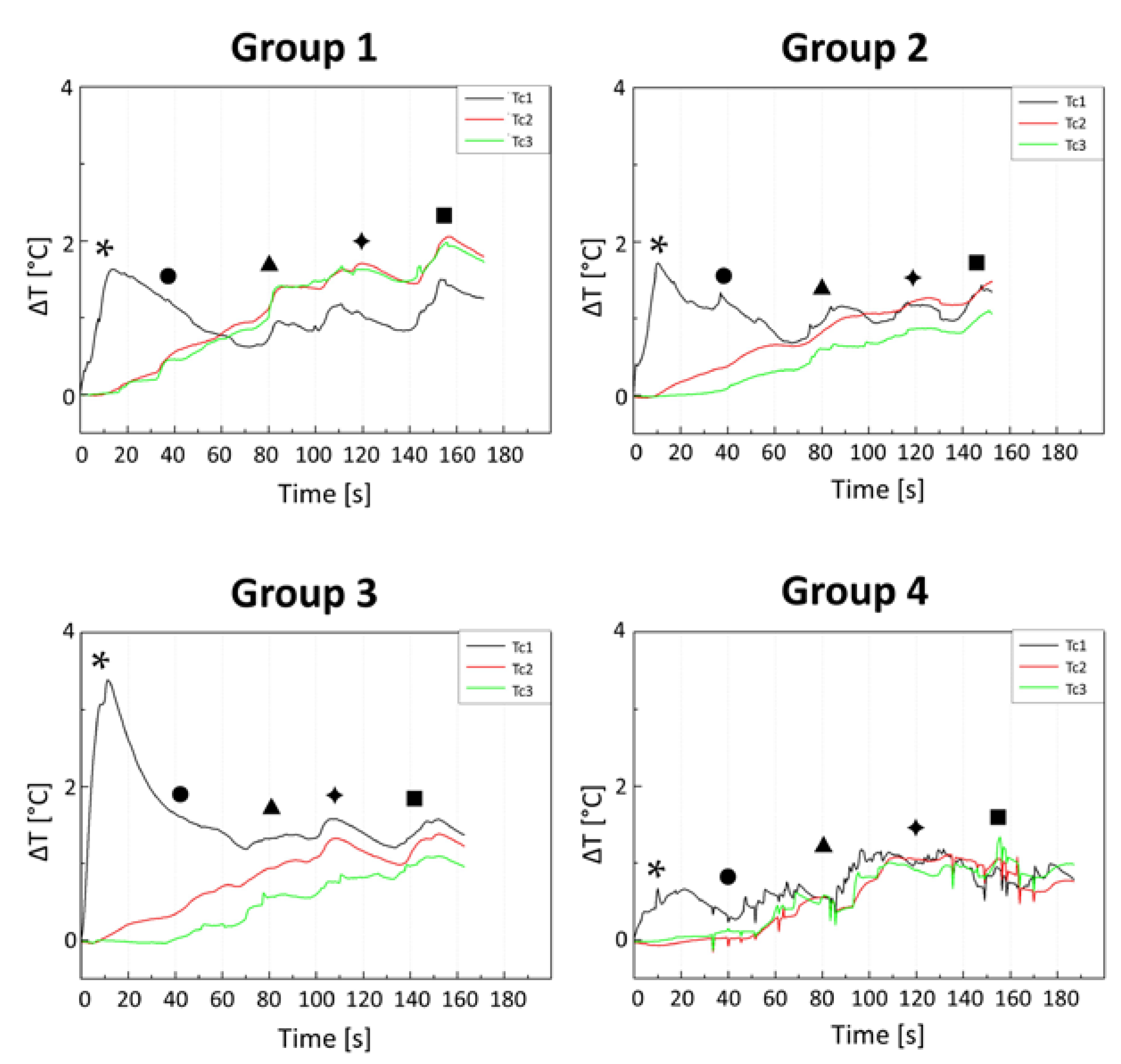
All outcome measures were continuous and were expressed as median and interquartile range (IQR). Since the outcome measures were not normally distributed (Normality assumption was checked using quantile-quantile plots), non-parametric testing was performed. Each outcome measure was compared between test group (Group 1) and each control group (groups 2, 3, and 4) using Wilcoxon signed rank test with Benjamini-Hochberg adjustment for multiple comparisons (adjusted p-values were indicated with padj). All tests were 2-sided and a p-value < 0.05 was considered statistically significant. Statistical analysis was performed using R 4.0 (R Foundation for Statistical Computing, Vienna, Austria) [26].

3. Results

The evolution of temperature variation over time as average of the twelve samples for each group is displayed in Figure 4.
The largest temperature variation at Tc1 showed a median of 3.0 °C (IQR 2.4–3.4 °C) in Group 1, which was not statistically different than Group 2 (median 2.3 °C, IQR 1.4–3.7 °C; padj = 0.83), Group 3 (median 3.2 °C, IQR 2.9–6.2 °C; padj = 0.81), or Group 4 (median 2.0 °C, IQR 1.5–3.3 °C; padj = 0.83) (Figure 5 and Table S1).
Among the secondary outcome measures, the largest temperature variation at Tc2 showed a median of 1.9 °C (IQR 1.5–3.2 °C) in Group 1, which was not statistically different than Group 2 (median 1.7 °C, IQR 1.2–2.5 °C; padj = 0.49), Group 3 (median 1.6 °C, IQR 1.0–3.4 °C; padj = 0.49) or Group 4 (median 2.0 °C, IQR 1.0–2.5 °C; padj = 0.49) (Figure 6 and Table S1).
The largest temperature variation at Tc3 showed a median of 2.3 °C (IQR 1.8–3.5 °C) in Group 1, which was not statistically different than Group 2 (median 0.9°C, IQR 0.7–2.2 °C; padj = 0.15), Group 3 (median 2.0 °C, IQR 0.9–2.5 °C; padj = 0.15) or Group 4 (median 1.3 °C, IQR 0.6–2.1 °C; padj = 0.27) (Figure 6 and Table S1).
Among the secondary outcome measures, the average temperature increment with respect to starting temperature is displayed in Figure 7 and Table S2. At Tc1, the average temperature increment showed a median of 1.0°C (IQR 0.7–1.6 °C) in Group 1, which was not statistically different than Group 2 (median 0.8 °C, IQR 0.5–1.1 °C; padj = 0.42), Group 3 (median 1.3 °C, IQR 0.8–2.3 °C; padj = 0.51) or Group 4 (median 0.8 °C, IQR 0.4–1.0 °C; padj = 0.48).
At Tc2, the average temperature increment showed a median of 0.9 °C (IQR 0.7–1.2 °C) in Group 1, which was higher with respect to Group 2 (median 0.5 °C, IQR 0.4–0.9 °C; padj = 0.03) but was not statistically different than Group 3 (median 0.7 °C, IQR 0.4–1.3 °C; padj = 0.19) or Group 4 (median 0.7 °C, IQR 0.3–0.8 °C; padj = 0.19).
At Tc3, the average temperature increment showed a median of 1.0 °C (IQR 0.7–1.1 °C) in Group 1, which was higher with respect to Group 2 (median 0.4 °C, IQR 0.1–0.7 °C; padj = 0.03), Group 3 (median 0.6 °C, IQR 0.1–0.7 °C; padj = 0.03) and Group 4 (median 0.3 °C, IQR 0.2–0.7 °C; padj = 0.03).
Post-experimental CBCT observations revealed the presence of radio-opaque material partially occluding the aperture of the internal irrigation tubes and also attached to the inner surface of the guide sleeves (Figure 8).

4. Discussion

The surgical guide with internal irrigation investigated in the present study has been proposed with the intent to provide an additional source of irrigation of the surgical field. The built-in irrigation tubes were designed to reach the body of the drill, underneath the guide sleeve. To the best of our knowledge, no other studies on this type of 3D-printed surgical guide has been published with the exception of a case report, where a similar guide was described [27]. Supposedly, a guide with such design should overcome one of the major issues of computer guided implant surgery, that is the risk of bone overheating.
This study, however, did not support the initial hypothesis that supplementary irrigation may reduce the bone temperature increase, as we did not find any significant difference in the largest temperature variation among the different groups.
It was observed that the median highest temperatures were found in the cortical bone (Tc1) with the use of the first drill, i.e., Cortical Perforator drill, in the majority of cases (Figure 4). This fact, which is in accordance with previous studies, may be explained by the impact of a relatively small drill on intact highly dense bone [17,28]. Cortical bone is denser and more resistant than trabecular bone; it has a high coefficient of friction and a low thermal conductivity, which hinders heat dissipation [29]. The initial drill, in fact, encounters greater resistance and must remove considerable quantities of cortical bone at the drill entrance point. Subsequent larger drills, on the other hand, induce lower temperature variations as they have to remove smaller quantities of cortical bone tissue and the larger flute spaces can facilitate irrigation and bone chips removal [18,30]. The lower temperature variations recorded at Tc2 and Tc3, which were located in the medullar bone, can be explained by the lower density and the lower cutting resistance while drilling this tissue compared to the cortical bone [31].
In a study by Liu et al., a conventional external irrigation from the hand-piece, an internal tube in the surgical guide and a drill with an internal cooling were compared. The findings showed that the guide with the internal tube was a superior irrigation method than the conventional external irrigation, while it was less efficient than the internal drill cooling [32]. In the present study, however, the use of the supplementary internal irrigation (Group 1) did not significantly reduce the temperature variation compared with an external irrigation only (Group 2). No significant differences on temperature variation were found between Group 3 and Group 4 as well. A tendency, however, can be outlined for the main outcome, presenting the Group 3 with the highest temperature increase and Group 4 with the lowest.
According to the assumption that the limit condition for bone regeneration is 47 °C for 1 min [8], the magnitude of temperature increase during the drilling procedure in this study seems to be rather safe in all groups. According to the temperature variation observed in this in vitro study, the threshold value was never exceeded. On the contrary the maximum temperature increase was lower than 5 °C in most cases. Such values are in accordance with previous in vitro and ex vivo studies. Investigating the effect of drilling speed and irrigation liquid temperature during guided and freehand implant surgery in bovine ribs, Barrack et al. recorded a maximum bone temperature increase of 2.10 °C [33]. In another ex vivo study in porcine ribs, bone temperature with open-flap and flapless guided implant surgery registered a maximum temperature increase of 4.81 °C [20].
It must be said that the actual maximum bone temperature at the site of drilling cannot be registered with thermocouples. The probe tips were located at 1.5 mm from the heat source, corresponding to the nominal implant surface. Bone has low thermal conductivity properties, and we could expect a low grade of heat distribution through the bone [34]. Therefore, temperatures at the immediate vicinity of an osteotomy may have been much higher, as demonstrated by Aghvami et al. [31].
In this study, the persistence of an increased temperature was assessed by the average temperature increment from the starting temperature. Values of Group 1 were significantly higher than Group 2 in Tc2 and higher than groups 2, 3, and 4 in Tc3. This difference, even though of limited amount, may indicate that there was a heat concentration in the medullar bone and more specifically at the apex of the preparation for Group 1. It was speculated that drilling preparation caused the clogging of the internal irrigation tubes aperture facing the bone, as previously reported [11]. This theory was confirmed by CT observations carried out after the experimental phase. As matter of fact, the final parts of the built-in irrigation tubes were impacted by condensed bone tissue (Figure 8). This obstructive material could have negatively affected the temperature variation in two ways, since it would have impeded the irrigation flow from the internal tube and increased the friction with the drill.
To reduce this phenomenon, it could be postulated that a more powerful irrigation should be performed. In the setting of the present study, the irrigation fluid flow originated from the hand-piece and it was distributed among the external and the internal built-in tubes. It can be speculated that the fluid pressure at the end of the tubes was too weak. Another source of irrigation, distinct from that provided in the hand-piece may be recommended. Moreover, the drilling method may have contributed to the creation of the clog. Even though a recent study demonstrated that the intermittent drilling movement during a conventional osteotomy did not decrease the bone temperature [35], this method may be relevant during guided surgery, since it increases the irrigation flow and remove a major amount of bone debris from the drilling site [36].
With the present study, the novel irrigation method was tested in an ex vivo model using bovine ribs. This model was chosen since it resembles the morphological characteristics and thermal properties similar to human bone [34]. 3D-printing technology was used to fabricate surgical guides specifically designed for each experimental ex vivo substrate. Thanks to this approach, thermocouples positioning, which usually requires a standardized in vitro condition, was closer to clinical reality. The results on temperature variation can hardly be reproduced in an in vivo setting, with a different basal temperature and in the absence of the blood flow. Another limitation may rely on the fact that biological substrates were used, which differed one from another in terms of cortical thickness and medullar density. The lack of a constant drill load may represent a limitation, but the use of a testing machine could have affected a passive insertion of the drills through the sleeves. Therefore a manual drilling by a single operator was adopted. To obtain a more precise temperature recording, each osteotomy was performed with one continuous movement and then 30 s were waited. It must be said that his method is rarely reproduced in the clinical scenario, since the lag time from one drill to the subsequent is usually shorter.
Further studies should test the guide under different conditions, such as an additional irrigation source and a different drilling method. A finite element analysis should be run to identify which elements (e.g., irrigation volumetric flow and pressure) should be modified to avoid the clogging of the tubes.

5. Conclusions

The additional internal irrigation was not associated with a decrease of the average bone temperature. Within the limitations of an ex vivo model, it was showed that the temperature increases never exceeded the threshold for bone damage. The highest temperature variation was associated with the use of the cortical perforator drill. Moreover, bone debris was found obstructing the internal irrigation tubes, thus reducing the cooling efficiency of the guide. The present findings highlight a need for further investigations on the need and the optimization of the supplementary irrigation system and to assess the effects of the drilling methods on bone heating.

Supplementary Materials

The following are available online at https://www.mdpi.com/2076-3417/11/6/2588/s1, Figure S1, Table S1, Table S2.

Author Contributions

Conceptualization, M.S. and G.B.; methodology, M.S.; software, L.B.; formal analysis, M.S. and F.C.; investigation, M.S., A.Z. and L.B.; data curation, M.S., F.C. and L.B.; writing—original draft preparation, M.S.; writing—review and editing, all authors; supervision, S.S. and L.B.; funding acquisition, S.S. All authors have read and agreed to the published version of the manuscript.

Funding

This research was partially funded by Biomax S.p.A. (Vicenza, Italy). The founder had no role in the data acquisition and analysis.

Institutional Review Board Statement

Not applicable.

Informed Consent Statement

Not applicable.

Data Availability Statement

Not applicable.

Acknowledgments

Biomax S.p.a. (Vicenza, Italy) for the materials (surgical guides) and financial support. Claudio Albi and Socrate Avesani for their precious technical support.

Conflicts of Interest

The authors declare no conflict of interests.

References

  1. Albrektsson, T.; Brånemark, P.I.; Hansson, H.A.; Lindström, J. Osseointegrated Titanium Implants. Requirements for Ensuring a Long-Lasting, Direct Bone-to-Implant Anchorage in Man. Acta Orthop. Scand. 1981, 52, 155–170. [Google Scholar] [CrossRef] [Green Version]
  2. Esposito, M.; Hirsch, J.M.; Lekholm, U.; Thomsen, P. Biological Factors Contributing to Failures of Osseointegrated Oral Implants. (I). Success Criteria and Epidemiology. Eur. J. Oral Sci. 1998, 106, 527–551. [Google Scholar] [CrossRef] [PubMed]
  3. Wagenberg, B.; Froum, S.J. A Retrospective Study of 1925 Consecutively Placed Immediate Implants from 1988 to 2004. Int. J. Oral Maxillofac. Implant. 2006, 21. [Google Scholar] [CrossRef]
  4. Sharawy, M.; Misch, C.E.; Weller, N.; Tehemar, S. Heat Generation During Implant Drilling: The Significance of Motor Speed. J. Oral Maxillofac. Surg. 2002, 60, 1160–1169. [Google Scholar] [CrossRef] [Green Version]
  5. Calvo-Guirado, J.L.; Delgado-Peña, J.; Maté-Sánchez, J.E.; Mareque Bueno, J.; Delgado-Ruiz, R.A.; Romanos, G.E. Novel Hybrid Drilling Protocol: Evaluation for the Implant Healing–Thermal Changes, Crestal Bone Loss, and Bone-to-Implant Contact. Clin. Oral Implant. Res. 2015, 26, 753–760. [Google Scholar] [CrossRef]
  6. Stocchero, M.; Jinno, Y.; Toia, M.; Ahmad, M.; Papia, E.; Yamaguchi, S.; Becktor, J.P. Intraosseous Temperature Change during Installation of Dental Implants with Two Different Surfaces and Different Drilling Protocols: An in Vivo Study in Sheep. J. Clin. Med. 2019, 8, 1198. [Google Scholar] [CrossRef] [Green Version]
  7. Eriksson, A.; Albrektsson, T.; Grane, B.; McQueen, D. Thermal Injury to Bone: A Vital-Microscopic Description of Heat Effects. Int. J. Oral Surg. 1982, 11, 115–121. [Google Scholar] [CrossRef]
  8. Eriksson, R.A.; Albrektsson, T. The Effect of Heat on Bone Regeneration: An Experimental Study in the Rabbit Using the Bone Growth Chamber. J. Oral Maxillofac. Surg. 1984, 42, 705–711. [Google Scholar] [CrossRef]
  9. Augustin, G.; Zigman, T.; Davila, S.; Udilljak, T.; Staroveski, T.; Brezak, D.; Babic, S. Cortical Bone Drilling and Thermal Osteonecrosis. Clin. Biomech. 2012, 27, 313–325. [Google Scholar] [CrossRef] [PubMed]
  10. Yoshida, K.; Katsumi, U.; Kimimitsu, O.; Takeyasu, M. Influence of Heat Stress to Matrix on Bone Formation. Clin. Oral Implant. Res. 2009, 20, 782–790. [Google Scholar] [CrossRef]
  11. Misir, A. Ferhat, Mahmut Sumer, Murat Yenisey, and Erol Ergioglu. Effect of Surgical Drill Guide on Heat Generated from Implant Drilling. J. Oral Maxillofac. Surg. 2009, 67, 2663–2668. [Google Scholar] [CrossRef] [PubMed]
  12. Oh, H.J.; Wikesjö, U.M.; Kang, H.S.; Ku, Y.; Eom, T.G.; Koo, K.T. Effect of Implant Drill Characteristics on Heat Generation in Osteotomy Sites: A Pilot Study. Clin. Oral Implant. Res. 2011, 22, 722–726. [Google Scholar] [CrossRef]
  13. Yacker, M.J.; Klein, M. The Effect of Irrigation on Osteotomy Depth and Bur Diameter. Int. J. Oral Maxillofac. Implant. 1996, 11, 634–638. [Google Scholar]
  14. Ercoli, C.; Funkenbusch, P.D.; Lee, H.J.; Moss, M.E.; Graser, G.N. The Influence of Drill Wear on Cutting Efficiency and Heat Production During Osteotomy Preparation for Dental Implants: A Study of Drill Durability. Int. J. Oral Maxillofac. Implant. 2004, 19, 335–349. [Google Scholar]
  15. Trisi, P.; Marco, B.; Antonello, F.; Michele, P.V.; Giorgio, P. Insufficient Irrigation Induces Peri-Implant Bone Resorption: An in Vivo Histologic Analysis in Sheep. Clin. Oral Implant. Res. 2014, 25, 696–701. [Google Scholar] [CrossRef]
  16. Dos Santos, P.L.; Pereira Queiroz, T.; Margonar, R.; de Souza Carvalho, A.C.G.; Betoni, W., Jr.; Rodrigues Rezende, R.R.; Garcia, R., Jr. Evaluation of Bone Heating, Drill Deformation, and Drill Roughness after Implant Osteotomy: Guided Surgery and Classic Drilling Procedure. Int. J. Oral Maxillofac. Implant. 2014, 29. [Google Scholar] [CrossRef] [Green Version]
  17. Gehrke, S.A.; Aramburú Júnior, J.S.; Pérez-Albacete Martínez, C.; Ramirez Fernandez, M.P.; Maté Sánchez de Val, J.E.; Calvo-Guirado, J.L. The Influence of Drill Length and Irrigation System on Heat Production During Osteotomy Preparation for Dental Implants: An Ex Vivo Study. Clin. Oral Implant. Res. 2018, 29, 772–778. [Google Scholar] [CrossRef] [PubMed]
  18. Tehemar, S.H. Factors Affecting Heat Generation During Implant Site Preparation: A Review of Biologic Observations and Future Considerations. Int. J. Oral Maxillofac. Implant. 1999, 14, 127–136. [Google Scholar] [PubMed]
  19. Tatakis, D.N.; Chien, H.H.; Parashis, A.O. Guided Implant Surgery Risks and Their Prevention. Periodontol. 2000 2019, 81, 194–208. [Google Scholar] [CrossRef]
  20. Migliorati, M.; Amorfini, L.; Signori, A.; Barberis, F.; Benedicenti, S. Internal Bone Temperature Change During Guided Surgery Preparations for Dental Implants: An in Vitro Study. Int. J. Oral Maxillofac. Implant. 2013, 28, 1464–1469. [Google Scholar] [CrossRef] [Green Version]
  21. Azari, A.; Nikzad, S. Flapless Implant Surgery: Review of the Literature and Report of 2 Cases with Computer-Guided Surgical Approach. J. Oral Maxillofac. Surg. 2008, 66, 1015–1021. [Google Scholar] [CrossRef]
  22. Kivovics, M.; Pénzes, D.; Németh, O.; Mijiritsky, E. The Influence of Surgical Experience and Bone Density on the Accuracy of Static Computer-Assisted Implant Surgery in Edentulous Jaws Using a Mucosa-Supported Surgical Template with a Half-Guided Implant Placement Protocol—A Randomized Clinical Study. Materials 2020, 13, 5759. [Google Scholar] [CrossRef] [PubMed]
  23. Vercruyssen, M.; Laleman, I.; Jacobs, R.; Quirynen, M. Computer-Supported Implant Planning and Guided Surgery: A Narrative Review. Clin. Oral Implant. Res. 2015, 26, 69–76. [Google Scholar] [CrossRef]
  24. Henprasert, P.; Dawson, D.V.; El-Kerdani, T.; Song, X.; Couso-Queiruga, E.; Holloway, J.A. Comparison of the Accuracy of Implant Position Using Surgical Guides Fabricated by Additive and Subtractive Techniques. J. Prosthodont. 2020, 29, 534–541. [Google Scholar] [CrossRef] [Green Version]
  25. Deeb, G.R.; Allen, R.K.; Hall, V.P.; Whitley, D., III; Laskin, D.M.; Bencharit, S. How Accurate Are Implant Surgical Guides Produced with Desktop Stereolithographic 3-Dimentional Printers? J. Oral Maxillofac. Surg. 2017, 75, 2559.e1–2559.e8. [Google Scholar] [CrossRef] [PubMed]
  26. Team R Core. R: A Language and Environment for Statistical Computing; Team R Core: Vienna, Austria, 2013. [Google Scholar] [CrossRef]
  27. Alevizakos, V.; Mitov, G.; von See, C. Guided Implant Placement Using an Internally Cooling Surgical Template–a Technical Note. J. Oral Implantol. 2020, 46, 533–535. [Google Scholar] [CrossRef]
  28. Bulloch, S.E.; Olsen, R.G.; Bulloch, B. Comparison of Heat Generation between Internally Guided (Cannulated) Single Drill and Traditional Sequential Drilling with and without a Drill Guide for Dental Implants. Int. J. Oral Maxillofac. Implant. 2012, 27, 1456–1460. [Google Scholar]
  29. Calttenburg, R.; Cohen, J.; Conner, S.; Cook, N. Thermal Properties of Cancellous Bone. J. Biomed Mater. Res. 1975, 9, 169–182. [Google Scholar] [CrossRef] [PubMed]
  30. Strbac, G.D.; Unger, E.; Donner, R.; Bijak, M.; Watzek, G.; Zechner, W. Thermal Effects of a Combined Irrigation Method during Implant Site Drilling. A Standardized in Vitro Study Using a Bovine Rib Model. Clin. Oral Implant. Res. 2014, 25, 665–674. [Google Scholar] [CrossRef] [PubMed]
  31. Aghvami, M.; Brunski, J.B.; Serdar Tulu, U.; Chen, C.H.; Helms, J.A. A Thermal and Biological Analysis of Bone Drilling. J. Biomech. Eng. 2018, 140. [Google Scholar] [CrossRef]
  32. Liu, Y.F.; Wu, J.L.; Zhang, J.X.; Peng, W.; Liao, W.Q. Numerical and Experimental Analyses on the Temperature Distribution in the Dental Implant Preparation Area When Using a Surgical Guide. J. Prosthodont. 2018, 27, 42–51. [Google Scholar] [CrossRef] [PubMed]
  33. Barrak, I.; Joób-Fancsaly, Á.; Braunitzer, G.; Varga, E., Jr.; Boa, K.; Piffkó, J. Intraosseous Heat Generation During Osteotomy Performed Freehand and through Template with an Integrated Metal Guide Sleeve: An in Vitro Study. Implant Dent. 2018, 27, 342–350. [Google Scholar] [CrossRef] [PubMed]
  34. Davidson, S.R.; James, D.F. Measurement of Thermal Conductivity of Bovine Cortical Bone. Med Eng. Phys. 2000, 22, 741–747. [Google Scholar] [CrossRef]
  35. Di Fiore, A.; Sivolella, S.; Stocco, E.; Favero, V.; Stellini, E. Experimental Analysis of Temperature Differences During Implant Site Preparation: Continuous Drilling Technique Versus Intermittent Drilling Technique. J. Oral Implantol. 2018, 44, 46–50. [Google Scholar] [CrossRef] [PubMed]
  36. Marković, A.; Lazić, Z.; Mišić, T.; Šćepanović, M.; Todorović, A.; Thakare, K.; Glišić, M. Effect of Surgical Drill Guide and Irrigans Temperature on Thermal Bone Changes During Drilling Implant Sites-Thermographic Analysis on Bovine Ribs. Vojnosanit. Pregl. 2016, 73, 744–750. [Google Scholar] [CrossRef] [Green Version]
Figure 1. Virtual planning (a) and images of both sides ((b) and (c)) of one of the 3D-printed guides utilized in the experiment. (1) Fixation pins; (2) thermocouple drill holes; (3) implant positions.
Applsci 11 02588 g001
Figure 2. (a) Schematic drawing of the procedure. A 4.1 × 13 mm dental implant osteotomy was planned. The bone temperature was recorded at 1.5 mm from the nominal implant surface at three osteotomy depths from the bone crest: 1.5 mm (Tc1), 7 mm (Tc2), and 12 mm (Tc3). (b) One of the 3D-printed guides fixed on its own experimental specimen, with three thermocouples (Tc) in place.
Applsci 11 02588 g002
Figure 3. Comparison of the 4 experimental conditions: (1) guided osteotomy using the surgical guide with external irrigation and an additional internal irrigation with the use of additional built-in tubes (Group 1); (2) guided osteotomy using the surgical guide with an external irrigation only (Group 2); (3) guided osteotomy using the surgical guide without irrigation (Group 3); (4) osteotomy finalized without the use of a surgical guide, with external irrigation (Group 4).
Applsci 11 02588 g003
Figure 4. Mean temperature variation (ΔT) over time. The initiation of each drilling sequence step is indicated as follows: (*) Cortical Perforator drill; (●) 2.0 mm; (▲) 2.75 mm; (✦) 3.00 mm; () 3.25 mm twist drills.
Applsci 11 02588 g004
Figure 5. Boxplot showing the largest temperature variation (ΔT) at Tc1 in the four groups.
Applsci 11 02588 g005
Figure 6. Boxplots showing the largest of temperature variation (ΔT) at Tc2 (a) and Tc3 (b) in the four groups.
Applsci 11 02588 g006
Figure 7. Boxplots showing the average temperature variation (ΔT) with respect to starting temperature at (a) Tc1, (b) Tc2, and (c) Tc3 in the four groups.
Applsci 11 02588 g007
Figure 8. Cone beam computerized tomography (CBCT) images of one of the surgical guides after the experimental phase. Condensed bone tissue partially occupies the final parts of the built-in irrigation tubes.
Applsci 11 02588 g008
Publisher’s Note: MDPI stays neutral with regard to jurisdictional claims in published maps and institutional affiliations.

