Article

Healing Capacity of Bone Surrounding Biofilm-Infected and Non-Infected Gutta-Percha: A Study of Rat Calvaria

1
Endodontics and Dental Trauma Department, School of Graduate Dentistry, Rambam Health Care Campus, P.O. Box 9602, Haifa 3109601, Israel
2
Ruth and Bruce Rappaport Faculty of Medicine, Israel Institute of Technology—Technion, Haifa 3525433, Israel
3
Department of Endodontology, The Goldschleger School of Dental Medicine, Tel Aviv University, Tel Aviv 6100000, Israel
4
Department of Pathology, Rambam Health Care Campus, Haifa 3500000, Israel
5
Clinical Microbiology Laboratory, Rambam Health Care Campus, Haifa 3500000, Israel
6
The Israeli National Center for Trauma and Emergency Medicine Research Gertner Institute, Tel Hashomer 5262000, Israel
*
Author to whom correspondence should be addressed.
Equal contribution.
Appl. Sci. 2021, 11(15), 6710; https://doi.org/10.3390/app11156710
Submission received: 16 June 2021 / Revised: 17 July 2021 / Accepted: 19 July 2021 / Published: 22 July 2021
(This article belongs to the Special Issue Dental Materials: Latest Advances and Prospects)

Abstract

:

Featured Application

It has been suggested that some root canal treatments fail because of non-microbial factors, including extruded root canal filling materials, which may cause a reaction to a foreign body. This study shows that overextension of sealing material, without other clinical or roentgen signs or symptoms, should not be considered an indication for endodontic surgery.

Abstract

This paper aims to evaluate the healing capacity of bony lesions around biofilm-infected and non-infected gutta-percha (GP) points. Bony defects were created in the calvaria of 28 Wistar rats. The rats were divided into three groups: Group 1—Implantation of infected GP particles in the bony defect; Group 2—Positive control implantation of non-infected GP particles in the bony defect; and Group 3—Negative control, in which no GP particles were implanted. The biofilm consisted of three strains of bacteria: Enterococcus faecalis, Streptococcus sanguis, and Porphyromonas gingivalis. The animals were sacrificed 60 days postoperation, and histological assessments were performed. In Group 1, the biofilm-infected group, we observed a mild foreign body reaction with a few inflammatory cells adjacent to the capsule and a newly woven bone matrix surrounded by osteoblasts and mature bone. In Group 2, the non-infected GP particles group, minimal inflammatory cell reactions were observed in the adjacent tissue, and a newly woven bone matrix was surrounded by osteoblasts. This study shows that bone healing is possible around both sterile and infected GP points. This contradicts the claim that some root canal treatments fail because of non-microbial factors, including extruded root canal filling materials, which may cause foreign body reactions. The healing observed suggests that overextension should not be considered an indication for endodontic surgery.

1. Introduction

Gutta-percha (GP) is the most popular root canal filling material [1]. The widely held view that GP has low toxicity and is therefore well tolerated by human tissues is inconsistent with the observations of outcome studies in which extruded GP is associated with the delayed healing of apical tissues [2,3,4,5,6]. According to Ng et al. (2008), the presence of the apical extent of root filling is a significant prognostic factor of endodontic treatment success [2].
There is consensus that microorganisms, mainly bacteria, cause primary apical periodontitis, but the reasons for persistent apical periodontitis lesions are debatable [7,8]. Secondary factors, such as foreign material in the periapical area of a tooth with apical periodontitis, may contribute to additional irritation and/or inflammatory reactions [8]. Nair (2008) claimed that extruded root canal filling might cause a foreign body reaction [9], but Haapasalo et al. (2008) suggested that a primary role for such factors, without the continued presence of bacteria, is lacking [8].
It has been suggested that this adverse effect (i.e., the foreign body reaction) could be a result of over-instrumentation and the subsequent transportation of contaminated debris periapically [10,11,12]. Siqueira (2001) stated that failure associated with overfilled teeth is usually caused by a concomitant intraradicular and/or extraradicular infection [10]. Over-instrumentation usually precedes overfilling and induces the displacement of infected debris into apical tissues in teeth with infected necrotic pulps [10,11,12,13].
The prevention of contamination becomes a problem when GP cones are used since this material does not readily lend itself to sterilization by moist or dry heat [14]. A one-minute immersion in 5.25% sodium hypochlorite may be used in clinical practice to sterilize GP cones and avoid this possible source of exogenous contamination [15]. Bacteria adherent to GP and growing as a biofilm may play a role in the delayed healing of apical tissues, but few studies have examined the biofilm potential on GP points as a cause for persistent apical periodontitis after endodontic treatment [16,17].
This study aimed to examine the healing capacity of bony lesions around biofilm-infected and non-infected GP points in a rat calvaria model. Our main hypothesis was that biofilm and GP would induce bone destruction, whereas bone defects alone would not.

2. Materials and Methods

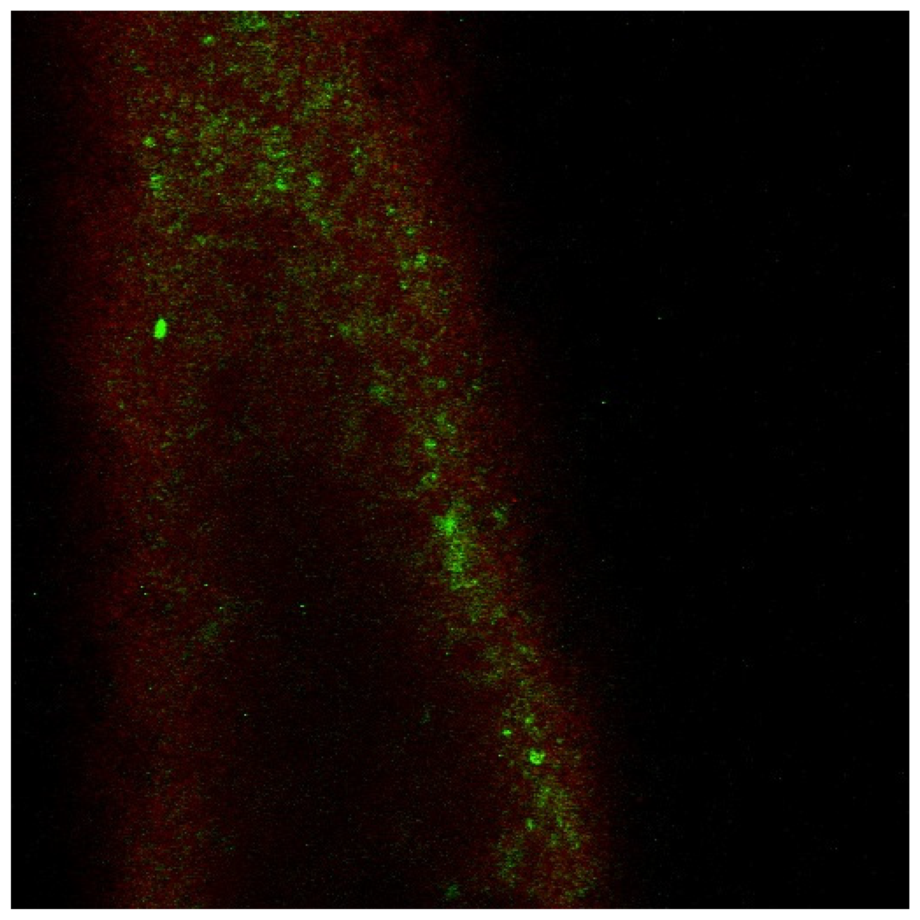
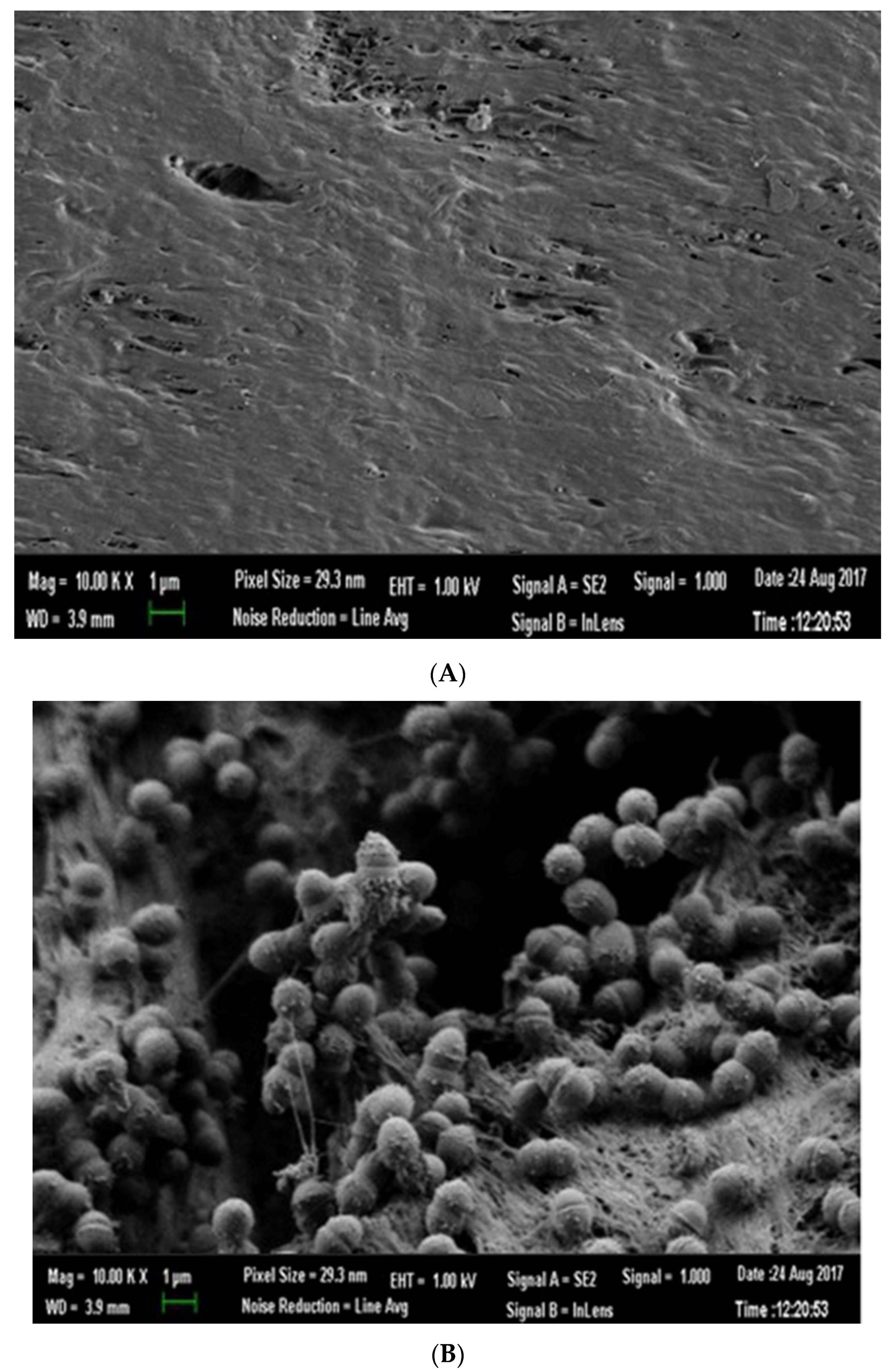
2.1. Biofilm Culture on GP

GP points 40/0.04 (VDW GmbH, Munich, Germany) were sterilized with plasma gas (low-temperature hydrogen peroxide gas plasma, Kingsport, TN, USA) and divided into two groups:
Sterile GP points—The GP points were cut into 1 × 1 mm GP particles using a sterile scalpel (no. 11) and an endodontic ruler under 2.5× magnification (Orascoptic Inc., Madison, WI, USA).
Infected GP points—The formation of biofilm on GP points was carried out according to Takemura et al. (2004). Three strains of bacteria, i.e., Enterococcus faecalis (E.F.) ATCC 29212, Streptococcus sanguis (S.S.) ATCC 10556, and Porphyromonas gingivalis (P.G.) ATCC 33277, were cultured in equal amounts on GP points. The infected GP points were suspended in a growth medium containing varying percentages of human serum [14]. Each bacterial strain was harvested during the stationary phase, and 100 µL of each bacterial suspension was inoculated into 900 µL of cell culture medium, 900 µL of human serum (Biological Industries, Israel), or 900 µL of cell culture medium supplemented with 5% or 50% (vol/vol) human serum. Formalin fixation was performed at the end of the incubation process (Tissue-Tek ® O.C.T. ™ Compound, Sakura, The Netherlands). The GP points were sliced at 20–40 μm (Leica CM1900 Cryostat, Germany). Several segments were stained according to the Strathmann et al. (2002) protocol [15]. Briefly, extracellular polymeric substances (EPS) were stained red with concanavalin. A conjugate to tetramethyl rhodamine isothiocyanate (TRITC) was used as a marker for biofilm, and DNA-binding stain SYTO 9 (green color) was used to visualize live bacterial cells. Slices were observed under a laser scanning confocal microscope (Carl Zeiss CLSM (LSM 510 META) with an Apo 60 × 1.40 objective) to confirm the generated biofilm. Then, 3D depth images of the biofilm generated over the GP were taken (Figure 1).
Non-infected and infected GP were incubated in primary fixative containing 2.5% GA and 2% PFA in 0.1 M sodium cacodylate buffer at pH 7.4 for 1 h at RT. Following washes in 0.1 M cacodylate buffer, the samples underwent a second fixation in 1% OsO4 for 20 min. The specimens were then dehydrated through a graded ethanol series, and the critical points were dried and coated with 10 nm chromium. A Zeiss ULTRA plus field emission scanning electron microscope was used to observe the samples (Figure 2A,B).
After slicing the infected GP points, several segments were transferred into 2 mL sterile tubes. The vials were then shaken on a Retsch MM401 bead mill for 2.5 min at 30 Hz. Aliquots of the bead-milled suspensions were collected and then inoculated into blood culture bottles. Additional aliquots were spread on blood agar plates and PolyViteX chocolate agar plates, both of which were incubated for 5 days at 37 °C in 5% CO2 and at 37 °C in an anaerobic atmosphere. Isolated bacteria were identified according to standard laboratory procedures. In all cases, viable E.F., S.S., and P.G. isolates were recovered from the infected GP points. The remaining GP points were cut using the same technique as the sterile GP points.

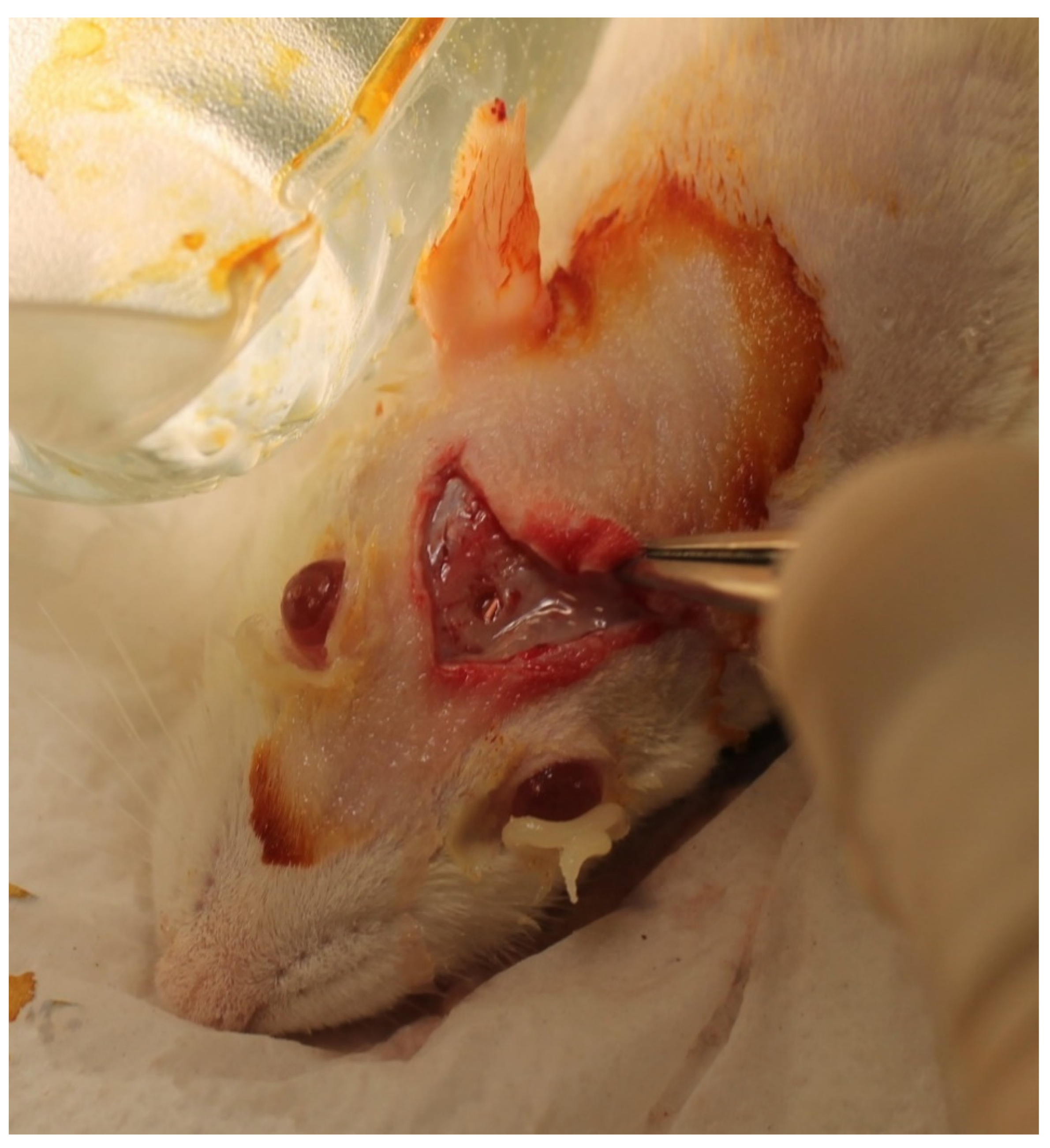
2.2. Animals and Surgery

The study consisted of 28 female, 8-month-old Wistar rats that were maintained in a room with a 12 h light/dark cycle. The animals had free access to tap water and standard laboratory food. The Animal Research Council approved this research (il-071-06-2009).
The rats were divided into 3 groups:
Group 1 (n = 12): Implantation of infected GP particles in the bony defects;
Group 2 (n = 12): Positive control group implantation of non-infected GP particles in the bony defects;
Group 3 (n = 4): Negative control group, in which no GP particles were implanted in the bony defects.
The animals were weighed and anesthetized with an intramuscular injection of ketamine chlorhydrate (90 mg/kg) and xylazine (2%** 10 mg/kg body weight), and then their heads were shaved.
A modification of Turnbull and Freeman’s (1974) original surgical approach was used [18]. A U-shaped incision was made in the scalp between the eyebrows that caudally connected two sagittal incisions extending posteriorly over the parietal bone to enable the elevation of a full-thickness flap and expose the soft tissues covering the calvarium. The fascia overlying the bone was dissected to expose the periosteum (Figure 3). A high-speed, water-cooled, diamond wheel-shaped bur was used under 2.5× magnification (Orascoptic, Middleton, WI, USA) to create a bony defect measuring 3 mm in diameter and 1 mm in depth while avoiding damage to the dura mater or puncturing the sagittal sinus.
Resorbable collagen membranes (4BONE Resorbable Collagen Membrane, MIS Implants Technologies Ltd., Bar Lev Industrial Park, Israel) were cut into 28 membrane discs (4 mm in diameter) and used to cover each bony defect.
The flaps were repositioned and sutured using 3–0 vicryl absorbable sterile surgical suture (Ethicon, Inc., Somerville, NJ, USA).
The animals were sacrificed at 60 days postoperation, then the calvariae were resected, fixed in 10% neutral buffered formalin, washed, dehydrated in ethanol and xylene, embedded in paraffin, and cut into transverse 5 mm sections. Sections were stained with hematoxylin and eosin (H&E) for examination under light microscopy.
The histological appearance was assessed regarding the degree and type of inflammation and the thickness of the connective tissue capsule surrounding GP particles.
The level of inflammation was classified as none/mild or moderate/severe, with the former describing an absence of inflammation or scattered inflammatory cells surrounding the particles. An intense concentration of inflammatory cells indicated a moderate/severe reaction to the GP particles. The inflammation types were defined as acute or chronic. Acute inflammation represented a predominance of polymorphonuclear leukocytes with few chronic inflammatory cells (e.g., macrophages, lymphocytes, or plasma cells), and chronic inflammation consisted of a predominance of chronic inflammatory cells with few polymorphonuclear neutrophils.

3. Results

3.1. Control (n = 4)

No remnants of the resorbable collagen membranes were found at the surgical site at 60 days. Fibrous connective tissue containing fibroblasts and blood vessels, a few inflammatory cells, and a newly woven bone matrix surrounded by osteoblasts were noted.

3.2. Non-Infected GP Points (n = 12)

No remnants of the resorbable collagen membranes were found at the surgical site at 60 days. The GP particles were surrounded by a fine thin fibrous capsule, fibroblasts, and a few chronic inflammatory cells. In adjacent tissues, minimal inflammatory cell reactions were observed. In some specimens, a newly woven bone matrix surrounded by osteoblasts was noted, similar to findings in the control group (Figure 4).
A newly woven bone matrix surrounded by active osteoblasts was also noted. H&E: hematoxylin & eosin stain, ×100.

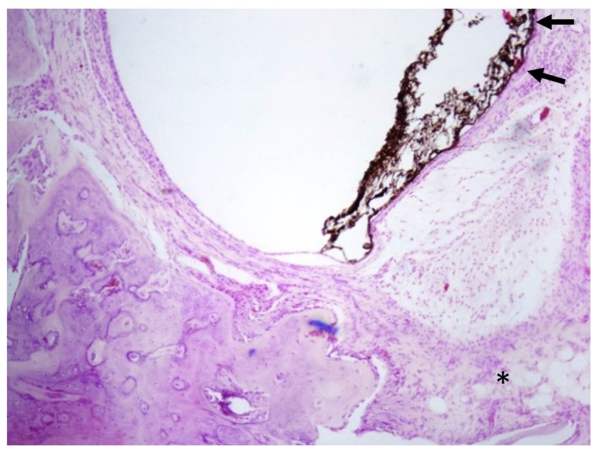
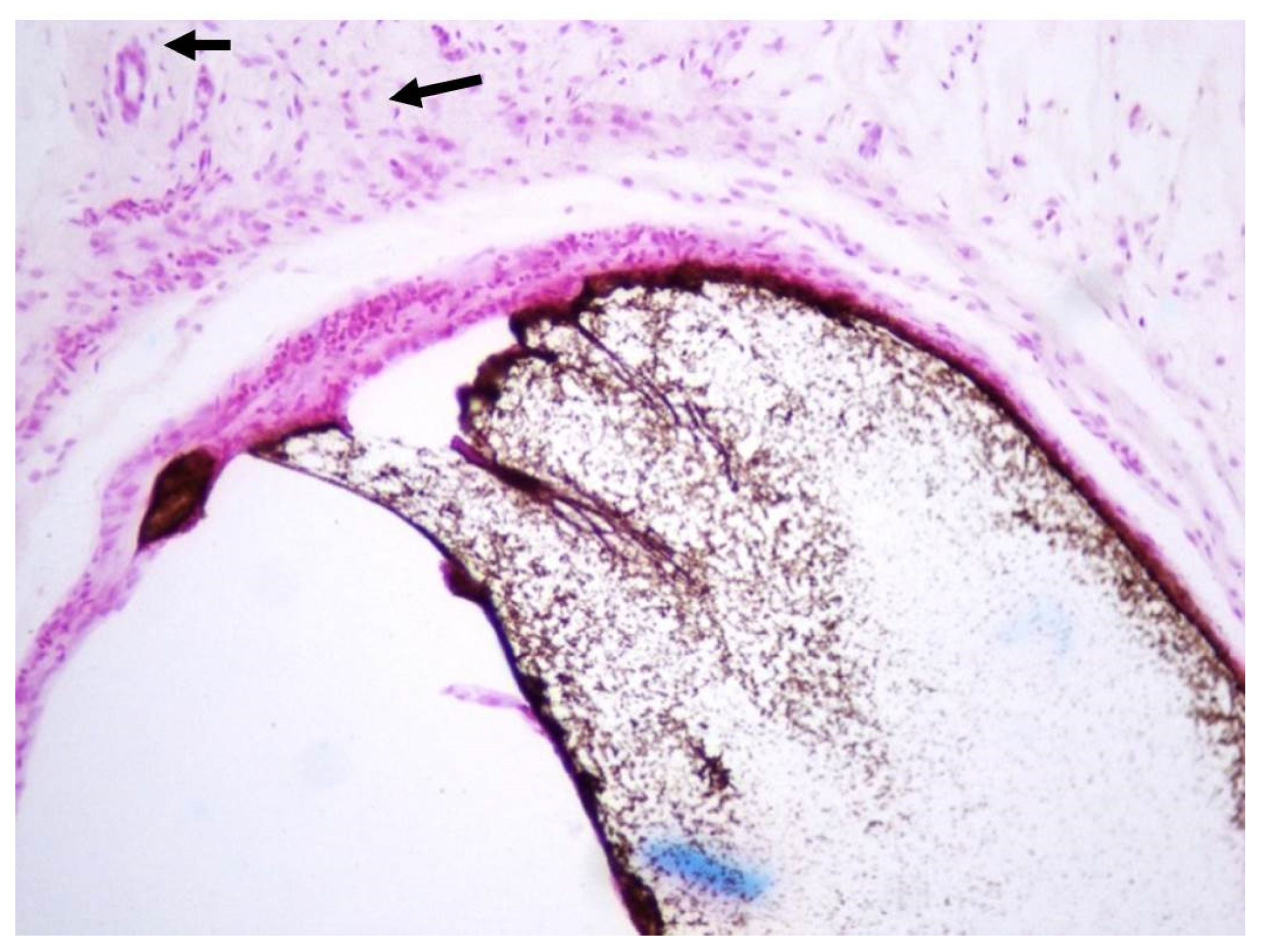
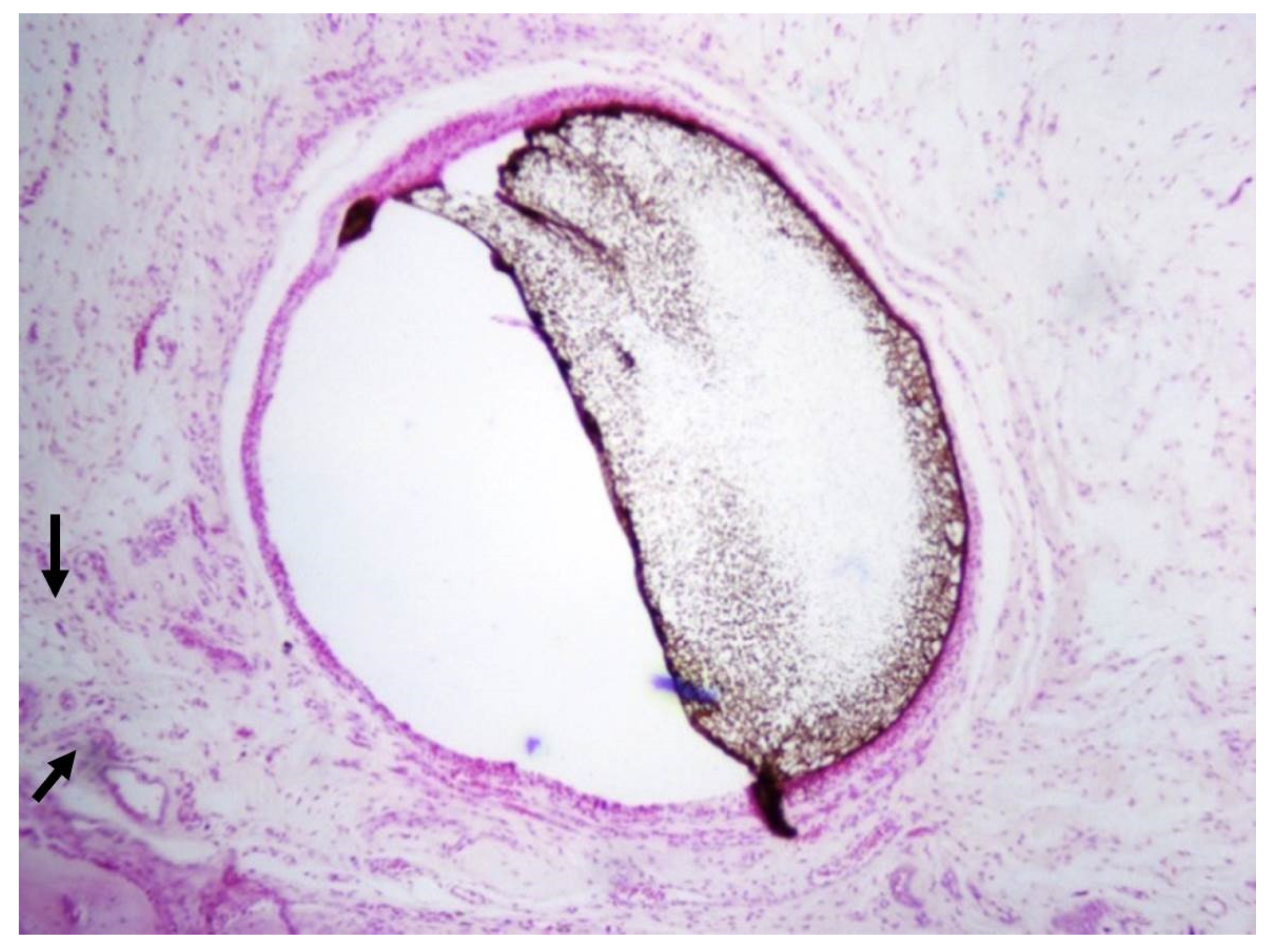
3.3. Infected GP Points (n = 12)

No remnants of the resorbable collagen membranes were found at the surgical site at 60 days. Two rats in Group 1 were excluded from the experiment. Adjacent to the capsule, a mild foreign body reaction with a few inflammatory cells was noted. In some specimens, a newly woven bone matrix surrounded by osteoblasts and mature bone was observed at 60 days (n = 10) (Figure 5 and Figure 6).

4. Discussion

Previous studies have shown that foreign bodies, such as GP (infected and non-infected), prevent wound healing and bone formation [19]. The aim of this pilot study was to test the hypothesis that biofilm and GP would induce bone destruction, whereas bone defects alone would not, using a rat calvarium model known in periodontology for its ability to show bone formation [20,21]. To the best of our knowledge, this is the first study testing this hypothesis using the methodology described above.
Root canal treatment aims to eliminate infection from the root canal and prevent reinfection by filling the root [22]. Despite the highest standards of care, some cases still result in failure due to persistent or secondary intraradicular infection or extraradicular infection [10]. It has been claimed that failures in other cases are due to non-microbial factors, such as extruded root canal filling materials; this may cause a foreign body reaction [10,23].
Contrary to our hypotheses, GP, per se, did not cause bone destruction at the calvaria, and surprisingly, biofilm-infected GP also did not cause bone destruction. Likewise, a mild inflammatory reaction was observed in the adjacent tissue in the non-infected group, and a newly woven bone matrix surrounded by osteoblasts was noted in some specimens. In the infected group, a mild foreign body reaction with a few inflammatory cells was observed adjacent to the capsule. A newly woven bone matrix surrounded by osteoblasts and mature bone was detected at 60 days.
GP points consist of approximately 65% zinc oxide, 20% GP, 10% radiopacifiers, and 5% plasticizers [24]. Wolfson and Seltzer (1975) studied the reaction of rat connective tissue to some formulations of GP in common use for up to 64 days and noticed that most of the specimens initially showed an acute response, followed by fibrous tissue encapsulation [25].
Sjogren et al. (1995) claimed that the size of the GP particles determines how the tissues react to the material [26]. These investigators studied the tissue reaction to various GP sizes subcutaneously implanted in guinea pigs and found that large particles (1–2 mm in size) were well encapsulated while the surrounding tissue was free of inflammation [26]. However, fine particles (50–100 µm in size) evoked an intense, localized tissue response characterized by the presence of macrophages and multinucleated giant cells [25]. Nair (2008) claimed that GP particles in apical tissue might gradually fragment into fine particles that induce a foreign body reaction [8]. In this research, we used a rat calvarium model, which is known in periodontology for its ability to evaluate bone formation [20,21]. The aim was to assess the ability of a foreign body, such as GP (infected and non-infected), in preventing wound healing and bone formation. GP, per se, did not cause bone destruction at the calvaria, but more surprisingly, infected GP with biofilm was also incapable of causing bone destruction. It seems this biofilm alone could not cause bone destruction unless there is a connection between the infected root and the periapical lesion. “It is possible that other types of bacteria are capable of causing such isolated infection; the independent extraradicular infection probably does not occur often and is usually associated with apical actinomycosis” [27]. Further research is warranted regarding GP fragmentation into fine particles in apical tissues and the response of apical tissues to fine particles.
The connection, or lack thereof, between the presence of GP particles in apical tissues and biofilm accumulation on GP points has not been sufficiently studied. Outcome studies showing associations between extruded GP and delayed apical tissue healing do not refer to this issue [2,3,4,5,6]. However, some studies state that over-instrumentation and apical transportation of contaminated debris might cause this adverse effect [2,10,11,12]. This might also be the result of an unusual root apex form or apical zipping that might leave residual bacteria around the apex.
The most common cause of failure is persistent intraradicular infection. Therefore, retreating failed teeth prior to surgery is recommended to exclude this possibility [10]. Retreatment of a failed root canal treatment requires the complete removal of the root canal filling. Poorly condensed GP fillings can be easily removed, but GP fragments may remain in the periapical tissue during attempts to remove overextended fillings [28]. Our findings suggest that healing was noticed in the non-infected and infected groups, even when GP particles remained in the apical tissue. Therefore, clinicians should not initiate surgical procedures as long as the tooth is functional, with no radiographic evidence of enlargement of apical rarefication. Note that the biofilm in this study was formed by two strains of Gram-positive facultative anaerobic bacteria (E.F. and S.S.) and one Gram-negative anaerobic bacterium (P.G.) that were capable of colonizing and forming extracellular matrices on GP. To the best of our knowledge, this is the first study on the biofilm-forming ability of these species on GP points. E.F. is the most frequently isolated strain from root-filled teeth with apical periodontitis [29]. Few species can overcome host defense mechanisms, flourish in the inflamed periapical tissues, and establish an extraradicular infection. Actinomyces and Propionibacterium propionicum participate in extraradicular infections that cause apical actinomycosis, which can be successfully treated only by periapical surgery [30,31,32].
Earlier studies suggested that some root canal treatments fail because of non-microbial factors, such as extruded root canal filling materials, which may cause foreign body reactions. In this study, healing was noticed in both the non-infected and infected groups, suggesting that overextension should not be considered an indication for endodontic surgery.

5. Conclusions

Our results contradict the hypothesis of this study. We have shown, contrary to common knowledge, that gutta-percha (both infected and non-infected) does not obstruct the healing of bony defects. Further study is needed to better understand the effect of different concentrations and strains of bacteria on intrabony wound healing.

Author Contributions

Conceptualization, S.L.; methodology, S.L.; investigation, Y.G., S.A., R.W. and S.L.; writing—original draft preparation, D.M., S.L. and R.W.; writing—review and editing, D.M. and S.L. All authors have read and agreed to the published version of the manuscript.

Funding

This study was funded in part by MIS Company Implants Technologies Ltd., (Bar Lev Industrial Park, Israel).

Institutional Review Board Statement

The Animal Research Council approved this research (il-071-06-2009).

Informed Consent Statement

Not applicable.

Data Availability Statement

Not applicable.

Acknowledgments

The authors would like to thank MIS Company Implants Technologies Ltd., (Bar Lev Industrial Park, Israel) for funding and supporting scientific research in Israel.

Conflicts of Interest

The authors declare that there were no conflicts of interest in this research.

References

  1. Johnson, W.; Kulild, J. Obturation of the Cleaned and Shaped Root Canal System; Elsevier: St. Louis, MO, USA, 2011; pp. 349–388. [Google Scholar]
  2. Ng, Y.L.; Mann, V.; Gulabivala, K. Outcome of secondary root canal treatment: A systematic review of the literature. Int. Endod. J. 2008, 41, 1026–1046. [Google Scholar] [CrossRef]
  3. Fristad, I.; Molven, O.; Halse, A. Nonsurgically retreated root filled teeth—Radiographic findings after 20–27 years. Int. Endod. J. 2004, 37, 12–18. [Google Scholar] [CrossRef] [PubMed]
  4. Bergenholtz, G.; Lekholm, U.; Milthon, R.; Engstrom, B. Influence of apical overinstrumentation and overfilling on re-treated root canals. J. Endod. 1979, 5, 310–314. [Google Scholar] [CrossRef]
  5. Bergenholtz, G.; Lekholm, U.; Milthon, R.; Heden, G.; Odesjö, B.; Engström, B. Retreatment of endodontic fillings. Scand. J. Dent. Res. 1979, 87, 217–224. [Google Scholar] [CrossRef] [PubMed]
  6. Van Nieuwenhuysen, J.P.; Aouar, M.; D’Hoore, W. Retreatment or radiographic monitoring in endodontics. Int. Endod. J. 1994, 27, 75–81. [Google Scholar] [CrossRef]
  7. Nair, P.N. On the causes of persistent apical periodontitis: A review. Int. Endod. J. 2006, 39, 249–281. [Google Scholar] [CrossRef]
  8. Haapasalo, M.; Shen, Y.A.; Ricucci, D. Reasons for persistent and emerging post-treatment endodontic disease: Reasons for persistent and emerging post-treatment endodontic disease. Endod. Top. 2008, 18, 31–50. [Google Scholar] [CrossRef]
  9. Ingle, J.I.; Bakland, L.K.; Baumgartner, J.C. Ingle’s Endodontics 6; BC Decker: Raleigh, NC, USA, 2008. [Google Scholar]
  10. Siqueira, J.F., Jr. Aetiology of root canal treatment failure: Why well-treated teeth can fail. Int. Endod. J. 2001, 34, 1–10. [Google Scholar] [CrossRef] [Green Version]
  11. Friedman, S. Prognosis of initial endodontic therapy. Endod. Top. 2002, 2, 59–88. [Google Scholar] [CrossRef] [Green Version]
  12. Farzaneh, M.; Abitbol, S.; Lawrence, H.P.; Friedman, S. Treatment outcome in endodontics-the Toronto Study. Phase II: Initial treatment. J. Endod. 2004, 30, 302–309. [Google Scholar] [CrossRef]
  13. Yusuf, H. The significance of the presence of foreign material periapically as a cause of failure of root treatment. Oral Surg. Oral Med. Oral Pathol. 1982, 54, 566–574. [Google Scholar] [CrossRef]
  14. Senia, E.S.; Marraro, R.V.; Mitchell, J.L.; Lewis, A.G.; Thomas, L. Rapid sterilization of gutta-percha cones with 5.25% sodium hypochlorite. J. Endod. 1975, 1, 136–140. [Google Scholar] [CrossRef]
  15. Takemura, N.; Noiri, Y.; Ehara, A.; Kawahara, T.; Noguchi, N.; Ebisu, S. Single species biofilm-forming ability of root canal isolates on gutta-percha points. Eur. J. Oral Sci. 2004, 112, 523–529. [Google Scholar] [CrossRef] [PubMed]
  16. Strathmann, M.; Wingender, J.; Flemming, H.C. Application of fluorescently labelled lectins for the visualization and biochemical characterization of polysaccharides in biofilms of Pseudomonas aeruginosa. J. Microbiol. Methods 2002, 50, 237–248. [Google Scholar] [CrossRef]
  17. George, S.; Basrani, B.; Kishen, A. Possibilities of gutta-percha-centered infection in endodontically treated teeth: An in vitro study. J. Endod. 2010, 36, 1241–1244. [Google Scholar] [CrossRef]
  18. Turnbull, R.S.; Freeman, E. Use of wounds in the parietal bone of the rat for evaluating bone marrow for grafting into periodontal defects. J. Periodontal. Res. 1974, 9, 39–43. [Google Scholar] [CrossRef]
  19. Ramachandran Nair, P.N. Light and electron microscopic studies of root canal flora and periapical lesions. J. Endod. 1987, 13, 29–39. [Google Scholar] [CrossRef]
  20. Moses, O.; Vitrial, D.; Aboodi, G.; Sculean, A.; Tal, H.; Kozlovsky, A.; Artzi, Z.; Weinreb, M.; Nemcovsky, C.E. Biodegradation of three different collagen membranes in the rat calvarium: A comparative study. J. Periodontol. 2008, 79, 905–911. [Google Scholar] [CrossRef]
  21. Kozlovsky, A.; Aboodi, G.; Moses, O.; Tal, H.; Artzi, Z.; Weinreb, M.; Nemcovsky, C.E. Bio-degradation of a resorbable collagen membrane (Bio-Gide) applied in a double-layer technique in rats. Clin. Oral Implant. Res. 2009, 20, 1116–1123. [Google Scholar] [CrossRef]
  22. Peters, O.A.; Peters, C.I. Chapter 9—Cleaning and Shaping of the Root Canal System. In Cohen’s Pathways of the Pulp, 10th ed.; Hargreaves, K.M., Cohen, S., Eds.; Mosby: St. Louis, Mo, USA, 2011; pp. 283–348. [Google Scholar]
  23. Nair, P.N.; Sjögren, U.; Krey, G.; Sundqvist, G. Therapy-resistant foreign body giant cell granuloma at the periapex of a root-filled human tooth. J. Endod. 1990, 16, 589–595. [Google Scholar] [CrossRef]
  24. Friedman, C.E.; Sandrik, J.L.; Heuer, M.A.; Rapp, G.W. Composition and physical properties of gutta-percha endodontic filling materials. J. Endod. 1977, 3, 304–308. [Google Scholar] [CrossRef]
  25. Wolfson, E.M.; Seltzer, S. Reaction of rat connective tissue to some gutta-percha formulations. J Endod. 1975, 1, 395–402. [Google Scholar] [CrossRef]
  26. Sjögren, U.; Sundqvist, G.; Nair, P.N. Tissue reaction to gutta-percha particles of various sizes when implanted subcutaneously in guinea pigs. Eur. J. Oral Sci. 1995, 103, 313–321. [Google Scholar] [CrossRef]
  27. Lin, L.M.; Huang, G.T.J. Chapter 14—Pathobiology of the Periapex. In Cohen’s Pathways of the Pulp, 10th ed.; Hargreaves, K.M., Cohen, S., Eds.; Mosby: St. Louis, Mo, USA, 2011; pp. 529–558. [Google Scholar]
  28. Metzger, Z.; Ben-Amar, A. Removal of overextended gutta-percha root canal fillings in endodontic failure cases. J. Endod. 1995, 21, 287–288. [Google Scholar] [CrossRef]
  29. Molander, A.; Reit, C.; Dahlén, G.; Kvist, T. Microbiological status of root-filled teeth with apical periodontitis. Int. Endod. J. 1998, 31, 1–7. [Google Scholar] [CrossRef] [PubMed]
  30. Happonen, R.P. Periapical actinomycosis: A follow-up study of 16 surgically treated cases. Endod. Dent. Traumatol. 1986, 2, 205209. [Google Scholar] [CrossRef] [PubMed]
  31. Siquera, J.F., Jr. Periapical Actinomycosis and infection with Propionibacterium Propionicum. Endod. Top. 2003, 6, 78–95. [Google Scholar] [CrossRef]
  32. Lin, S.; Sela, G.; Sprecher, H. Periopathogenic bacteria in persistent periapical lesions: An in vivo prospective study. J. Periodontol. 2007, 78, 905–908. [Google Scholar] [CrossRef]
Figure 1. Confocal laser scanning microscopy image of bacteria and biofilm generated over a gutta-percha segment.
Applsci 11 06710 g001
Figure 2. (A) Scanning electron microscope image of biofilm on infected GP. (B) Scanning electron microscope image of sterilized GP.
Applsci 11 06710 g002
Figure 3. Intra-operative view of the rat caldarium operation.
Applsci 11 06710 g003
Figure 4. Histologic micrograph showing foreign material surrounded by a thin fibrous connective tissue capsule (arrows) and mild chronic inflammatory cell infiltrate composed primarily of lymphocytes (asterisk).
Applsci 11 06710 g004
Figure 5. Histologic micrograph of foreign material surrounded by a thin fibrous connective tissue capsule. The matrix contained mild inflammatory cell infiltrate composed primarily of lymphocytes and few scattered small blood vessels (arrows). H&E: hematoxylin & eosin stain, ×200.
Applsci 11 06710 g005
Figure 6. Magnified view showing mild inflammatory cell infiltrate composed primarily of lymphocytes and few scattered small blood vessels (arrows). H&E: hematoxylin & eosin stain, ×200.
Applsci 11 06710 g006
Publisher’s Note: MDPI stays neutral with regard to jurisdictional claims in published maps and institutional affiliations.

