Article

Changes in Crystal Phase, Morphology, and Flexural Strength of As-Sintered Translucent Monolithic Zirconia Ceramic Modified by Femtosecond Laser

1
Second Clinical Division, Peking University Hospital of Stomatology, Beijing 100081, China
2
Department of Prosthodontics, Peking University School and Hospital of Stomatology, Beijing 100081, China
3
Center of Digital Dentistry, Department of Prosthodontics, Peking University School and Hospital of Stomatology, Beijing 100081, China
*
Author to whom correspondence should be addressed.
These authors contributed to this work equally.
Appl. Sci. 2021, 11(15), 6925; https://doi.org/10.3390/app11156925
Submission received: 31 May 2021 / Revised: 18 July 2021 / Accepted: 22 July 2021 / Published: 28 July 2021
(This article belongs to the Special Issue Dental Materials: Latest Advances and Prospects)

Abstract

:
Conventional bonding technology suitable for silica-based ceramics is not applicable to zirconia, due to its polycrystalline phase composition, chemical stability, and acid corrosion resistance. The development of an effective treatment to improve its surface roughness and mechanical properties remains an unresolved problem. Therefore, to solve this problem, this in vitro study evaluated the changes in surface morphology and flexural strength of translucent monolithic zirconia surfaces treated with femtosecond laser technology. As-sintered translucent zirconia specimens were subjected to airborne particle abrasion and femtosecond laser treatments, while control group specimens received no treatment. After treatment, the roughness and morphology of the treated zirconia surfaces were examined. The flexural strength and X-ray diffraction of the treated specimens were measured and analyzed. Statistical inferential analysis included one-way analysis of variance at a set significance level of 5%. The surface roughness after femtosecond laser treatment was significantly improved when compared with the control group and the group that received the airborne particle abrasion treatment (p < 0.05). In comparison with the airborne particle abrasion group, the flexural strength of the group that received the femtosecond laser treatment was significantly improved (p < 0.05). The femtosecond laser approach using appropriate parameters enhanced the roughness of the zirconia without reducing its flexural strength; therefore, this approach offers potential for the treatment of zirconia surfaces.

1. Introduction

Zirconia is highly popular in clinical use for dental restorations, due to its exceptional flexural strength, chemical resistance, and good aesthetics [1]. Depending on whether a glass-matrix phase is present or absent, or whether the material contains an organic matrix highly filled with ceramic particles, all-ceramic materials can be classified into three families: (1) glass-matrix ceramics, (2) polycrystalline ceramics, and (3) resin-matrix ceramics [2]. However, unlike other dental ceramic materials, zirconia has a polycrystalline phase composition without a glass phase composition, as well as good chemical stability and acid corrosion resistance. Owing to this, conventional bonding technology suitable for silica-based ceramics is not effective for zirconia. Thus, an important scientific problem in the use of zirconia materials is the effective treatment of zirconia to improve its surface roughness and mechanical properties [3].
Contemporary surface treatment technologies used for zirconia ceramics include airborne particle abrasion (APA), acid etching, laser etching, silicon coating, pre-treatment agents, and other technologies and combinations thereof. APA with 50 µm Al2O3 under 0.2 MPa pressure at a distance of 10 mm from the zirconia surface has been found to be an effective method for improving bond strength [4]. However, this approach may lead to sub-surface damage to the zirconia, resulting in microcracks and debris, which may reduce the mechanical properties of yttria-stabilized tetragonal zirconia polycrystal (Y-TZP), thus affecting its longevity. Studies have shown that APA may cause fractures in zirconia restorations and influence the long-term durability of the bond strength between zirconia ceramics and resin [5,6,7]. The reduced retention rate after APA may be related to microcracks in the zirconia, and APA may, therefore, have an adverse effect on flexural strength and other long-term mechanical properties of restorations [3].
Other surface modification methods are therefore required to replace aluminum oxide APA and to avoid adverse effects on the mechanical properties and long-term durability of zirconia [4]. Recently, scholars worldwide have trialed advanced technologies, such as thermal acid etching solutions [7,8], plasma technology [6,9], tribochemical silica coating [10,11], ultrashort-pulse lasers [12,13], and fusion sputtering technology [14,15], to treat zirconia surfaces.
Thermal chemical etching solutions have been employed for pre-treating zirconia ceramics. It has been found that this method improves roughness and, therefore, increases the zirconia–resin cement bond strength [7]; however, the configuration of a safe and effective thermal acid etching solution remains unclear, as thermal acid etching may also affect the physical properties of zirconia [8].
The application of plasma technology for the surface modification of zirconia ceramics has also been studied. Fernandes et al. [6] reported that non-thermal plasma modification without significant damage promoted adequate adhesion, but the bond strength was not found to be significantly different from that under aluminum oxide APA. Plasma modification resulted in a significant increase in the surface free energy of the zirconia ceramic, but no significant changes in surface roughness were observed. The application of plasma treatment in zirconia bonding, therefore, cannot replace APA [9].
Tribochemical silica coating is a commonly used silicon coating technology at present, which uses 30 μm alumina particles covered with silica for sandblasting at 0.23 MPa on the zirconia surface [10]. By increasing the silicon content on the surface of the zirconia, the silica layer can react with the cement-containing silane [11]. As a result, through copolymerization between the silane and the resin cement, the bond strength between the zirconia resin can be improved.
In addition, it has been reported that application of the fusion sputtering technique promotes a rough surface and significantly enhances the zirconia–resin microshear bond strength [14,15]. However, the influence of fusion sputtering technology on the crystal phase change and mechanical properties of zirconia, as well as the long-term bond strength remains unclear.
Holthaus et al. [16] found that the application of laser treatment could potentially replace traditional surface treatment by APA, due to the high speed and precise control of the laser. A more regular micro-texture and a reduction in contamination were obtained through the application of a femtosecond laser (FSL), compared with CO2 and Nd-YAG lasers, for zirconia surface micromachining [12]. Ruja et al. [13] evaluated the use of an ultrashort-pulse laser to irradiate the zirconia ceramic surface, so as to improve adhesive properties in the resin–zirconia interface. The results showed that the topography of the zirconia ceramic surface was regularly roughened and wettability was increased, while an improvement in microtensile bond strength was promoted without a significant tetragonal–monoclinic phase transformation [13]. This may be attributed to the fact that the energy of the laser was absorbed by the surface of the zirconia, and the thermal induction process produced shell-like ruptures on the surface [17].
Through the use of FSLs, the surfaces of zirconia ceramics could be effectively modified without inducing thermal or mechanical damage [17,18]. While limited research has been conducted on the surface pre-treatment of zirconia ceramics using laser technology, the ideal effect of zirconia modification by laser, in order to improve the bond strength, has not yet been achieved [19]. Therefore, the changes in surface morphology and flexural strength upon applying the FSL technique require further exploration.
This study aims to evaluate the effect of zirconia surface modification using the FSL method. The null hypothesis was that the application of the FSL would not affect the surface morphology and flexural strength of translucent monolithic zirconia ceramic material.

2. Materials and Methods

2.1. Zirconia Specimen Preparation

A total of 36 disc-shaped fully sintered translucent monolithic zirconia ceramic (UPCERA, ST, Shenzhen, China) specimens (10 mm diameter × 2 mm thickness) and 36 rectangular fully sintered translucent monolithic zirconia specimens (25 mm long × 4 mm wide × 3 mm thick) were prepared from a dental zirconia blank (in which the content of Y2O3 was 4.5–6%). A precision cutting machine was used to prepare the specimens, which were sintered using a programmable furnace according to the manufacturer’s instructions. The temperature of the furnace was heated from room temperature, at a rate of 8 °C/min, to 1200 °C, then increased at a rate of 2 °C/min from 1200 °C to 1450 °C, maintained at 1450 °C for 2 h, and finally cooled at a rate of 10 °C/min to room temperature. Depending on the employed surface pre-treatment method, the 36 disc-shaped fully sintered zirconia specimens were randomly divided into three groups, with each group containing 12 specimens. Disc-shaped specimens were employed for observation of surface topography and assessment of roughness. Rectangular specimens were used for flexural strength testing. In order to avoid potential failure of specimens due to edge defects, a 45° diagonal angle was introduced on the edges of all the rectangular specimens. Specimen preparation was carried out in accordance with the requirements of the ISO/CD 6872:2015 standard [20]. The calculation of the sample size of this study was carried out with reference to similar studies [21].

2.2. Zirconia Surface Treatment

The 72 sintered translucent zirconia specimens were classified into Groups I, II, and III, with each group containing 12 circular and 12 rectangular specimens. Group I was the control, with no treatment (NT) of the surface of the specimens. Group II underwent APA, whereby the surfaces of the specimens were air abraded (Renfert GmbH, Hilzingen, Germany) with 50 μm aluminum oxide (Al2O3) particles at a perpendicular distance of 10 mm from the surface under 0.20 MPa of pressure for 20s. Group III underwent FSL treatment, whereby the surfaces of the specimens were microtextured with FSL (Amplitude Systemes, Tangerine laser head, Bordeaux, France) having a wavelength of 1030 nm, pulse width of 400 fs, repetition frequency of 200 kHz, peak power of 5 W, and single-pulse energy of 25 µJ. The FSL ablation was controlled using a three-axis numerically controlled laser galvanometer scanning system (175 mm lens focal length, ~80 mm spot diameter, 2000 mm/s light spot scanning speed, 0.1 µm minimum step size along the z-axis, and 10 mm maximum step size; see Figure 1).

2.3. Surface Structure and Roughness Assessment

The surface roughness of the specimens was measured using a confocal three-dimensional (3D) laser scanning microscope (VK-X210, Osaka, Japan). For each specimen, the surface roughness of three areas was measured under 200× magnification (n = 12/group). The 3D surface roughness parameter Ra was calculated using VK Analyzer software. The acquisition Ra (μm) value was derived from the average of three analyzed areas, using a simple average-type smoothing filter. The disc-shaped specimens of each group were sputter-coated with gold (108AUTO sputter coater, Cressington, Watford, UK), and the surface topographies of the specimens (n = 3/group) were observed using a scanning electron microscope (SEM, SU8010, Tokyo, Japan) under vacuum conditions and secondary electron detector of SE(UL), with 5.0 kV working voltage, at 500× and 10,000× magnifications.

2.4. X-ray Diffraction Analysis (XRD)

The crystalline phases of the specimens were determined using an X-ray powder diffractometer (XRD) system (Bruker D8 ADVANCE, Karlsruhe, Germany), with the following measurement conditions: Cu K-α radiation, tube voltage of 40 kV, tube current of 40 mA, scanning angle range of 2θ = 25–60°, scanning speed of 17.7 s/step, and step size of 0.02°.

2.5. Flexural Strength Test (Three-Point Bending Test)

Flexural strength tests were conducted using a universal testing machine (5969R9273, Instron, Norwood, MA, USA) with 1 mm/min crosshead speed and 13 mm span, according to a three-point bending test. During a flexural strength test, the treated surface was placed in contact with the loading stylus (compressive loading zone). The upper limit of the load cell value applied was 2500 N [20]. The fracture loads of the specimens are expressed in Newtons, and their flexural strength was measured in megapascals, using the equation [20] σ = 3 P l / 2 ω b 2 , where P is the fracture load (N), l is the span (mm), ω is the specimen width (mm), and b is the specimen height (mm).

2.6. Statistical Analysis

Descriptive statistics and inferential statistic measures were employed to analyze the flexural strength (MPa) and surface roughness (Ra). The mean and standard deviation were calculated, and the normality of the data distribution was confirmed by the Shapiro–Wilk test. A one-way analysis of variance (ANOVA) and Least Significance Difference (LSD) post hoc tests were performed, in order to analyze the results among all groups (NT, APA, and FSL). All calculations were performed using SPSS 20 statistical software (SPSS Inc., Chicago, IL, USA). The significance level was set at α = 0.05.

3. Results

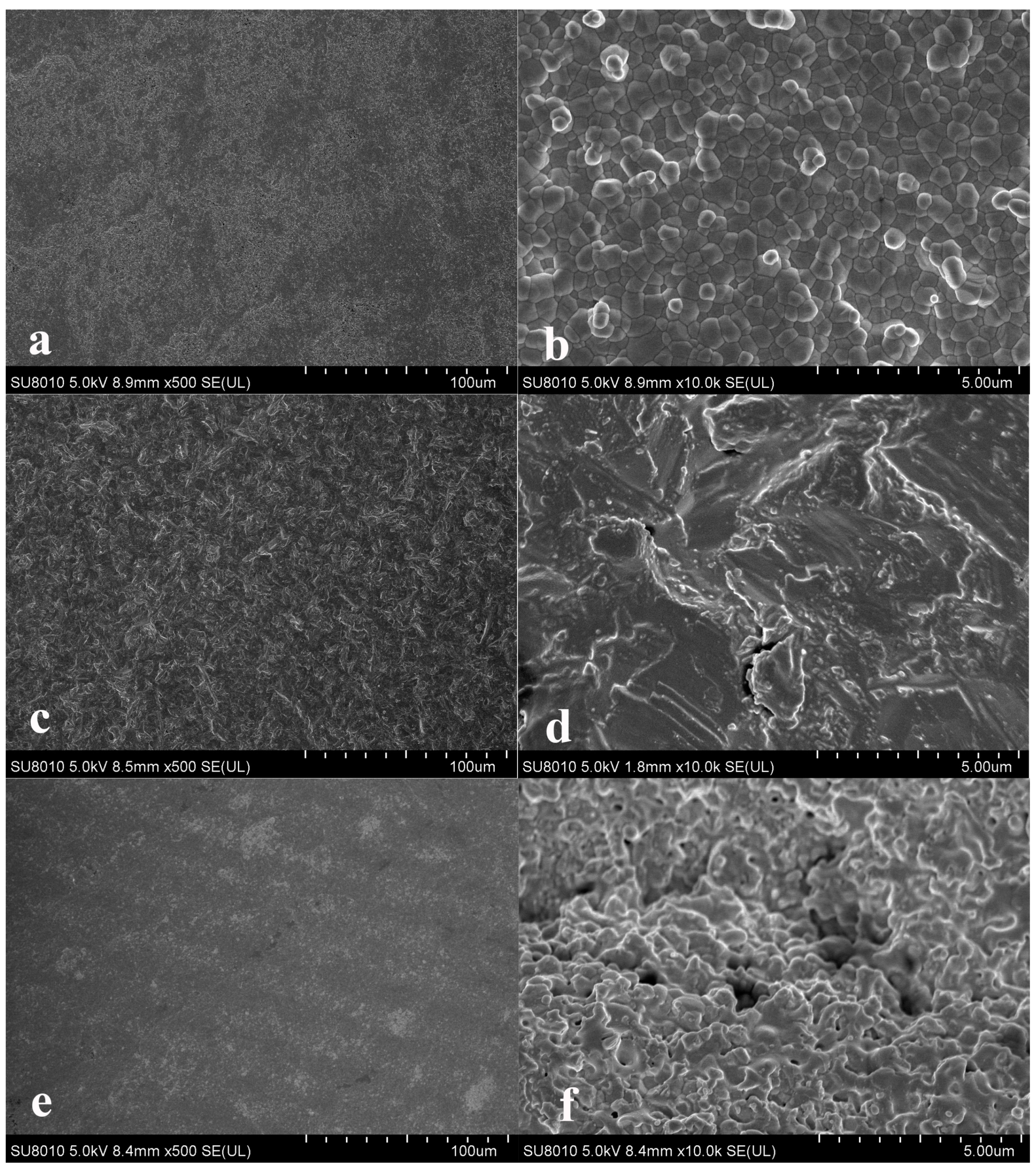
3.1. Scanning Electron Microscopy Observations

The SEM images shown in Figure 2 present the surface topographies of the specimens that underwent the different surface treatment methods. Specifically, Figure 2a,b represents the NT group, where Figure 2a shows that the surface was relatively flat, and Figure 2b reveals the unit cell-like microstructure of the zirconia sample after heat treatment. Many grain boundaries can be observed, and the arrangement is dense. There are no voids between the grains. Figure 2c, which represents the APA group, shows that the surface was uniformly rough with sharp edges and corners. A few microcracks can be observed in the image shown in Figure 2d. Finally, the image of the FSL group, depicted in Figure 2e, shows that the surface was uniform and flat without deep grooves. Figure 2f shows an irregular structure with rounded edges and a few pores.

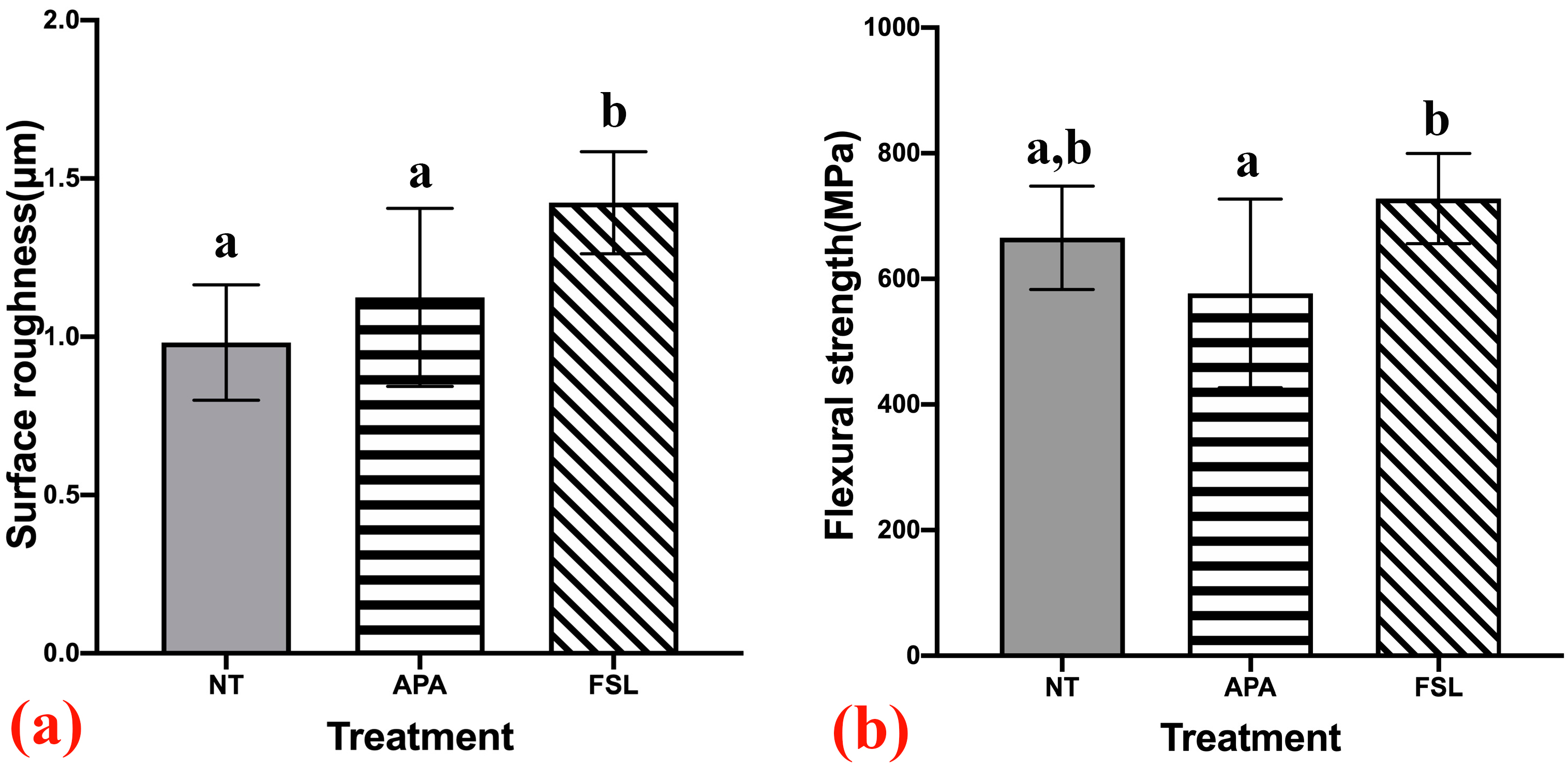
3.2. Surface Roughness Evaluation

The surface roughness results (measured by the Ra amplitude value) are recorded in Table 1. One-way ANOVA analysis showed that there were statistical differences between the three groups (p < 0.05; Figure 3a). Compared with the NT group (Ra = 0.98 ± 0.18 µm) and the APA group (Ra = 1.12 ± 0.28 µm), it was found that the FSL group achieved significantly superior roughness (Ra = 1.42 ± 0.16 µm; Table 1). Therefore, the highest roughness of the zirconia surface was due to the FSL treatment (Figure 3a).

3.3. Flexural Strength
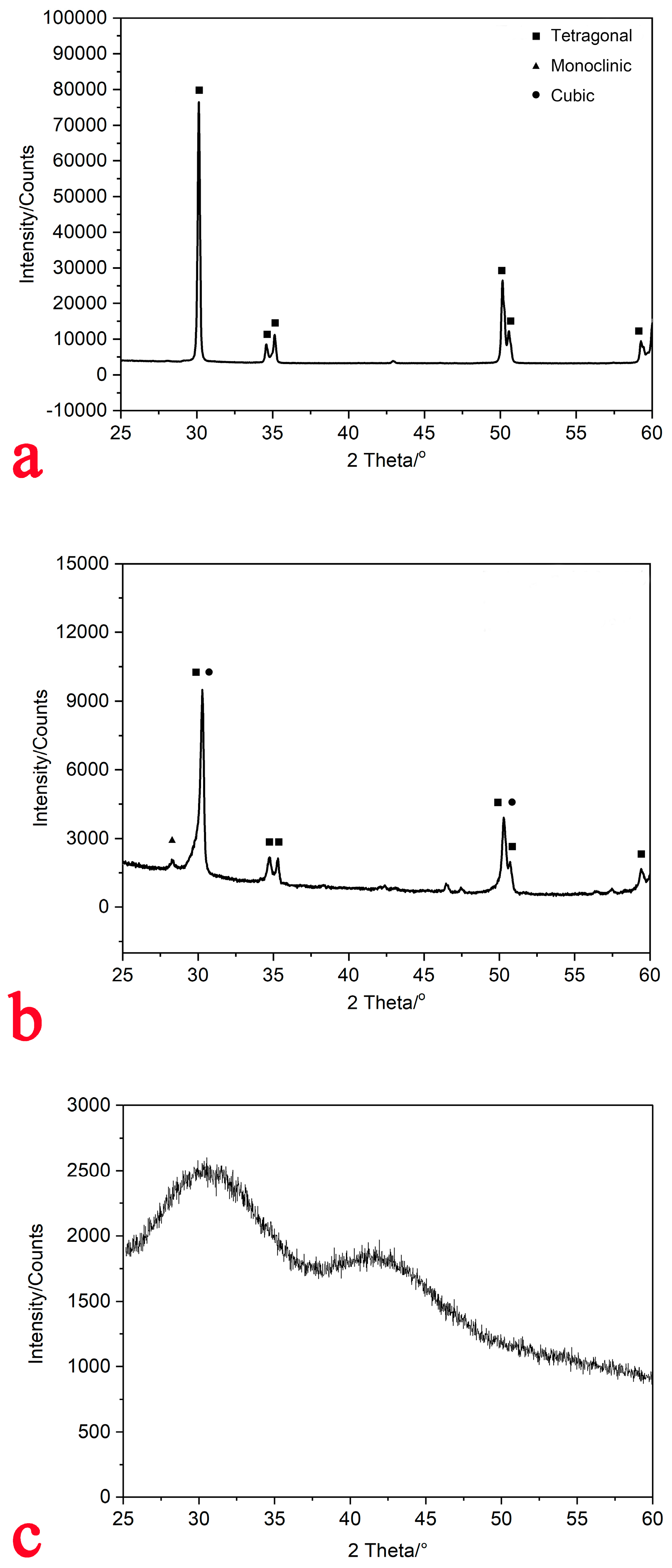
Table 2 shows the mean flexural strength values (MPa), along with the standard deviation, for the three groups. The one-way ANOVA analysis indicated a statistical difference among the three groups (p < 0.05; Figure 3b). No significant difference was found between FSL and NT or between APA and NT, but a significant difference was found between FSL and APA (Figure 3b). The effect of the FSL treatment was, therefore, found to be superior to that of the APA treatment (Figure 3b).

3.4. XRD

Through analysis of the XRD pattern (Figure 4) on zirconia, we found that strong diffraction peaks of the tetragonal phase were identified in both the NT and APA groups. The zirconia surface with no treatment was completely tetragonal-phase (Figure 4a). Compared with the APA group, the diffraction peaks of the tetragonal phase for the NT group were dominant. The tetragonal phase ratio of the surface treated with APA was low, with a small amount of monoclinic phase (about 9.23%) and cubic phase (4.10%), as shown by the small peaks of monoclinic crystal and cubic phases (Figure 4b); however, the XRD of the sample treated with FSL suggested that there may be an amorphous phase, as it was also possible to form salt crystals on the surface of the ceramic treated by FSL (Figure 4c).

4. Discussion

The purpose of this study was to evaluate the effect of femtosecond laser treatment on the surface roughness and flexural strength of translucent zirconia. Compared with the no treatment group, the surface roughness of translucent zirconia was significantly increased in the femtosecond laser group, while the flexural strength measured via three-point bending of zirconia modified by the femtosecond laser was found not to be significantly different from that of samples in the no treatment group. Thus, the null hypothesis of the study was partly rejected.
The bending strength and fatigue resistance of translucent zirconia are higher than those of glass ceramics [22]. Thus, translucent zirconia is a material that offers both mechanical strength and aesthetic performance, and its clinical applications are becoming more and more extensive [22]. However, translucent zirconia may be adversely affected by the use of high-pressure airborne abrasion, as microcracks and defects are generated, and flexural strength is reduced [23]. To date, limited studies have evaluated the effect of FSL modification on Y-TZP surfaces and, to the best of our knowledge, no existing study has evaluated the effect of FSL for treating the surface of translucent zirconia.
Studies have reported that the application of FSL forms groove- or pit-like structures on the surface of zirconia specimens, thereby increasing their surface roughness; however, this was accompanied by a reduction in flexural strength [24]. This approach may lead to sub-surface damage to the zirconia due to 50 μm alumina blasting, which may cause microcracks that limit the life of zirconia restorations [25]. In this study, we explored a specific parameter of FSL in the surface modification of translucent zirconia, in order to improve surface roughness without producing significant grooves or pits. Even though no statistical significance was found, the results in this study revealed that surface roughness was improved. As studies incorporating the FSL method continue to improve, FSL may become more suitable as a surface treatment for zirconia materials.
Although the use of APA with Al2O3 particles is popular for the treatment of zirconia surfaces in order to increase the bond strength, studies have shown that the increased roughness imparted by this technique is accompanied by an increased fracture risk, thereby weakening the structure through the introduction of microcracks [26,27,28]. The results of our study demonstrated that FSL-treated translucent zirconia showed significantly higher mean Ra values than those in the APA and NT groups, and there was no significant difference in the Ra value between the APA and NT groups. These findings are consistent with those of the study of Inokoshi et al. [29], where the surface roughness of highly translucent Y-PSZ modified using Al2O3 APA was not significantly enhanced, except for that of specimens comprising KATANA UTML (Kuraray Noritake, Japan). In contrast, FSL ablation of zirconia ceramic significantly enhanced surface roughness and improved the zirconia ceramic–resin bond strength, due to the presence of groove-like structures [30]. In our study, the translucent zirconia surface obtained following the FSL treatment presented uniform irregular structures without groove-like structures or pits, as confirmed in the SEM image shown in Figure 2e. Surface topography can be modified by femtosecond laser surface treatment and surface roughness of the zirconia can be increased; thus, the bond strength can be improved [31].
The main height parameters for evaluating surface roughness are average roughness (Ra), the root mean square of the height of each point of the contour (Rq), and the ten-point height of microscopic unevenness (Rz). The Ra value can represent the arithmetic mean deviation of the surface roughness profile amplitude parameter. Consequently, the Ra value was used in this study to evaluate the surface roughness [32]. Compared with the control group, roughness in the APA group did not increase significantly, which may have been due to the particular cutting texture produced during the processing of the untreated translucent zirconia specimens, which yielded a certain roughness after sintering. The surfaces of the specimens were not polished in this experiment, in order to maintain the original surface morphologies of the final sintered translucent zirconia. On one hand, this allowed for simulation of the surface of a final sintered zirconia crown without polishing; on the other hand, when the surface of a zirconia specimen is highly polished, FSL irradiates a smooth surface, producing reflections, which may affect the treatment of the zirconia surface. In this study, the surfaces of the zirconia specimens were not polished, allowing the FSL to fully exert its plasma effect at a lower energy density. This may also contribute to the discrepancy in the roughness results between APA and FSL treatments reported in this study when compared with those in other studies. Various surface analysis systems, such as SEM and Atomic Force Microscopy (AFM), are useful for qualitative analysis, but three-dimensional microscopy was used for quantitative evaluation of the surface roughness variation.
The results of XRD showed that the monoclinic content of the APA group was increased, which indicated that aluminum airborne abrasion may lead to t–m phase transformation. At present, there is controversy about the influence of sandblasting on mechanical properties [33]. Some scholars have reported that t–m phase transformation can increase volume and produce protective residual compressive stress, thus preventing the further expansion of microcracks and, consequently, leading to enhanced mechanical strength. This is called the phase transformation toughening mechanism [34]. However, other scholars have indicated that small grain size (within 200 nm) can have a negative impact on the phase transformation and toughening mechanism, consequently reducing the mechanical strength of translucent ZrO2 [35]. In this experiment, the XRD results indicated that the sintered translucent zirconia specimen appeared to be amorphized after the FSL treatment, which may have been caused by the phenomenon of “avalanche ionization” during interaction of the high-power and high-repetition FSL with the zirconia specimen surface, resulting in high-speed motion [36]. The hypothesis is that the plasma carries a certain element, which is deposited on the surface of the specimen to form a coating, resulting in amorphization [37]. It is also possible to form salt crystals on the surface of the ceramic; however, the interfacial topography between the “amorphized” zirconia layer after FSL and the substrate was not investigated, and further experimental verification is required.
Air particle abrasion may lead to sub-surface damage of the zirconia surface by 50 μm alumina, which may lead to microcracks that limit the life of zirconia restorations [38]. It has been reported that impact-induced defects were observed on zirconia surfaces modified by APA treatment; thus, the longevity of APA-treated zirconia ceramic prostheses may be shortened [39]. Consistent with previous studies, in our study, the surface treated with APA presented a number of microcracks and defects as revealed in Figure 2d, which may lead to a reduction in the flexural strength of the zirconia specimens. In contrast, in a study reported by Wang et al. [40], APA enhanced the flexural strength of zirconia, regardless of the particle size, air pressure, or blasting time. Song et al. [41] reported that flexural strength was significantly higher in the group of air-abraded zirconia specimens than that in the group without any treatment. The content of the monoclinic phase of the lower zirconia surface determines the mechanical behavior of the zirconia specimens, as this is where tensile stress is dispersed. Furthermore, this study indicated that APA of the inner surface of zirconia specimens, in order to improve their bonding performance, might also enhance their fracture strength [40]. In this experiment, we applied 50 µm Al2O3 particles to treat the surface of the translucent zirconia specimen, in order to reduce tetragonal–monoclinic phase transformation.
There are two main methods for testing the bending strength of ceramics: uniaxial bending and biaxial bending. In uniaxial bending tests, a cuboid specimen is supported by two points and loaded vertically at one point (i.e., a three-point bending test) or two points (i.e., a four-point bending test). In a biaxial bending test, a thin disk is supported by a ring or three balls close to it, and a load is applied through a ball or a piston in its central area, or a smaller ring in its center. The above methods have been recognized in international standards [20]. Therefore, a three-point bending test was used to evaluate the used methods after surface treatment in this study. New possible methods for the mechanical analysis of materials have been reported as a future perspective for classic dynamometer systems, such as Dynamic Mechanical Analysis (DMA) and Brillouin’s micro-spectroscopy [42].
Importantly, the flexural strength results presented herein were consistent with the observed SEM images. However, further investigations are required to evaluate the long-term stability of zirconia treated using different methods. It should also be noted that, in the experimental setup, line-patterning of zirconia surfaces was achieved; thus, tuning the FSL parameters should allow for independent variation of the pattern depth, overall roughness, and surface finish. More specifically, increasing both the fluence and the number of pulses will allow for deeper patterning, with the maximum achievable depth being 1 µm. However, increasing the number of pulses can have a detrimental effect on the quality of the lines produced, and surface damage can occur (e.g., intergranular cracking, open porosity, and nanodroplet formation), depending on the FSL parameters employed [43]. In future experiments, our research team will try to design an integrated processing device featuring a femtosecond laser. After the zirconia restorations are sintered, the dental technician can hold the working end of the device to modify the tissue surface of dental zirconia restorations, in order to achieve a clean, efficient, and damage-free effect.
One limitation of this study is that the long-term effects of FSL modification technology on the flexural strength of zirconia were not investigated. Therefore, an evaluation of resin–zirconia bond strength and durability using different surface modification methods will be reported in a future article.

5. Conclusions

  • Femtosecond laser technology offers potential for zirconia surface treatment.
  • Through the employment of appropriate parameters, femtosecond laser treatment can be used to modify the surface of zirconia, in order to enhance its roughness without decreasing flexural strength.

Author Contributions

Conceptualization, F.Y.; methodology, F.Y. and S.L.; software, H.Y.; validation, F.Y., S.L. and H.Y.; formal analysis, F.Y.; investigation, S.L.; resources, S.L.; data curation, H.Y.; writing—original draft preparation, S.L.; writing—review and editing, F.Y.; visualization, F.Y.; supervision, F.Y.; project administration, F.Y.; funding acquisition, F.Y. All authors have read and agreed to the published version of the manuscript.

Funding

This research was funded by the National Key R&D Program of China [Grant no. 2020YFB1312801], and the State Key Lab of Advance Metals and Materials [Grant no. 2021-Z08].

Institutional Review Board Statement

Not applicable.

Informed Consent Statement

Not applicable.

Data Availability Statement

The data presented in this study are available on request from the corresponding author.

Conflicts of Interest

The authors declare no conflict of interest.

References

  1. Denry, I.; Kelly, J.R. State of the art of zirconia for dental applications. Dent. Mater. 2008, 24, 299–307. [Google Scholar] [CrossRef]
  2. Gracis, S.; Thompson, V.P.; Ferencz, J.L.; Silva, N.R.; Bonfante, E.A. A new classification system for all-ceramic and ceramic-like restorative materials. Int. J. Prosthodont. 2015, 28, 227–235. [Google Scholar] [CrossRef] [Green Version]
  3. Scaminaci Russo, D.; Cinelli, F.; Sarti, C.; Giachetti, L. Adhesion to Zirconia: A Systematic review of current conditioning methods and bonding materials. Dent. J. 2019, 7, 74. [Google Scholar] [CrossRef] [Green Version]
  4. Thompson, J.Y.; Stoner, B.R.; Piascik, J.R.; Smith, R. Adhesion/cementation to zirconia and other non-silicate ceramics: Where are we now? Dent. Mater. 2011, 27, 71–82. [Google Scholar] [CrossRef] [PubMed] [Green Version]
  5. Hallmann, L.; Ulmer, P.; Wille, S.; Polonskyi, O.; Köbel, S.; Trottenberg, T.; Bornholdt, S.; Haase, F.; Kersten, H.; Kern, M. Effect of surface treatments on the properties and morphological change of dental zirconia. J. Prosthet. Dent. 2016, 15, 341–349. [Google Scholar] [CrossRef] [PubMed]
  6. Fernandes, V.V.B., Jr.; Dantas, D.C.B.; Bresciani, E.; Huhtala, M.F.R.L. Evaluation of the bond strength and characteristics of zirconia after different surface treatments. J. Prosthet. Dent. 2018, 120, 955–959. [Google Scholar] [CrossRef]
  7. Casucci, A.; Monticelli, F.; Goracci, C.; Mazzitelli, C.; Cantoro, A.; Papacchini, F.; Ferrari, M. Effect of surface pre-treatments on the zirconia ceramic–resin cement microtensile bond strength. Dent. Mater. 2011, 27, 1024–1030. [Google Scholar] [CrossRef] [PubMed]
  8. Lv, P.; Yang, X.; Jiang, T. Influence of hot-etching surface treatment on Zirconia/Resin shear bond strength. Materials 2015, 8, 8087–8096. [Google Scholar] [CrossRef] [Green Version]
  9. Lükemann, N.; Eichberger, M.; Stawarczyk, B. Different surface modifications combined with universal adhesives: The impact on the bonding properties of zirconia to composite resin cement. Clin. Oral. Investig. 2019, 23, 3941–3950. [Google Scholar] [CrossRef]
  10. Wu, X.; Xie, H.; Meng, H.; Yang, L.; Chen, B.; Chen, Y.; Chen, C. Effect of tribochemical silica coating or multipurpose products on bonding performance of a CAD/CAM resin-based material. J. Mech. Behav. Biomed. Mater. 2019, 90, 417–425. [Google Scholar] [CrossRef]
  11. Chen, B.; Yan, Y.; Xie, H.; Meng, H.; Zhang, H.; Chen, C. Effects of tribochemical silica coating and alumina-particle air abrasion on 3Y-TZP and 5Y-TZP: Evaluation of surface hardness, roughness, bonding, and phase transformation. J. Adhes. Dent. 2020, 22, 373–382. [Google Scholar] [CrossRef] [PubMed]
  12. Bitencourt, S.B.; Ferreira, L.C.; Mazza, L.C.; Dos Santos, D.M.; Pesqueira, A.A.; Theodoro, L.H. Effect of laser irradiation on bond strength between zirconia and resin cement or veneer ceramic: A systematic review and meta-analysis. J. Indian. Prosthodont. Soc. 2021, 21, 125–137. [Google Scholar] [CrossRef] [PubMed]
  13. Ruja, M.A.; De Souza, G.M.; Finer, Y. Ultrashort-pulse laser as a surface treatment for bonding between zirconia and resin cement. Dent. Mater. 2019, 35, 1545–1556. [Google Scholar] [CrossRef]
  14. Aboushelib, M.N. Fusion sputtering for bonding to zirconia-based materials. J. Adhes. Dent. 2012, 14, 323–328. [Google Scholar] [CrossRef] [PubMed]
  15. Hussein, N.A.; El Kady, A.S.; Aboushelib, M.S. The effect of fusion sputtering surface treatment on microshear bond strength of zirconia and MDP-containing resin cement. Dent. Mater. 2019, 35, e107–e112. [Google Scholar] [CrossRef]
  16. Holthaus, M.G.; Treccani, T.; Rezwan, K. Comparison of micropatterning methods for ceramic surfaces. J. Eur. Ceram. Soc. 2011, 31, 2809–2817. [Google Scholar] [CrossRef]
  17. Akpinar, Y.Z.; Kepceoglu, A.; Yavuz, T.; Aslan, M.A.; Demirtag, Z.; Kılıc, H.S.; Usumez, A. Effect of femtosecond laser beam angle on bond strength of zirconia-resin cement. Lasers. Med. Sci. 2015, 30, 2123–2128. [Google Scholar] [CrossRef] [PubMed]
  18. Aivazi, M.; Hossein Fathi, M.; Nejatidanesh, F.; Mortazavi, V.; HashemiBeni, B.; Matinlinna, J.P.; Savabi, O. The evaluation of prepared microgroove pattern by femtosecond laser on alumina-zirconia nano-composite for endosseous dental implant application. Lasers Med. Sci. 2016, 31, 1837–1843. [Google Scholar] [CrossRef]
  19. Gomes, A.L.; Ramos, J.C.; Santos-del Riego, S.; Montero, J.; Albaladejo, A. Thermocycling effect on microshear bond strength to zirconia ceramic using Er:YAG and tribochemical silica coating as surface conditioning. Lasers Med. Sci. 2015, 20, 787–795. [Google Scholar] [CrossRef] [PubMed] [Green Version]
  20. International Organization for Standardization. ISO 6872:2015. Dentistry-Ceramic materials. Geneva: International Organization for Standardization. 2015. Available online: https://www.iso.org/standard/59936.html (accessed on 30 June 2015).
  21. Mao, L.; Kaizer, M.R.; Zhao, M.; Guo, B.; Song, Y.F.; Zhang, Y. Graded ultra-translucent Zirconia (5Y-PSZ) for strength and functionalities. J. Dent. Res. 2018, 97, 1222–1228. [Google Scholar] [CrossRef] [PubMed]
  22. Kwon, S.J.; Lawson, N.C.; McLaren, E.E.; Nejat, A.H.; Burgess, J.O. Comparison of the mechanical properties of translucent zirconia and lithium disilicate. J. Prosthet. Dent. 2018, 120, 132–137. [Google Scholar] [CrossRef] [PubMed]
  23. Zhang, X.; Liang, W.; Jiang, F.; Wang, Z.; Zhao, J.; Zhou, C.; Wu, J. Effects of air-abrasion pressure on mechanical and bonding properties of translucent zirconia. Clin. Oral. Investig. 2021, 25, 1979–1988. [Google Scholar] [CrossRef] [PubMed]
  24. Vicente, M.; Gomes, A.L.; Montero, J.; Rosel, E.; Seoane, V.; Albaladejo, A. Influence of cyclic loading on the adhesive effectiveness of resin-zirconia interface after femtosecond laser irradiation and conventional surface treatments. Lasers. Surg. Med. 2016, 48, 36–44. [Google Scholar] [CrossRef] [PubMed]
  25. Okada, M.; Taketa, H.; Torii, Y.; Irie, M.; Matsumoto, T. Optimal sandblasting conditions for conventional-type yttria-stabilized tetragonal zirconia polycrystals. Dent. Mater. 2019, 35, 169–175. [Google Scholar] [CrossRef] [PubMed]
  26. Guess, P.C.; Zhang, Y.; Kim, J.-W.; Rekow, E.D.; Thompson, V.P. Damage and reliability of Y-TZP after cementation surface treatment. J. Dent. Res. 2010, 89, 592–596. [Google Scholar] [CrossRef] [Green Version]
  27. Aung, S.S.M.P.; Takagaki, T.; Lyann, S.K.; Ikeda, M.; Inokoshi, M.; Sadr, A.; Nikaido, T.; Tagami, J. Effects of alumina-blasting pressure on the bonding to super/ultra-translucent zirconia. Dent. Mater. 2019, 35, 730–739. [Google Scholar] [CrossRef] [PubMed]
  28. Zhao, P.; Yu, P.; Xiong, Y.; Yue, L.; Arola, D.; Gao, S. Does the bond strength of highly translucent zirconia show a different dependence on the airborne-particle abrasion parameters in comparison to conventional zirconia? J. Prosthodont. Res. 2020, 64, 60–70. [Google Scholar] [CrossRef] [PubMed]
  29. Inokoshi, M.; Shimizu, H.; Nozaki, K.; Takagaki, T.; Yoshihara, K.; Nagaoka, N.; Zhang, F.; Vleugels, J.; Van Meerbeek, B.; Minakuchi, S. Crystallographic and morphological analysis of air abraded highly translucent dental zirconia. Dent. Mater. 2018, 34, 508–518. [Google Scholar] [CrossRef]
  30. Prieto, M.V.; Gomes, A.L.C.; Martín, J.M.; Lorenzo, A.A.; Mato, V.S.; Martínez, A.A. The effect of femtosecond laser treatment on the effectiveness of resin-zirconia adhesive: An in vitro study. J. Lasers Med. Sci. 2016, 7, 214–219. [Google Scholar] [CrossRef] [Green Version]
  31. Okutan, Y.; Kandemir, B.; Gundogdu, Y.; Kilic, H.S.; Yucel, M.T. Combined application of femtosecond laser and air-abrasion protocols to monolithic zirconia at different sintering stages: Effects on surface roughness and resin bond strength. J. Biomed. Mater. Res. B Appl. Biomater. 2021, 109, 596–605. [Google Scholar] [CrossRef]
  32. Tonietto, L.; Gonzaga, L., Jr.; Veronez, M.R.; Kazmierczak, C.S.; Arnold, D.C.M.; Costa, C.A.D. New method for evaluating surface roughness parameters acquired by laser scanning. Sci. Rep. 2019, 9, 15038. [Google Scholar] [CrossRef] [Green Version]
  33. Botelho, M.G.; Dangay, S.; Shih, K.; Lam, W.Y.H. The effect of surface treatments on dental zirconia: An analysis of biaxial flexural strength, surface roughness and phase transformation. J. Dent. 2018, 75, 65–73. [Google Scholar] [CrossRef]
  34. Ozer, F.; Naden, A.; Turp, V.; Mante, F.; Sen, D.; Blatz, M.B. Effect of thickness and surface modifications on flexural strength of monolithic zirconia. J. Prosthet. Dent. 2018, 119, 987–993. [Google Scholar] [CrossRef]
  35. Zhang, F.; Spies, B.C.; Vleugels, J.; Reveron, H.; Wesemann, C.; Müller, W.D.; van Meerbeek, B.; Chevalier, J. High-translucent yttria-stabilized zirconia ceramics are wear-resistant and antagonist-friendly. Dent. Mater. 2019, 35, 1776–1790. [Google Scholar] [CrossRef] [PubMed]
  36. Joglekar, A.P.; Liu, H.H.; Meyhöfer, E.; Mourou, G.; Hunt, A.J. Optics at critical intensity: Applications to nanomorphing. Proc. Natl. Acad. Sci. USA 2004, 101, 5856–5861. [Google Scholar] [CrossRef] [Green Version]
  37. Zheng, Q.; Fan, Z.; Jiang, G.; Pan, A.; Yan, Z.; Lin, Q.; Cui, J.; Wang, W.; Mei, X. Mechanism and morphology control of underwater femtosecond laser microgrooving of silicon carbide ceramics. Opt. Express. 2019, 27, 26264–26280. [Google Scholar] [CrossRef] [PubMed]
  38. Okada, M.; Taketa, H.; Hara, E.S.; Torii, Y.; Irie, M.; Matsumoto, T. Improvement of mechanical properties of Y-TZP by thermal annealing with monoclinic zirconia nanoparticle coating. Dent. Mater. 2019, 35, 970–978. [Google Scholar] [CrossRef] [PubMed]
  39. Qeblawi, D.M.; Muñoz, C.A.; Brewer, J.D.; Monaco, E.A. The effect of zirconia surface treatment on flexural strength and shear bond strength to a resin cement. J. Prosthet. Dent. 2010, 103, 210–220. [Google Scholar] [CrossRef]
  40. Wang, H.; Aboushelib, M.N.; Feilzer, A.J. Strength influencing variables on CAD/CAM zirconia frameworks. Dent. Mater. 2008, 24, 633–638. [Google Scholar] [CrossRef]
  41. Song, J.Y.; Park, S.W.; Lee, K.; Yun, K.D.; Lim, H.P. Fracture strength and microstructure of Y-TZP zirconia after different surface treatments. J. Prosthet. Dent. 2013, 110, 274–280. [Google Scholar] [CrossRef]
  42. Pagano, S.; Lombardo, G.; Caponi, S.; Costanzi, E.; Di Michele, A.; Bruscoli, S.; Xhimitiku, I.; Coniglio, M.; Valenti, C.; Mattarelli, M.; et al. Bio-mechanical characterization of a CAD/CAM PMMA resin for digital removable prostheses. Dent. Mater. 2021, 37, 118–130. [Google Scholar] [CrossRef] [PubMed]
  43. Roitero, E.; Lasserre, F.; Anglada, M.; Mücklich, F.; Jiménez-Piqué, E. A parametric study of laser interference surface patterning of dental zirconia: Effects of laser parameters on topography and surface quality. Dent. Mater. 2017, 33, e28–e38. [Google Scholar] [CrossRef] [PubMed] [Green Version]
Figure 1. Schematic of the FSL surface treatment system and laser trajectory during surface treatment of the specimen. The red line indicates the propagation path of the FSL in the system, and the blue line indicates the light from the surface treatment result received by the charge-coupled device, which monitored the real-time processing results as the laser processed the specimen surface.
Applsci 11 06925 g001
Figure 2. SEM images of zirconia surfaces from different groups at 500× and 10,000× magnifications: (a,b) NT; (c,d) APA; and (e,f) FSL. NT: no treatment group; APA: airborne particle abrasion group; FSL: femtosecond laser group.
Applsci 11 06925 g002
Figure 3. (a) The mean surface roughness values (Ra), where the Ra value for the FSL group was significantly the highest (p < 0.05); (b) the mean flexural strength values (p < 0.05), where the result in the FSL group was significantly higher than that in the APA group (p < 0.05). NT: no treatment group; APA: airborne particle abrasion group; FSL: femtosecond laser group.
Applsci 11 06925 g003
Figure 4. XRD images of zirconia surface from different groups: (a) For NT (no treatment group), the zirconia surface for NT was completely tetragonal-phase; (b) for APA (airborne particle abrasion group), the tetragonal phase ratio of the surface was low, with a small amount of monoclinic and cubic phases; and (c) for FSL (femtosecond laser group), there may be an amorphous phase, as it was also possible to form salt crystals on zirconia surfaces treated with FSL.
Applsci 11 06925 g004
Table 1. Statistical description of surface roughness of the three surface treatment groups (µm).
GroupNMeanSDSE95% Confidence Interval of the MeanMinimumMaximum
Lower BoundUpper Bound
NT120.98200.18210.05260.86631.09770.711.28
APA121.12480.28130.08120.94611.30350.891.81
FSL121.42370.16130.04661.32131.52621.221.69
The mean difference is significant at the 0.05 level. NT: no treatment group; APA: airborne particle abrasion group; FSL: femtosecond laser group.
Table 2. Statistical description of the flexural strengths of the three surface treatments (Mpa).
GroupNMeanSDSE95% Confidence Interval of the MeanMinimumMaximum
Lower BoundUpper Bound
NT12665.460482.251823.7441613.2001717.7207563.78825.14
APA12577.0494150.084243.3256481.6905672.4083415.92927.46
FSL12727.789071.736020.7084682.2101773.3678611.99887.62
The mean difference is significant at the 0.05 level. NT: no treatment group; APA: airborne particle abrasion group; FSL: femtosecond laser group.
Publisher’s Note: MDPI stays neutral with regard to jurisdictional claims in published maps and institutional affiliations.

