Article

Probiotic Streptococcus salivarius Reduces Symptoms of Denture Stomatitis and Oral Colonization by Candida albicans

by
Claudio Passariello
1,
Francesco Di Nardo
2,
Antonella Polimeni
3,
Dario Di Nardo
3,* and
Luca Testarelli
3
1
Department of Public Health and Infectious Diseases, ‘Sapienza’ University of Rome, P.le Aldo Moro, 5, 00185 Rome, Italy
2
Azienda Sanitaria Locale di Novara, Viale Roma, 7-28100 Novara, Italy
3
Department of Oral and Maxillo-Facial Sciences, ‘Sapienza’ University of Rome, Via Caserta, 6-00161 Rome, Italy
*
Author to whom correspondence should be addressed.
Appl. Sci. 2020, 10(9), 3002; https://doi.org/10.3390/app10093002
Submission received: 31 March 2020 / Revised: 22 April 2020 / Accepted: 23 April 2020 / Published: 25 April 2020
(This article belongs to the Special Issue Applied Sciences in Oral Pathology)

Abstract

:
Denture stomatitis (DS) is an inflammatory status of oral mucosae frequently observed in denture wearers, and mainly associated with oral overgrowth of Candida albicans. DS is the cause of multiple visits to the dental office and is thought to enhance the risk of systemic infections. The treatment of DS mainly relies upon improvement of oral hygiene measures and prescription of topical or systemic antifungal agents, and disinfectants that, although effective, are not without drawbacks. Since, in recent years, some probiotics were investigated as a means to contrast oral colonization by Candida spp., this study was designed to preliminarily evaluate the effects of probiotic strain Streptococcus salivarius K12, in subjects affected by DS, and the duration of these effects. Fifty adult denture wearers affected by DS were enrolled and randomly divided into two groups: the experimental group was instructed to perform careful oral and denture hygiene and to assume the probiotic preparation for 30 days; the control group received only oral hygiene instructions. Patients were evaluated for signs of DS at the beginning of the study, at the end of treatment and 30 days later. Microbiological samples were obtained at the beginning of the study and at the end of treatment to quantify Candida albicans cells. Experimental treatment reduced clinical signs and symptoms of DS and the count of C. albicans. The clinical effects of experimental treatment were still evident after 30 days, suggesting that administration of probiotic strain Streptococcus salivarius K12 could be a promising approach in the treatment of DS.

1. Introduction

Life expectancy significantly increased in industrialized countries in the last century, while natality progressively reduced [1]; consequently, the proportion of elderly subjects significantly increased, with evident healthcare issues in all fields of medicine, including dentistry [2,3]. Edentulism (both partial and total) is among the most frequent dental problems of an aged population, and in many cases it is treated by removable dentures [4]. An inflammatory status of the oral mucosal areas covered by the denture is frequently observed in denture wearers. This inflammatory condition is commonly known as denture stomatitis (DS). The prevalence of DS was reported to range from 15% to over 70%, depending on variables including: continuous use of the denture, quality of oral and denture hygiene (frequently limited by dexterity of the subject), adequateness of the denture, and co-morbidities such as diabetes [5].
The etiology of DS is multifactorial, but Candida albicans and other fungi of the genus Candida are believed to be key etiologic factors. Several members of the genus Candida are common members of oral biofilms and are able to overgrow in the denture microenvironment, inducing inflammation and acting as promoters of opportunistic polymicrobial infections, frequently observed in DS [5,6,7,8,9]. Clinically DS is characterized by soreness, pain, discomfort, denture mobilization and is consequently the cause of multiple visits to the dental office. DS is, moreover, believed to be a condition potentially enhancing the risk of pulmonary and other systemic infections, particularly in institutionalized elderly subjects [10,11,12].
The prevention and treatment of DS in many cases relies only upon an examination of the adequacy of the denture, improvement of oral hygiene measures and a discontinuation of nocturnal wearing of the denture [13]. Therapeutic strategies based upon the administration of topical or systemic antifungal agents and disinfectants [14], or upon incorporation of slow release antifungal/antimicrobial agents into denture base materials [15] were also proposed. Although antifungal drugs proved effective in relieving clinical signs and symptoms of DS, they are not without drawbacks. In fact, high rates of early post-treatment relapse and recurrence were reported [16], while prolonged therapeutic regimens or multiple cycles of treatment greatly enhance the risk of potential systemic adverse effects [17] and of induction of antifungal drug resistance in resident fungal strains [18], making the treatment of DS by their means challenging. In recent years the administration of specific probiotic preparations was investigated as a means to contrast oral colonization by Candida spp. in denture wearers both in the presence and in the absence of DS, although with contrasting results [19,20,21,22].
The present work was consequently designed to preliminarily evaluate if a probiotic preparation, containing Streptococcus salivarius strain K12 could prove effective in reducing clinical signs and symptoms of DS and in reducing oral colonization by C. albicans, and if the duration of clinical effects continued after the end of treatment.

2. Experimental Section

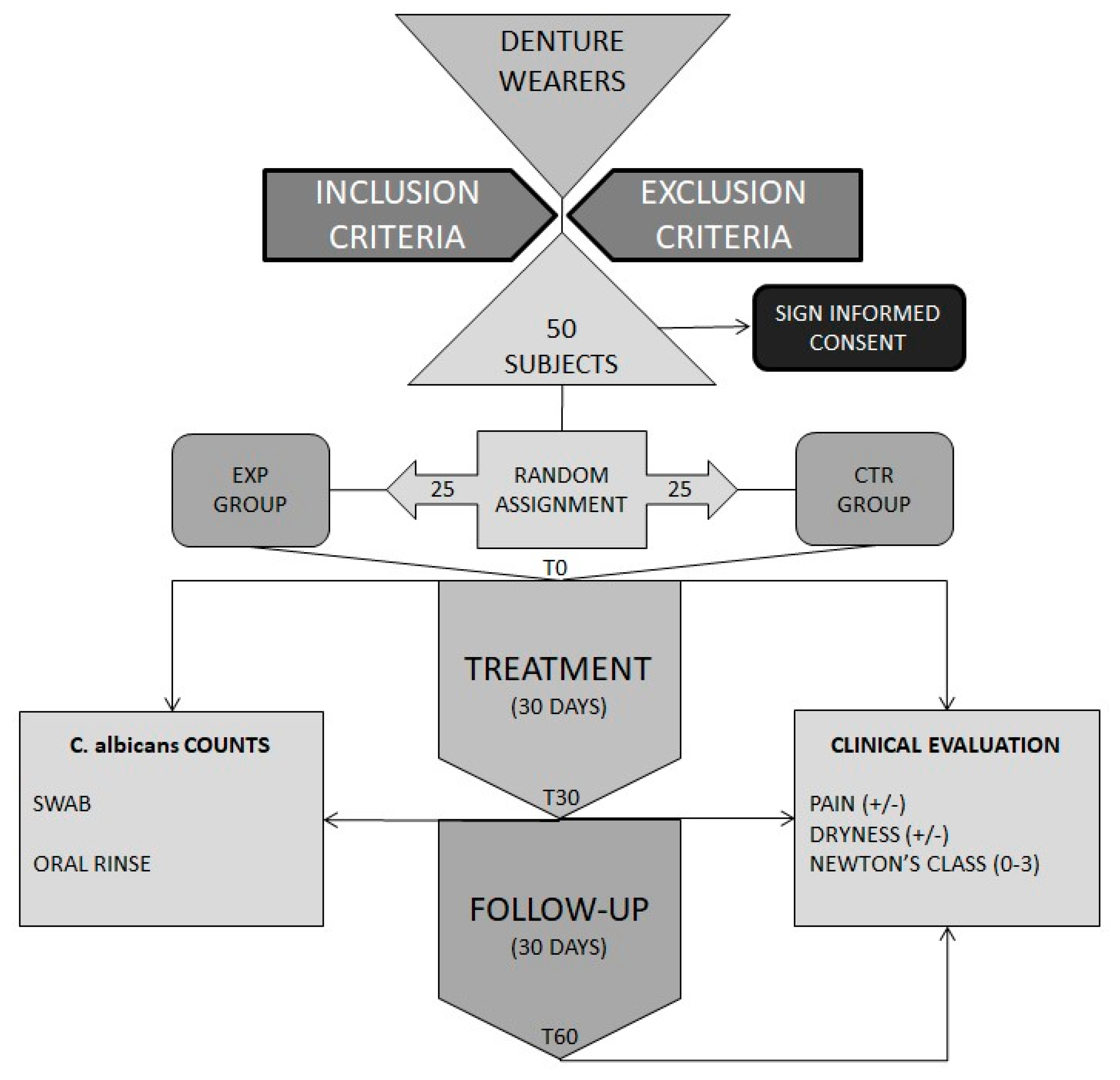
2.1. Studied Subjects and Study Design

Fifty adult human subjects of both sexes were enrolled for this study from a larger population of denture wearers attending or referred to the Department of Oral and Maxillo-Facial Sciences of “Sapienza” University of Rome. Inclusion criteria were as follows: presence of at least one totally edentulous dental arch, presence of a full arch, well-fitting removable acrylic denture, presence of subjective discomfort/pain in relation to denture, and presence of clinical signs of DS. Exclusion criteria were as follows: use of adhesives for denture stabilization, use of antibiotics in the last 30 days, presence of systemic diseases influencing homeostasis of the oral mucosae (i.e., lupus erythematosus, lichen planus, xerostomia) or significantly influencing immunity (malignancies, transplantations requiring immunosuppressive therapy).
Upon enrollment in the study patients were informed of the scope of the study and were asked to sign an informed consent in accordance with criteria of the Helsinki Declaration of 1975, as revised in 2000. The present study was approved by the Ethical Committee of Policlinico Umberto I of Rome (n. 4790) and registered with the trial identification number ISRCTN14751782. A written informed consent form was signed by all the patients before their enrollment.
All patients were instructed to perform scrupulous mechanic denture and oral hygiene daily. More precisely they were instructed to clean the denture with a dedicated brush under a constant flux of tap water and to carefully brush their teeth, the oral mucosae and tongue with their toothbrush. They were also instructed to refrain from wearing the denture 24 h a day and invited not to use oral rinses containing antimicrobial substances. Patients enrolled in the study were subjected to baseline (T-0) clinical evaluation and microbiological sampling and randomly assigned to either the experimental group (25 subjects) or to the control group (25 subjects). Patients assigned to the experimental group (EXP) were invited to perform mechanic denture and oral hygiene as previously explained, and to take 1 tablet of Bactoblis® (Pharmaextracta Spa, Pontenure, Piacenza, Italy), containing 109 CFU of the probiotic strain S. salivarius K12 for 30 days in the evening, just before going to sleep. Patients were instructed to remove the denture and allow the tablet to dissolve in the mouth, and to refrain from drinking for the next 60 min.
Patients assigned to the control group (CTR) were invited to perform mechanic denture and oral hygiene as previously explained for 30 days. All subjects of both groups were instructed not to wear the denture while sleeping and to maintain the denture within a dedicated box during the night. At the end of the period of 30 days all patients were subjected to a second (T-30) clinical evaluation and microbiological sampling, and further instructed to reinstate previous oral and denture hygiene measures for the next 30 days. At the end of this period they were subjected to further clinical evaluation (T-60) (Figure 1).

2.2. Clinical Evaluation

Clinical evaluation included recording of two subjective parameters and one objective index. Subjective feeling of pain in the oral mucosa and dryness of the mouth were recorded for each patient. Answers were recorded as presence (1) or absence (0). Each patient was inspected by an experienced clinician in order to determine the presence and extension of clinical signs of DS according to Newton’s classification [23]. This classification of DS describes three steps of stomatitis according to diffusion and intensity of clinical signs of inflammation. Grade 1: presence of pin-point hyperemic lesions (localized simple inflammation); grade 2: presence of diffuse erythema involving most of the mucosa contacting the denture (generalized simple inflammation); grade 3: presence of inflammation associated with a granular surface of the mucosa contacting the denture (inflammatory papillary hyperplasia).
All clinical evaluations were performed separately by three experienced clinicians (C.P., D.D.N., and L.T.), and calibrated to provide consistent determination of Newton’s class as follows: twenty subjects were selected among those participating in the study. Each examiner independently evaluated and scored each subject; measures were repeated after 24 h on the same subjects. Values recorded by the three examiners at the same time were used to calculate inter-examiner reproducibility. Values recorded by each examiner at different times were used to calculate intra-examiner reproducibility. Inter- and intra-examiner reproducibility was measured through Cohen’s weighted kappa; values obtained suggest an almost perfect agreement among the three examiners [24].

2.3. Microbiological Samples

Microbiological samples for detection and quantification of C. albicans cells were obtained from all patients at T0 and T30 by two methods [25]: (i) by streaking a cotton swab along the apex of the gingival process for a length of 30 mm in areas with signs of DS; the swab was then inserted in a sterile polystyrene tube containing 1 mL of Stuarts transport medium; (ii) by instructing each patient to remove the denture and to rinse vigorously with 10 mL of sterile physiologic saline for 1 min. The rinse was collected in a sterile 50 mL polypropylene tube. Samples were always stored on ice until further processing.

2.4. Quantification of Candida Albicans in Samples

Tubes containing swabs for enumeration of C. albicans cells were vortexed for 1 min before transferring the transport medium to a sterile 1.5 mL polypropylene centrifuge tube. Samples were then centrifuged at 10,000× g at 4 °C for 5 min; the supernatant was removed, and cellular pellets were suspended in 1 mL of sterile physiologic saline. Tubes containing rinses were centrifuged at 10,000× g at 4 °C for 5 min; the supernatant was then discarded, and pellets were suspended in 1 mL of sterile physiologic saline. Tenfold dilutions were prepared from all samples, and 0.1 mL of each sample and of its dilution was plated on BBL Chromagar Candida Medium (Becton Dikinson GmbH, Heidelberg, Germany). Following incubation for 48 h at 37 °C, plates were inspected and colonies of C. albicans (i.e., green colored colonies) were counted. The development of colonies with phenotypes typical of non-albicans Candida spp. was always recorded.

2.5. Statistical Analysis

Differences in mean counts of C. albicans in the swab and rinse cultures between the study and control group at T0 and T30 were analyzed using the Student’s t-test. Differences in terms of DS according to the Newton’s class were compared using the Mann–Whitney U test. Differences between groups in the distribution of pain and dryness were compared using the Fisher’s exact test. Statistical analysis was performed with SPSS 13.0 for Windows. The significance level was set at p ≤ 0.05.

3. Results

3.1. Studied Population

Fifty adult denture wearers of both sexes were selected for this study and randomly assigned to two groups of 25 subjects. The two groups were comparable for age (EXP mean age 74.3 ± 3.8 years, age range 68–82, CTR mean age 73.1 ± 3.6 years, age range 67–83, p = 0.26), and gender (males/females: EXP 13/12, CTR 11/14, p = 0.58) (Table 1). At T0 the EXP and CTR groups were comparable with respect to clinical parameters, and C. albicans counts. With respect to the degree of DS, as determined by Newton’s classification, at T0 the EXP group showed a mean score of 2.1 ± 0.6, and the CTR group a mean score of 2.2 ± 0.6 (p = 0.66). All patients in both groups suffered pain at T0, while 36% of patients of the EXP group suffered from dryness, as compared to 44% in the CTR group (p = 0.77). No significant differences were observed at T0 between groups in the mean C. albicans counts in the swab culture (191.2 CFU/mL in the EXP group vs. 183.8 in the CTR group, p = 0.63) and in the rinse culture (477.1 vs. 463.9, p = 0.64).

3.2. Probiotic S. salivarius Improved Clinical Conditions

Clinical parameters recorded for the two groups at T0, T30 and T60 are reported in Table 1 and Table 2. Although careful mechanic denture and oral hygiene according to a standard protocol was able to significantly improve clinical conditions in the CTR group between T0 and T30, the adjunctive administration of the probiotic preparation Bactoblis® caused a significantly greater improvement of objective clinical conditions (Table 1) in the same time lapse. Moreover, when patients of both groups discontinued therapeutic regimens, and resumed their usual oral hygiene behavior for 30 days (T60), subjects of the EXP group showed further improvement of clinical conditions while those of the CTR group showed partial worsening (Table 2).
With respect to the degree of DS, no significant difference was observed at T0, while differences were statistically significant at T30 (p = 0.005) and at T60 (p < 0.001), with DS being more severe in the CTR group (Table 1). All patients in both groups suffered pain at T0. At T30 7 out of 25 patients reported pain in the EXP group (28.0%) against 14 out of 25 in the CTR group (56.0%, p = 0.085). Differences in pain were significant at T60, with four (16.0%) patients reporting it in the EXP group against 17 in the CTR group (68.0%, p < 0.001, Table 3).
No significant differences in dryness of mouth were observed between groups at T0 (p = 0.77), T30 (p = 0.42), and T60 (p = 0.14, Table 3). The higher number of patients suffering from dryness at T30 and T60 in the CTR group can be explained by the higher starting number registered at T0. No patient in the EXP group reported any adverse effect related to the assumption of Bactoblis®.

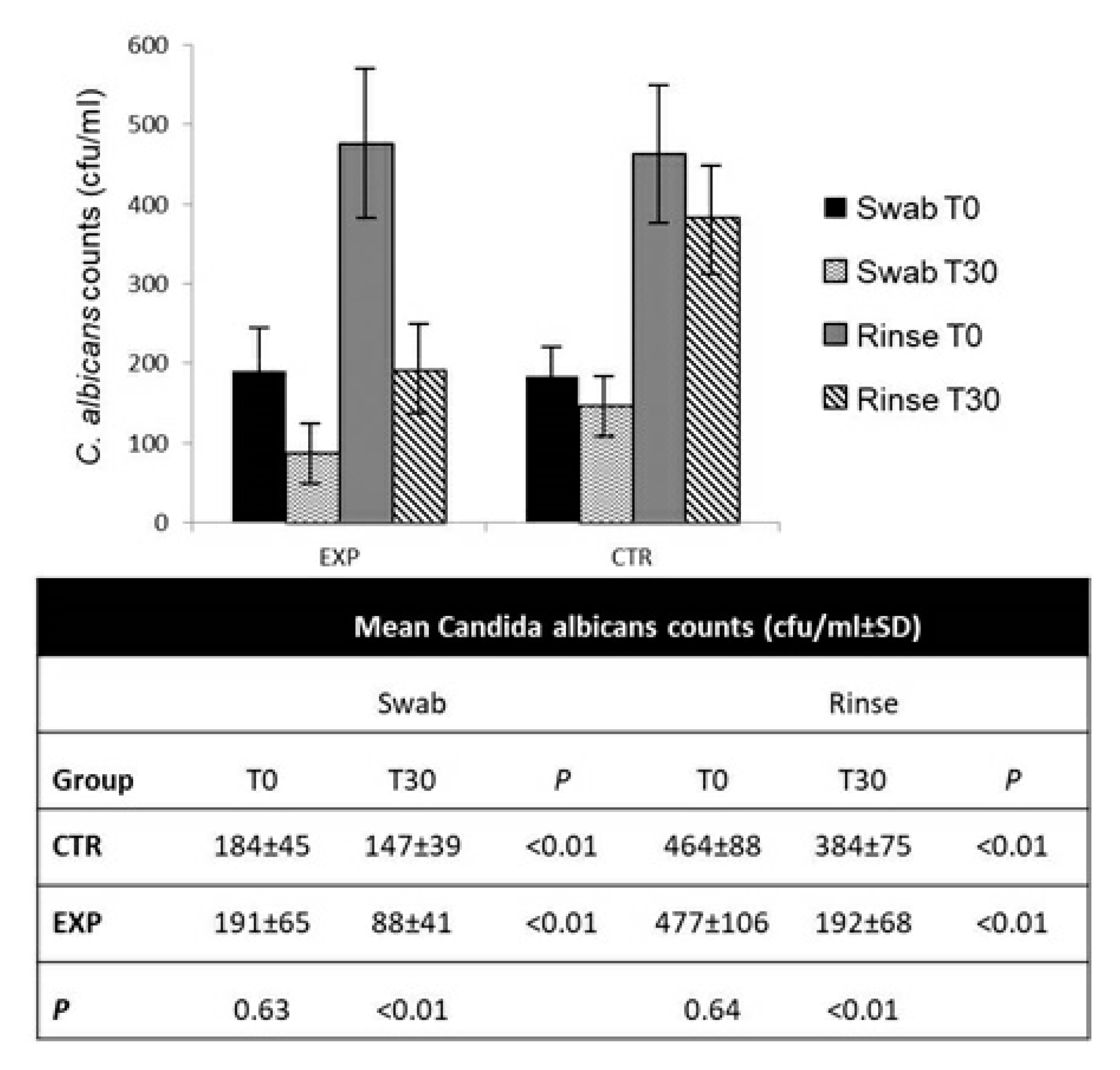
3.3. Probiotic S. salivarius Reduced C. albicans Counts

Counts of C. albicans were obtained from all patients at T0 and T30 by collecting two different samples, a swab streaked for a length of 30 mm on the diseased mucosa underlying the denture and an oral rinse with 10 mL of sterile physiologic saline. As expected from previous reports [25], counts obtained from oral rinses were, on average, 2.5 times higher than those obtained from swabs.
While counts of C. albicans obtained at T0 from samples of patients of both the EXP and CTR group were comparable, analysis of both swab and rinse samples showed that the EXP group underwent a reduction of about 60% of C. albicans counts between T0 and T30, whereas the reduction in the CTR group, although significant, was only about 20% (Figure 2). Colonies with phenotypes typical of non-albicans Candida spp. were observed in many cases but their counts were always low so that they were considered as not relevant for the purposes of the study.

4. Discussion

The present work was designed to preliminarily evaluate the potential effectiveness of combining scrupulous oral and denture hygiene with the administration of a probiotic preparation, containing Streptococcus salivarius K12 for the treatment of DS in patients wearing well-fitting full arch dentures. DS is a multifactorial inflammatory disease of the oral mucosal areas covered by the denture, in which an overgrowth of C. albicans and other opportunistic fungal species normally found in the oral biofilm, is believed to play a key etiologic role [5,6,26]. DS is the cause of multiple visits to the dental office for pain, sensation of dryness of the mouth, discomfort and denture mobilization and is also believed to enhance the risk of pulmonary and other systemic infections [10,11,12]. C. albicans, as many other fungal species, is a normal colonizer of mucosal surfaces including the oral mucosa [8], although in healthy subjects its counts are low. Some clinical conditions, as for example the presence of acrylic bases of removable prostheses and a diminished hygienic skill favored by ageing, are known to promote a significant overgrowth of resident fungal species and of C. albicans in particular, thus promoting the onset of DS [6,7,8,16].
The availability of effective preventative and therapeutic approaches for DS is consequently important. As a direct consequence of the multifactorial nature of DS, its treatment may rely on different approaches, including examination and correction of the denture base, discontinuation of nocturnal wearing of the denture, improvement of oral and denture hygiene measures [13]. Other strategies could be the administration of topical or systemic antifungal agents and disinfectants [14], or the incorporation of slow release antifungal/antimicrobial agents into denture base materials [15]. Despite its multifactorial etiology, the main etiological factor and the principal cause of extensive inflammation observed in DS is certainly the colonization of the denture base and of the underlying mucosa by Candida spp. and other fungal species [26]. Consequently, the administration of antifungal drugs is highly effective in relieving clinical signs and symptoms of DS. Nevertheless, the use of such drugs is not without drawbacks, since short term treatments are affected by high rates of early post-treatment relapse and recurrence [16]. Furthermore, prolonged or repeated therapeutic regimens enhance the risk of systemic adverse effects [17] and are likely to induce antifungal drug resistance in resident fungal strains [18]. It is well known that the administration of probiotics can be beneficial for patients suffering for mucosal candidiasis [27].
Previous studies have already shown that the assumption of some, but not all [22], probiotic preparations can reduce symptoms of DS [19,20,21], and Candida colonization levels in healthy denture wearers [19,20] and in subjects affected by DS [21]. However, common probiotic strains of the genus Lactobacillus have few chances to exert prolonged protective activity since they are not adapted to be oral colonizers. To overcome this limitation, studies were recently performed to isolate probiotic Lactobacillus strains directly from the oral cavity [28]. As an alternative strategy, probiotic strains were searched among normal pioneer colonizers of the oral cavity as S. salivarius [29]. S. salivarius strain K12 is considered as the prototype probiotic strain of this species. It was originally selected for its ability to inhibit Streptococcus pyogens, but it was subsequently shown to possess several other interesting activities, including inhibitory activity on Candida spp. [29,30]. Although data on this matter are still incomplete, several probiotic strains were shown to be able to inhibit biofilm growth, and expression of virulence and resistance genes of C. albicans [20,30,31,32,33].
These considerations prompted us to preliminarily evaluate the effectiveness of a probiotic preparation containing S. salivarius strain K12 in the treatment of DS. Inclusion/exclusion criteria were designed to minimize the influence of factors other than microbial dysbiosis due to inadequate oral hygiene on the presence of clinical signs and symptoms of DS. Subjects wearing not-well-fitting dentures were excluded, as well as subjects affected by diseases altering immunity and oral homeostasis. In the selection of patients, attention was paid to minimize the possible interference of potential confounders. Consequently, in addition to the rigid application of inclusion/exclusion criteria, attention was dedicated to include only patients in apparent good conditions: diabetic patients with high glycemic levels, patients with arterial hypertension not well controlled by drugs (i.e., pressure levels > 140/90 mmHg) and patients smoking >10 cigarettes/day were excluded from the study. Nevertheless diabetes, arterial hypertension and smoking were not included as variables in the statistical analysis.
Due to the preliminarily nature of this study, the CTR group was not designed to receive a placebo; this could have influenced perception of subjective symptoms between the two groups of patients.
As expected, our experimental data confirm that, in the absence of repeated trauma due to inadequacy of the denture base or too prolonged denture wearing, improvement of the oral and denture hygiene procedure reduces clinical signs and symptoms of DS and reduces oral colonization by C. albicans [13,14]. Moreover, data show that the effectiveness of oral and denture hygiene alone is limited in time. In fact, when (after T30) patients of the CTR group were invited to resume their previous habits with the dentures, their clinical conditions worsened significantly. Although microbiological samples were not analyzed at T60, it can be considered plausible, in accordance with data from the literature [25], that clinical relapse was paralleled by an increase in oral C. albicans counts.
Experimental data show that when improved oral and denture hygiene procedures were flanked by the administration of a preparation of the probiotic strain S. salivarius K12, a significantly greater improvement of clinical conditions and a higher reduction of oral C. albicans counts were obtained in the EXP group at T30. Interestingly and unexpectedly, clinical evaluations performed at T60, when patients of the EXP group had discontinued treatment for 30 days and had resumed previous oral and denture hygiene habits, revealed a further improvement of clinical conditions, as opposed to the partial relapse observed in the CTR group. These data suggest that S. salivarius K12 could be able to colonize oral mucosa at least for some weeks after discontinuation of treatment and consequently exhibit a prolonged anti-Candida action. Unfortunately, microbiological sampling was not planned for T60 and consequently this aspect will deserve future further investigations. A prolonged presence of the probiotic strain would be in accordance with the well-known role of S. salivarius as a pioneer colonizer of the human oral cavity [29]. The effective duration of oral colonization by S. salivarius K12 after 30 days of treatment and the consequent duration of its anti-Candida activity will be the object of future investigations using specific molecular methods that were recently made available [34]. Future studies will also be necessary to elucidate the cellular and molecular mechanisms underlying the clinical and microbiological effects of S. salivarius K12 on DS described in this paper.

5. Conclusions

The present work was designed to preliminarily evaluate the clinical and microbiological effects of a probiotic preparation, containing S. salivarius K12, in Candida albicans-positive subjects affected by DS, and the duration of clinical effects.
Data presented in this paper confirm that, at least in subjects wearing well-fitting full-arch dentures and affected by DS, reinforcement of oral and denture hygiene procedures coupled with administration of a probiotic preparation containing S. salivarius K12 is effective in reducing symptoms and clinical signs of DS, and significantly reduces oral colonization by C. albicans. The clinical effects associated with the administration of S. salivarius K12 persist for at least 30 days after discontinuation of treatment. The tested probiotic preparation could consequently prove as a valid and safe aid in combination with the improvement of oral and denture hygiene measures for the treatment of DS.

Author Contributions

C.P., A.P. and L.T. were responsible for study design, C.P., L.T. and D.D.N. performed clinical examinations and collected microbiological samples, C.P. performed microbiological analyses, F.D.N. performed statistical analyses, all authors analyzed and discussed data, and cooperated at writing the paper. All authors have read and agreed to the published version of the manuscript.

Funding

This research received no external funding.

Acknowledgments

Pharmaextracta Spa contributed by supplying patients with the amounts of BactoBlis necessary for the study.

Conflicts of Interest

The authors declare no conflict of interest. Pharmaextracta Spa had no role in the design of the study; in the collection, analyses, or interpretation of data, and in the writing of the manuscript.

References

  1. Bongaarts, J. Human population growth and the demographic transition. Philos. Trans. R. Soc. B Biol. Sci. 2009, 364, 2985–2990. [Google Scholar] [CrossRef] [PubMed] [Green Version]
  2. Griffin, S.O.; Jones, J.A.; Brunson, D.; Griffin, P.M.; Bailey, W.D. Burden of Oral Disease Among Older Adults and Implications for Public Health Priorities. Am. J. Public Health 2012, 102, 411–418. [Google Scholar] [CrossRef] [PubMed]
  3. Meurman, J.H.; Hämäläinen, P. Oral health and morbidity-implications of oral infections on the elderly. Gerodontology 2006, 23, 3–16. [Google Scholar] [CrossRef] [PubMed]
  4. Kassebaum, N.; Bernabe, E.; Dahiya, M.; Bhandari, B.; Murray, C.; Marcenes, W. Global Burden of Severe Tooth Loss. J. Dent. Res. 2014, 93, 20S–28S. [Google Scholar] [CrossRef] [PubMed] [Green Version]
  5. Gendreau, L.; Loewy, Z.G. Epidemiology and Etiology of Denture Stomatitis. J. Prosthodont. 2011, 20, 251–260. [Google Scholar] [CrossRef]
  6. Gauch, L.M.R.; Pedrosa, S.S.; Silveira-Gomes, F.; Esteves, R.A.; Marques-Da-Silva, S.H. Isolation of Candida spp. from denture-related stomatitis in Pará, Brazil. Braz. J. Microbiol. 2017, 49, 148–151. [Google Scholar] [CrossRef]
  7. Chevalier, M.; Ranque, S.; Precheur, I. Oral fungal-bacterial biofilm models in vitro: A review. Med. Mycol. 2017, 56, 653–667. [Google Scholar] [CrossRef] [Green Version]
  8. Morales, D.; Hogan, D.A. Candida albicans Interactions with Bacteria in the Context of Human Health and Disease. PLoS Pathog. 2010, 6, e1000886. [Google Scholar] [CrossRef]
  9. Shirtliff, M.; Peters, B.M.; Jabra-Rizk, M.A. Cross-kingdom interactions: Candida albicans and bacteria. FEMS Microbiol. Lett. 2009, 299, 1–8. [Google Scholar] [CrossRef] [Green Version]
  10. Gendron, R.; Grenier, D.; Maheu-Robert, L.-F. The oral cavity as a reservoir of bacterial pathogens for focal infections. Microbes Infect. 2000, 2, 897–906. [Google Scholar] [CrossRef]
  11. O’Donnell, L.E.; Smith, K.; Williams, C.; Nile, C.J.; Lappin, D.F.; Bradshaw, D.; Lambert, M.; Robertson, D.; Bagg, J.; Hannah, V.; et al. Dentures are a Reservoir for Respiratory Pathogens. J. Prosthodont. 2015, 25, 99–104. [Google Scholar] [CrossRef] [PubMed] [Green Version]
  12. Coulthwaite, L.; Verran, J. Potential pathogenic aspects of denture plaque. Br. J. Biomed. Sci. 2007, 64, 180–189. [Google Scholar] [CrossRef] [PubMed]
  13. Hasan, S.; Kuldeep; Shamimul, H. Denture Stomatitis: A Literature Review. J. Orofac. Health Sci. 2015, 6, 65. [Google Scholar] [CrossRef]
  14. Hilgert, J.B.; Giordani, J.M.D.A.; De Souza, R.F.; Wendland, E.M.; D’Avila, O.P.; Hugo, F.N. Interventions for the Management of Denture Stomatitis: A Systematic Review and Meta-Analysis. J. Am. Geriatr. Soc. 2016, 64, 2539–2545. [Google Scholar] [CrossRef] [PubMed]
  15. Bueno, M.; Urban, V.; Barbério, G.; Da Silva, W.J.; Porto, V.; Pinto, L.; Neppelenbroek, K. Effect of antimicrobial agents incorporated into resilient denture relines on the Candida albicans biofilm. Oral Dis. 2013, 21, 57–65. [Google Scholar] [CrossRef] [PubMed]
  16. Ramage, G.; Tomsett, K.; Wickes, B.L.; Lopez-Ribot, J.L.; Redding, S.W. Denture stomatitis: A role for Candida biofilms. Oral Surg. Oral Med. Oral Pathol. Oral Radiol. Endodontol. 2004, 98, 53–59. [Google Scholar] [CrossRef] [PubMed]
  17. Perfect, J.R.; Lindsay, M.H.; Perfect, J.R.; Drew, R.H. Adverse Drug Reactions to Systemic Antifungals. Drug Saf. 1992, 7, 323–363. [Google Scholar] [CrossRef]
  18. White, T.C.; Marr, K.A.; Bowden, R.A. Clinical, Cellular, and Molecular Factors That Contribute to Antifungal Drug Resistance. Clin. Microbiol. Rev. 1998, 11, 382–402. [Google Scholar] [CrossRef] [Green Version]
  19. Ishikawa, K.; Mayer, M.P.A.; Miyazima, T.Y.; Matsubara, V.H.; Silva, E.G.; Paula, C.R.; Campos, T.T.; Nakamae, A. A Multispecies Probiotic Reduces Oral Candida Colonization in Denture Wearers. J. Prosthodont. 2014, 24, 194–199. [Google Scholar] [CrossRef]
  20. Song, Y.-G.; Lee, S.-H. Inhibitory effects of Lactobacillus rhamnosus and Lactobacillus casei on Candida biofilm of denture surface. Arch. Oral Biol. 2017, 76, 1–6. [Google Scholar] [CrossRef]
  21. Lee, X.; Vergara, C.; Lozano, C.P. Severity of Candida-associated denture stomatitis is improved in institutionalized elders who consume Lactobacillus rhamnosus SP1. Aust. Dent. J. 2019, 64, 229–236. [Google Scholar] [CrossRef] [PubMed]
  22. Sutula, J.; Coulthwaite, L.; Thomas, L.; Verran, J. The effect of a commercial probiotic drink on oral microbiota in healthy complete denture wearers. Microb. Ecol. Health Dis. 2012, 23, 175. [Google Scholar] [CrossRef]
  23. Newton, A.V. Denture sore mouth. A possible etiology. Br. Dent. J. 1962, 112, 357–360. [Google Scholar]
  24. Koch, J.R.L.G. The Measurement of Observer Agreement for Categorical Data. Biometrics 1977, 33, 159. [Google Scholar] [CrossRef] [Green Version]
  25. Tooyama, H.; Matsumoto, T.; Hayashi, K.; Kurashina, K.; Kurita, H.; Uchida, M.; Kasuga, E.; Honda, T. Candida concentrations determined following concentrated oral rinse culture reflect clinical oral signs. BMC Oral Health 2015, 15, 150. [Google Scholar] [CrossRef] [PubMed] [Green Version]
  26. Zomorodian, K.; Haghighi, N.N.; Rajaee, N.; Pakshir, K.; Tarazooie, B.; Vojdani, M.; Sedaghat, F.; Vosoghi, M.; Vossoughi, M. Assessment ofCandidaspecies colonization and denture-related stomatitis in complete denture wearers. Med. Mycol. 2011, 49, 208–211. [Google Scholar] [CrossRef] [Green Version]
  27. Salazar, S.B.; Simões, R.S.; Pedro, N.A.; Pinheiro, M.J.; Carvalho, M.F.N.N.; Mira, N.P. An Overview on Conventional and Non-Conventional Therapeutic Approaches for the Treatment of Candidiasis and Underlying Resistance Mechanisms in Clinical Strains. J. Fungi 2020, 6, 23. [Google Scholar] [CrossRef] [Green Version]
  28. Rossoni, R.D.; De Barros, P.P.; De Alvarenga, J.A.; Ribeiro, F.D.C.; Velloso, M.D.S.; Fuchs, B.B.; Mylonakis, E.; Jorge, A.O.C.; Junqueira, J.C. Antifungal activity of clinical Lactobacillus strains against Candida albicans biofilms: Identification of potential probiotic candidates to prevent oral candidiasis. Biofouling 2018, 34, 212–225. [Google Scholar] [CrossRef] [Green Version]
  29. Wescombe, P.A.; Hale, J.D.; Heng, N.C.; Tagg, J.R. Developing oral probiotics fromStreptococcus salivarius. Futur. Microbiol. 2012, 7, 1355–1371. [Google Scholar] [CrossRef]
  30. Hu, L.; Mao, Q.; Zhou, P.; Lv, X.; Hua, H.; Yan, Z. Effects of Streptococcus salivarius K12 with nystatin on oral candidiasis—RCT. Oral Dis. 2019, 25, 1573–1580. [Google Scholar] [CrossRef]
  31. Aarti, C.; Ameer, K.; Varghese, R.; Arasu, M.V.; Agastian, P.; Al-Dhabi, N.A.; Ilavenil, S.; Choi, K.C. In vitro investigation on probiotic, anti-Candida, and antibiofilm properties of Lactobacillus pentosus strain LAP1. Arch. Oral Biol. 2018, 89, 99–106. [Google Scholar] [CrossRef] [PubMed]
  32. Kim, H.; Kang, S.-S. Antifungal activities against Candida albicans, of cell-free supernatants obtained from probiotic Pediococcus acidilactici HW01. Arch. Oral Biol. 2019, 99, 113–119. [Google Scholar] [CrossRef] [PubMed]
  33. Oliveira, V.; Dos Santos, S.S.F.; Silva, C.; Jorge, A.; Leão, M. Lactobacillus is able to alter the virulence and the sensitivity profile of Candida albicans. J. Appl. Microbiol. 2016, 121, 1737–1744. [Google Scholar] [CrossRef] [PubMed]
  34. Reid, P.; Heng, N.C.; Hale, J.D.; Krishnan, D.; Crane, J.; Tagg, J.R.; Milne, T.J. A TaqMan™-based quantitative PCR screening assay for the probiotic Streptococcus salivarius K12 based on the specific detection of its megaplasmid-associated salivaricin B locus. J. Microbiol. Methods 2020, 170, 105837. [Google Scholar] [CrossRef]
Figure 1. Experimental study design.
Applsci 10 03002 g001
Figure 2. Candida albicans counts before (T0) and after 30 days treatment regimens (T30) with (EXP) and without (CTR) probiotic S. salivarius BLIS®K12. C. albicans counts obtained by two distinct sampling procedures (swab and rinse) are represented in the plot and reported in the explanatory table as colony forming units/ml (CFU/mL) means (±standard deviation). Significance of differences between single groups of data was calculated by the Student T test and is reported in the table as values of P.
Applsci 10 03002 g002
Table 1. Main characteristics of patients enrolled in the study, divided according to treatment group.
Group
ExperimentalControl
Age (mean ± SD)74.3 ± 3.873.1 ± 3.6
Age range68–8267–83
Gender (Males/Females)13/1211/14
Smokers a (%)2420
Diabetes b (%)3628
Arterial hypertension c (%)6452
a Only subjects smoking <10 cigarettes/day were enrolled in the study; b only subjects with diabetes in good control were enrolled in the study; c only patients under therapy and with arterial pressure level <140/90 mmHg were enrolled in the study.
Table 2. Distribution of denture stomatitis (DS) (according to Newton’s classification) between the study and control groups at T0 (p = 0.665), T30 (p = 0.005) and T60 (p < 0.001).
Group
ControlExperimental
CountColumn N %CountColumn N %
Newton class at T0000.0%00.0%
128.0%416.0%
21768.0%1560.0%
3624.0%624.0%
Newton class at T300624.0%1560.0%
11664.0%1040.0%
2312.0%00.0%
300.0%00.0%
Newton class at T600416.0%1872.0%
11352.0%728.0%
2832.0%00.0%
300.0%00.0%
Table 3. Distribution of pain and dryness between groups before treatment (T0), after 30 days of treatment (T30) and after 30 days after treatment had stopped (T60).
Group
ControlExperimental
CountColumn N %CountColumn N %
Pain T0No00.0%00.0%
Yes25100.0%25100.0%
Pain T30No1144.0%1872.0%
Yes1456.0%728.0%
Pain T60No832.0%2184.0%
Yes1768.0%416.0%
Dryness T0No1456.0%1664.0%
Yes1144.0%936.0%
Dryness T30No2080.0%2392.0%
Yes520.0%28.0%
Dryness T60No1872.0%2392.0%
Yes728.0%28.0%

