Article

Ag-Sensitized NIR-Emitting Yb3+-Doped Glass-Ceramics

1
CNR-ISP Istituto di Scienze Polari, c/o campus scientifico Università Ca’ Foscari Venezia, via Torino 155, 30172 Mestre-Venezia, Italy
2
Dipartimento di Scienze Molecolari e Nanosistemi, Università Ca’ Foscari Venezia, via Torino 155, 30172 Mestre-Venezia, Italy
3
Museo Storico della Fisica e Centro Studi e Ricerche “Enrico Fermi”, Piazza del Viminale 1, 00184 Roma, Italy
4
CNR-IFAC Istituto di Fisica Applicata Nello Carrara, Via Madonna del Piano 10, 50019 Sesto Fiorentino, Firenze, Italy
5
Division of Materials Science, Department of Engineering Sciences and Mathematics, Luleå University of Technology, 971 87 Luleå, Sweden
*
Author to whom correspondence should be addressed.
Appl. Sci. 2020, 10(6), 2184; https://doi.org/10.3390/app10062184
Submission received: 11 February 2020 / Revised: 9 March 2020 / Accepted: 14 March 2020 / Published: 23 March 2020
(This article belongs to the Special Issue Photonic Glass-Ceramics: Fabrication, Properties and Applications)

Abstract

:

Featured Application

downshifting layers for silicon solar cells, NIR emitting devices and lasers.

Abstract

The optical photoluminescent (PL) emission of Yb3+ ions in the near infrared (NIR) spectral region at about 950–1100 nm has many potential applications, from photovoltaics to lasers and visual devices. However, due to their simple energy-level structure, Yb3+ ions cannot directly absorb UV or visible light, putting serious limits on their use as light emitters. In this paper we describe a broadband and efficient strategy for sensitizing Yb3+ ions by Ag codoping, resulting in a strong 980 nm PL emission under UV and violet-blue light excitation. Yb-doped silica–zirconia–soda glass–ceramic films were synthesized by sol-gel and dip-coating, followed by annealing at 1000 °C. Ag was then introduced by ion-exchange in a molten salt bath for 1 h at 350 °C. Different post-exchange annealing temperatures for 1 h in air at 380 °C and 430 °C were compared to investigate the possibility of migration/aggregation of the metal ions. Studies of composition showed about 1–2 wt% Ag in the exchanged samples, not modified by annealing. Structural analysis reported the stabilization of cubic zirconia by Yb-doping. Optical measurements showed that, in particular for the highest annealing temperature of 430 °C, the potential improvement of the material’s quality, which would increase the PL emission, is less relevant than Ag-aggregation, which decreases the sensitizers number, resulting in a net reduction of the PL intensity. However, all the Ag-exchanged samples showed a broadband Yb3+ sensitization by energy transfer from Ag aggregates, clearly attested by a broad photoluminescence excitation spectra after Ag-exchange, paving the way for applications in various fields, such as solar cells and NIR-emitting devices.

Graphical Abstract

1. Introduction

Rare earth ions (RE3+) have many optical applications due to their unique spectral properties, in relation to the features of their electronic energy levels, covering UV, visible and IR [1]. This makes them excellent candidates for lighting [2,3,4], displays [5], biosensing [6,7,8], optical amplification [9], anticounterfeiting [10,11] and solar cells [12,13,14]. Among them, Yb3+ ions provide near infrared (NIR) emission around 950–1100 nm, which is interesting for many of the previously cited applications. However, the absorption and emission of Yb3+ ions are due to transitions between only two levels: 2F7/2 (ground state) and 2F5/2 (excited state). Therefore, Yb3+ ions are not able to directly absorb UV or visible photons, and other lanthanides are often added as codopants to provide an alternative excitation path by energy transfer, and sometimes to offer the additional possibility of photon multiplication by quantum cutting [15,16,17,18,19]. Nevertheless, codoping with other RE3+ ions still do not solve the limited excitation and absorption bandwidths and their small excitation cross sections, which are major limitations for their implementation in thin film downconverting devices and other specific applications.
In the past, silicon [20,21,22,23] or silver aggregates [24,25,26,27] were proven as broadband efficient sensitizers for Er3+ ions. Ag sensitization was successfully observed also for other RE3+ ions (Eu, Tb, Yb, Dy, Sm) [28,29,30,31,32,33,34,35,36]. In this paper, we will further investigate the interaction between Ag aggregates and Yb3+, analyzing the role of the glass-ceramic (GC) matrix and its crystalline structure on the optical properties of the composite material. A GC is a homogeneous dispersion of ceramic nanocrystals in a glass. EXAFS studies suggest that RE3+ ions should be preferentially located in the nanocrystals [37], resulting in better spectroscopic properties. Additionally, zirconia has lower phonon energy and higher refractive index than silica [38]. By combining Ag enhancement with Yb3+-doped silica–zirconia GC, the possibility to obtain a more efficient optical device is discussed in this paper. Compositional, structural and optical measurements were combined for retrieving information on the role and nature of Ag-aggregates controlled by Ag+↔Na+ ion exchange, followed by annealing at different temperatures, and their interaction with Yb3+ ions for obtaining efficient sensitization and photoluminescence boosting.

2. Materials and Methods

Silica–zirconia (70%–30%) GC films were synthesized by following a sol–gel and dip-coating procedure, starting from Tetraethyl orthosilicate Si(OC2H5)4 (TEOS) and zirconium propoxide Zr(OC3H7)4 (ZPO) precursors, as described in [32]. As usual in sol–gel synthesis, all dopants were calculated and put in addition to this composition: 5 mol% Na was added as sodium acetate for allowing the introduction of Ag by ion exchange; 4 mol% YbNO3 was added for Yb-doped samples. In short, three solutions were prepared as follows:
(1)
TEOS, ethanol (EtOH), H2O and HCl (TEOS:HCl:H2O:EtOH = 1:0.01:2:25), adding 4 mol% YbNO3 for Yb-doped samples;
(2)
ZPO, acetylacetone (Acac), ethanol (ZPO:Acac:EtOH = 1:0.5:50);
(3)
sodium acetate in methanol (60 mg/mL)
Solutions (1) and (2) were mixed together, then (3) was added dropwise and left stirring overnight (16 h). Multi-layer films were deposited on fused silica, with each layer annealed in air at 700 °C for 3 min. After 10 layers, 400 nm total thickness was achieved. A final annealing in air at 1000 °C for 1 h induced the crystallization of zirconia in the glass-ceramic (GC) matrix.
Undoped (label GC0) and 4 mol% Yb-doped (label GC4) films were then immersed for 1 h in a molten salt bath (1 mol% AgNO3 in NaNO3) at 350 °C to obtain Ag+⇿Na+ ion exchange (label GC0A and GC4A). Finally, post-exchange annealing in air at 380 °C (GC0B and GC4B) and 430 °C (GC0C and GC4C) were used for controlling Ag migration and aggregation [32].
The film composition was studied by Rutherford backscattering spectrometry (RBS), by 2.2 MeV 4He+ beam at 160° backscattering angle in IBM geometry, and simulating the experimental spectra by the RUMP code [39]. Areal density (the natural unit of measurement for RBS) to film thickness conversion was obtained, considering a molar density of the film equal to a weighted average between silica (2.00 g cm-3) and zirconia (5.68 g cm-3), according to the nominal stoichiometric composition of the matrix (70 SiO2 – 30 ZrO2), which was confirmed by RBS analysis.
The crystal phase identification was obtained by X-ray diffraction (XRD) at room temperature, with an X’Pert PRO diffractometer (Panalytical) using a Cu anode (Kα, λ = 1.54056 Å). Diffractograms were collected in Bragg–Brentano geometry in the 2θ range 10°–100°. Nanocrystal size was determined by Line Broadening Analysis (LBA) [40] by the Warren–Averbach method.
Photoluminescence excitation (PLE) and emission (PL) spectra were recorded by a FLS980 (Edinburgh Instruments). The excitation was provided by a xenon lamp coupled to a double-grating monochromator, while the emission from the sample was analyzed by a double-grating monochromator and recorded by a photon counting R928P (Hamamatsu) cooled at –20 °C (visible region) or by a photon counting R5509-73 (Hamamatsu) cooled at −80 °C (NIR region). The excitation wavelength for recording PL emission spectra was 330 nm, which is not absorbed by Yb3+ ions, while the emission wavelength for acquiring PL excitation spectra was set at 975 nm, the peak wavelength for Yb3+ ions 2F5/22F7/2 transition.

3. Results and discussion

The film composition was confirmed by RBS analysis in agreement with the nominal values for Si, Zr and Yb. The Ag concentration depth profile (see examples in Figure 1) revealed a reduction from about 2 mol% (at surface) to 1 mol% (inner part of the film) both for the Ag-exchanged and the annealed samples. This indicates a small silver mobility in the glass ceramic matrix. Figure 1 presents the RBS spectra for the undoped samples before and after Ag-exchange, and the RUMP simulation of the latter (Figure 1a). In Figure 1b, a selected region corresponding to the Zr and Ag signals is reported, including a depth profile scale for the Zr signal, obtained from the RBS signal combined with the weighted average molar density between silica and zirconia, as described in the Experimental section. In Figure 1c, the calculated Ag depth profiling is reported for the samples GC0A, GC0B and GC0C. Post-exchange heat treatment changes only slightly the Ag profile, with no difference between sample GC0B and GC0C, within the experimental uncertainty of the RBS.
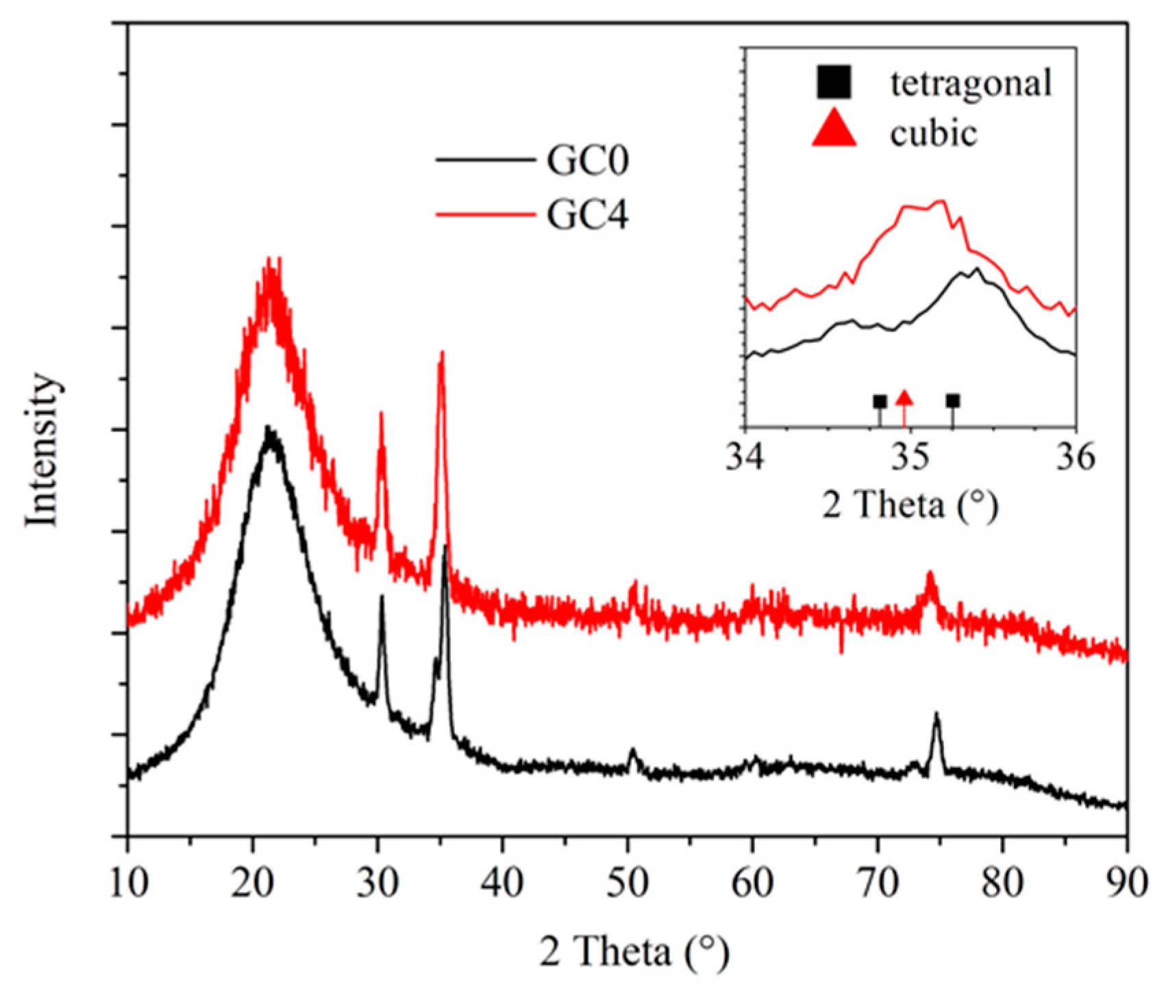
XRD reflections before Ag+↔Na+ ion exchange are reported in Figure 2, showing the presence of ZrO2 tetragonal nanocrystals in the undoped samples and cubic nanocrystals in Yb-doped films, attested by a double peak (tetragonal) or a single peak (cubic) at 2θ ≈ 35.5° and 2θ ≈ 75°. Noteworthy, deeper analysis which goes beyond the scope of this paper can be further exploited to corroborate the observed structural behavior and to quantify the relative phases [41]. The effect of Ag-exchange and subsequent annealing was not observed on XRD spectra. Indeed, it should be noted that crystal formation in similar materials occurs at 1000 °C. Previous studies highlighted that below that temperature, the sol-gel remains in a glassy state (see, for example, Zur et al. [19]). Ag-exchange and the following annealing processes were done at 350 °C and up to 430 °C. Therefore, it can be reasonably concluded that the energies involved at these treatment temperatures are not enough for modifying the crystalline features of the glass-ceramic material. Finally, proper line broadening analysis (LBA) of XRD diffraction peaks [40] obtained by the Warren–Averbach method showed that nanocrystal size was about 14 nm for tetragonal zirconia in our undoped samples (GC0), and about 12 nm for cubic zirconia in Yb-doped samples (GC4).
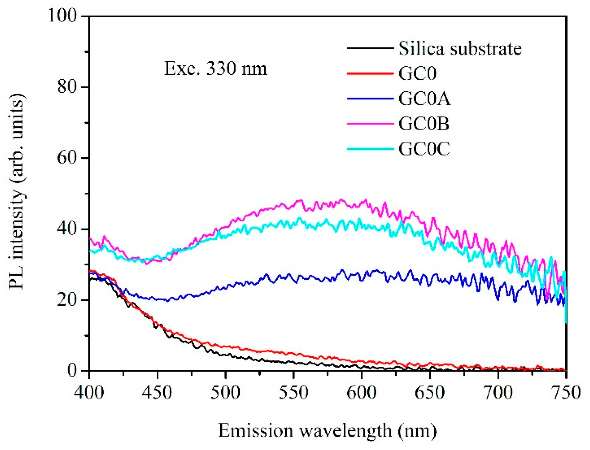
It is known from previous studies that the optical properties of silver-doped silicate glasses have broad luminescence emissions under UV light [42,43,44,45] related to isolated Ag+ ions (Em. 330–370 nm), Ag+–Ag+ pairs (Em. 430–450 nm), formation of (Ag3)2+ trimers, multimers, and aggregates (Em. 550–650 nm and above). Studies of the PL emission under 280 nm excitation before (GC0) and after Ag-exchange (GC0A) and 430 °C annealing (GC0C) have been reported elsewhere [32], showing a main peak at 425 nm, reasonably related to Ag+–Ag+ pairs, with a broad emission extending to the red spectral region, attributed to trimers and multimers. The modification of the curves after annealing is related to the evolution of the Ag species within the material. After annealing, the maximum peak at 425 nm slightly decreases, while the red emission contribution increases as a consequence of the decreasing of the number of dimers towards the formation of multimers and small aggregates. The optical properties of these Ag-aggregates can be better investigated by 330 nm excitation, which is reported in Figure 3. Additionally, there is also higher interest in analyzing near UV and blue excitation because sources like lasers, LEDs, or lamps for these wavelengths are much more available and cheaper than deeper UV light sources. As expected, the PL spectra of Ag-exchanged GC samples under 330 nm excitation, before (GC0A) and after annealing at 380 °C (GC0B) and 430 °C (GC0C), show a broadband emission related to multimers and aggregates, covering up to 750 nm. As previously observed [32], the intensity of this emission increases by annealing, as a possible consequence of matrix recovery with the removal of non-radiative defects, and of the increase of the number of multimers at the expense of single ions and dimers, at least for 380 °C annealing. A further thermal treatment at 430 °C seems detrimental with respect to 380 °C annealing. The reduction of PL intensity suggests a decrease in the effective number of emitting species, which is corroborated by the following PL analysis in the NIR spectral range.
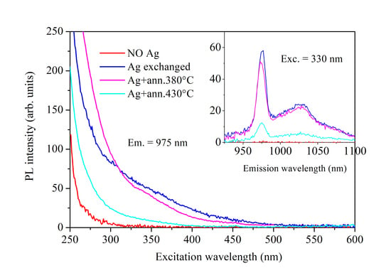
The NIR PL emission under 330 nm excitation is reported in Figure 4b for Yb3+-doped GC samples before Ag-exchange (GC4), after exchange (GC4A), and after annealing at 380 °C (GC4B) and 430 °C (GC4C). The direct excitation of Yb3+ ions by 330 nm light is completely absent. After Ag ion exchange, a strong Yb3+ PL emission peak at 975 nm is detected, possibly due to dimers, multimers or small aggregates formed during the ion exchange process, which act as efficient sensitizers. In agreement with the previous PL analysis of Ag aggregates, annealing is expected to improve the quality of the matrix, which should increase the PL intensity. However, annealing also decreases the number of sensitizers due to the formation of multimers and bigger aggregates, and this additionally increases the average distance between them and Yb3+ ions, reducing the efficiency of energy transfer. The overall combination of these positive and negative effects is almost balanced at 380 °C, while it results in a significant decrease in the NIR PL signal at 430 °C. Therefore, Ag as-exchanged samples are actually the best performing NIR emitters.
Deeper investigation of the excitation behavior of Yb3+-doped GC samples, before and after Ag-exchange and annealing, is reported in Figure 4a. The intensity of the PLE curves agrees well with the PL analysis described above, with a further remarkable feature: the possibility for efficient and broadband excitation on Ag-exchanged samples, covering the whole spectral range from UV to blue, up to about 500 nm. On the contrary, the PLE spectrum of Yb3+-doped samples before Ag-exchange (GC4) gives no signal above 300 nm excitation. Below that wavelength, the possible excitation of Yb3+ ions is due to the occurrence of charge-transfer processes from the matrix itself.

4. Conclusions

The synthesis and characterization of efficient NIR emitting glass-ceramic films is presented and discussed. Yb3+-doped silica-zirconia-soda materials are prepared by sol-gel and dip-coating. Annealing in air at 1000 °C induces the precipitation of zirconia nanocrystals. Different crystalline phases have been observed, depending on the presence of the RE dopant: tetragonal for undoped, and cubic for Yb3+-doped glass-ceramics. The following Ag+↔Na+ ion exchange allows the introduction of Ag species as dimers, multimers, and aggregates in the film, which can be excited in the UV spectral region, and act as sensitizers for Yb3+ ions. As a consequence, Yb3+ NIR emission around 950–1100 nm is significantly enhanced after Ag ion exchange, not only in intensity, but also in the broadness of the excitation band, covering spectral regions not allowed for the direct absorption of Yb3+ ions. Ag-sensitized Yb3+-doped films could have applications as spectral downshifters for photovoltaic (PV) solar cells and NIR light-emitting sources. Furthermore, the high versatility and optical quality of sol-gel glass-ceramic waveguides make these materials also suitable for realizing solar concentrators and integrated optical devices, such as optical amplifiers and lasers.

Author Contributions

Conceptualization by F.E.; data acquisition and analysis by F.E., E.C., T.F., P.R., E.T., A.V.; resources by F.E., E.C., P.R., G.C.R., E.T., A.V.; writing by F.E.; funding acquisition by F.E., G.C.R., A.V. All authors have read and agreed to the published version of the manuscript.

Funding

The research was partially supported by Centro Fermi through the MiFo project and the Italy-South Africa PLESC bilateral project (Ministero Affari Esteri e Coop. Internazionale, MAECI). F.E. acknowledges VINNOVA for support through the Nano2Solar project (Vinnmer Marie Curie Incoming, Ref. N. 2016-02011), and A.V. acknowledges the Knut & Alice Wallenberg Foundation and the Kempe Foundation, the European Union’s Horizon 2020 R&I (grant agreement N. 654002), and INFN-LNL, Legnaro, Italy for RBS analyses.

Conflicts of Interest

The authors declare no conflicts of interest.

References

  1. Spectroscopic Properties of Rare Earths in Optical Materials; Liu, G.; Jacquier, B. (Eds.) Springer-Verlag: Berlin, Germany, 2006; ISBN 978-3-540-28209-9. [Google Scholar]
  2. Jia, X.; Puthen-Veettil, B.; Xia, H.; Yang, T.C.J.; Lin, Z.; Zhang, T.; Wu, L.; Nomoto, K.; Conibeer, G.; Perez-Wurfl, I. All-silicon tandem solar cells: Practical limits for energy conversion and possible routes for improvement. J. Appl. Phys. 2016, 119, 233102. [Google Scholar] [CrossRef]
  3. Lin, Y.C.; Karlsson, M.; Bettinelli, M. Inorganic phosphor materials for lighting. Top. Curr. Chem. 2016, 374, 1–47. [Google Scholar] [CrossRef] [Green Version]
  4. Marin, R.; Sponchia, G.; Riello, P.; Sulcis, R.; Enrichi, F. Photoluminescence properties of YAG:Ce 3+,Pr 3+ phosphors synthesized via the Pechini method for white LEDs. J. Nanoparticle Res. 2012, 14. [Google Scholar] [CrossRef]
  5. Kim, C.H.; Kwon, I.E.; Park, C.H.; Hwang, Y.J.; Bae, H.S.; Yu, B.Y.; Pyun, C.H.; Hong, G.Y. Phosphors for plasma display panels. J. Alloys Compd. 2000, 311, 33–39. [Google Scholar] [CrossRef]
  6. Liu, Y.; Tu, D.; Zhu, H.; Chen, X. Lanthanide-doped luminescent nanoprobes: Controlled synthesis, optical spectroscopy, and bioapplications. Chem. Soc. Rev. 2013, 42, 6924–6958. [Google Scholar] [CrossRef] [PubMed]
  7. Enrichi, F.; Riccò, R.; Meneghello, A.; Pierobon, R.; Cretaio, E.; Marinello, F.; Schiavuta, P.; Parma, A.; Riello, P.; Benedetti, A. Investigation of luminescent dye-doped or rare-earth-doped monodisperse silica nanospheres for DNA microarray labelling. Opt. Mater. (Amst.) 2010, 32. [Google Scholar] [CrossRef]
  8. Enrichi, F. Luminescent amino-functionalized or erbium-doped silica spheres for biological applications. Annals of the New York Academy of Sciences 2008, 1130, 262–266. [Google Scholar] [CrossRef] [PubMed]
  9. Desurvire, E. Erbium-Doped Fiber Amplifiers: Principles and Applications; John Wiley & Sons Inc.: New York, NY, USA, 1994; ISBN 0471589772. [Google Scholar]
  10. Moretti, E.; Pizzol, G.; Fantin, M.; Enrichi, F.; Scopece, P.; Nuñez, N.O.; Ocaña, M.; Benedetti, A.; Polizzi, S. Deposition of silica protected luminescent layers of Eu:GdVO4 nanoparticles assisted by atmospheric pressure plasma jet. Thin Solid Films 2016, 598. [Google Scholar] [CrossRef] [Green Version]
  11. Moretti, E.; Pizzol, G.; Fantin, M.; Enrichi, F.; Scopece, P.; Ocaña, M.; Polizzi, S. Luminescent Eu-doped GdVO4 nanocrystals as optical markers for anti-counterfeiting purposes. Chem. Pap. 2017, 71. [Google Scholar] [CrossRef]
  12. Chiappini, A.; Zur, L.; Enrichi, F.; Boulard, B.; Lukowiak, A.; Righini, G.C.; Ferrari, M. Glass ceramics for frequency conversion. In Solar Cells and Light Management: Materials, Strategies and Sustainability; Enrichi, F., Righini, G.C., Eds.; Elsevier: Amsterdam, The Netherlands, 2019; ISBN 9780081027622. [Google Scholar]
  13. Trupke, T.; Green, M.A.; Würfel, P. Improving solar cell efficiencies by down-conversion of high-energy photons. J. Appl. Phys. 2002, 92, 1668–1674. [Google Scholar] [CrossRef]
  14. Richards, B.S. Enhancing the performance of silicon solar cells via the application of passive luminescence conversion layers. Sol. Energy Mater. Sol. Cells 2006, 90, 2329–2337. [Google Scholar] [CrossRef]
  15. Enrichi, F.; Armellini, C.; Belmokhtar, S.; Bouajaj, A.; Chiappini, A.; Ferrari, M.; Quandt, A.; Righini, G.C.; Vomiero, A.; Zur, L. Visible to NIR downconversion process in Tb3+-Yb3+codoped silica-hafnia glass and glass-ceramic sol-gel waveguides for solar cells. J. Lumin. 2018, 193, 44–50. [Google Scholar] [CrossRef]
  16. Bouajaj, A.; Belmokhtar, S.; Britel, M.R.; Armellini, C.; Boulard, B.; Belluomo, F.; Di Stefano, A.; Polizzi, S.; Lukowiak, A.; Ferrari, M.; et al. Tb3+/Yb3+ codoped silica-hafnia glass and glass-ceramic waveguides to improve the efficiency of photovoltaic solar cells. Opt. Mater. (Amst.) 2016, 52. [Google Scholar] [CrossRef] [Green Version]
  17. Lakshminarayana, G.; Qiu, J. Near-infrared quantum cutting in RE3+/Yb3+ (RE = Pr, Tb, and Tm): GeO2-B2O3-ZnO-LaF3 glasses via downconversion. J. Alloys Compd. 2009, 481, 582–589. [Google Scholar] [CrossRef]
  18. Katayama, Y.; Tanabe, S. Downconversion for 1µm luminescence in lanthanide and Yb3+-codoped phosphors. In Solar Cells and Light Management: Materials, Strategies and Sustainability; Enrichi, F., Righini, G.C., Eds.; Elsevier: Amsterdam, The Netherlands, 2019; ISBN 9780081027622. [Google Scholar]
  19. Zur, L.; Armellini, C.; Belmokhtar, S.; Bouajaj, A.; Cattaruzza, E.; Chiappini, A.; Coccetti, F.; Ferrari, M.; Gonella, F.; Righini, G.C.; et al. Comparison between glass and glass-ceramic silica-hafnia matrices on the down-conversion efficiency of Tb3+/Yb3+ rare earth ions. Opt. Mater. (Amst.) 2019, 87, 102–106. [Google Scholar] [CrossRef]
  20. Falconieri, M.; Borsella, E.; De Dominicis, L.; Enrichi, F.; Franzò, G.; Priolo, F.; Iacona, F.; Gourbilleau, F.; Rizk, R. Study of the Si-nanocluster to Er3+ energy transfer dynamics using a double-pulse experiment. Opt. Mater. (Amst.) 2006, 28. [Google Scholar] [CrossRef]
  21. Gourbilleau, F.; Dufour, C.; Levalois, M.; Vicens, J.; Rizk, R.; Sada, C.; Enrichi, F.; Battaglin, G. Room-temperature 1.54 μm photoluminescence from Er-doped Si-rich silica layers obtained by reactive magnetron sputtering. J. Appl. Phys. 2003, 94. [Google Scholar] [CrossRef]
  22. Enrichi, F.; Mattei, G.; Sada, C.; Trave, E.; Pacifici, D.; Franzò, G.; Priolo, F.; Iacona, F.; Prassas, M.; Falconieri, M.; et al. Study of the energy transfer mechanism in different glasses co-doped with Si nanoaggregates and Er3+ ions. Opt. Mater. (Amst.) 2005, 27. [Google Scholar] [CrossRef]
  23. Enrichi, F.; Mattel, G.; Sada, C.; Trave, E.; Pacific, D.; Franzò, G.; Priolo, F.; Iacona, F.; Prassas, M.; Falconieri, M.; et al. Evidence of energy transfer in an aluminosilicate glass codoped with Si nanoaggregates and Er3+ ions. J. Appl. Phys. 2004, 96. [Google Scholar] [CrossRef]
  24. Trave, E.; Back, M.; Cattaruzza, E.; Gonella, F.; Enrichi, F.; Cesca, T.; Kalinic, B.; Scian, C.; Bello, V.; Maurizio, C.; et al. Control of silver clustering for broadband Er3+luminescence sensitization in Er and Ag co-implanted silica. J. Lumin. 2018, 197, 104–111. [Google Scholar] [CrossRef]
  25. Martucci, A.; De Nuntis, M.; Ribaudo, A.; Guglielmi, M.; Padovani, S.; Enrichi, F.; Mattei, G.; Mazzoldi, P.; Sada, C.; Trave, E.; et al. Silver-sensitized erbium-doped ion-exchanged sol-gel waveguides. Appl. Phys. A Mater. Sci. Process. 2005, 80. [Google Scholar] [CrossRef] [Green Version]
  26. Strohhöfer, C.; Polman, A. Silver as a sensitizer for erbium. Appl. Phys. Lett. 2002, 81. [Google Scholar] [CrossRef] [Green Version]
  27. Mazzoldi, P.; Padovani, S.; Enrichi, F.; Mattei, G.; Sada, C.; Trave, E.; Guglielmi, M.; Martucci, A.; Battaglin, G.; Cattaruzza, E.; et al. Sensitizing effects in Ag-Er co-doped glasses for optical amplification. In Proceedings of the SPIE—The International Society for Optical Engineering, Volume 5451, Photonics Europe, Strasbourg, France, 18 August 2004. [Google Scholar]
  28. Ye, S.; Guo, Z.; Wang, H.; Li, S.; Liu, T.; Wang, D. Evolution of Ag species and molecular-like Ag cluster sensitized Eu3+emission in oxyfluoride glass for tunable light emitting. J. Alloys Compd. 2016, 685, 891–895. [Google Scholar] [CrossRef]
  29. Jiménez, J.A.; Lysenko, S.; Liu, H.; Fachini, E.; Cabrera, C.R. Investigation of the influence of silver and tin on the luminescence of trivalent europium ions in glass. J. Lumin. 2010, 130, 163–167. [Google Scholar] [CrossRef]
  30. Abbass, A.E.; Swart, H.C.; Kroon, R.E. Effect of silver ions on the energy transfer from host defects to Tb ions in sol-gel silica glass. J. Lumin. 2015, 160, 22–26. [Google Scholar] [CrossRef]
  31. Li, J.; Wei, R.; Liu, X.; Guo, H. Enhanced luminescence via energy transfer from Ag^+ to RE ions (Dy^3+, Sm^3+, Tb^3+) in glasses. Opt. Express 2012, 20, 10122–10127. [Google Scholar] [CrossRef]
  32. Enrichi, F.; Cattaruzza, E.; Ferrari, M.; Gonella, F.; Ottini, R.; Riello, P.; Righini, G.C.; Trave, E.; Vomiero, A.; Zur, L. Ag-Sensitized Yb3+ Emission in Glass-Ceramics. Micromachines 2018, 9, 380. [Google Scholar] [CrossRef] [Green Version]
  33. Enrichi, F.; Belmokhtar, S.; Benedetti, A.; Bouajaj, A.; Cattaruzza, E.; Coccetti, F.; Colusso, E.; Ferrari, M.; Ghamgosar, P.; Gonella, F.; et al. Ag nanoaggregates as efficient broadband sensitizers for Tb3+ ions in silica-zirconia ion-exchanged sol-gel glasses and glass-ceramics. Opt. Mater. (Amst.) 2018, 84, 668–674. [Google Scholar] [CrossRef]
  34. Lin, H.; Chen, D.; Yu, Y.; Zhang, R.; Wang, Y. Molecular-like Ag clusters sensitized near-infrared down-conversion luminescence in oxyfluoride glasses for broadband spectral modification. Appl. Phys. Lett. 2013, 103. [Google Scholar] [CrossRef]
  35. Enrichi, F.; Armellini, C.; Battaglin, G.; Belluomo, F.; Belmokhtar, S.; Bouajaj, A.; Cattaruzza, E.; Ferrari, M.; Gonella, F.; Lukowiak, A.; et al. Silver doping of silica-hafnia waveguides containing Tb3+/Yb3+ rare earths for downconversion in PV solar cells. Opt. Mater. (Amst.) 2016, 60. [Google Scholar]
  36. Enrichi, F.; Cattaruzza, E.; Ferrari, M.; Gonella, F.; Martucci, A.; Ottini, R.; Riello, P.; Righini, G.C.; Trave, E.; Vomiero, A.; et al. Role of Ag multimers as broadband sensitizers in Tb3+/Yb3+ co-doped glass-ceramics. SPIE Proceedings. Fiber Lasers Glas. Photonics Mater. Appl. 2018, 10683. [Google Scholar]
  37. Afify, N.D.; Dalba, G.; Rocca, F. XRD and EXAFS studies on the structure of Er3+-doped SiO 2-HfO2 glass-ceramic waveguides: Er3+-activated HfO2 nanocrystals. J. Phys. D Appl. Phys. 2009, 42, 115416. [Google Scholar] [CrossRef]
  38. Zhao, X.; Vanderbilt, D. Phonons and lattice dielectric properties of zirconia. Phys. Rev. B—Condens. Matter Mater. Phys. 2002, 65, 075105. [Google Scholar] [CrossRef] [Green Version]
  39. Doolittle, L.R. Algorithms for the rapid simulation of Rutherford backscattering spectra. Nucl. Instrum. Methods Phys. Res. Sect. B Beam Interact. Mater. Atoms. 1985, 9, 344–351. [Google Scholar] [CrossRef]
  40. Enzo, S.; Polizzi, S.; Benedetti, A. Applications of fitting techniques to the Warren-Averbach method for X-ray line broadening analysis. Zeitschrift fur Krist.—New Cryst. Struct. 1985, 170, 275–287. [Google Scholar]
  41. Wiemer, C.; Lamperti, A.; Lamagna, L.; Salicio, O.; Molle, A.; Fanciulli, M. Detection of the Tetragonal Phase in Atomic Layer Deposited La-Doped ZrO2 Thin Films on Germanium. J. Electrochem. Soc. 2011, 158, G194. [Google Scholar] [CrossRef]
  42. Cattaruzza, E.; Caselli, V.M.; Mardegan, M.; Gonella, F.; Bottaro, G.; Quaranta, A.; Valotto, G.; Enrichi, F. Ag+↔Na+ion exchanged silicate glasses for solar cells covering: Down-shifting properties. Ceram. Int. 2015, 41, 7221–7226. [Google Scholar] [CrossRef]
  43. Cattaruzza, E.; Mardegan, M.; Trave, E.; Battaglin, G.; Calvelli, P.; Enrichi, F.; Gonella, F. Modifications in silver-doped silicate glasses induced by ns laser beams. Appl. Surf. Sci. 2011, 257, 5434–5438. [Google Scholar] [CrossRef]
  44. Borsella, E.; Battaglin, G.; Garcìa, M.A.; Gonella, F.; Mazzoldi, P.; Polloni, R.; Quaranta, A. Structural incorporation of silver in soda-lime glass by the ion-exchange process: A photoluminescence spectroscopy study. Appl. Phys. A 2000, 132, 125–132. [Google Scholar]
  45. Borsella, E.; Gonella, F.; Mazzoldi, P.; Quaranta, A.; Battaglin, G.; Polloni, R. Spectroscopic investigation of silver in soda-lime glass. Chem. Phys. Lett. 1998, 284, 429–434. [Google Scholar] [CrossRef]
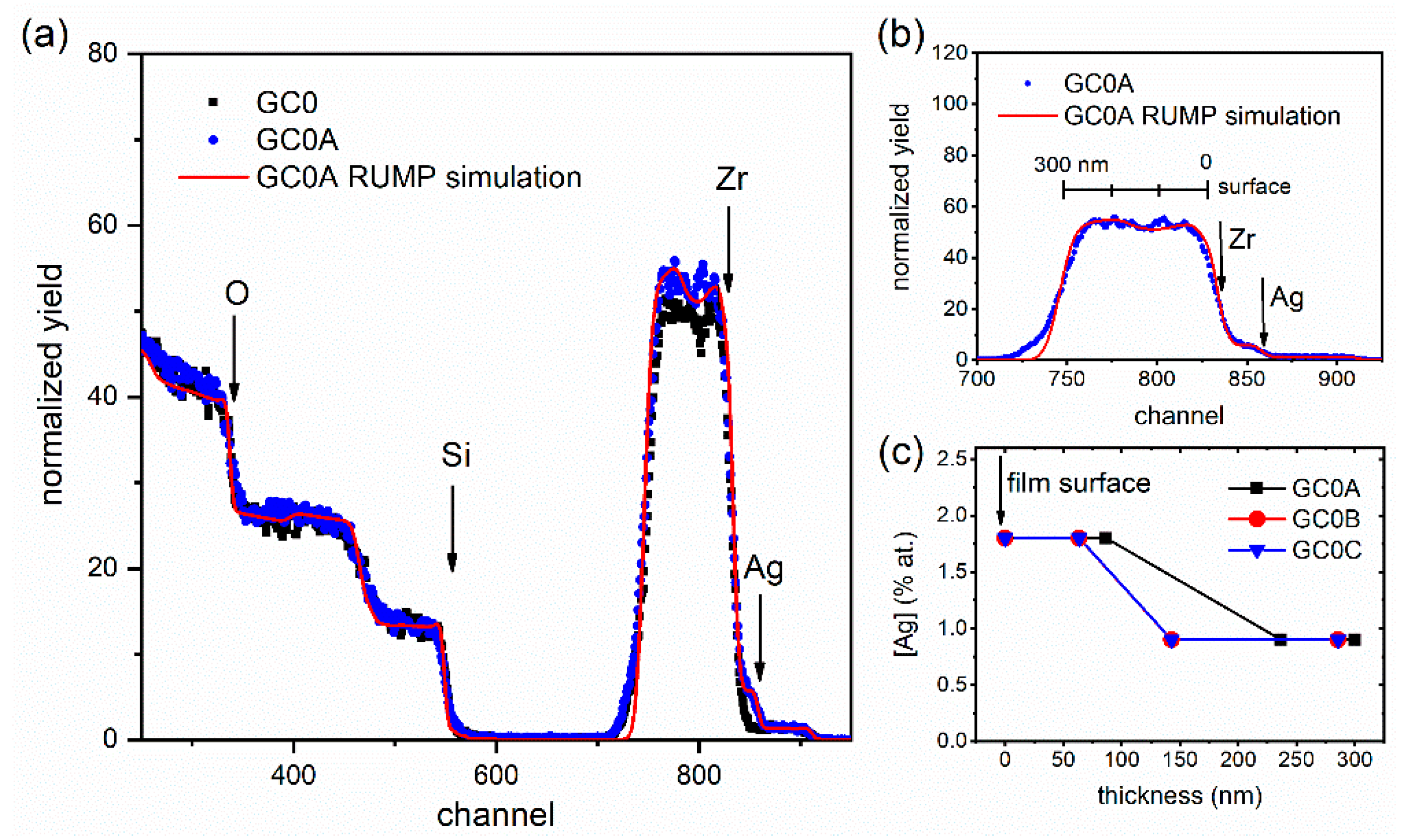
Figure 1. Full Rutherford backscattering spectrometry (RBS) spectra (a) and selected spectral region corresponding to Zr and Ag (b) for undoped samples before and after Ag-exchange (GC0 and GC0A). Ag concentration profile (c) as a function of film depth for samples GC0A, GC0B and GC0C. The RUMP code simulation of the GC0A spectrum is shown. In (b), the corresponding film depth is reported for the Zr signal.
Applsci 10 02184 g001
Figure 2. XRD comparison between undoped (GC0) and Yb-doped (GC4) silica-zirconia samples. The inset reports a selected region of the diffraction pattern around 2θ ≈ 35°, attesting to different crystal structures of the two samples: tetragonal-phase (diffraction standard reference ICSD #85322) zirconia nanocrystals in undoped GC0 and cubic-phase (diffraction standard reference PDF #49-1642) zirconia nanocrystals in Yb-doped GC4.
Applsci 10 02184 g002
Figure 3. Photoluminescent (PL) emission by 330 nm excitation of undoped samples before Ag-exchange (GC0), after exchange (GC0A), and after annealing at 380 °C (GC0B) and 430 °C (GC0C). The broad emission, up to 750 nm and more, is reasonably attributed to Ag multimers and small aggregates.
Applsci 10 02184 g003
Figure 4. PL excitation (a) and near-infrared (NIR) emission (b) of Yb-doped samples before Ag-exchange (GC4), after exchange (GC4A), and after annealing at 380 °C (GC4B) and 430 °C (GC4C). A broadband excitation up to about 500 nm can be observed after Ag-exchange. Moreover, 330 nm wavelength is not absorbed by Yb3+ ions, while it provides a strong PL emission in Ag-exchanged samples.
Applsci 10 02184 g004