Article

A Combination of Biphasic Calcium Phosphate (Maxresorb®) and Hyaluronic Acid Gel (Hyadent®) for Repairing Osseous Defects in a Rat Model

by
Abeer G. Ahmed
1,
Fatin A. Awartani
1,
Abdurahman A. Niazy
2,
John A. Jansen
3 and
Hamdan S. Alghamdi
1,2,*
1
Department of Periodontics and Community Dentistry, College of Dentistry, King Saud University, Riyadh 11545, Saudi Arabia
2
Molecular and Cell Biology Laboratory, College of Dentistry, King Saud University, Riyadh 11454, Saudi Arabia
3
Department of Dentistry—Biomaterials, Radboudumc, 6525EX Nijmegen, The Netherlands
*
Author to whom correspondence should be addressed.
Appl. Sci. 2020, 10(5), 1651; https://doi.org/10.3390/app10051651
Submission received: 11 February 2020 / Revised: 23 February 2020 / Accepted: 27 February 2020 / Published: 1 March 2020
(This article belongs to the Special Issue Permanent and Long-Term Biodegradable Biomaterials)

Abstract

:

Featured Application

Irrespective of the presence of hyaluronic acid (Hyadent®, HyA), biphasic calcium phosphate (Maxresorb®, BCP) was capable of bone regeneration within the defects as early as four weeks with sustained bone remodeling even at 10 weeks. Further in vivo studies are required to evaluate whether the dose and molecular weight of HyA could potentially benefit osseous regeneration with bone graft materials.

Abstract

The aim of this in vivo study was to evaluate the efficacy of biphasic calcium phosphate (Maxresorb®, BCP) used in combination with hyaluronic acid (Hyadent®, HyA) gel for regeneration of osseous defects in a rat model. Bilateral femoral condylar bone defects (3 mm diameter and 3 mm depth) were created in 40 male Wistar rats. The defects were grafted as group I (BCP only), group II (BCP + HyA), group III (HyA only), and group IV (empty control). At four weeks and 10 weeks, the bone specimens were evaluated using histological and histomorphometrical analyses to identify the newly formed bone area (NF-BA (%)), as well as the remaining BCP particles (R-BCP (%)). Light microscopic examination indicated the absence of an inflammatory reaction within the bone defects after four weeks or 10 weeks of implantation. Significant new bone regeneration was present in the bone defects grafted with BCP or BCP + HyA biomaterials, as early as four weeks, compared to control groups. The addition of HyA to BCP did not significantly improve bone regeneration at four weeks or 10 weeks. Nevertheless, its role in bone healing and regeneration warrants further investigation.

1. Introduction

Periodontal disease caused by bacterial colonization of the subgingival biofilm evokes an inflammatory response, which finally leads to destruction of the periodontal tissues [1] such as the periodontal ligament and alveolar bone [2]. The goal of contemporary periodontal therapy has always aimed at eliminating infection and restoring the periodontal bone defect [3]. Guided tissue regeneration and bone-grafting procedures have been used to regenerate bone within osseous defects, including that of the alveolar bone [4]. Bone-grafting materials are capable of providing a structural scaffold which stimulates and supports bone formation within osseous defects [4,5]. Additionally, bone grafts have also been reported to promote the formation of cementum and periodontal ligament (PDL), especially when used in periodontal defects [4,6].
Commonly used bone graft materials are classified as autologous cancellous or cortical bone, allogeneic mineralized or demineralized bone matrix, and synthetic or animal-derived bone substitutes [6]. Autologous bone (autograft) is considered as the “gold standard” bone substitute material, as it contains viable osteoblasts and osteoprogenitor cells, which are capable of inducing new bone formation (i.e., osteogenesis). On the one hand, demineralized allogeneic bone (allograft) is considered an osteoinductive material due to the presence of biologically active growth factors and induces differentiation of the host osteoprogenitor cells into osteoblasts [7]. On the other hand, synthetic (alloplast) and animal-derived bone grafts (xenograft) are only osteoconductive scaffolds, which guide the formation of new bone into the osseous defect, from the borders of the defect [4].
Although bone autografts completely satisfy the tenets of bone regeneration and could be termed as the “gold standard”, they have several clinical shortcomings, [7] including limited supply intra-orally and need for a second surgery at the donor site. In addition, donor site morbidity and restricted availability of autologous bone are significant clinical disadvantages [8]. While banked cadaveric bone allografts and animal-derived xenografts are readily available and are commonly used, their use is constrained by the potential risk of disease transmission and socio-ethnic restrictions [9]. Therefore, it has become desirable to synthesize bone-graft substitutes, which could be used for treating osseous defects. During the last decade, different bone-graft substitutes have been produced, including calcium phosphate based ceramics [10].
Calcium phosphate (CaP) ceramics have generated greater interest in bone regeneration, because of their similarities to the biological composition of natural bone mineral [11,12]. CaP-based materials, such as hydroxyapatite (HA), tricalcium phosphate (TCP), and biphasic calcium phosphate (BCP) are capable of being synthesized in the form of granules, blocks, or cements [13]. However, one major disadvantage of CaP ceramics is that they are only osteoconductive [14]. Therefore, advances in biomaterials research have focused on the development of composite bone substitutes, in which osteogenic cells or biological factors (e.g., mesenchymal stem cells, bone morphogenic proteins, platelet derived growth factor etc.) are incorporated with the synthetic bone graft materials to induce osteogenesis [14,15]. Nevertheless, the use of osteogenic cells or biological factors along with osteoconductive bone substitute materials are either technically sensitive or are expensive [15,16].
Studies have shown that bone regeneration using synthetic bone substitutes can be enhanced through less complicated therapeutic strategies such as application of hyaluronic acid (HyA) [15,17]. HyA has been reported to be capable of increasing the function of the osteogenic cells [15,18]. HyA is a critical component of extracellular matrix (ECM) in connective tissue and bone marrow, and it has been proven to play a key role in tissue repair and regeneration [17,19]. Moreover, it exhibits anti-inflammatory properties through inhibition of tissue destruction and accelerated wound healing [20] HyA has been routinely used in dentistry for the treatment of gingivitis and periodontitis as it is commercially available (e.g., Hyadent®) in a liquid or gel form at various concentrations [21]. Therefore, it could be hypothesized that the application of HyA to CaP-based bone substitutes could result in favorable bone regeneration within osseous defects. However, evidences in the literature have not been evaluated the same in clinical or in vivo scenarios.
Thus, the aim of the present in vivo study was to evaluate bone regeneration within femoral condylar defects, in a rat model, grafted with a combination biphasic calcium phosphate (BCP, Maxresorb®) and hyaluronic acid gel (HyA, Hyadent®), at four weeks and 10 weeks post-implantation.

2. Materials and Methods

2.1. Animal Model

The experimental animal model consisted of healthy male Wistar rats (N = 40) aged 12 weeks with a mean weight of 350 g. The animals were housed under veterinary supervision and acclimatized to the laboratory environment for 10 days pre-operatively. The animals were maintained under standard laboratory conditions (light-dark cycle 12:12 h, temperature 20–22 °C, and relative humidity 45% to 55%) and had ad libitum access to food and water. The experimental protocol was formulated by the “Molecular and Cell Biology laboratory” and ethically approved by the College of Dentistry Research Centre (CDRC), King Saud University, Riyadh, Saudi Arabia (Ref. # PR0038).

2.2. Experimental Groups

The bilateral femoral condylar bone defects made in the study animals were randomly grouped into the following groups based on the grafting material used:
Group I (BCP) 
Bone defects (n = 10 per group per time period) were grafted using 0.5 mL BCP (Maxresorb®, Botiss Biomaterials GmbH, Zossen, Germany) mixed with 1.0 mL sterile saline.
Group II (BCP + HyA) 
Bone defects (n = 10 per group per time period) were grafted using 0.5 mL BCP (Maxresorb®, Botiss Biomaterials GmbH, Zossen, Germany) mixed with a commercially available hyaluronic acid (HyA) (Hyadent®, BioScience GmbH, Dümmer, Germany). HyA was provided as hydrogel in sterile 1.0 mL syringe. β-TCP granules extemporaneously mixed with HyA. Then, the mixture was immediately pushed into the bony defect with a spatula.
Group III (HyA) 
Bone defects (n = 10 per group per time period) were filled with 1.0 mL HyA gel (Hyadent®, BioScience GmbH, Dümmer, Germany).
Group IV (control) 
Bone defects (n = 10 per group per time period) were left empty as a control for natural healing.

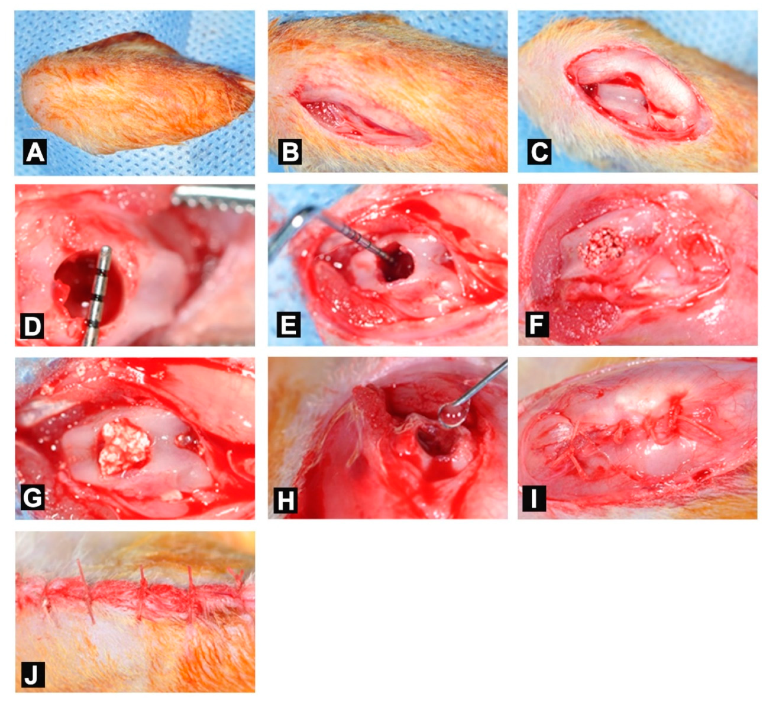
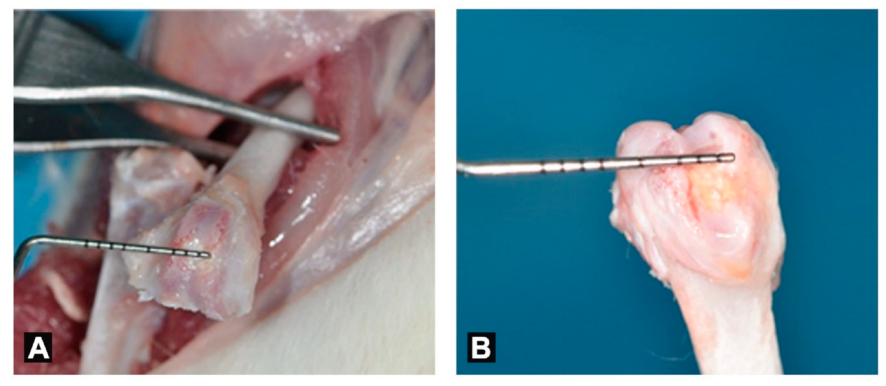
2.3. Surgical Procedure

All surgical procedures were carried out under general anesthesia (GA), administered by intramuscular (IM) injection of a mixture of xylazine 10 mg/kg (Xilagesic®, Laboratorios Calier S.A., Barcelona, Spain) and ketamine 100 mg/kg (HIKMA Pharmaceutical, Amman, Jordan). Intraoperative monitoring of respiratory rate and heart rate of the animals was done throughout the surgical procedure. The animals were positioned supine with the knee joint in a flexed position and the hind limbs were shaved and disinfected with 10% povidone iodine solution (Povidone®, Avalon Pharma Mecp, Riyadh, Saudi Arabia). A parapatellar incision was made on the skin overlying the knee joint and the joint capsule was incised longitudinally. The knee joint was completely exposed by elevating and retracting the patellar ligament laterally, facilitated by a slight extension of the knee joint.
A cylindrical hole measuring 3 mm in diameter and 3 mm in depth was, then, made at the intercondylar notch, parallel to the long axis of the femur. Drilling was done using calibrated dental drills on a rotary surgical motor (Elcomed 100; W&H Dentalwerk Burmoos) at a slow speed (800 rpm) and continuous external cooling with sterile saline. Each bone defect was grafted according to the experimental grouping mentioned in Section 2.2. Following grafting of the bone defects, overlying soft tissues and skin were closed in layers using 4-0 resorbable sutures (Vicryl, Ethicon, Johnson & Johnson, Norderstedt, Germany). Immediately after the surgery, the animals were monitored until recovery and, then, returned to their housing. Post-operative antibiotics (Amoxicillin 15 mg/kg; Norbrook Laboratories Ltd., Northern Ireland, UK) and analgesics (Analgivet® 50 mL/kg; Can Tho City, Vietnam) were administered IM once daily for 5 days for all the study animals, in addition to monitoring for signs of infection, inflammation, and loss of function. Representative images of the surgical protocol are shown in Figure 1.

2.4. Retrieval of Samples

Animals were euthanized at four weeks and 10 weeks post-surgery using an overdose of ketamine. The femoral condyles were carefully dissected and sectioned from the femoral diaphysis. Any soft tissue overlying the condylar specimen was thoroughly cleaned (Figure 2) and immediately fixed in 10% freshly prepared, neutral buffered formaldehyde solution for 48 h. Following fixation, the samples were stored in 70% ethanol, in preparation for histological assessment.

2.5. Analyses

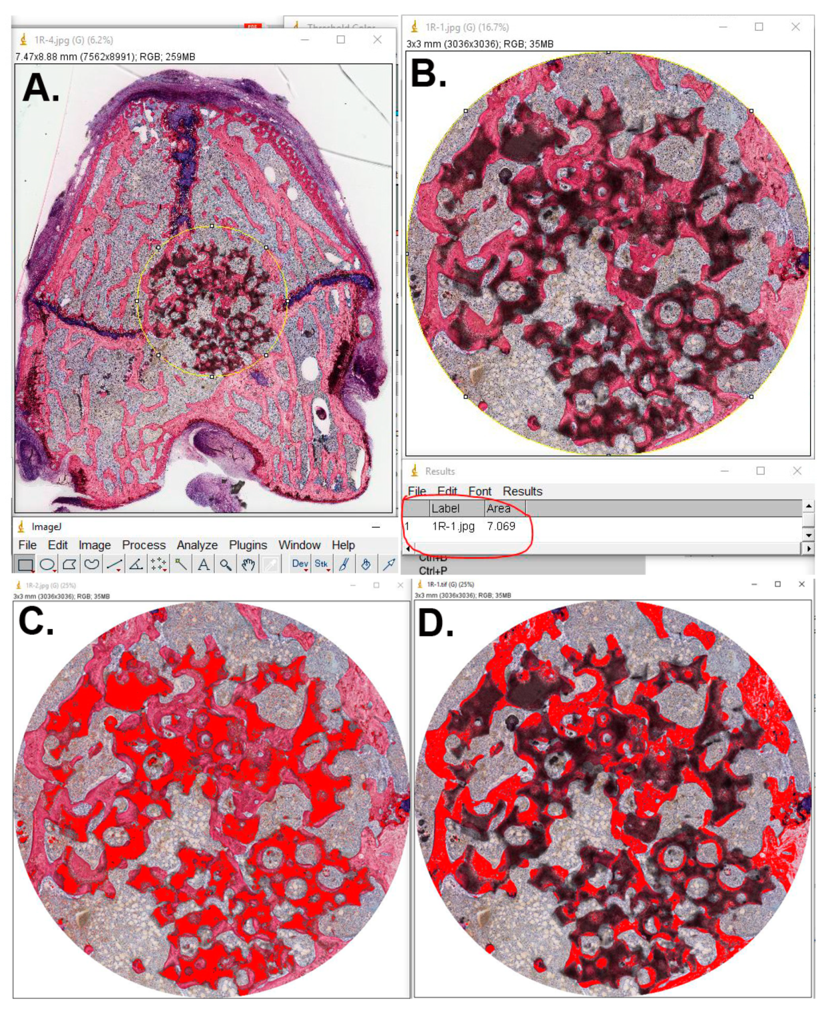
2.5.1. Histological Preparations

The specimens were dehydrated in ascending grades of alcohol from 70% to 100% and subsequently embedded in poly-(methyl methacrylate) (pMMA) resin, prepared by mixing 600 mL of methyl methacrylate monomer (Acros Organics BVBA, Geel, Belgium), 60 mL dibutyl phthalate (Merck KGaA, Darmstadt, Germany), and 1.25 g perkadox (AkzoNobel, Amersfoort, The Netherlands). After polymerization, the tissue blocks were cut in serial transverse histological sections with a thickness of ~10 μm, using a modified sawing microtome technique, as described previously [22]. Then, sections were stained using methylene blue and basic fuchsin.

2.5.2. Histological and Histomorphometrical Evaluation

To evaluate the bone formation within the defects, qualitative histologic and quantitative histomorphometrical analyses were performed. All stained sections were scanned and stored as digital images using an image scanner (Aperio Scanscope ATTurbo, Aperio Technologies, Vista, CA, USA) and examined using a histological image viewing software (Aperio Imagescope Version 12, Aperio Technologies, Vista, CA, USA). The histological sections were qualitatively assessed for the presence of inflammatory infiltrate, granulation tissue, fibrous tissue, in addition to newly formed bone and remaining grafted biomaterials.
As showed in Figure 3, quantitative histomorphometrical evaluation was performed using image analysis software (ImageJ® Version 1.3.8, National Institutes of Health, Bethesda, MD, USA). The threshold for assessment was predetermined on the image analyzer for the three components of interest in each section, namely newly formed bone, remaining BCP granules, and soft tissue. The surfaces areas for newly formed bone (NF-BA) and remaining BCP materials (R-BCP) were determined and expressed as a percentage of the total bone defect surface area, in relation to a 3 mm region of interest (ROI), corresponding to a cross-section of the defect. Results were expressed as mean and standard deviation for the NF-BA (%) and R-BCP (%) for three sections in each specimen.

2.6. Statistical Analysis

Quantitative variables, namely NF-BA (%) and R-BCP (%) were evaluated using statistical software (GraphPad Prism® Version 6.01, GraphPad Software Inc. San Diego, CA, USA). One-way analysis of variance (ANOVA) with Tukey’s post-hoc test, at a level of significance (p < 0.05), was used for comparison of outcomes between the groups at different time periods.

3. Results

3.1. General Observations and Monitoring of Animals

All the study animals tolerated the surgical procedures without any morbidity or mortality. While the animals recovered from GA shortly after the operation, they could walk freely within the first post-operative day with normal range of motion in the knee. No signs of inflammation or adverse tissue reaction were observed at the surgical site. However, wound dehiscence was observed during the early post-operative period in three animals and this was managed through disinfection and resuturing.

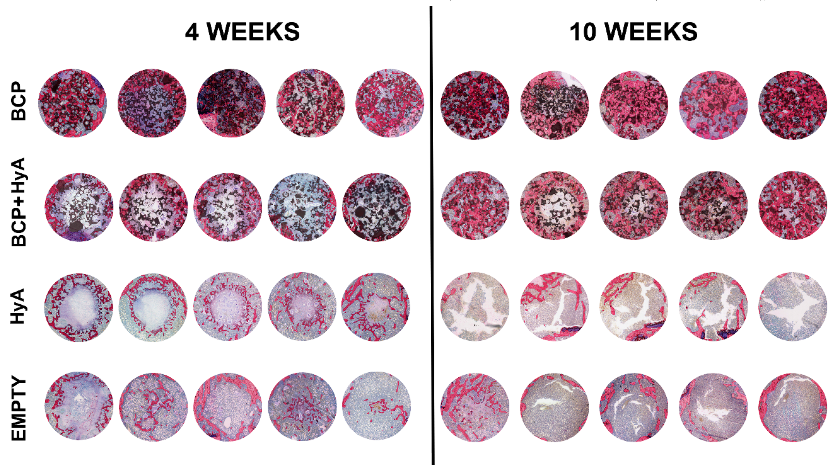
3.2. Qualitative Histological Evaluation

Representative light microscopic images of the non-decalcified histological sections are shown in Figure 4. Histologic evaluation revealed new bone formation within the defects in all groups and at both time periods, without any remarkable inflammatory reaction. There was an increase in new bone formation from the fourth to the 10th week, along with a decrease in the grafted BCP particles.
At four weeks, BCP material was still evident within the BCP and BCP + HyA treated defects and the BCP particles were predominantly surrounded by newly formed bone. In the groups of HyA and control, bone formation was less and observed either at the edges of the defect or as small islands within the center of the defect with sparse trabeculae. The remaining defect space was filled with bone marrow-like tissue.
At 10 weeks, there was a discernible increase in new bone formation within the defects grafted with BCP and BCP + HyA as compared with that of the four weeks. Although new bone was found to be evenly distributed throughout the defect, it was still distinguishable from the surrounding normal bone. This could possibly be attributed to the presence of the BCP particles. Nevertheless, resorption of BCP particles was more evident at 10 weeks than at four weeks. However, defects in the control and HyA groups were still largely open, with very little new bone formation, predominantly close to the edges of the defect.

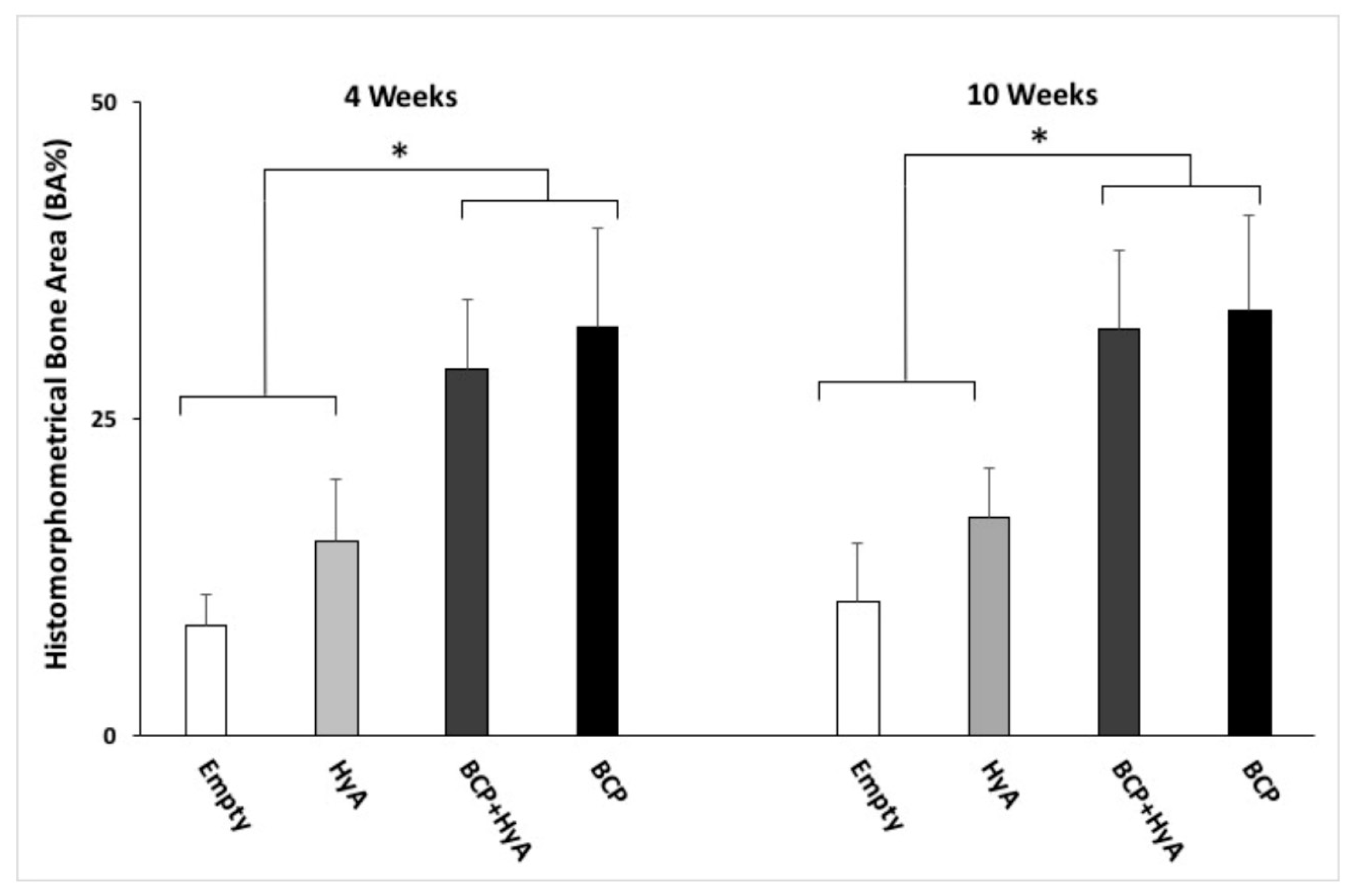
3.3. Quantitative Histomorphometrical Evaluation

In Table 1 and Figure 5, histomorphometrical evaluation revealed an overall increase in the NF-BA (%) from four weeks to 10 weeks in all the groups. At four weeks, the highest NF-BA (%) was observed in the BCP group followed by the BCP + HyA, HyA, and control groups. Statistically significant differences in the NF-BA (%) were observed between the BCP grafted groups and the non-grafted groups (p < 0.05). Similarly, at 10 weeks, the NF-BA (%) in the BCP and BCP + HyA groups was significantly higher than that of the HyA and control groups (p < 0.05). Interestingly, the addition of HyA to BCP did not significantly increase new bone formation as evidenced by the histomorphometrical findings. Along with an increase in NF-BA (%), there was a concomitant decrease in the R-BCP (%) in both the BCP and BCP + HyA groups.

4. Discussion

The present study aimed to evaluate the beneficial effect of adding HyA to BCP for regeneration of femoral condylar defects in a rat model. Osseous defects grafted with BCP, in the present study, displayed significantly higher new bone formation as compared with the control defects, both at four weeks and at 10 weeks. The addition of HyA to BCP did not improved new bone formation or enhanced BCP resorption.
The management of periodontal osseous defects is still a clinical challenge [4]. Primarily, it is because the healing of large bone defects usually requires grafting with a sufficient amount of autogenous bone harvested from a suitable donor site [8]. Alternatively, several types of allogeneic bone grafts, including freeze-dried, demineralized, and fresh frozen cadaveric bone have been used clinically, notwithstanding their associated risk of disease transmission [23]. Similarly, deproteinized xenografts have also been used clinically to treat bone defects, in spite of their biological inferiority to autografts and allografts [4]. Several synthetic bone substitutes have been used in clinical dentistry for regeneration of osseous defects, with considerable success and absolutely no risk of disease transmission [11,12]. The primary goal of synthetic bone material scaffolds such as calcium phosphate ceramics (CaP) is to augment bone defects and facilitate osteoconductive bone regeneration within those defects [6].
CaP-based biomaterials can undergo ceramolysis leading to their resorption and increasing bioavailability of calcium and phosphorous ions, which together permit new bone matrix formation and its early mineralization [24]. Furthermore, CaP ceramic granules are reportedly capable of adsorbing proteins and growth factors essential for bone regeneration, thereby exerting indirect osteoinductive properties [24]. Nevertheless, research evidence is still inconclusive as to whether CaP bone graft has a sufficient osteogenic ability in large osseous defects [25].
One of the most commonly used CaP ceramics, beta-tricalcium phosphate (β-TCP) undergoes rapid degradation, thereby limiting its ability to promote bone regeneration in large osseous defects and over a longer period of time [24]. However, hydroxyapatite (HA) is known for its slow degradation and presence at grafted bone defect sites even after a very long time [13]. Therefore, biphasic bone substitutes (HA/β-TCP) with a controlled rate of degradation and the ability to promote osteoconductive bone regeneration and remodeling over a longer period of time were considered favorable [12]. BCP composed of HA and β-TCP combines the advantages of rapid degradation of β-TCP for early bone matrix deposition and the persistence of HA for the long term mineralization of the new bone [24]. The results of the present study were coherent to the above findings and revealed minimal inflammatory response associated with BCP grafting along with new bone formation as early as four weeks and persistent remodeling even at 10 weeks.
While there has been controversy surrounding the optimum ratio of HA to β-TCP in BCP preparations, Nery et al. [26] reported that HA and β-TCP used in vivo, at concentrations of 60% and 40%, respectively, improved osseous defect healing in dogs. Moreover, they reported that BCP (60% HA + 40% β-TCP) was capable of better recruitment of osteogenic cells and acted as an adequate scaffold for bone regeneration [26]. The BCP combination used in the present study, namely Maxresorb®, was a porous granulate form (particle size 0.5 to 2 mm), primarily indicated for dental applications. Maxresorb® consists of a mixture of 60% HA and 40% β-TCP, [27] and owing to its significant biocompatibility, has been reported to allow direct bone bonding without development of an intervening soft tissue layer [11,12]. This was clearly evident from the histologic findings of the present study, wherein, new bone matrix deposition was evident not only at the edges of the defect, but also within the defect, especially surrounding the BCP particles.
Hyaluronic acid (HyA) is a component of tissue extracellular matrix (ECM) which plays a key role in cell adhesion, chemotaxis, differentiation, and proliferation, signaled through several macromolecules and especially during wound healing and tissue regeneration [28,29]. Aslan et al. [19] reported that HyA stimulated bone healing by accelerating the proliferation and migration of mesenchymal cells. Thus, it was suggested that HyA has a molecular mode of action which can improve the osteogenic ability of bone graft materials [19]. Furthermore, HyA of a specific molecular weight when used in vitro, was reported to significantly increase alkaline phosphatase activity and stimulate osteoblastic cell proliferation and differentiation [28]. It has also been suggested that HyA combined with synthetic bone grafts could prevent connective tissue ingrowth towards the core of the grafted biomaterials [30].
The mechanisms by which HyA stimulates bone formation are mainly attributed to its physicochemical properties, whereby it forms a suitable substrate for the migration and differentiation of osteoprogenitor cells [31] and for binding proteins crucial to bone healing, such as fibrinogen, fibrin, fibronectin, and collagen [32] Moreover, HyA could protect the surgical site from bacterial contamination and reduce post-surgical inflammation associated with bone healing [20]. Contrary to the reported studies, histomorphometrical evaluation in the present in vivo study did not reveal any superior performance of adding HyA to BCP granules, grafted in the rat femoral condylar defects, at four weeks and at 10 weeks. This could possibly be attributed to the low molecular weight (<103 kDa) of the HyA used in the present study. Interestingly, in previous studies, it has been documented that HyA of higher molecular weight (>103 kDa) promoted mesenchymal stem cells (MSCs) proliferation and differentiation. It showed that the use of a higher molecular weight of HyA enhanced the formation of calcium nodules, as well as the gene expressions of ALP, RUNX-2, and OCN. For instance, Huang et al. [28] reported that the capability of HyA to enhance the osteogenic and osteoinductive properties of bone graft materials was dependent on its dose and molecular weight. On the basis of their in vitro experiment, they reported that high molecular weight HyA (900 kDa and 2300 kDa), when used at a dose of 1 mg/mL, resulted in significantly pronounced osteogenic activity and mineralization on rat calvarium derived cell cultures as compared with low molecular weight of HyA and other doses [28].
The HyA biomaterial used in the present study, namely Hyadent®, was a hydrogel consisting of long molecular chain HyA at a concentration of 14 mg/mL and has been reportedly used in the successful management of periodontal tissue loss [21]. Although the present study hypothesis of enhanced bone regeneration within rat femoral condylar bone defects using a combination of HyA and BCP was based on previous research findings, the results of the present study did not support the same. Nevertheless, it would be alluring to evaluate through future studies, whether HyA used at a specific dose and molecular weight could still enhance bone regeneration when used along with osteoconductive bone graft materials.

5. Conclusions

Within the limitations of the present in vivo study, the addition of HyA to BCP did not significantly enhance bone regeneration within the femoral condylar bone defects in rats. Irrespective of the presence of HyA, BCP was capable of bone regeneration within the defects as early as four weeks with sustained bone remodeling even at 10 weeks. While the addition of HyA hydrogel to bone defects did not have any adverse effects, further in vivo studies are required to evaluate whether the dose and molecular weight of HyA could potentially benefit osseous regeneration with bone graft materials.

Author Contributions

Conceptualization, A.G.A., F.A.A. and H.S.A.; methodology, A.G.A. and H.S.A.; software, A.G.A.; validation, F.A.A., A.A.N. and H.S.A.; formal analysis, F.A.A., A.G.A., and H.S.A.; investigation, F.A.A., A.G.A., and H.S.A.; resources, A.A.N.; data curation, A.G.A., A.A.N. and H.S.A.; writing—original draft preparation, F.A.A. and H.S.A.; writing—review and editing, J.A.J. and H.S.A.; visualization, H.S.A.; supervision, J.A.J. and H.S.A.; project administration, F.A.A.; funding acquisition, H.S.A. All authors have read and agreed to the published version of the manuscript.

Funding

The authors extend their appreciation to the Deanship of Scientific Research at King Saud University for funding this work through research group No (RG-1439-026).

Acknowledgments

The authors extend their appreciation to the Deanship of Scientific Research at King Saud University for funding this work through research group no. (RG-1439-026). The authors would like to acknowledge Vincent Cuijpers and Terrence Sumague for their assistance with histomorphometrical analysis, and Natasja van Dijk and Mary Grace Vigilla for their assistance with the histological sectioning.

Conflicts of Interest

No competing financial interests exist.

References

  1. Pihlstrom, B.L.; Michalowicz, B.S.; Johnson, N.W. Periodontal diseases. Lancet 2005, 366, 1809–1820. [Google Scholar] [CrossRef] [Green Version]
  2. Offenbacher, S. Periodontal diseases: Pathogenesis. Ann. Periodontol. 1996, 1, 821–878. [Google Scholar] [CrossRef] [PubMed]
  3. Van Dyke, T.; Serhan, C. Resolution of inflammation: A new paradigm for the pathogenesis of periodontal diseases. J. Dent. Res. 2003, 82, 82–90. [Google Scholar] [CrossRef] [PubMed]
  4. Reynolds, M.A.; Aichelmann-Reidy, M.E.; Branch-Mays, G.L. Regeneration of periodontal tissue: Bone replacement grafts. Dent. Clin. N. Am. 2010, 54, 55–71. [Google Scholar] [CrossRef]
  5. Shiau, H.J.; Reynolds, M.A. Sex differences in destructive periodontal disease: Exploring the biologic basis. J. Periodontol. 2010, 81, 1505–1517. [Google Scholar] [CrossRef]
  6. Kao, R.T.; Nares, S.; Reynolds, M.A. Periodontal regeneration-intrabony defects: A systematic review from the AAP regeneration workshop. J. Periodontol. 2015, 86, S77–S104. [Google Scholar] [CrossRef]
  7. Rosenberg, E.; Rose, L.F. Biologic and clinical considerations for autografts and allografts in periodontal regeneration therapy. Dent. Clin. N. Am. 1998, 42, 467–490. [Google Scholar]
  8. Sen, M.K.; Miclau, T. Autologous iliac crest bone graft: Should it still be the gold standard for treating nonunions? Injury 2007, 38, S75–S80. [Google Scholar] [CrossRef]
  9. Tomford, W.W.; Thongphasuk, J.; Mankin, H.; Ferraro, M. Frozen musculoskeletal allografts. A study of the clinical incidence and causes of infection associated with their use. J. Bone Jt. Surg. Am. 1990, 72, 1137–1143. [Google Scholar] [CrossRef]
  10. Sutherland, D.; Bostrom, M. Grafts and bone graft substitutes. In Bone Regeneration and Repair; Springer: Berlin/Heidelberg, Germany, 2005; pp. 133–156. [Google Scholar]
  11. Knaack, D.; Goad, M.; Aiolova, M.; Rey, C.; Tofighi, A.; Chakravarthy, P.; Lee, D.D. Resorbable calcium phosphate bone substitute. J. Biomed. Mater. Res. 1998, 43, 399–409. [Google Scholar] [CrossRef]
  12. Kolk, A.; Handschel, J.; Drescher, W.; Rothamel, D.; Kloss, F.; Blessmann, M.; Heiland, M.; Wolff, K.D.; Smeets, R. Current trends and future perspectives of bone substitute materials–from space holders to innovative biomaterials. J. Cranio-Maxillofac. Surg. 2012, 40, 706–718. [Google Scholar] [CrossRef] [PubMed]
  13. Giuliani, A.; Manescu, A.; Mohammadi, S.; Mazzoni, S.; Piattelli, A.; Mangano, F.; Iezzi, G.; Mangano, C. Quantitative Kinetics Evaluation of Blocks Versus Granules of Biphasic Calcium Phosphate Scaffolds (HA/beta-TCP 30/70) by Synchrotron Radiation X-ray Microtomography: A Human Study. Implant Dent. 2016, 25, 6–15. [Google Scholar] [CrossRef] [PubMed]
  14. Finkemeier, C.G. Bone-grafting and bone-graft substitutes. J Bone Jt. Surg. Am. 2002, 84, 454–464. [Google Scholar] [CrossRef]
  15. Kim, J.; Kim, I.S.; Cho, T.H.; Lee, K.B.; Hwang, S.J.; Tae, G.; Noh, I.; Lee, S.H.; Park, Y.; Sun, K. Bone regeneration using hyaluronic acid-based hydrogel with bone morphogenic protein-2 and human mesenchymal stem cells. Biomaterials 2007, 28, 1830–1837. [Google Scholar] [CrossRef]
  16. Diker, N.; Koroglu, T.; Gulsever, S.; Akcay, E.Y.; Oguz, Y. Effects of hyaluronic acid and alloplastic bone graft combination on bone regeneration. Int. J. Oral Maxillofac. Surg. 2015, 44, e55. [Google Scholar] [CrossRef]
  17. Prestwich, G.D. Hyaluronic acid-based clinical biomaterials derived for cell and molecule delivery in regenerative medicine. J. Control. Release 2011, 155, 193–199. [Google Scholar] [CrossRef] [PubMed] [Green Version]
  18. Patterson, J.; Siew, R.; Herring, S.W.; Lin, A.S.; Guldberg, R.; Stayton, P.S. Hyaluronic acid hydrogels with controlled degradation properties for oriented bone regeneration. Biomaterials 2010, 31, 6772–6781. [Google Scholar] [CrossRef] [Green Version]
  19. Aslan, M.; Şimşek, G.; Dayi, E. The effect of hyaluronic acid-supplemented bone graft in bone healing: Experimental study in rabbits. J. Biomater. Appl. 2006, 20, 209–220. [Google Scholar] [CrossRef]
  20. Hahn, S.K.; Jelacic, S.; Maier, R.V.; Stayton, P.S.; Hoffman, A.S. Anti-inflammatory drug delivery from hyaluronic acid hydrogels. J. Biomater. Sci. Polym. Ed. 2004, 15, 1111–1119. [Google Scholar] [CrossRef]
  21. Awartani, F.A.; Tatakis, D.N. Interdental papilla loss: Treatment by hyaluronic acid gel injection: A case series. Clin. Oral Investig. 2016, 20, 1775–1780. [Google Scholar] [CrossRef]
  22. Van der Lubbe, H.; Klein, C.; De Groot, K. A simple method for preparing thin (10 μm) histological sections of undecalcified plastic embedded bone with implants. Stain Technol. 1988, 63, 171–176. [Google Scholar] [CrossRef] [PubMed]
  23. Contar, C.M.; Sarot, J.R.; Bordini, J., Jr.; Galvao, G.H.; Nicolau, G.V.; Machado, M.A. Maxillary ridge augmentation with fresh-frozen bone allografts. J. Oral Maxillofac. Surg. Off. J. Am. Assoc. Oral Maxillofac. Surg. 2009, 67, 1280–1285. [Google Scholar] [CrossRef] [PubMed]
  24. Lobo, S.E.; Livingston Arinzeh, T. Biphasic calcium phosphate ceramics for bone regeneration and tissue engineering applications. Materials 2010, 3, 815–826. [Google Scholar] [CrossRef] [Green Version]
  25. Fellah, B.H.; Gauthier, O.; Weiss, P.; Chappard, D.; Layrolle, P. Osteogenicity of biphasic calcium phosphate ceramics and bone autograft in a goat model. Biomaterials 2008, 29, 1177–1188. [Google Scholar] [CrossRef] [Green Version]
  26. Nery, E.B.; Legeros, R.Z.; Lynch, K.L.; Lee, K. Tissue response to biphasic calcium phosphate ceramic with different ratios of HA/βTCP in periodontal osseous defects. J. Periodontol. 1992, 63, 729–735. [Google Scholar] [CrossRef]
  27. Jelusic, D.; Zirk, M.L.; Fienitz, T.; Plancak, D.; Puhar, I.; Rothamel, D. Monophasic ß-TCP vs. biphasic HA/ß--TCP in two-stage sinus floor augmentation procedures—A prospective randomized clinical trial. Clin. Oral Implants Res. 2016, 28, e175–e183. [Google Scholar] [CrossRef]
  28. Huang, L.; Cheng, Y.; Koo, P.; Lee, K.M.; Qin, L.; Cheng, J.C.Y.; Kumta, S.M. The effect of hyaluronan on osteoblast proliferation and differentiation in rat calvarial—Derived cell cultures. J. Biomed. Mater. Res. 2003, 66, 880–884. [Google Scholar] [CrossRef]
  29. Kuo, J.; Prestwich, G. Hyaluronic acid. In Comprehensive Biomaterials; Elsevier: Amsterdam, The Netherlands, 2011. [Google Scholar]
  30. ELkarargy, A. Alveolar sockets preservation using hydroxyapatite/beta tricalcium phosphate with hyaluronic acid (histomorphometric study). J. Am. Sci. 2013, 9, 556–563. [Google Scholar]
  31. Sasaki, T.; Watanabe, C. Stimulation of osteoinduction in bone wound healing by high-molecular hyaluronic acid. Bone 1995, 16, 9–15. [Google Scholar] [CrossRef]
  32. Stern, M.; Schmidt, B.; Dodson, T.B.; Stern, R.; Kaban, L.B. Fetal cleft lip repair in rabbits: Histology and role of hyaluronic acid. J. Oral Maxillofac. Surg. 1992, 50, 263–268. [Google Scholar] [CrossRef]
Figure 1. Representative surgical images of bone defect preparation and bone-grafting procedures. (A) Surgical site preparation of the skin over the femoral condyle; (B) parapatellar incision over the knee joint capsule; (C) soft tissue reflection to expose the underlying femoral condyle; (D,E) experimental bone defect measuring 3 mm in diameter and 3 mm in depth; (FH) bone defects filled according to the experimental grouping protocol; (I) closure of subcutaneous tissues; and (J) skin closure.
Applsci 10 01651 g001
Figure 2. Representative images showing retrieval of the femoral condylar samples without any associated overlying soft tissue. (A) Sectioning the condylar bone from the remaining femur; (B) experimental bone defect with any remaining biomaterials if grafted.
Applsci 10 01651 g002
Figure 3. Representative histological sections showing histomorphometrical measurements using imageJ software. (A,B) A region of interest (ROI) measuring 3 mm in diameter and corresponding to the defect cross-section; (C) for selecting and measuring the percentage (%) of remaining biomaterials; (D) for identifying bone area in (%).
Applsci 10 01651 g003
Figure 4. Representative histological images stained with methylene blue and basic fuchsin, showing non-decalcified (pMMA embedded) cross-sections of the different experimental bone defects during each time period.
Applsci 10 01651 g004
Figure 5. Bar-chart with mean and standard deviation based on histomorphometrical assessment showing bone area (BA%) for all study groups at 4 weeks and 10 weeks. (* indicates p < 0.05)
Applsci 10 01651 g005
Table 1. Quantitative (mean ± SD) histomorphometrical evaluation of newly formed bone area (%) and remaining BCP (%) in the different groups during the two time periods of evaluation.
Study Groups (n = 10 Per Group Per Time Period)Bone Healing Time
4 Weeks10 Weeks
Newly Formed Bone Area (%)Remaining BCP (%)Newly Formed Bone Area (%)Remaining BCP (%)
BCP32.2 ± 7.728.7 ± 7.633.5 ± 7.423.9 ± 6.3
BCP + HyA28.8 ± 5.622.6 ± 5.132.0 ± 6.221.6 ± 3.7
HyA15.3 ± 4.9-17.1 ± 3.9-
Control8.6 ± 2.5-10.6 ± 4.5-
BCP, biphasic calcium phosphate and HyA, hyaluronic acid.

