Article

Postural Control in Children with Cerebellar Ataxia

by
Veronica Farinelli
1,
Chiara Palmisano
2,3,
Silvia Maria Marchese
1,*,
Camilla Mirella Maria Strano
1,
Stefano D’Arrigo
4,
Chiara Pantaleoni
4,
Anna Ardissone
5,
Nardo Nardocci
5,
Roberto Esposti
1 and
Paolo Cavallari
1
1
Human Physiology Section of the Department of Pathophysiology and Transplantation, Università degli Studi di Milano, 20133 Milano, Italy
2
Movement Biomechanics and Motor Control Lab, Department of Electronic Information and Bioengineering, Politecnico di Milano, 20133 Milano, Italy
3
Department of Neurology, University Hospital and Julius-Maximilian University Würzburg, 97080 Würzburg, Germany
4
Developmental Neurology Department, Fondazione IRCCS Istituto Neurologico C. Besta, 20133 Besta, Italy
5
UOC Neuropsichiatria Infantile, Fondazione IRCCS Istituto Neurologico C. Besta, 20133 Besta, Italy
*
Author to whom correspondence should be addressed.
Appl. Sci. 2020, 10(5), 1606; https://doi.org/10.3390/app10051606
Submission received: 5 December 2019 / Revised: 10 February 2020 / Accepted: 20 February 2020 / Published: 28 February 2020
(This article belongs to the Special Issue Movement Biomechanics and Motor Control)

Abstract

:
Controlling posture, i.e., governing the ensemble of involuntary muscular activities that manage body equilibrium, represents a demanding function in which the cerebellum plays a key role. Postural activities are particularly important during gait initiation when passing from quiet standing to locomotion. Indeed, several studies used such motor task for evaluating pathological conditions, including cerebellar disorders. The linkage between cerebellum maturation and the development of postural control has received less attention. Therefore, we evaluated postural control during quiet standing and gait initiation in children affected by a slow progressive generalized cerebellar atrophy (SlowP) or non-progressive vermian hypoplasia (Joubert syndrome, NonP), compared to that of healthy children (H). Despite the similar clinical evaluation of motor impairments in NonP and SlowP, only SlowP showed a less stable quiet standing and a shorter and slower first step than H. Moreover, a descriptive analysis of lower limb and back muscle activities suggested a more severe timing disruption in SlowP. Such differences might stem from the extent of cerebellar damage. However, literature reports that during childhood, neural plasticity of intact brain areas could compensate for cerebellar agenesis. We thus proposed that the difference might stem from disease progression, which contrasts the consolidation of compensatory strategies.

1. Introduction

Postural adjustments are involuntary muscular activities that accompany the voluntary movement. These activities spread over adjacent muscles and thus create “chains” that reach the available support points (in many cases, the ground). Such chains allow fine-tuning the body equilibrium, in order to adapt it to the mechanical needs of the ensuing movement. For example, when flexing both arms at the shoulder, postural actions develop in a dorsal muscle chain, including Erector Spinae (ES), Biceps Femoris (BF), and Soleus (SOL), to counteract the reaction force due to arm movement [1]. Instead, when rising on tiptoes, involuntary bursts of activity develop in Tibialis Anterior (TA) muscles, so as to induce a forward fall of the Centre of Mass (COM); in this way, COM reaches the forefoot, and the voluntary contraction of Soleus muscles (SOL) rises the body [2]. Otherwise, simply recruiting SOL would produce a backward fall. Whenever the mechanical needs of action may be estimated beforehand, like when programming a voluntary movement, appropriate postural actions are usually produced in advance of the movement itself, witnessing that such Anticipatory Postural Adjustments (APAs) are programmed in a feed-forward way [3,4,5,6].
APAs are particularly evident in gait initiation, in which they maintain the body’s dynamic balance and create the propulsive forces to move the COM forwards. In healthy adults performing gait initiation [7,8,9,10,11,12], the Center of Pressure (CoP), i.e., the barycenter of the ground reaction forces, first moves backward and towards the future swing foot. The onset of such a CoP shift is usually called APA onset, while its time period is called the imbalance phase. Indeed, the ensuing horizontal gap between CoP and COM (where the gravity force vector is applied) produces an “imbalance” torque that pushes COM forwards and towards the future stance foot. Then, CoP moves laterally towards the stance foot, continuing to promote the forward acceleration of COM while braking its lateral fall. At the same time, this CoP shift withdraws body weight from the swing foot, hence the name unloading phase. Finally, as COM proceeds, CoP travels forwards along the stance foot, from toe-off to heel-strike of the swing foot (first swing) [13]. Considering the muscular actions that drive gait initiation, before the beginning of the imbalance phase, an inhibition occurs in the background EMG (electromyographic) activity of both SOL muscles, which are tonically active during quiet stance. SOL inhibitions are shortly followed by the recruitment of TA muscles, which are silent during quiet stance and activate close to the APA onset. In particular, in the stance leg, the SOL inhibition precedes TA excitation by about 100 ms [7]. A drop in the background activity is also observed in other dorsal muscles, like BF and ES, while bursts of activity occur in ventral muscles, like Rectus Femoris (RF) [14].
Less literature is available on APAs during gait initiation in children. A systematic survey by Ledebt et al. (1998) [15] showed that APAs start developing at 2–3 years of age but complete their maturation well after the age of 8, a result in line with the observations carried out on toddlers up to 5 years old children [16] and in 4–6 years old children [17]. Another interesting study was published by Isaias et al. (2014) [18], who analyzed SOL and TA in 10 ± 3 years-old children and reported inhibition-excitation patterns like in adults, but with a lower time interval between SOL inhibition and TA excitation.
Several studies showed altered gait initiation in those neurological diseases characterized by poor motor control, as Rett syndrome [18], Parkinson’s disease [19], and cerebellar pathologies [20,21]. In particular, Timmann and Horak [21] reported that adults with cerebellar deficits showed a decreased force production and a significant reduction of the length and peak velocity of the first step, accompanied by impairments in the predictive adaptation of APAs to the mechanical needs of gait initiation. Despite these authors also found that the temporal parameters of APAs were overall preserved in patients with cerebellar disease, several other works [22,23,24,25,26] addressed the role of cerebellum in postural control and provided evidence that such structure is involved not only in modulating rate and force of muscle activities but also in determining their relative timing. Indeed, cerebellar deficits often lead to dysfunctional co-contractions [22,24,26]. In this regard, it is worth recalling the involvement of the cerebellum in building up the temporal pattern of APAs, in particular, its ability to create and store internal models of body mechanics. This is proved by the contribution of the cerebellum in modulating sensory-motor interactions and integrating feed-forward and feed-back modes [27].
The cerebellum is also known to play an important role in many developmental disorders [28]. Nevertheless, very little attention has been given to the linkage between the development of postural control and the maturation of such neural structure. Aiming to elucidate this topic, we explored quiet stance and gait initiation in children affected by Pediatric Cerebellar Ataxia (PCA), used as a model of cerebellar dysfunction vs. a healthy control group of comparable age.
PCAs are a heterogeneous group of cerebellar developmental disorders characterized by dysfunctional motor coordination and very early cerebellar symptoms. The first clinical signs are marked hypotonia, wobbling gait, dysmetria, dysarthria, and a significant developmental delay. Most children show also marked speech impairment and cognitive deficits. In some cases, the cerebellum degenerates with time, but so slowly that it becomes difficult to classify the disorder as progressive or not [29]. In this framework, we studied a group of children with generalized cerebellar atrophy and clinical evidence of slow progression during follow-up (SlowP). In other cases, the disease has a proven non-progressive course, as in Joubert syndrome, which is characterized by cerebellar hypoplasia limited to the vermis and peduncles [30]. Thus, we also considered the second group of children (non-progressive, NonP) affected by this kind of pathology. It is also important to note that the onset of the SlowP pathology is clinically indistinguishable from that of the NonP forms; therefore, practically, both diseases are present since birth.
In order to characterize quiet stance and gait initiation, we measured the classical posturographic parameters [31,32] and the first step length and velocity (as measures of performance). Besides, we also calculated the shifts of CoP and COM, as well as the horizontal distance (gap) between these two points in the imbalance and unloading phases, to highlight the net effect on COM dynamics. In order to document possible changes in muscular APAs, we evaluated the EMG activities that accompany the APA onset. Should we observe significant differences between each pathological group and healthy children, this would point out the involvement of the cerebellum also during the key phase of human maturation, in which the central nervous system learns gait initiation dynamics and how to optimize this motor process. Moreover, these findings would be fruitful in tailoring rehabilitation for such pathologies. Finally, a different motor pattern in children with SlowP vs. NonP would suggest possible compensation mechanisms. In particular, taking into consideration that children with SlowP suffer from generalized cerebellar damage, better motor behavior in SlowP vs. NonP could suggest the involvement of extracerebellar regions. On the other side, better behavior in children of the NonP group could as well stem from a compensatory involvement of the cerebellar hemispheres, which are unaffected by Joubert syndrome.

2. Materials and Methods

2.1. Participants

Thirteen participants with PCA were recruited at the “Istituto Neurologico Carlo Besta” of Milan: seven of them had radiological signs of generalized cerebellar atrophy and clinical evidence of slow progression during follow-up (SlowP, mean age: 12 ± 3 years), while the remaining six suffered from Joubert syndrome, i.e., a proven non-progressive pathology (NonP, mean age: 12 ± 3 years). All of them underwent clinical evaluation, including the administration of the Scale for the Assessment and Rating of Ataxia (SARA, [33]), an MRI scan for establishing the cerebellar alteration (atrophy and/or hypoplasia), and genetic screening. In particular, all children with Joubert syndrome showed a unique cerebellar and brainstem malformation known as the “molar tooth sign” [30]. The demographic and clinical data of each patient are reported in Table 1.
Seven healthy children, free from neurological or psychological pathologies and typically developing, were enrolled as a control group, from the primary school “FAES” in Milan (H, 4 males and 3 females, mean age: 10 ± 3 years). The experimental procedure was carried out in accordance with the standards of the Declaration of Helsinki. The Ethical Committee “Comitato Etico di Ateneo dell’Università degli Studi di Milano” approved the study and the written consent procedure, on 15 February 2016 (counsel 5/16). Before each acquisition, the child neuropsychiatrist and the experimenters explained the aim of the study and the details of the experimental procedure to the parents and to their child. Only those children who agreed with the study participated in the experiments. The parents of each participant, as her/his legal representatives, signed the consent procedure. All children were perfectly aware of the task since no one of them failed in accomplishing it.

2.2. Experimental Protocol

Subjects were asked to perform a gait initiation task several times. They were instructed to stand quietly on a force plate for 30 s and then to walk at their natural speed after a vocal prompt, self-selecting the leading limb [35]. After three to five steps, subjects stopped and returned to the initial position.
Each subject repeated the task until three valid trials were collected (i.e., trials in which the subject did not move the feet, arms, or head during the quiet stance preceding gait initiation). Subjects were allowed to rest 2 min before repeating the task. A maximum of nine trials was required to satisfy the above criterion; the average number of trials per subject was 6.6 ± 2.7. At the end of the resting periods between motor tasks, the experimenter asked the child, “Do you feel fatigued? Are you ready to start again?”. Moreover, since the parents assisted at the trials, they could report to the experimenter any possible discomfort of their child. Neither children nor their parents complained about fatigue. The width of the base of support was self-selected by each subject in the first trial, then marked on the platform with adhesive tape and kept fixed for all further trials. The distance between lateral malleoli during quiet stance was comparable among groups (SlowP: median = 162.8 mm, range = 115.6 to 205.6 mm; NonP: 165.6 mm, 152.3 to 198.5 mm; H: 150.3 mm, 147.5 to 242.3 mm), with no significant differences (Kruskal–Wallis p = 0.566).

2.3. Recordings

Body kinematics was recorded by means of a six-cameras optoelectronic system (SMART-E, BTS, Milan, Italy) using a full-body marker set [36], which allowed estimating the Centre of Mass (COM) and its trajectory, according to Isaias et al. (2014) [18]. A dynamometric force plate (9286AA, KISTLER, Winterthur, Switzerland) was used to compute the Center of Pressure (CoP) position. Wireless probes (FREEEMG 1000, BTS, Milan, Italy) were employed bilaterally to record the surface electromyographic (EMG) activity of Tibialis Anterior (TA), Soleus (SOL), Rectus Femoris (RF), Biceps Femoris (BF), and Erector Spinae (ES). Electrodes were placed according to the Surface Electromyography for the Non-Invasive Assessment of Muscles (SENIAM) guidelines [37]. Synchronous data acquisition was accomplished by the SMART-E workstation; sampling rate being 60 Hz for optoelectronic cameras, 960 Hz for dynamometric signals, and 1000 Hz for EMGs.

2.4. Data Processing

During the 30 s quiet standing period, the statokinesigram, i.e., the trajectory of the CoP in the horizontal plane, was used to extract specific indexes of balance control. These indexes were: the area and the eccentricity of the ellipse containing 95% of CoP positions, the total length of CoP trajectory (CoP length), the average CoP velocity, and the peak-to-peak Mediolateral and Anteroposterior CoP displacements (ML and AP ranges, respectively) [31,32]. In particular, the ellipse area (A) and eccentricity ( e ) were calculated according to the following formulae:
A = a b π
e = |   a 2 b 2   | a
in which a and b were the semi-major axis (i.e., half of the ellipse longest diameter) and the semi-minor axis (i.e., half of the shortest diameter), respectively.
Gait initiation was subdivided into three phases [13]: the imbalance phase, in which CoP moves backward and towards the future swing foot; the unloading phase, in which CoP moves laterally towards the stance foot, and the first swing, in which CoP moves forwards along the stance foot, from toe-off to heel-strike of the swing foot. The temporal events delimiting each phase were determined by visual inspection of the CoP trajectory; in particular, the onset of the CoP backward shift represented the APA onset.
For the imbalance and unloading phases, separately, we measured the phase duration, the length of the CoP trajectory, the maximum AP and ML shifts of both CoP and COM, and the distance from CoP to COM projection on the horizontal plane, at the end of the phase.
The first swing phase was evaluated by measuring the length of the first step, normalized to the lower limb length (LL), and its velocity (v), expressed in Froude number (Fr = v g L L g being gravity acceleration [38]); also these measurements were obtained from kinematic data.
For each subject, the kinematic and dynamometric variables were averaged over the three valid trials recorded. Data normality was evaluated by means of the Shapiro–Wilk test. Considering that data were not normally distributed, the differences among SlowP, NonP, and H groups were analyzed non-parametrically by using Kruskal–Wallis tests followed by Dunn post hoc, with Bonferroni adjustment. The level of significance was set to 0.05.
The analysis of EMG recordings regarded the timing of muscle activation or inhibition, surrounding the APA onset at gait initiation. Raw EMG data were high-pass filtered (fcut = 50 Hz) with a zero-phase shift 6th-order elliptic filter, to remove movement artifacts, and then the signals were rectified. For each muscle, the traces of the recorded trials were time-aligned to the APA onset and averaged across trials. For each averaged EMG track, the period from 1 s to 0.5 s before the APA onset—where no EMG changes were observed—was assumed as reference. The signal was integrated (time constant = 11 ms), and the mean level in the reference period was subtracted. The onset of an excitatory or inhibitory EMG change was identified by a software algorithm, which searched the first time point in which the track fell above or below 2 SD of the reference signal (excitation or inhibition, respectively) and remained there for at least 50 ms. Whenever the criterion was met, the algorithm searched backward the point in which the signal started to deviate from the mean reference value [39]. No statistical analysis was performed on EMG timings because of the many cases in which no clear inhibitory or excitatory changes could be identified.

3. Results

3.1. Postural Parameters

The analysis of quiet stance (static posturography) highlighted alterations of postural control in subjects with cerebellar deficits. In fact, children of both the SlowP and NonP groups showed an ellipse area greater than H (Kruskal–Wallis p = 0.039), mainly due to a greater CoP displacement in the mediolateral direction (Kruskal–Wallis p = 0.022).
However, post hoc showed that this difference was statistically significant only in subjects with SlowP vs. H (Table 2). In particular, the latter group revealed an inversion of the normal ellipse configuration, with mediolateral oscillation as the preferred direction (Figure 1), which led to a reduction of the ellipse eccentricity.

3.2. Gait Initiation Parameters

The spatial and temporal parameters during the imbalance and unloading phases were not significantly affected by the pathology (Table 3). First step length and velocity were instead different among the three groups (Kruskal–Wallis, p = 0.005 for length and p = 0.019 for velocity). However, post hoc tests showed that such difference was statistically significant only in children with SlowP vs. H (Table 4).

3.3. EMG

Postural EMG changes accompanying APA onset, defined as the first CoP backward shift, could not be detected in all recorded muscles and for all subjects. A descriptive analysis of electromyographic recordings allowed appreciating the development of an inhibitory postural chain involving ES, BF, and SOL, followed by an excitatory chain in RF and TA. Such a general pattern was observed in both the stance and swing sides, irrespectively from the healthy or pathological status. Nevertheless, a different timing distribution of the muscular actions was found in the three groups (Table 5). In the stance limb side, healthy subjects showed a clear craniocaudal progression, for both the inhibitory (first ES and BF, then SOL) and excitatory (first RF, then TA) chains. Such a progression was lost both in children with NonP and in those affected by SlowP; moreover, in SlowP, the recruitment of the excitatory chain was delayed. On the contralateral side with respect to the swing limb, both chains had a caudocranial progression in the H group (first SOL, then BF and ES; first TA, then RF), while children with NonP displayed a disrupted progression of the inhibitory chain, where SOL de-activated after BF and ES, followed by an almost synchronous activation of RF and TA. Instead, in children with SlowP, the inhibitory chain still maintained a caudocranial progression but was overall delayed. Also, in this group, the excitatory actions in RF and TA were synchronous.
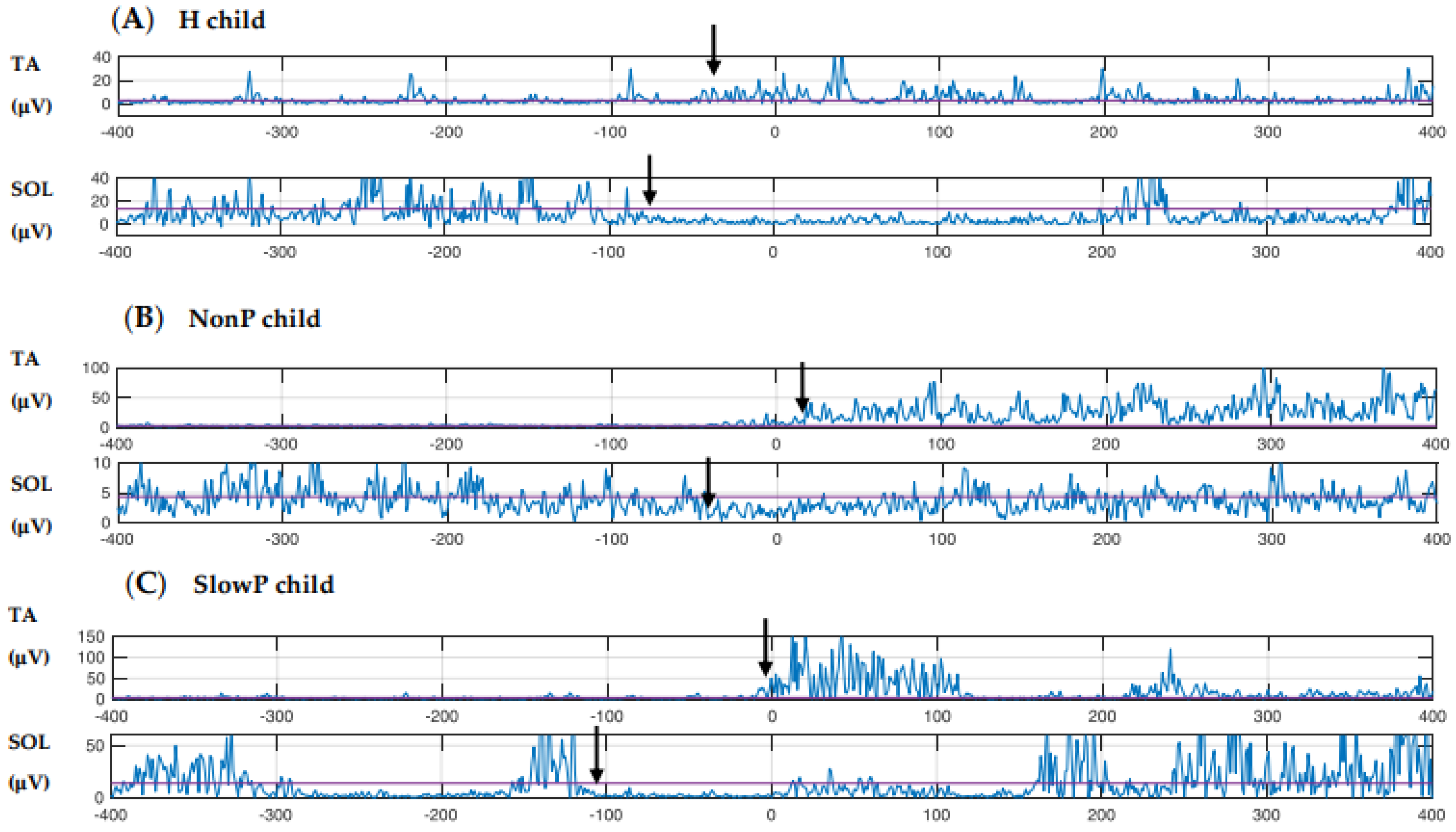
Of note, in the control group, the inhibition of the stance leg SOL started about 40 ms prior to TA excitation. While this timing was overall preserved in the children of the NonP group (about 60 ms), it was effectively increased in children with SlowP (about 100 ms, Figure 2). Similar changes were detected also for the swing leg.

4. Discussion

The aim of this study was to describe the postural control adopted by children with PCA during quiet stance and gait initiation, in order to draw considerations on the role of the cerebellum in the development of postural control. As a main result, we observed that in patients with slow progressive PCA, i.e., SlowP, both the static and dynamic components of postural control were disturbed, while the postural behavior of children with non-progressive PCA, i.e., NonP, was much similar to that of healthy children.
During the maintenance of upright posture, children with SlowP showed an increased ellipse area, mainly due to large mediolateral oscillations of the CoP. Considering CoP oscillations in anteroposterior direction too, this resulted in a general reduction of the ellipse eccentricity, outlining an omnidirectional decrease of stability. This finding was in agreement with the results described in adults with cerebellar lesions [26]. No statistical posturographic differences were instead found between children with NonP and H participants.
Gait initiation parameters during the imbalance and unloading phases remained substantially unchanged in patients of both pathological groups compared to H controls. Also, this observation fitted with previous results obtained in adults with cerebellar ataxia [21,40]. First step length and velocity showed instead a marked reduction in children with SlowP with respect to H, possibly reflecting a compensatory strategy for their poor balance control, and in agreement with what has been previously described in adults [21].
Electromyographic data, despite the roughness of the descriptive approach, suggested that patients with NonP and SlowP suffered more alterations in the temporal (when) than in the spatial distribution (to what muscle) and in the sign of activity (how, i.e., excitation or inhibition). This aspect agreed with the general view that assigns to the cerebellum the role of a “timing-machine” [41,42,43,44,45] and leaves the pattern selection to other brain structures, like the basal ganglia. Such a perspective has been confirmed also for what regards APAs in adults [46,47].
A short comment also deserves TA and SOL reciprocal activation. Despite our analysis was descriptive, patients with PCA and healthy children displayed the classical anticipatory postural pattern, characterized by SOL inhibition followed by TA activation of the stance limb (see Introduction). However, the latency between SOL and TA activity in the healthy group (about 40 ms) was consistent with what reported by Isaias et al. (2014) [18] and much lower than what has been found in adults (about 100 ms, [7]). This observation supported the choice of devoting a paper to gait initiation in children and, at the same time, suggested that the present H group, despite scarce, well represents the underlying population. On the contrary, indications of altered timing were observed in patients with PCA, in which the SOL-TA latency slightly increased to about 60 ms in children with NonP and attained about 100 ms in children with SlowP. This suggested a framework of abnormal feed-forward muscle synergies [21,24].
In summary, the reported differences in postural behavior between children with typical development and children affected by PCA support our hypothesis that the cerebellum plays a role also during the key phase of human maturation, in particular, in building internal models of gait initiation dynamics. This finding further stresses the importance of including postural training exercises in the rehabilitation programs for these pathologies [48]. Moreover, the observation that the gait initiation protocol allowed distinguishing motor deficits in children with SlowP vs. NonP, although the corresponding SARA scores were comparable, suggests that such protocol may be useful to monitor the evolution of motor deficits over time. The following two subsections are devoted to discussing the putative reasons for the different motor patterns we observed in patients of the two groups, as well as the resulting clues about the compensation mechanisms.

4.1. Disease Progression and Postural Behavior

When looking to the present results as a whole, children with SlowP seem to have a worse postural behavior with respect to both children of the NonP and H groups. This result is unlikely related to the severity of the pathology since all patients had a homogeneous SARA score, which, in turn, indicates comparable motor deficits in clinical terms. Therefore, the difference might stem either from the kind of cerebellar lesion (generalized atrophy vs. vermian hypoplasia) or from the progressive or non-progressive nature of the pathology. In this regard, children with SlowP suffered from generalized cerebellar atrophy, which represents macroscopic neuronal death, and received an ascertained clinical and/or radiological diagnosis of slow progression. On the contrary, Joubert syndrome, affecting children of the NonP group, is a congenital malformation that causes anomalous organogenesis of both the cerebellar vermis and peduncles. Therefore, it has an intrinsically stable nature along with the growth of the subject. In fact, once the organogenesis is completed, the vermian hypoplasia remains stable throughout the patient’s lifetime.
It could be argued that our observation of worse postural control in children with SlowP may be related to the larger extent of their cerebellar compromission (generalized atrophy vs. vermian hypoplasia). However, literature reports an emblematic case that contrasts with this interpretation. In fact, Titomanlio et al. (2005) [49] published a case report in which a 17-year-old subject with complete cerebellar agenesis showed only mild ataxia with slight dysmetria and moderate mental retardation, but no difficulty in attaining very complex motor tasks. Such evident functional compensation could be explained only through the plasticity of the remaining brain areas, which had to cope with a lesion that is stable since embryogenesis. This report suggests restricting the hypothesis to the progressive nature of the pathology.
Returning to the present study, we envisage that children with NonP could use the plasticity of their intact brain areas, which may include the cerebellar hemispheres, to effectively compensate for their stable lesion and attain an almost normal psychomotor development. On the contrary, children with SlowP suffer from a continuous cerebellar degeneration, which conflicts with the consolidation of compensatory functional strategies. This perspective not only fits with the gradual worsening of postural deficits we documented here when passing from healthy children to patients with NonP and to patients with SlowP but would also explain why patients with adult-onset cerebellar lesions show even more pronounced postural deficits [46]. Indeed, neural plasticity gradually but consistently decreases over the lifespan [50].

4.2. Putative Compensatory Network

Finally, it remains to figure out which neural substrate could be involved in functional compensation. In this regard, it is interesting to highlight recent evidence showing subcortical bidirectional connections between the basal ganglia and the cerebellum [51,52,53].
The functional role of the basal ganglia to cerebellum connections has been deeply investigated. Indeed, it has been observed that patients with Parkinson’s disease (PD) show abnormal functioning also in the cerebellum [54,55]. A SPECT study in patients with PD confirms an increased cerebellar activity when the effect of the anti-parkinsonian drug extinguishes [56]. Considering the reciprocal connectivity, it is of interest that functional MRI has shown increased putamen-cerebellar activity in patients with PD performing simple motor tasks and that greater putamen-cerebellar connectivity is significantly correlated with better motor performance. On the contrary, the administration of levodopa, which compensates the low endogenous dopamine production in patients with PD, has reduced this connectivity, relieving the cerebellum from its compensatory task [57]. It has also been observed that the compensatory role of the cerebellum contributes to preventing the full manifestation of the typical motor symptoms during the initial stage of PD; this compensatory ability saturates with time, leading these patients to develop cerebellar symptoms too [58].
These pieces of evidence allow arguing that, reciprocally, intact basal ganglia may compensate for cerebellar deficits. This hypothesis is still to be demonstrated, but should it be proved, it would provide a straightforward explanation for the graded postural impairments we found in children affected by PCA, as well as for the worse impairments reported in adult patients with cerebellar ataxia [46]. Evidence in this regard might come from functional MRI and diffusion tensor imaging techniques.

4.3. Limitations of the Study

The two main limits of the present study were the small number of participants and the low number of valid trials recorded in each of them. In particular, we could not observe APAs in all subjects, and this prevented a statistical analysis of EMG data. While it could be feasible to recruit more H subjects, the rarity of PCAs limited the number of children that precisely fall within the SlowP or NonP groups. With regard to the low number of valid trials, it could be increased only by prolonging the experimental session, which would quickly become burdensome for children, and especially for those affected by PCA.

5. Conclusions

Although all children with PCA showed clinically similar motor impairments, only children with SlowP were less stable in standing and showed a significantly shorter and slower first step than healthy children. Also, the descriptive EMG analysis in lower limb and back muscles pointed to a worse postural control in children of the SlowP group. On the basis of recent literature, we proposed that such different behavior stems from the disease progression, which interferes with the consolidation of compensatory strategies in children with SlowP but not in those affected by NonP.

Author Contributions

Conceptualization, funding acquisition, and supervision, P.C.; formal analysis, V.F., C.P. (Chiara Palmisano), S.M.M., C.M.M.S., and R.E.; investigation V.F., S.M.M., and C.M.M.S.; software, V.F., C.P. (Chiara Palmisano), and R.E.; clinical resources, S.D., C.P. (Chiara Pantaleoni), A.A., and N.N.; writing—original draft preparation, V.F., S.M.M., and C.P. (Chiara Palmisano); writing—review and editing, V.F., C.P. (Chiara Palmisano), S.M.M., R.E., P.C., S.D., C.P. (Chiara Pantaleoni), A.A., and N.N. All authors have read and agreed to the published version of the manuscript.

Funding

This study was supported by Fondazione Mariani for Child Neurology.

Conflicts of Interest

The authors declare no conflict of interest.

References

  1. Aruin, A.S.; Latash, M.L. The role of motor action in anticipatory postural adjustments studied with self-induced and externally triggered perturbations. Exp. Brain Res. 1995, 106, 291–300. [Google Scholar] [CrossRef] [PubMed]
  2. Ito, T.; Azuma, T.; Yamashita, N. Anticipatory control related to the upward propulsive force during the rising on tiptoe from an upright standing position. Eur. J. Appl. Physiol. 2004, 92, 10–186. [Google Scholar] [CrossRef] [PubMed]
  3. Bouisset, S.; Zattara, M. Biomechanical study of the programming of anticipatory postural adjustments associated with voluntary movement. J. Biomech. 1987, 20, 735–742. [Google Scholar] [CrossRef]
  4. Cordo, P.J.; Nashner, L.M. Properties of postural adjustments associated with rapid arm movements. J. Neurophysiol. 1982, 47, 287–302. [Google Scholar] [CrossRef]
  5. Marsden, C.D.; Merton, P.A.; Morton, H.B. Human postural responses. Brain 1981, 104, 513–534. [Google Scholar] [CrossRef]
  6. Massion, J. Movement, posture and equilibrium: Interaction and coordination. Prog. Neurobiol. 1992, 35–56. [Google Scholar] [CrossRef]
  7. Crenna, P.; Frigo, C. A motor programme for the initiation of forward-oriented movements in humans. J. Physiol. 1991, 437, 635–653. [Google Scholar] [CrossRef]
  8. Nissan, M.; Whittle, M.W. Initiation of gait in normal subjects: A preliminary study. J. Biomed. Eng. 1990, 12, 165–171. [Google Scholar] [CrossRef]
  9. Burleigh, A.L.; Horak, F.B.; Malouin, F. Modification of postural responses and step initiation: Evidence for goal-directed postural interactions. J. Neurophysiol. 1994, 72, 2892–2902. [Google Scholar] [CrossRef]
  10. Burleigh, A.; Horak, F. Influence of instruction, prediction, and afferent sensory information on the postural organization of step initiation. J. Neurophysiol. 1996, 75, 1619–1628. [Google Scholar] [CrossRef]
  11. McIlroy, W.E.; Maki, B.E. Do anticipatory postural adjustments precede compensatory stepping reactions evoked by perturbation? Neurosci. Lett. 1993, 164, 199–202. [Google Scholar] [CrossRef]
  12. Yiou, E.; Caderby, T. Balance control during gait initiation: State-of-the-art and research perspectives. World J. Orthop. 2017, 8, 815–828. [Google Scholar] [CrossRef] [PubMed]
  13. Crenna, P.; Carpinella, I.; Rabuffetti, M.; Rizzone, M.; Lopiano, L.; Lanotte, M.; Ferrarin, M. Impact of subthalamic nucleus stimulation on the initiation of gait in Parkinson’s disease. Exp. Brain Res. 2006, 172, 519–532. [Google Scholar] [CrossRef] [PubMed]
  14. Wang, Y.; Zatsiorsky, V.M.; Latash, M.L. Muscle synergies involved in shifting the center of pressure while making a first step. Exp. Brain Res. 2005, 167, 196–210. [Google Scholar] [CrossRef] [PubMed]
  15. Ledebt, A.; Bril, B.; Brenière, Y. The build-up of anticipatory behaviour: An analysis of the development of gait initiation in children. Exp. Brain Res. 1998, 120, 9–17. [Google Scholar] [CrossRef] [PubMed]
  16. Assaiante, C.; Woollacott, M.; Amblard, B. Development of postural adjustment during gait initiation: Kinematic and EMG analysis. J. Mot. Behav. 2009, 32, 211–226. [Google Scholar] [CrossRef]
  17. Malouin, F.; Richards, C.L. Preparatory adjustments during gait initiation in 4–6-year-old children. Gait Posture 2000, 11, 239–253. [Google Scholar] [CrossRef]
  18. Isaias, I.U.; Dipaola, M.; Michi, M.; Marzegan, A.; Volkmann, J.; Roidi, M.L.R.; Frigo, C.A.; Cavallari, P. Gait initiation in children with rett syndrome. PLoS ONE 2014, 9, e92736. [Google Scholar] [CrossRef]
  19. Dipaola, M.; Frigo, C.A.; Cavallari, P.; Iasias, I.U. Alterations of load transfer mechanism during gait initiation in Parkinson’s disease. In Proceedings of the EHB 2017 IEEE International Conference on e-Health and Bioengineering, Sinaia, Romania, 22–24 June 2017; pp. 579–582. [Google Scholar] [CrossRef]
  20. Timmann, D.; Horak, F.B. Perturbed step initiation in cerebellar subjects 1. Modifications of postural responses. Exp. Brain Res. 1998, 119, 73–84. [Google Scholar] [CrossRef]
  21. Timmann, D.; Horak, F.B. Perturbed step initiation in cerebellar subjects: 2. Modification of anticipatory postural adjustments. Exp. Brain Res. 2001, 141, 110–120. [Google Scholar] [CrossRef]
  22. Martino, G.; Ivanenko, Y.P.; Serrao, M.; Ranavolo, A.; D’Avella, A.; Draicchio, F.; Conte, C.; Casali, C.; Lacquaniti, F. Locomotor patterns in cerebellar ataxia. J. Neurophysiol. 2014, 112, 2810–2821. [Google Scholar] [CrossRef] [PubMed]
  23. Ilg, W.; Golla, H.; Thier, P.; Giese, M.A. Specific influences of cerebellar dysfunctions on gait. Brain 2007, 130, 786–798. [Google Scholar] [CrossRef] [PubMed]
  24. Asaka, T.; Wang, Y. Feedforward postural muscle modes and multi-mode coordination in mild cerebellar ataxia. Exp. Brain Res. 2011, 210, 153–163. [Google Scholar] [CrossRef] [PubMed]
  25. Serrao, M.; Pierelli, F.; Ranavolo, A.; Draicchio, F.; Conte, C.; Don, R.; Di Fabio, R.; Lerose, M.; Padua, L.; Sandrini, G.; et al. Gait pattern in inherited cerebellar ataxias. Cerebellum 2012, 11, 194–211. [Google Scholar] [CrossRef] [PubMed]
  26. Morton, S.M.; Bastian, A.J. Cerebellar control of balance and locomotion. Neuroscientist 2004, 10, 247–259. [Google Scholar] [CrossRef]
  27. Bastian, A.J. Learning to predict the future: The cerebellum adapts feedforward movement control. Curr. Opin. Neurobiol. 2006, 645–649. [Google Scholar] [CrossRef]
  28. Stoodley, C.J. The cerebellum and neurodevelopmental disorders. The Cerebellum 2017, 15, 34–37. [Google Scholar] [CrossRef]
  29. Valence, S.; Cochet, E.; Rougeot, C.; Garel, C.; Chantot-Bastaraud, S.; Lainey, E.; Afenjar, A.; Barthez, M.A.; Bednarek, N.; Doummar, D.; et al. Exome sequencing in congenital ataxia identifies two new candidate genes and highlights a pathophysiological link between some congenital ataxias and early infantile epileptic encephalopathies. Genet. Med. 2019, 21, 553–563. [Google Scholar] [CrossRef]
  30. Romani, M.; Micalizzi, A.; Valente, E.M. Joubert syndrome: Congenital cerebellar ataxia with the molar tooth. Lancet Neurol. 2013, 12, 894–905. [Google Scholar] [CrossRef] [Green Version]
  31. Prieto, T.E.; Myklebust, J.B.; Hoffmann, R.G.; Lovett, E.G.; Myklebust, B.M. Measures of postural steadiness: Differences between healthy young and elderly adults. IEEE Trans. Biomed. Eng. 1996, 43, 956–966. [Google Scholar] [CrossRef]
  32. Rocchi, L.; Chiari, L.; Horak, F.B. The effects of deep brain stimulation and levodopa on postural sway in subjects with Parkinson’s disease. J. Neurol. Neurosurg. Psychiatry 2002, 73, 240. [Google Scholar] [CrossRef] [PubMed]
  33. Schmitz-Hübsch, T.; Du Montcel, S.T.; Baliko, L.; Berciano, J.; Boesch, S.; Depondt, C.; Giunti, P.; Globas, C.; Infante, J.; Kang, J.S.; et al. Scale for the assessment and rating of ataxia: Development of a new clinical scale. Neurology 2006, 66, 1717–1720. [Google Scholar] [CrossRef] [PubMed]
  34. Orphanet. Available online: https://www.orpha.net (accessed on 27 January 2020).
  35. Hiraoka, K.; Hatanaka, R.; Nikaido, Y.; Jono, Y.; Nomura, Y.; Tani, K.; Chujo, Y. Asymmetry of anticipatory postural adjustment during gait initiation. J. Hum. Kinet. 2014, 42, 7–14. [Google Scholar] [CrossRef] [Green Version]
  36. Ferrari, A.; Benedetti, M.G.; Pavan, E.; Frigo, C.; Bettinelli, D.; Rabuffetti, M.; Crenna, P.; Leardini, A. Quantitative comparison of five current protocols in gait analysis. Gait Posture 2008, 28, 207–216. [Google Scholar] [CrossRef] [PubMed]
  37. Hermens, H.J.; Freriks, B.; Disselhorst-Klug, C.; Rau, G. Development of recommendations for SEMG sensors and sensor placement procedures. J. Electromyogr. Kinesiol. 2000, 10, 361–374. [Google Scholar] [CrossRef]
  38. Hof, A.L. Scaling gait data to body size. Gait Posture 1996, 222–223. [Google Scholar] [CrossRef]
  39. Marchese, S.M.; Esposti, R.; Bolzoni, F.; Cavallari, P. Transcranial Direct Current Stimulation on Parietal Operculum Contralateral to the Moving Limb Does Not Affect the Programming of Intra-Limb Anticipatory Postural Adjustments. Front. Physiol. 2019, 10, 1–9. [Google Scholar] [CrossRef]
  40. Mummel, P.; Timmann, D.; Krause, U.W.H.; Boering, D.; Thilmann, A.F.; Diener, H.C.; Horak, F.B. Postural responses to changing task conditions in patients with cerebellar lesions. J. Neurol. Neurosurg. Psychiatry 1998, 65, 734–742. [Google Scholar] [CrossRef]
  41. Ivry, R.B.; Keele, S.W. Timing functions of the cerebellum. J. Cogn. Neurosci. 1989, 1, 136–152. [Google Scholar] [CrossRef]
  42. Ivry, R.B.; Spencer, R.M.; Zelaznik, H.N.; Diedrichsen, J. The cerebellum and event timing. Ann. N. Y. Acad. Sci. 2002, 302–317. [Google Scholar] [CrossRef]
  43. Cerri, G.; Esposti, R.; Locatelli, M.; Cavallari, P. Coupling of hand and foot voluntary oscillations in patients suffering cerebellar ataxia: Different effect of lateral or medial lesions on coordination. Prog. Brain Res. 2005, 148, 227–241. [Google Scholar] [CrossRef] [PubMed]
  44. D’Angelo, E. Rebuilding cerebellar network computations from cellular neurophysiology. Front. Cell. Neurosci. 2010, 4, 131. [Google Scholar] [CrossRef] [Green Version]
  45. Bareš, M.; Apps, R.; Avanzino, L.; Breska, A.; D’Angelo, E.; Filip, P.; Gerwig, M.; Ivry, R.B.; Lawrenson, C.L.; Louis, E.D.; et al. Consensus paper: Decoding the contributions of the cerebellum as a time machine. from neurons to clinical applications. Cerebellum 2019, 18, 266–286. [Google Scholar] [CrossRef] [PubMed] [Green Version]
  46. Bruttini, C.; Esposti, R.; Bolzoni, F.; Vanotti, A.; Mariotti, C.; Cavallari, P. Temporal disruption of upper-limb anticipatory postural adjustments in cerebellar ataxic patients. Exp. Brain Res. 2015, 233, 197–203. [Google Scholar] [CrossRef] [PubMed]
  47. Bolzoni, F.; Esposti, R.; Marchese, S.M.; Pozzi, N.G.; Ramirez-Pasos, U.E.; Isaias, I.U.; Cavallari, P. Disrupt of intra-limb APA pattern in Parkinsonian patients performing index-finger flexion. Front. Physiol. 2018, 9, 1745. [Google Scholar] [CrossRef] [PubMed]
  48. Marquer, A.; Barbieri, G.; Pérennou, D. The assessment and treatment of postural disorders in cerebellar ataxia: A systematic review. Ann. Phys. Rehabil. Med. 2014, 57, 67–78. [Google Scholar] [CrossRef] [Green Version]
  49. Titomanlio, L.; Romaniello, R.; Borgatti, R. Cerebellar agenesis. Handb. Cereb. Cereb. Disord. 2005, 1855–1872. [Google Scholar] [CrossRef] [Green Version]
  50. Lu, C.; Huffmaster, S.L.A.; Harvey, J.C.; MacKinnon, C.D. Anticipatory postural adjustment patterns during gait initiation across the adult lifespan. Gait Posture 2017, 57, 182–187. [Google Scholar] [CrossRef]
  51. Bostan, A.C.; Dum, R.P.; Strick, P.L. The basal ganglia communicate with the cerebellum. Proc. Natl. Acad. Sci. USA 2010, 107, 8452–8456. [Google Scholar] [CrossRef] [Green Version]
  52. Bostan, A.C.; Dum, R.P.; Strick, P.L. Cerebellar networks with the cerebral cortex and basal ganglia. Trends Cogn. Sci. 2013, 17, 241–254. [Google Scholar] [CrossRef] [Green Version]
  53. Hoshi, E.; Tremblay, L.; Féger, J.; Carras, P.L.; Strick, P.L. The cerebellum communicates with the basal ganglia. Nat. Neurosci. 2005, 8, 1491–1493. [Google Scholar] [CrossRef] [PubMed]
  54. Wu, T.; Hallett, M. The cerebellum in Parkinson’s disease. Brain 2013, 136, 696–709. [Google Scholar] [CrossRef] [PubMed] [Green Version]
  55. Yu, H.; Sternad, D.; Corcos, D.M.; Vaillancourt, D.E. Role of hyperactive cerebellum and motor cortex in Parkinson’s disease. Neuroimage 2007, 35, 222–233. [Google Scholar] [CrossRef] [PubMed] [Green Version]
  56. Rascol, O.; Sabatini, U.; Fabre, N.; Brefel, C.; Loubinoux, I.; Celsis, P.; Senard, J.M.; Montastruc, J.L.; Chollet, F. The ipsilateral cerebellar hemisphere is overactive during hand movements in akinetic parkinsonian patients. Brain 1997, 120, 103–110. [Google Scholar] [CrossRef] [Green Version]
  57. Simioni, A.C.; Dagher, A.; Fellows, L.K. Compensatory striatal-cerebellar connectivity in mild-moderate Parkinson’s disease. NeuroImage Clin. 2016, 10, 54–62. [Google Scholar] [CrossRef] [Green Version]
  58. Martinu, K.; Monchi, O. Cortico-basal ganglia and cortico-cerebellar circuits in Parkinson’s disease: Pathophysiology or compensation? Behav. Neurosci. 2013, 127, 222–236. [Google Scholar] [CrossRef]
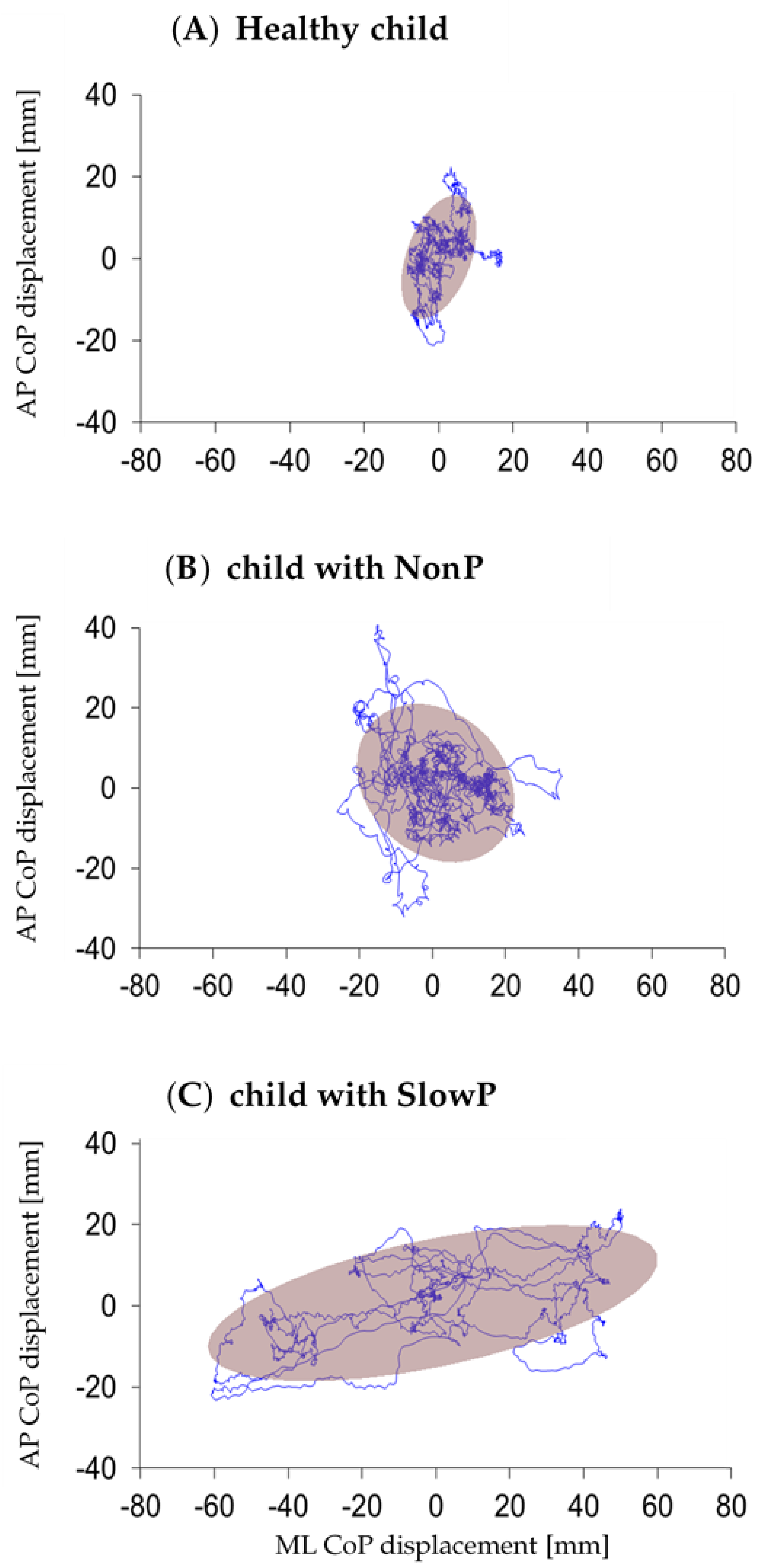
Figure 1. Statokinesigram, with 95% confidence ellipse, for representative children of the three groups: healthy (H, panel A), with non-progressive PCA (NonP, B), and with slow progressive PCA (SlowP, C). ML: mediolateral; AP: anteroposterior; PCA: pediatric cerebellar ataxia.
Applsci 10 01606 g001
Figure 2. EMG (electromyographic) of shank muscles of the stance leg. Comparison among healthy children H, panel (A), children with non-progressive PCA NonP, (B) and children with slow progressive PCA SlowP, (C). One representative subject for each group. Time 0: APA (anticipatory postural adjustment) onset, defined as the first backward shift of the CoP (center of pressure). Black arrows show SOL (soleus) inhibition and the following TA (tibialis anterior) excitation. Note that the time delay between these two reciprocal actions gradually increased in children with NonP and SlowP with respect to H children.
Applsci 10 01606 g002
Table 1. Demographic and clinical characteristics of children with PCA. SlowP: slowly progressive ataxia; NonP: non-progressive ataxia (Joubert syndrome); EXOSC3: exosome component 3; KCNC3: potassium voltage-gated channel subfamily C member 3; ADCK3: aarF domain-containing kinase 3; NPHP1: nephrocystin 1; AHI1: Abelson helper integration site 1; SUFU: negative regulator of hedgehog signaling; PCA: pediatric cerebellar ataxia. Details about a molecular diagnosis can be found at [34].
PatientAgeGenderMolecular DiagnosisSARA
SlowP_019Mmutation in a candidate gene8
SlowP_028Mmutation in a candidate gene14
SlowP_0312MEXOSC3: c.572G > A15
SlowP_0413FKCNC3: c.1268G > A17
SlowP_0513Fto be evaluated13
SlowP_0616FADCK3: c547C > T; c1042C > T13
SlowP_0717Mto be evaluated18
NonP_0110MNPHP1: c.1358G > T; c.1438-4C > T12
NonP_0212Fto be evaluated15
NonP_0318Mto be evaluated14
NonP_049MAHI1: c.1829G > C; c.2671C > T15
NonP_0512FSUFU: c.1217T > C11
NonP_069MSUFU: c.1217T > C15.5
Table 2. Postural parameters during quiet stance. Data are shown as median value (range) for children with slow progressive PCA (SlowP), children with non-progressive PCA (NonP), and healthy children (H). ML: mediolateral; AP: anteroposterior; CoP: center of pressure. * Significant difference between SlowP and H groups, Dunn test p < 0.05.
SlowPNonPH
CoP length (mm)1924(1222 to 2983)1481(893 to 2911)1594(895 to 2309)
Average CoP velocity (mm/s)64(40 to 99)49(29 to 110)53(29 to 76)
ML range (mm)* 37 (24 to 97)29.72(13 to 85)* 19(11 to 33)
AP range (mm)39(28 to 57)31(18.39 to 77.96)26(15 to 60)
Ellipse area (mm2)* 564 (354 to 2857)447(123 to 2075)* 208(54 to 609)
Ellipse eccentricity0.68(0.48 to 0.87)0.73(0.65 to 0.93)0.79(0.51 to 0.83)
Table 3. Postural parameters during the imbalance and unloading phases. Data are shown as median value (range) for children with slow progressive PCA (SlowP), children with non-progressive PCA (NonP), and healthy children (H). ML: mediolateral, positive towards the swing foot; AP: anteroposterior, positive when forwards; CoP→COM: horizontal distance from CoP to COM.
SlowPNonPH
IMBALANCEPhase duration (s)0.41(0.25 to 1.03)0.42(0.31 to 1.90)0.31(0.25 to 0.53)
CoP length (mm)34(24 to 100)70(33 to 134)42(17 to 68)
ML CoP shift (mm)15.99(13 to 28)39.80(−6 to 67)25.95(8 to 49)
AP CoP shift (mm)−18(−43 to −5)−25(−43 to −11)−14(−41 to −8)
ML COM shift (mm)−7.33(−14.67 to −2)−6(−9.67 to 2)−7(−13.67 to −2.33)
AP COM shift (mm)4.33(−0.67 to 20.33)6(2 to 8.67)6(2.33 to 11.67)
ML CoP→ COM (mm)−25(−54.67 to −16.67)−30.75(−49.25 to 54)−37.33(−56.67 to −18)
AP CoP → COM (mm)24.67(2.67 to 62.67)28.83(12 to 47.33)21(12 to 56)
UNLOADINGPhase duration (s)0.46(0.21 to 1.53)0.33(0.23 to 1.20)0.41(0.21 to 0.56)
CoP length (mm)172(112 to 202)134(62 to 175)143(90 to 172)
ML CoP shift (mm)−104(−147 to −48)−127(−168 to −9)−121(−152 to −76)
AP CoP shift (mm)9(−12 to 28)−1(−26 to 17)−9(−59 to 9)
ML COM shift (mm)−34.67(−66.67 to −21)−34.37(−49 to −17)−35.67(−53.33 to −22)
AP COM shift (mm)43(21.67 to 47.33)35.08(12 to 55.5)38.67(28 to 55.33)
ML CoP → COM (mm)44.25(33.67 to 90.33)50.96(−7 to 62.67)46.67(23 to 61.67)
AP CoP → COM (mm)51(37.33 to 94.33)67.75 (29.33 to 106)80(57.33 to 108.5)
Table 4. First swing parameters. Data are shown as median value (range) for children with slow progressive PCA (SlowP), children with non-progressive PCA (NonP), and healthy children (H). LL: lower limb length; Fr: Froude number; * significant difference between SlowP and H groups, Dunn test p < 0.05.
SlowPNonPH
First step length (%LL)* 47.34 (38.39 to 58.70)62.93(51.83 to 72.67)* 72.99(47.46 to 83.61)
First step velocity (Fr)* 0.24(0.12 to 0.32)0.31(0.24 to 0.39)* 0.39(0.26 to 0.49)
Table 5. Latencies (ms) of postural EMG changes with respect to the APA onset (time 0). Median, minimum, and maximum values, together with the number of subjects (n) in which APAs could be identified, for children with slow progressive PCA (SlowP), children with non-progressive PCA (NonP), and healthy children (H). EMG: electromyographic; APA: anticipatory postural adjustment.
SlowP NonP H
STANCEErector spinaeinhibition−68(−85 to −52)n = 2−79(−109 to −49)n = 2−102(−115 to −80)n = 3
Biceps femorisinhibition−70(−70 to −70)n = 4−68(−84 to −64)n = 3−100(−187 to 119)n = 7
Soleusinhibition−72(−199 to 63)n = 6−98(−130 to −63)n = 3−72(−146 to −25)n = 5
Rectus femorisexcitation15(−32 to 141)n = 4−35(−112 to 22)n = 5−72(−110 to 57)n = 5
Tibialis anteriorexcitation31(−52 to 66)n = 5−41(−49 to 26)n = 5−31(−49 to 108)n = 7
SWINGErector spinaeinhibition−55(−99 to 2)n = 3−111(−127 to −95)n = 2−90(−263 to 100)n = 4
Biceps femorisinhibition−71(−90 to −39)n = 4−136(−160 to −113)n = 2−94(−199 to 70)n = 5
Soleusinhibition−98(−184 to −35)n = 6−53(−100 to −24)n = 4−121(−259 to −111)n = 5
Rectus femorisexcitation3(−40 to 24)n = 3−11(−83 to 162)n = 468(−80 to 113)n = 3
Tibialis anteriorexcitation−2(−51 to 145)n = 46(−67 to 45)n = 4−61(−101 to 6)n = 6