Article

Exercise Training of Secreted Protein Acidic and Rich in Cysteine (Sparc) KO Mice Suggests That Exercise-Induced Muscle Phenotype Changes Are SPARC-Dependent

1
Department of Molecular Medicine, Faculty of Medicine, Laval University, Québec, QC G1V 0A6, Canada
2
Functional Genomics Laboratory, Endocrinology and Nephrology Axis, CHU de Québec-Université Laval Research Center, Québec, QC G1V 4G2, Canada
*
Author to whom correspondence should be addressed.
Appl. Sci. 2020, 10(24), 9108; https://doi.org/10.3390/app10249108
Submission received: 1 December 2020 / Revised: 14 December 2020 / Accepted: 15 December 2020 / Published: 20 December 2020
(This article belongs to the Special Issue New Trends in Sport and Exercise Medicine)

Abstract

:

Featured Application

This work highlights secreted protein acidic and rich in cysteine (SPARC) and its pathways as pharmacological targets/tools for conditions and diseases in which muscle properties enhancement would provide therapeutic benefits.

Abstract

We previously identified secreted protein acidic and rich in cysteine (Sparc) as an exercise-induced gene in young and elderly individuals. Via this animal experiment, we aim to identify selected implications of SPARC mainly within the muscle in the contexts of exercise. Mice were divided into eight groups based on three variables (age, genotype and exercise): Old (O) or young (Y) × Sparc knock-out (KO) or wild-type (WT) × sedentary (Sed) or exercise (Ex). The exercised groups were trained for 12 weeks at the lactate threshold (LT) speed (including 4 weeks of adaptation period) and all mice were sacrificed afterwards. Body and selected tissues were weighed, and lactate levels in different conditions measured. Expression of skeletal muscle (SM) collagen type I alpha 1 chain (COL1A1) and mitochondrially encoded cytochrome c oxidase I (MT-CO1) in addition to SM strength (grip power) were also measured. Ageing increased the body and white adipose tissue (WAT) weights but decreased SM weight percentage (to body weight) and MT-CO1 expression (in WT). Exercise increased SM COL1A1 in WT mice and MT-CO1 expression, as well as weight percentage of the tibialis anterior muscle, and decreased WAT weight (trend). Compared to WT mice, Sparc KO mice had lower body, muscle and WAT weights, with a decrease in SM MT-CO1 and COL1A1 expression with no genotype effect on lactate levels in all our blood lactate measures. Sparc KO effects on body composition, adiposity and metabolic patterns are toward a reduced WAT and body weight, but with a negative metabolic and functional phenotype of SM. Whereas such negative effects on SM are worsened with ageing, they are relatively improved by exercise. Importantly, our data suggest that the exercise-induced changes in the SM phenotype, in terms of increased performance (metabolic, strength and development), including lactate-induced changes, are SPARC-dependent.

1. Secreted Protein Acidic and Rich in Cysteine as an Exercise-Induced Gene

The modern lifestyle, characterized by the lack of physical activity combined with an unhealthy diet, leads to an increase in health problems typical of our era such as obesity and diabetes. In addition, the improvement of health care systems increased the life expectancy and, therefore, geriatric health problems, such as sarcopenia, are also increasing. Interestingly, exercise is considered as a “panacea” for many of these problems [1]. Whereas exercise benefits have been widely documented, many exercise-related molecular mechanisms are yet to be fully elucidated. In addition to its direct metabolic implications, skeletal muscles (SM) represent secretary organs producing myokine, such as secreted protein acidic and rich in cysteine (SPARC) [2]. Within the exercise context, functional genomics studies (mainly but not only in the energy metabolism context [3]) have identified genes related to physical activity among which we have SPARC/Sparc. Indeed, beyond the known implications of SPARC in wound healing and tissue repair [4,5], this gene was characterized (for the first time) as an exercise-induced gene [6] and also as an electrical pulse stimulation (considered as an in vitro model of exercise)-induced gene in muscular cells [7]. Moreover, SPARC secretion is induced by exercise [8,9,10] after which the concentrations of myokines (including SPARC, interleukin 6 and fibroblast growth factor) increase in the circulation [11]. Therefore, we hypothesize that at least some of the exercise benefits and biological consequences, mainly the muscular phenotype adaptation to exercise, would be mediated by SPARC or the pathways it controls. Therefore, in this study we aim to explore the implication of Sparc (via it knock-out (KO)) in mice with a focus on exercise effects on muscles. In addition, age was also introduced as a variable in this study. Therefore, we would find out the combinatory impacts of Sparc KO, exercise and age on selected patterns related to SM physiological properties and metabolic performance. We explore the lactate levels and their implications with the SM phenotype changes (both structural and metabolic) in a SPARC-dependent way.

2. Animal Experimental Design, Material and Methods

Our study was carried out on male mice and involved both wild-type (WT) mice (C57BL/6J, the most commonly used strain for genetic and/or transgenic study that also consistently showed the highest level of voluntary wheel-running [12]) and Sparc KO mice (129/Sv-C57BL/6J) fed with chow diet (Teklad global 18% protein rodent diets [13]). Mice had access to food and water ad libitum during the whole experimental period (except for fasting periods during which they had access to water only). WT mice were from the Jackson Laboratory (https://www.jax.org/) and Sparc KO mice were generated via in vitro fertilization using Sparc KO mice sperm generously provided by Dr. Amy D. Bradshaw. Sparc KO mice of Dr. Amy D. Bradshaw were generated as previously described [14,15]. Each age-group of mice (young (Y) and old (O)) was divided based on the genotype (KO or WT) to obtain 4 groups: Y-KO, Y-WT, O-KO, O-WT. Finally, each of these 4 groups was further subdivided into two groups according to whether they were exercising (Ex) or sedentary (Sed) mice. Therefore, our experimental design included 8 groups: Y-WT-Sed, Y-WT-Ex, Y-KO-Sed, Y-KO-Ex, O-WT-Sed, O-WT-Ex, O-KO-Sed and O-KO-Ex. Each group had 11 to 12 mice (n). Mice were housed at the animal facility of the CHU de Québec-Université Laval Research Center (12-h light/dark cycle) and periodically checked by animal care technicians for health and wellness. The exercise groups were trained during the dark phase.
The exercising mice were trained during 12 weeks (starting at the age of 9 weeks for Y mice and 66 weeks for O mice) on running wheels (Lafayette instrument Co, Lafayette, IN, USA) placed horizontally (no angle adjustment). Whereas Y mice were sacrificed at the age of 21 weeks, old mice were sacrificed at the age of 78 weeks. Mice were sacrificed following a 12-h fasting (postprandial period) by cardiac puncture following isoflurane inhalation anesthesia. The coming sub-sections detail the measures performed before, during and after the training, as well as on and after the sacrifice day.
All animal experimentation was conducted in accord with the guidelines of the Canadian Council on Animal Care and approved by the Animal Protection Committee of Laval University (Identifications: 2014165 and 2014168). Mice with any type of illness were immediately euthanized by cervical dislocation and excluded from the study. Mice found with anatomical abnormalities (during the sacrifice) were also excluded from the study.

2.1. Mice Exercise Protocol and Running Speed Determination

At the beginning of the training, mice had an adaptation period of 4 weeks. During those 4 weeks, mice performed the incremental exercise. They were trained through a progressive (gradual) increase in both running speed and duration (up to their maximum endurance) throughout this adaptation step, at the end of which we determined the speed at the lactate threshold (LT). The LT level [16] is a parameter indicating the level of physical activity corresponding to the metabolic point at which the muscle production of lactate starts to increase and overcome the blood clearance of lactate. This indicates that the energy produced via oxidative phosphorylation is insufficient to meet energetic needs and, therefore, the muscles trigger anaerobic energy production that generates lactate at a level superior to its blood clearance. At the end of the adaptation period, LT levels were determined following a measure of running speed-dependent blood lactate level curves, based on previously reported protocols [12,17]. Briefly, the mouse run at a determined speed for 4 min, after which we immediately measure the blood lactate level (within 1 min), after that it ran at the next speed (higher) and again the blood lactate speed was measured. We repeated this procedure until the mouse was not able to run (cannot maintain the speed). At the end, we obtained the curve representing blood lactate levels corresponding to the different running speeds, based on which we obtained the speed at the LT. The blood lactate levels were measured, as described in the next section (1.2). The LT speed was chosen as a parameter for our study, based on evidence showing that exercise at LT generates metabolic and functional benefits, including improved insulin sensitivity, peripheral glucose effectiveness, lipid profile, blood pressure, physiological fitness [18,19,20,21] and body fat weight percentage decrease [6]. The LT was determined for each mouse of the exercise groups. After that, for each set of mice trained at the same period, the running speed of the 8 remaining weeks of training was a value chosen among the average values (range) of all the mice of that set.
The training was at those values close to the LT levels (LT speed) because it was the LT level that was the speed used during the study in which SPARC has been characterised as an exercise-induced gene [6]. In addition, the exercise frequency of our study (60 min/day, five times/week) was also similar to the same study [6]. However, we extended the duration from 6 weeks to 12 weeks to be able to easily see the impacts with significant differences between groups. The literature reported studies exploring the effect of exercise in which mice were both trained for longer periods (over 12 weeks) with the same frequency (60 min/day, five times/week) [22] and also at least as young and as old (3 and 19 months of age) [23] as the mice in this study. In addition, the life span for C57BL/6 mice is around 104 weeks (26 months) [24,25]. Therefore, our choices of mice ages, exercise speed and frequency were within a range of the mice’s abilities and did not damage their muscles, nor were they limited by physiological parameters. Importantly, since LT speed was chosen, based on evidence showing that exercise at LT generates metabolic and functional benefits, our study will provide additional data for molecular and biochemical explanations of such training benefits.
During mouse training, and unlike other protocols, no electrical [17] or any potentially harmful stimulations were used to force the mice to run. Only a light air stimulation (using a small hand air pump) was applied for mice in some cases to ensure, as much as possible, that all mice ran during the same period and at the same speed throughout the same training period (optimize the protocol for similar exercise amounts). Moreover, mice were always handled gently when taken from the cage to the training device and vice versa. In addition, sedentary mice were also transported to the exercise training room and kept in their cages, while exercising mice were trained so that all mice received a similar environmental (light, noise, etc.) stimuli. Thus, any possible impacts of stress or environmental stimuli on the performed measures were reduced to minimum.
The last training session was 48 h prior to the sacrifice so that the measures we obtained during the sacrifice and those performed on tissues afterwards did not reflect any possible acute effect of exercise, such as dehydration or neuroendocrine changes that could impact gene expression, post-exercise energy intake or expenditure, etc. Our study aimed to investigate the effects of the 12 weeks of exercise (chronic).

2.2. Fasting Lactate and Oral Glucose Gavage-Dependant Lactate Levels

As a resting metabolic indicator in the muscle, blood lactate levels were measured before and after glucose (prepared from 45% solution, Sigma-Aldrich Canada Co., Oakville, ON, Canada) oral gavage (2 mg of glucose per 1 g of body weight). Mice were fasted for 6 h prior to the glucose gavage. Each measure had 5 time points (0, 15, 30, 60 and 120 min after glucose gavage) allowing us to obtain a curve and calculate the area under the curve (AUC). This test was performed three times (a total of three AUCs)—before the training, at week 5 and the end of week 12 of the training. In addition, we also measured blood lactate (single measure) at the sacrifice day (following 12 h fasting) and at the end of the last training session. Blood lactate levels were measured via tail pricking with a needle to collect blood samples on lactate test strips that were then inserted into the lactate meter (Lactate scout, Sports Resource Group, Inc., Minneapolis, MN, USA).

2.3. Grip Power Test

At the end of the training period, the muscle strengths of all the mice were evaluated through performing a grip power test with a grip strength meter (Columbus Instruments International, Columbus, OH, USA). The grip strength was measured by allowing a mouse to grab (with four limbs) pull bar assemblies attached to the force transducer while the mouse was pulled horizontally by the tail away from the bars, similar to what has been previously described [26,27]. The peak force applied by the mouse (g) was then shown on a digital display.
This test was conducted five times (5 min apart) for each mouse, after which the forces (both mean and maximum) were calculated both as absolute values as well as being normalized to the body weights of the corresponding mice.

2.4. Body and Tissue Weights

Body and selected tissues were weighted at the sacrifice day. The selected tissues were the brain, pituitary gland, hypothalamus, liver, heart, aorta, white adipose tissue (WAT), SMs (gastrocnemius, soleus, tibialis anterior and extensor digitorum longus muscles). The values are reported both as tissue weights as well as a weight percentages (normalized) to the body weights of the corresponding mice.

2.5. Western Blotting

We measured the SM (tibialis anterior muscle) expression of two proteins—collagen alpha 1 type I (COL1A1) and mitochondrially encoded cytochrome c oxidase I (MT-CO1). Whereas COL1A1 is important in the structure of the muscle [28], MT-CO1 is an indicator of mitochondrial oxidative phosphorylation [29]. At the day of sacrifice, the tibialis anterior muscle was removed and quickly put in liquid nitrogen (snap frozen) then moved to −80 °C and kept until the protein extraction procedure. To measure the expression of both COL1A1 and MT-CO1, total proteins were extracted from the tibialis anterior muscle, using a radio-immunoprecipitation assay (RIPA) buffer and protease inhibitors cocktail (Sigma-Aldrich Canada Co., Oakville, ON, Canada) and followed by a protein quantification of each protein extract using Bio-Rad protein assay (Bio-Rad Laboratories Ltd., Mississauga, ON, Canada). Fifteen (MT-CO1) or ten (COL1A1) micrograms of proteins were separated by sodium dodecyl sulfate polyacrylamide gel electrophoresis (SDS-PAGE) using the TGX Stain-Free FastCast acrylamide solutions (Bio-Rad Laboratories Ltd., Mississauga, ON, Canada), and the trihalo compound in the gels was activated under UV light. Then, total proteins were transferred to polyvinylidene fluoride (PVDF) membranes (Bio-Rad Laboratories Ltd., Mississauga, ON, Canada), and gels (before and after the transfer) and membranes were visualized under UV light by using the AlphaImager TM 1220 (Alpha Innotech Co., San Leandro, CA, USA).
Membranes were blocked using the Pierce™ Protein-Free (TBS) blocking buffer (Life Technologies Inc., Burlington, ON, Canada), incubated with 1/400 (sc-8784R for COL1A1 and sc-48143 for MT-CO1) dilution of primary antibodies (Santa Cruz Biotechnology Inc., Dallas, TX, USA) and secondary antibodies (sc-2004 for COL1A1 and sc-2350 for MT-CO1, 1/10000 dilution: Santa Cruz Biotechnology Inc., Dallas, TX, USA), and finally visualized with the Clarity™ Western ECL Blotting Substrate on a film (Bio-Rad Laboratories Ltd., Mississauga, ON, Canada). The visualized total proteins on the membranes and target proteins on the films were quantified using ImageJ software (ImageJ bundled with 64-bit Java 1.8.0_172, U. S. National Institutes of Health, Bethesda, MD, USA) [30]. The methodology of lane and band quantifications, followed by expression evaluations, was performed according to Taylor et al. [31,32] as we have detailed in one of our previous works [33].

2.6. Statistical Analyses and Sample Size Determination

The data were analyzed by three-way (age, genotype and exercise) and the four-way (for the lactate AUC) ANOVA. When the ANOVA revealed a significant interaction between two or three variables, the Tukey Kramer post hoc test was performed to identify the significant difference between the groups (p < 0.05). A trend corresponds to 0.05 ≤ p < 0.1. In the results section, all the effects are significant (p < 0.05), unless mentioned as a trend.
The number of mice (11–12 mice per experimental condition) was based on the results of power analysis by setting the statistical power at 80% (α = 0.05 and β = 0.2) with our previous study, which used the same strain of WT mice [34].

3. Results

3.1. Exercise Patterns, Running Speed and Lactate Concentrations

Table 1 and Table 2 report the data collected during the 4 weeks of incremental exercise, the weeks of the LT speed training and the day of sacrifice. During the 4 weeks of incremental exercise, we have the effect of age on both LT speed (Y > O) and blood lactate level at rest (O > Y). However, for lactate at rest, this genotype effect is attributed to the Sparc KO mice since, for the WT, there is no difference between Y and O mice, but for the Sparc KO mice, O mice have a higher lactate concentration at rest than Y mice (significant effects of genotype×age interaction). We also have the effect of age (Y > O) for both the mean exercise speed as well as the total exercise distance during the 12 weeks of training. The same effect of age (Y > O) is observed during the last training session for both speed and lactate concentration, measured at the end of that last running hour (Table 1).
In Table 2, we notice effect of age (Y > O) for blood lactate level (trend) on the sacrifice day (measured after 12 h of fasting). For the curve of post glucose–gavage lactate concentrations at different time points (0, 15, 30, 60 and 120 min), we only have an effect of the age (Y > O). The value of the AUC was measured three times—before the training and at week 5 and the end of the week 12 of the training. Each time, mice had a 6 h fasting period prior to glucose gavage.

3.2. Body and Tissue Weights

Mice were weighed the morning of the sacrifice. During the sacrifice, tissues were removed and weighed as well (Table 3). Analyzed data are both as absolute values (weight) and percentages of the tissues weights to the body weight.
We found the effect of age (O > Y) on body weight as well as on the weights of pituitary gland, hypothalamus, liver, heart, and WAT, in addition to the weight percentage of WAT; the opposite effect of age (Y > O) on the weight percentages (to the body weight) of the brain, heart, aorta, SM and tibialis anterior muscle. We also found an effect (trend) of age (Y > O) on the weights of both SM and the tibialis anterior muscle.
We found an effect of genotype (WT > KO) on the body, aorta, WAT, SM and tibialis anterior muscle weights, and another effect (KO > WT) on the brain weight and weight percentage, liver and heart (both weight percentage). Coming to the last variable, exercise, we also report these exercise effects—Sed > Ex (trend) for body weight and liver weight percentage and WAT weight. Ex > Sed (trend) for weight percentages of both the brain and the hypothalamus. Ex > Sed for tibialis anterior muscle weight and Sed > Ex for liver weight.

3.3. Muscle Strength (Grip Power Tests)

As a measure of muscle strength for the four limbs (simultaneously), the grip power tests results (Table 4) show effect of the age (Y > O) for the both the mean and the maximum grip power as well as for the percentage of each of these two values (mean and the maximum grip power) on body weight. We also have an effect of the genotype (WT > KO) for both mean and the maximum grip powers. For the effect of exercise, we have a trend (Ex > Sed) for the percentage of the maximum grip power to the body weight.

3.4. COL1A1 and MT-CO1 Expressions in Tibialis Anterior Muscle

Protein expression of both COL1A1 and MT-CO1 was measured in the SM tibialis anterior muscle. The results (Figure 1) indicate an effect of genotype (WT > KO) on both proteins and an effect of exercise (Ex > Sed) on COL1A1 (trend) and MT-CO1. For the interactions, we found one between genotype and exercise for COL1A1 (Ex > Sed in WT) and one between age and genotype for MT-CO1 (high in Y-WT).

4. Discussion and Interpretation

As per Table 1, there is no effect in the genotype for the LT speed (in all the performed measures both during the 4 weeks of adaptation and during the 8 weeks of LT training), exercise speed, exercise time, exercise distance and even lactate concentrations during exercise. This has a key importance, since it means that mice of the two different genotypes (KO and WT) had equal amounts of exercise training (speed, distance, time and frequency) and blood lactate levels. Therefore, genotypes effects seen for the other measures will be, indeed, due to the genotype itself (consequence of Sparc KO) rather than difference in the exercise amount.
SPARC (osteonectin or BM-40) is a three-modular-domain [35,36] calcium binding extracellular matrix-associated glycoprotein [37,38]. The Sparc gene localized to the central region of chromosome 11 in mice [39] and in the chromosomal site at 5q31–q33 in humans [40]. It is well known for its roles in extracellular matrix (ECM) organization, growth, cellular differentiation, cell–matrix communication, wound healing, cell cycle and tissue response to injury [35,36,41,42,43]. SPARC is also implicated in metabolism [44,45], cancer [46] and inflammatory [47] homeostasis. Importantly, for the SM, a key metabolic tissue and the key organ for the exercise performance, SPARC represents an important element for its development [28] and function [7].
Indeed, SPARC is known for its importance is SM development and regeneration (satellite cells/myoblasts. myotubes and muscle fibers) [48]. Moreover, whereas during embryogenesis SPARC is highly expressed, its expression is mainly restricted to tissues undergoing changes and remodeling during adulthood [35,49,50,51] which indicates its importance for exercising muscle; which does undergo remodeling as an adaptation to exercise [52,53]. Importantly, SPARC modulates actin cytoskeleton within the SM structure which results in defective force recovery following in vitro fatigue stimulation in muscle from Sparc KO mice [54]; but in normal and uninjured muscles, SPARC is not detectable [48]. This further indicates the importance of SPARC in the context of healing, repair, remodeling and development, especially that the ECM (important for the cellular remodeling, for instance) repair, disassembly and degradation is mediated by SPARC [55]. These impacts on regeneration and during embryogenesis suggest that SPARC deficiency could impact some tissue development and growth, as illustrated by the loss of bone mass (osteopenia) in Sparc KO mice [56].
Another structural importance for SPARC in SM derives from its ability to interact with collagens. It interacts with collagen I and procollagen I [57], binds to fibrillar collagens [58], maintains SM stiffness (collagen accumulation regulation) [59] and specifically binds several molecules, including collagen types I, III and IV [60]. SPARC deficiency has also been shown to reduce the expression of different types of collagen such as collagen type I in mesangial cells [61], collagen in skin [62] and fibrillar collagen accumulation in tibialis anterior muscle [59].
Moving from SPARC-related structural muscle properties to metabolic implications, SPARC has been shown to be required for the expression of the exercise-induced (in vitro model of) mitochondrial enzymes (oxidative phosphorylation) [28] and is suggested to enhance the muscle mitochondrial biogenesis [63] as supported by the fact that small interfering RNA (siRNA) of SPARC reduces 5-aminoimidazole-4-carboxamide-1-β-Dribofuranoside (AICAR)-stimulated adenosine monophosphate-activated protein kinase (AMPK) phosphorylation [64], which is known to induce mitochondrial biogenesis via the activation/induction of peroxisome proliferator-activated receptor gamma coactivator 1 alpha (PPARGC1A, also known as PGC1α) [65,66,67], a master regulator of mitochondrial biogenesis. Importantly, knowing the importance of the mitochondria during regeneration [68,69], SPARC would impact regeneration. In addition, SPARC regulates glucose transporter type 4 expression [64] and improves glucose tolerance [70]. These are selected illustrations of SPARC importance and implications for the metabolism, mainly for the SM that we focus on in our study.
For other tissues, the implication of SPARC in tissue regeneration and development (including tissue repair, cell turnover, cellular differentiation and remodeling) [35,38,44,71,72], especially with the known implications of SPARC in the functions of stem cells [73,74] and other types of cells such as erythroid progenitors [74], could indicate that SPARC-deficient mice could exhibit impairments in terms of development for certain tissues under selected conditions.

4.1. Lactate Concentrations among the Indicators of Muscles Metabolic Performance

Lactate is not just produced by SM and WAT [75] but it is also consumed by muscles [76] with special metabolic patterns [77] and serves as a gluconeogenic precursor [78]. Therefore, the blood lactate levels represent the outcome of the balance between the production and the consumption (clearance) of lactate [79] mainly (but not only) by SM [80]. The production of lactate by SM does not always mean insufficient energy production through oxidative phosphorylation, but could also be due to the lack of oxygen [81], as illustrated by the production of lactate in the adipose tissue of obese subjects as a consequence of hypoxia in this adipose tissue [75]. Importantly, exercise-produced lactate both upregulates the expression cytochrome oxidase gene and protein expression and is a mitochondrial biogenesis activation signal [79]. All these changes seem to result from negative feedback, aiming to increase the oxidative phosphorylation ability and, therefore, reduce lactate production and increase its clearance (usage). The liver and heart also contribute to lactate clearance and, whereas myocardia oxidases lactate as a fuel, the brain also takes it when its levels increase in the blood and the liver uptakes it to form glucose [80]. The fact that no genotype effect has been seen for lactate levels, at similar amount of exercise indicates that Sparc KO mice are able to maintain the lactate concentrations at a homeostatic level (similar to that of WT mice) in spite of the impaired muscular functions (compared to WT mice), suggesting a compensatory effect of other tissues to re-balance blood lactate (as we detail below). This compensatory pathway highlights the importance of lactate blood homeostasis.

4.2. Body and Tissue Weights (Table 3)

The importance of SPARC is tissue development, embryogenesis, regeneration, its interaction with collagen and ECM, in addition to its role in collagen accumulation [59] would explain why Sparc KO reduces body weight and SM weights, including the tibialis anterior muscle (correlated with what Omi et al. reported [59]) in addition to other tissues (aorta and WAT), as a result of regeneration and development deficiency, similar to the decrease in bone mass (osteopenia) as a result of SPARC deficiency [56]. However, the observed increased weights or weight percentages of other tissues, such as the brain, liver and heart in Sparc KO mice, could result from feedback signals. Indeed, the reduced development and metabolic deficiency in Sparc KO mice would lead to the production of signals aiming to correct this developmental and metabolic deficiency (resulting from muscle low oxidation capacity, myokines secretion reduction, etc). Such signal effects would target selected tissues (those increased with Sparc KO, such as the brain, which is the center of numerous neuroendocrine signals and in which Compolongo et al., have shown that the neuronal activity levels of Sparc KO mice are increased in the brain region dentate gyrus [82] which could support the hypothesis of such signals in Sparc KO mice) and either be nonspecific or with insufficient impacts on other tissues (those for which Sparc KO does not reduce the weights). For instance, the increased heart weight percentage in Sparc KO mice could be adaptive to the fact that these mice have reduced oxidative phosphorylation ability (as shown by the low MT-CO1 expression) and would have more muscle-produced lactate. The developed heart could be an adaptation to the increased lactate production in order to increase the circulation and, therefore, increase lactate clearance, which could be taken by the liver to form glucose [80], which could also explain the increased weight (percentage) of the liver in Sparc KO mice. Therefore, although Sparc KO mice SM produced more lactate (weak oxidation capacity), they have the same blood lactate levels as WT mice because they would compensate via increased lactate blood clearance through enhanced blood circulation (increased heart weight percentage) combined to an increased intake by the liver (increased weight percentage), the brain (increased weight and weight percentage) and probably other tissues leading to that weight/weight percentage increase in those tissues in Sparc KO mice. This correlates with the liver weight percentage for which we have Ex-KO>Ex-WT, meaning that, in the exercised groups, the liver (weight percentage) of Sparc KO mice is superior to the liver (weight percentage) of WT mice (even though both had similar amount of training). This could indicate more tissue glycogen storage [83] (in a hydrated form that adds more water weight to the liver [84]) built from glucose made of the taken lactate because the Sparc KO mice SM would produce more lactate (weak oxidative phosphorylation reflected by the decrease in MT-CO1 expression in the Sparc KO mice) but clear it better through an increased blood circulation (increased heart weight percentage in Sparc KO) combined with lactate uptake (clearance) by the liver [80] and also by the brain [80] (that also increased in weight in Sparc KO mice) to compensate the low oxidation ability of the SM (supposed to contribute to lactate clearance but remains insufficient in terms of lactate clearance in Sparc KO mice). Overall, there is no genotype-related difference in lactate level because there would be compensation. Indeed, whereas WT mice have good muscle lactate clearance (with low lactate production), Sparc KO mice (although they have higher lactate production) have increased lactate clearance via the liver, brain, heart (that have increased weight percentage, compared to those in WT mice), etc.
Furthermore, the known implications of SPARC in the functions of erythroid progenitors [74] could suggest that Sparc KO mice would have reduced hemoglobin (low blood cells cancer) and, therefore, reduced oxygen transport ability. This would require one to increase the blood supply to different tissues to compensate low blood oxygenation via increased blood circulation that would require a developed cardiac pump and, thus, explains the increased weight (percentage) of the heart in Sparc KO mice; such low oxygenation further worsens the weak oxidative phosphorylation capacity in SM that Sparc KO mice already have.
The other tissues patterns (age- and exercise-dependant) are in accordance with the known effects of both ageing and exercise on diverse tissues. For instance, the increased brain and hypothalamus weight percentages (trend) with exercise fits with the ability of exercise to enhance neurogenesis [23,85,86], the exercise also reduces (trend) both body weight and liver weight percentage and WAT weight, whereas it increases the tibialis anterior muscle weight percentage. All these elements correlate with the ability of exercise to increase energy usage (WAT lipids and liver glycogen) as well as muscle weight. Regarding the tibialis anterior muscle, in addition to its increase (weight percentage) with exercise, it decreases with both age (weight percentage) and Sparc KO (weight). It is for these patterns in changes according to genotype, age and exercise that we have chosen the tibialis anterior muscle to measure the expression of COL1A1 and MT-CO1; which allowed us to make a correlation between the genotype-dependent changes in muscle weight and power and the corresponding changes in the expression of these two proteins, depending on SPARC expression. Additionally, the decrease in brain weight percentage with ageing correlates with age-related neurodegeneration and related diseases [87,88], which are improved by exercise [89,90,91] and that, also, correlate with our data, showing an increase (trend) in the brain weight percentage with exercise.
Interleukin 6 (among other myokine) is produced by the muscles during exercise [92,93], which reduces appetite [94] and WAT [93]. This correlates with our results, indicating an effect (trend) of exercise on reducing the body weight and WAT weight percentage, but without any interaction effect on genotype and exercise. This indicates that SPARC absence would not impact the ability of exercise to reduce adiposity. The possibilities could be whether the effect of SPARC is partial since (in Sparc KO mice) the WAT weight is lower in Y mice compare to O mice, or there are other SPARC-independent pathways linking myokine to adiposity reduction, such as IL-6 or also because both WT and Sparc KO mice spent similar amounts of exercise, leading to similar exercise-induced energy expenditure (would have similar impacts of reducing the WAT).
For the WAT, both for the age and age × genotype (both in WT and Sparc KO mice), we always found an increase in adiposity (weight and weight percentage) with ageing. In addition, there is also a reduction in the muscle mass (percentage) with age and for both WT and Sparc KO mice, which corresponds to the classical ageing profile (decreased muscle mass and increased adiposity) along with increased body weight with ageing [95,96,97,98], as our data show. It is worth noting that, while looking into the effects of genotype × age, both for the decrease in SM mass percentage and the increase in WAT weight (as well as weight percentage), we notice that these ageing-induced changes (musculature decrease and adiposity decrease), are more important in O mice than in Y mice. This could be explained by the implication of SPARC in these changes. Indeed, since SPARC expression is downregulated by ageing [8], the consequences of its KO would be more important in Y mice compared to O mice, where its expression is already reduced by ageing.
This ageing effect on SM explains the results of Table 1, showing that ageing reduces the LT speed (adaptation phase), mean exercise speed, total exercise distance (12 weeks of training) and both running speed and the lactate concentration of the last running session at the end of the 12 weeks training. However, the lactate at rest level (week 4 of the adaptation phase), which increases with age, indicates a reduced aerobic metabolic performance of the muscle. Importantly, the effect of genotype × age interaction reveals that the age effect comes from the Sparc KO mice rather than WT mice, meaning that it is the Sparc KO in O mice that leads to an increase in the resting lactate compared to both WT mice and KO-Y mice. This also explains, in part, how ageing is both a risk factor for numerous diseases and health conditions [99,100,101,102,103].
Since Norose et al. reported that, when handled, Sparc KO mice reduced physical activity [104], we deduce that our Sparc KO mice had reduced energy expenditure (compared to WT mice), but with a lower body and WAT weights they most probably had less food intake compared to the WT mice. This could indicate an effect (direct or indirect) of Sparc KO on appetite. This appetite (in addition to the physical activity patter), both impacting the body weight, could be explained by the increased levels of anxiety and reduced depression-related behaviors in Sparc KO mice [82]. Such variations in mood states would impact food intake and energy balance [105,106] and, therefore, body and tissue weights.

4.3. Protein Expressions (Figure 1) and Muscle Strength (Table 4)

The reduced expression of COL1A1 in Sparc KO mice fits with what Omi et al. reported [59] and confirms the importance of SPARC for COL1A1 expression, as we have previously shown [28], and as reported by Norose et al. [104] and Bradshaw et al. [107].
In addition, MT-CO1 decreased expression with Sparc KO highlights the implication of SPARC in mitochondrial enzyme expression [28] and mitochondrial regeneration [63], whereas MT-CO1 increases expression with exercise, which fits with our previous gene expression studies, showing an increase in oxidative phosphorylation genes with training at LT intensity [6], which further validates the choice of the exercise speed in this study.
Sparc KO mice have been reported as passive and with reduced physical activity compared to WT mice [104], this would indicate weak muscles and correlates with reduced grip power (both mean and maximum) in Sparc KO mice compared to WT mice of our study. The importance of SPARC in myoblast fusion [28] and, more important, the interaction of SPARC with actin in SM (actin cytoskeleton modulator) [54] also support our data, indicating a decrease in muscle strength with SPARC deficiency. This SPARC deficiency also reduced COL1A1 expression; indicating an impact on muscle structure (for which collage is a key element) and correlated with the Sparc KO-induced muscle strength decrease.
The effect of age (Y > O) on the muscle strength and the effect of exercise (Ex > Sed, trend of the percentage of maximum grip power) is an additional illustration of how ageing worsened the effects of SPARC deficiency (reduce the muscle power) while exercise improved it (increase in both SM strength and expression of both COL1A1 and MT-CO1 with exercise represents muscle adaptation to exercise).

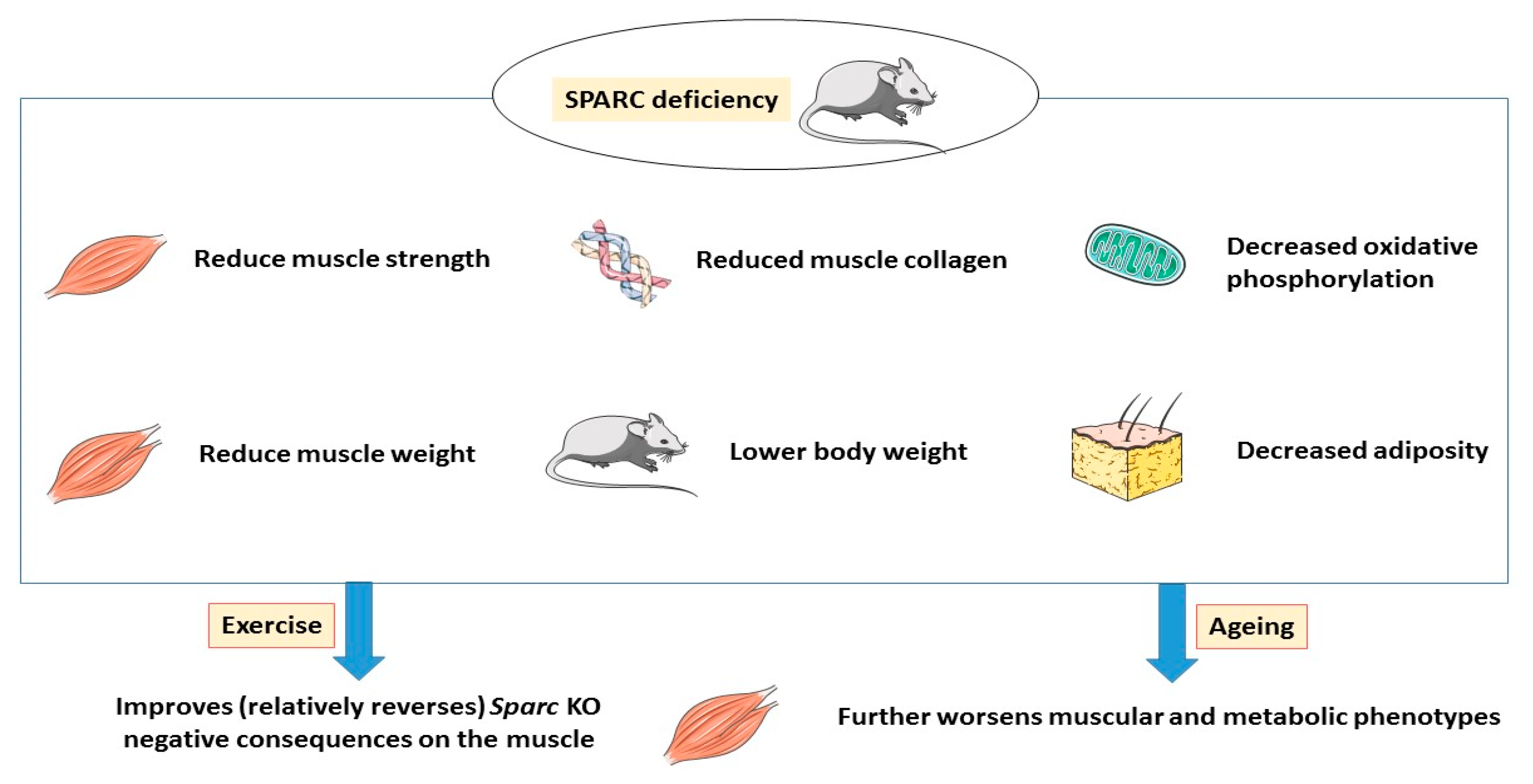
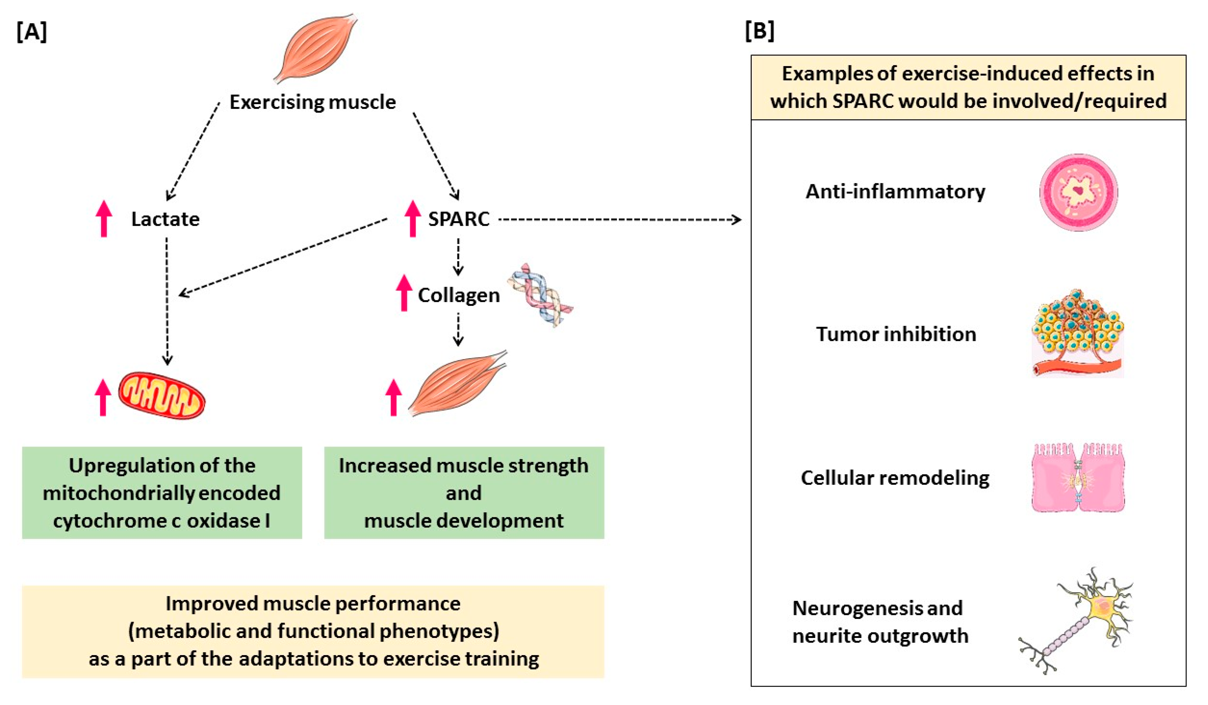
5. Conclusions and Hypothetical Mechanisms

Sparc KO effects are toward a reduced body and WAT weights with a negative SM phenotype (metabolism and strength). Such negative effects worsen with ageing but relatively improve through exercise (Figure 2). While exercise reduces risk factor for many diseases, ageing increases those risks [108].
Within this context, Aoi et al. reported 24 genes (including SPARC) that are both upregulated by exercise and downregulated by ageing [8] suggesting, once more, that some of the exercise-induced benefits such as mitochondrial biogenesis [109] could be SPARC-dependent or partially mediated by SPARC. It is within this perspective that we developed our hypothesis.
The exercise-produced lactate induces both an up-regulation of the expression of the cytochrome oxidase gene and protein, as well as a mitochondrial biogenesis activation signal [79], and since Sparc KO did not induce any genotype effect on the different lactate levels we measured (both WT and Sparc KO mice had statistically similar lactate levels) but reduced the expression of the MT-CO1 (WT and Sparc KO mice have similar lactate levels but WT mice express more MT-CO1 than Sparc KO mice), we hypothesize that the exercise-produced lactate-induced cytochrome oxidase upregulation and mitochondrial biogenesis activation do require SPARC (Figure 3A). This would, at least in part, explain why SPARC deficiency reduces tumor growth. Indeed, cancer cells require glycolytic energy and produce lactate, leading to lower pH, compared to normal tissue extracellular pH [110,111], and lactate would be an attempt to increase oxidative phosphorylation capacity via improving mitochondrial biogenesis. However, in the absence of SPARC this lactate-induced mitochondrial biogenesis remains limited, which worsens the tumor bioenvironment and results in tumor progress inhibition. This concept of lactate-related signaling correlates with the theory presenting lactate as a signaling molecule “lactormone” in the context of lactate shuttle [112,113] of lactate formation, utilization and exchange between tissues [114].
The results of Figure 1B further support our hypothesis that SPARC-dependent exercise impacts SM. Indeed, whereas there is a significant effect of exercise (increase in COL1A1 expression) in WT mice (WT-Ex > WT-Sed), there is no such effect in Sparc KO mice (both KO-Ex and KO-Sed mice have statistically similar expression of COL1A1). This also correlates with the genotype-induced decrease in COL1A1 expression (WT > KO) although both WT and Sparc KO mice had equal amounts of exercise (as detailed in the introduction of Section 4), therefore, indicating that exercise-induced COL1A1 is SPARC-dependent (Figure 3A). Moreover, since exercise-induced COL1A1 seems SPARC-dependent, and based on the importance of collagens in the SM structure (and fibrillar collagen I is reported to bind SPARC [115]) and development [116], this could also explain, in part, the low tibialis anterior muscle weight in Sparc KO mice compared to WT mice, even though all mice had similar amounts of exercise. Thus, this also suggests that SM development (tibialis anterior weight percentage increase; Table 3), as a part to the adaptation to exercise, would also be SPARC-dependent (Figure 3A).
These conclusions are based on the fact that, as per Table 1, there is no effect of the genotype for LT speed, exercise speed, exercise time, exercise distance and lactate concentrations during exercise. This means that mice of two different genotypes (Sparc KO and WT) had equal amounts of training (similar speed, distance, time and frequency). Therefore, genotype effects seen for MT-CO1, COL1A1 (Figure 1) and grip power (Table 4) are, indeed, due to the genotype itself (Sparc KO) rather than the difference in the exercise amount; suggesting that these exercise-induced changes are SPARC-dependent/mediated. Such exercise-induced effects in the muscle represent a part of the adaptation via increasing the respiratory capacity, mitochondrial content [52] and contractile properties of the SM [53].
In addition to these SPARC-mediated effects, the similarity of exercise benefits and the effects shown to be regulated or modulated by SPARC, such as inflammation [22,47], cancer growth [8,46], metabolic and structural remodeling of the SM [53], and even neurite outgrowth [117,118] and neurogenesis [85,119], all suggest that some of the exercise benefits would indeed be mediated by or dependent on SPARC expression (Figure 3B).

6. Implications and Perspectives

Our data suggest that the benefits of exercise would also be reduced in Sparc KO mice, not only because some of exercise benefits are directly mediated via SPARC, but also because physical performance and muscle performance is reduced as a result of Sparc KO (indirect effects). As an illustration, Sparc KO increases tumor growth [120,121], loss of SPARC increases cancer progression [122] and the tumorigenesis is prevented and suppressed by exercise-induced SPARC [8,123,124]. More generally, since SPARC is also a myokine secreted during exercise [2,8], exercise benefits including metabolic benefits and inflammation regulation would also be reduced with the Sparc KO since SPARC has been shown to play roles in metabolism [44], inflammation regulation [47] and cancer homeostasis [46]. Thus, its absence from circulation would impact tissues other than the SM. For instance, even some of the mechanisms underlying the beneficial effects of exercise that have been shown to involve factors other than SPARC, such as tumor growth suppression through interleukin 6 and epinephrine [92], would also be deficient in Sparc KO mice, since the absence of SPARC would reduce the ability of SM to correctly secrete the other myokines involved in the related pathways.
Overall, SM, both as a secretory organ [2] and a metabolic engine, represents the key tissue upon which exercise benefits depend. SPARC represents a “booster” of the SM, with beneficial effects on some other tissues as well. Therefore, SPARC and the pathways it governs would represent good targets to pharmacologically mimic the effects of SPARC, including improved muscle strength and metabolic performances. This is of a particular importance for individuals suffering from health problems, such as heart failure or physical handicap and, therefore, are unable to perform the required physical activity although they need it (reduce obesity, treat lipid disorders, etc.). In such a scenario we could imagine an “exercise pill”, targeting SPARC-related pathways and inducing exercise-like effects that would also be of a high therapeutic importance for diseases, such as sarcopenia. Such a therapeutic goal still requires further investigations into the implications of SPARC, not only within the SM but also on other tissues and in the diverse aspects of homeostasis. This will both extend the expected benefits of such an “exercise pill” and anticipate the side effects to decide whether it would be better given as a systemic drug or rather target a specific tissue (that would be the SM based on this study) to optimize clinical efficiency. Importantly, the results obtained from studying SPARC/Sparc in mice would be expected to be valid in humans, due the high homology between mouse and human SPARC [104], which would reduce the bridge between results in animals and future clinical studies. Perspectives of the future studies on SPARC implications in SM metabolism and contractile properties, both for sedentary and exercise individuals, would be of great clinical importance not only for SM diseases but also for ageing-related health deterioration and, due to the importance of the SM in the energy homeostasis, energy balance-related pathologies such as obesity and diabetes.
Our study was conducted only on male mice. Therefore, we acknowledge the limitation of sex-determined factors. Similar studies in female subjects, as well as studies comparing male and female subjects, will add significant data of clinical importance, especially with the sex-related difference effects on SM and exercise patterns, such as exercise capacity [125], pinch force reproduction [126], maximal oxygen uptake [127], cardiac adaptation to exercise [125], as well as metabolism, including lactate levels [128,129], aerobic oxidation and anaerobic glycolysis [130]. The parameters illustrated by patterns, including differences between men/male animals and women/female animals in red pepper-induced metabolic phenotype (carbohydrate oxidation Vs lipid oxidation) [131,132], beta-oxidation [130], type I fiber percentage [133] and enzyme activities [134], explain beyond such sex-related differences in exercise and SM properties. Based on such sex-related differences, our results could also indicate that SPARC involvement in exercise-induced muscle phenotype changes could also be sex-dependent and points to a possible interaction between SPARC activities and sexual hormones based on the known impacts of sexual hormones on exercise patterns and the adaptation to exercise [135,136,137].
Our data indicate that the impacts of Sparc KO on body composition, adiposity and metabolic patterns point toward a reduced WAT and body weight, but with negative metabolic and functional phenotypes of SM. Whereas such negative effects on SM worsen with ageing, they are relatively improved by exercise. Importantly, we report, for the first time, evidence suggesting that the exercise-induced changes in the SM phenotype in terms of increased performance (metabolic, strength and development), including lactate-induced changes, are SPARC-dependent. Such important implications of SPARC highlight SPARC and its pathways as pharmacological targets/tools for conditions and diseases in which muscle properties enhancement would provide therapeutic benefits.

Author Contributions

Data curation, A.G., A.M., M.Y. and J.S.-A.; Formal analysis, A.G., A.M. and M.Y.; Funding acquisition, J.S.-A.; Investigation, A.G., A.M., M.Y. and J.S.-A.; Methodology, A.G., A.M., M.Y. and J.S.-A.; Project administration, M.Y. and J.S.-A.; Supervision, M.Y. and J.S.-A.; Validation, M.Y. and J.S.-A.; Visualization, A.G., A.M., M.Y. and J.S.-A.; Writing—original draft, A.G.; Writing—review & editing, A.G., A.M., M.Y. and J.S.-A. All authors have read and agreed to the published version of the manuscript.

Funding

This work was supported by the Canadian Institutes of Health Research (CIHR), Number: 201309MOP-311306-BCA-CFBA-40425.

Acknowledgments

We thank Amy D. Bradshaw for providing our research group with sperm of Sparc KO mice. Abdelaziz Ghanemi received a Merit scholarship for foreign students from the Ministry of Education and Higher Education of Quebec, Canada, The Fonds de recherche du Québec—Nature et technologies (FRQNT) is responsible for managing the program (Bourses d’excellence pour étudiants étrangers du Ministère de l’Éducation et de l’Enseignement supérieur du Québec, Le Fonds de recherche du Québec—Nature et technologies (FRQNT) est responsable de la gestion du programme). Figure 2 and Figure 3 were created using images from http://smart.servier.com. Servier Medical Art by Servier is licensed under a Creative Commons Attribution 3.0 Unported License.

Conflicts of Interest

The authors declare no conflict of interest.

References

  1. Pedersen, B.K. The physiology of optimizing health with a focus on exercise as medicine. Annu. Rev. Physiol. 2019, 81, 607–627. [Google Scholar] [CrossRef] [PubMed]
  2. Aoi, W.; Sakuma, K. Skeletal muscle: Novel and intriguing characteristics as a secretory organ. BioDiscovery 2013, 7, e8942. [Google Scholar] [CrossRef]
  3. Ghanemi, A.; Melouane, A.; Yoshioka, M.; St-Amand, J. Exercise and high-fat diet in obesity: Functional genomics perspectives of two energy homeostasis pillars. Genes 2020, 11, 875. [Google Scholar] [CrossRef] [PubMed]
  4. Berryhill, B.L.; Kane, B.; Stramer, B.M.; Fini, M.E.; Hassell, J.R. Increased SPARC accumulation during corneal repair. Exp. Eye Res. 2003, 77, 85–92. [Google Scholar] [CrossRef]
  5. Phan, E.; Ahluwalia, A.; Tarnawski, A.S. Role of SPARC—Matricellular protein in pathophysiology and tissue injury healing. Implications for gastritis and gastric ulcers. Med. Sci. Monit. 2007, 13, RA25–RA30. [Google Scholar]
  6. Riedl, I.; Yoshioka, M.; Nishida, Y.; Tobina, T.; Paradis, R.; Shono, N.; Tanaka, H.; St-Amand, J. Regulation of skeletal muscle transcriptome in elderly men after 6 weeks of endurance training at lactate threshold intensity. Exp. Gerontol. 2010, 45, 896–903. [Google Scholar] [CrossRef]
  7. Melouane, A.; Yoshioka, M.; Kanzaki, M.; St-Amand, J. Sparc, an EPS-induced gene, modulates the extracellular matrix and mitochondrial function via ILK/AMPK pathways in C2C12 cells. Life Sci. 2019, 229, 277–287. [Google Scholar] [CrossRef]
  8. Aoi, W.; Naito, Y.; Takagi, T.; Tanimura, Y.; Takanami, Y.; Kawai, Y.; Sakuma, K.; Hang, L.P.; Mizushima, K.; Hirai, Y.; et al. A novel myokine, secreted protein acidic and rich in cysteine (SPARC), suppresses colon tumorigenesis via regular exercise. Gut 2013, 62, 882–889. [Google Scholar] [CrossRef]
  9. Matsuo, K.; Sato, K.; Suemoto, K.; Miyamoto-Mikami, E.; Fuku, N.; Higashida, K.; Tsuji, K.; Xu, Y.; Liu, X.; Iemitsu, M.; et al. A mechanism underlying preventive effect of high-intensity training on colon cancer. Med. Sci. Sports Exerc. 2017, 49, 1805–1816. [Google Scholar] [CrossRef]
  10. Norheim, F.; Raastad, T.; Thiede, B.; Rustan, A.C.; Drevon, C.A.; Haugen, F. Proteomic identification of secreted proteins from human skeletal muscle cells and expression in response to strength training. Am. J. Physiol. Endocrinol. Metab. 2011, 301, E1013–E1021. [Google Scholar] [CrossRef] [Green Version]
  11. Garneau, L.; Parsons, S.A.; Smith, S.R.; Mulvihill, E.E.; Sparks, L.M.; Aguer, C. Plasma myokine concentrations after acute exercise in non-obese and obese sedentary women. Front. Physiol. 2020, 11, 18. [Google Scholar] [CrossRef] [PubMed] [Green Version]
  12. Billat, V.L.; Mouisel, E.; Roblot, N.; Melki, J. Inter- and intrastrain variation in mouse critical running speed. J. Appl. Physiol. (1985) 2005, 98, 1258–1263. [Google Scholar] [CrossRef] [PubMed] [Green Version]
  13. Available online: https://insights.envigo.com/hubfs/resources/data-sheets/2018s-datasheet-0915.pdf (accessed on 9 August 2020).
  14. Norose, K.; Lo, W.K.; Clark, J.I.; Sage, E.H.; Howe, C.C. Lenses of SPARC-null mice exhibit an abnormal cell surface-basement membrane interface. Exp. Eye Res. 2000, 71, 295–307. [Google Scholar] [CrossRef] [PubMed]
  15. Nie, J.; Bradshaw, A.D.; Delany, A.M.; Sage, E.H. Inactivation of SPARC enhances high-fat diet-induced obesity in mice. Connect. Tissue Res. 2011, 52, 99–108. [Google Scholar] [CrossRef]
  16. Faude, O.; Kindermann, W.; Meyer, T. Lactate threshold concepts: How valid are they? Sports Med. 2009, 39, 469–490. [Google Scholar] [CrossRef]
  17. Schefer, V.; Talan, M.I. Oxygen consumption in adult and AGED C57BL/6J mice during acute treadmill exercise of different intensity. Exp. Gerontol. 1996, 31, 387–392. [Google Scholar] [CrossRef]
  18. Nishida, Y.; Tokuyama, K.; Nagasaka, S.; Higaki, Y.; Shirai, Y.; Kiyonaga, A.; Shindo, M.; Kusaka, I.; Nakamura, T.; Ishibashi, S.; et al. Effect of moderate exercise training on peripheral glucose effectiveness, insulin sensitivity, and endogenous glucose production in healthy humans estimated by a two-compartment-labeled minimal model. Diabetes 2004, 53, 315–320. [Google Scholar] [CrossRef] [Green Version]
  19. Tanaka, H.; Shindo, M. The benefits of the low intensity training. Ann. Physiol. Anthr. 1992, 11, 365–368. [Google Scholar] [CrossRef] [Green Version]
  20. Motoyama, M.; Sunami, Y.; Kinoshita, F.; Kiyonaga, A.; Tanaka, H.; Shindo, M.; Irie, T.; Urata, H.; Sasaki, J.; Arakawa, K. Blood pressure lowering effect of low intensity aerobic training in elderly hypertensive patients. Med. Sci. Sports Exerc. 1998, 30, 818–823. [Google Scholar]
  21. Sunami, Y.; Motoyama, M.; Kinoshita, F.; Mizooka, Y.; Sueta, K.; Matsunaga, A.; Sasaki, J.; Tanaka, H.; Shindo, M. Effects of low-intensity aerobic training on the high-density lipoprotein cholesterol concentration in healthy elderly subjects. Metabolism 1999, 48, 984–988. [Google Scholar] [CrossRef]
  22. Kawanishi, N.; Yano, H.; Mizokami, T.; Takahashi, M.; Oyanagi, E.; Suzuki, K. Exercise training attenuates hepatic inflammation, fibrosis and macrophage infiltration during diet induced-obesity in mice. Brain Behav. Immun. 2012, 26, 931–941. [Google Scholar] [CrossRef] [PubMed]
  23. Van Praag, H.; Shubert, T.; Zhao, C.; Gage, F.H. Exercise enhances learning and hippocampal neurogenesis in aged mice. J. Neurosci. 2005, 25, 8680–8685. [Google Scholar] [CrossRef] [PubMed]
  24. Rowlatt, C.; Chesterman, F.C.; Sheriff, M.U. Lifespan, age changes and tumour incidence in an ageing C57BL mouse colony. Lab. Anim. 1976, 10, 419–442. [Google Scholar] [CrossRef] [PubMed]
  25. Kunstyr, I.; Leuenberger, H.G. Gerontological data of C57BL/6J mice. I. Sex differences in survival curves. J. Gerontol. 1975, 30, 157–162. [Google Scholar] [CrossRef] [PubMed]
  26. Smith, J.P.; Hicks, P.S.; Ortiz, L.R.; Martinez, M.J.; Mandler, R.N. Quantitative measurement of muscle strength in the mouse. J. Neurosci. Methods 1995, 62, 15–19. [Google Scholar] [CrossRef]
  27. Takeshita, H.; Yamamoto, K.; Nozato, S.; Inagaki, T.; Tsuchimochi, H.; Shirai, M.; Yamamoto, R.; Imaizumi, Y.; Hongyo, K.; Yokoyama, S.; et al. Modified forelimb grip strength test detects aging-associated physiological decline in skeletal muscle function in male mice. Sci. Rep. 2017, 7, 42323. [Google Scholar] [CrossRef]
  28. Melouane, A.; Carbonell, A.; Yoshioka, M.; Puymirat, J.; St-Amand, J. Implication of SPARC in the modulation of the extracellular matrix and mitochondrial function in muscle cells. PLoS ONE 2018, 13, e0192714. [Google Scholar] [CrossRef] [Green Version]
  29. Zong, S.; Wu, M.; Gu, J.; Liu, T.; Guo, R.; Yang, M. Structure of the intact 14-subunit human cytochrome c oxidase. Cell Res. 2018, 28, 1026–1034. [Google Scholar] [CrossRef] [Green Version]
  30. Schneider, C.A.; Rasband, W.S.; Eliceiri, K.W. NIH Image to ImageJ: 25 years of image analysis. Nat. Methods 2012, 9, 671–675. [Google Scholar] [CrossRef]
  31. Taylor, S.C.; Berkelman, T.; Yadav, G.; Hammond, M. A defined methodology for reliable quantification of Western blot data. Mol. Biotechnol. 2013, 55, 217–226. [Google Scholar] [CrossRef] [Green Version]
  32. Taylor, S.C.; Posch, A. The design of a quantitative western blot experiment. Biomed. Res. Int. 2014, 2014, 361590. [Google Scholar] [CrossRef] [PubMed]
  33. Ghanemi, A.; Melouane, A.; Mucunguzi, O.; Yoshioka, M.; St-Amand, J. Energy and metabolic pathways in trefoil factor family member 2 (Tff2) KO mice beyond the protection from high-fat diet-induced obesity. Life Sci. 2018, 215, 190–197. [Google Scholar] [CrossRef]
  34. De Giorgio, M.R.; Yoshioka, M.; Riedl, I.; Moreault, O.; Cherizol, R.G.; Shah, A.A.; Blin, N.; Richard, D.; StAmand, J. Trefoil factor family member 2 (Tff2) KO mice are protected from high-fat diet-induced obesity. Obesity (Silver Spring) 2013, 21, 1389–1395. [Google Scholar] [CrossRef] [PubMed]
  35. Bradshaw, A.D.; Sage, E.H. SPARC, a matricellular protein that functions in cellular differentiation and tissue response to injury. J. Clin. Investig. 2001, 107, 1049–1054. [Google Scholar] [CrossRef] [PubMed] [Green Version]
  36. Brekken, R.A.; Sage, E.H. SPARC, a matricellular protein: At the crossroads of cell-matrix communication. Matrix Biol. 2001, 19, 816–827. [Google Scholar] [CrossRef]
  37. Lane, T.F.; Sage, E.H. The biology of SPARC, a protein that modulates cell-matrix interactions. FASEB J. 1994, 8, 163–173. [Google Scholar] [CrossRef] [PubMed]
  38. Yan, Q.; Sage, E.H. SPARC, a matricellular glycoprotein with important biological functions. J. Histochem. Cytochem. 1999, 47, 1495–1506. [Google Scholar] [CrossRef] [Green Version]
  39. Mason, I.J.; Murphy, D.; Münke, M.; Francke, U.; Elliott, R.W.; Hogan, B.L. Developmental and transformation-sensitive expression of the Sparc gene on mouse chromosome 11. EMBO J. 1986, 5, 1831–1837. [Google Scholar] [CrossRef]
  40. Swaroop, A.; Hogan, B.L.; Francke, U. Molecular analysis of the cDNA for human SPARC/osteonectin/BM-40: Sequence, expression, and localization of the gene to chromosome 5q31-q33. Genomics 1988, 2, 37–47. [Google Scholar] [CrossRef]
  41. Brekken, R.A.; Sage, E.H. SPARC, a matricellular protein: At the crossroads of cell-matrix. Matrix Biol. 2000, 19, 569–580. [Google Scholar] [CrossRef]
  42. Francki, A.; Motamed, K.; McClure, T.D.; Kaya, M.; Murri, C.; Blake, D.J.; Carbon, J.G.; Sage, E.H. SPARC regulates cell cycle progression in mesangial cells via its inhibition of IGF-dependent signaling. J. Cell. Biochem. 2003, 88, 802–811. [Google Scholar] [CrossRef]
  43. Basu, A.; Kligman, L.H.; Samulewicz, S.J.; Howe, C.C. Impaired wound healing in mice deficient in a matricellular protein SPARC (osteonectin, BM-40). BMC Cell Biol. 2001, 2, 15. [Google Scholar] [CrossRef] [PubMed]
  44. Ghanemi, A.; Melouane, A.; Yoshioka, M.; St-Amand, J. Secreted protein acidic and rich in cysteine and bioenergetics: Extracellular matrix, adipocytes remodeling and skeletal muscle metabolism. Int. J. Biochem. Cell Biol. 2019, 117, 105627. [Google Scholar] [CrossRef] [PubMed]
  45. Ghanemi, A.; Yoshioka, M.; St-Amand, J. Secreted protein acidic and rich in cysteine: Metabolic and homeostatic properties beyond the extracellular matrix structure. Appl. Sci. 2020, 10, 2388. [Google Scholar] [CrossRef] [Green Version]
  46. Ghanemi, A.; Yoshioka, M.; St-Amand, J. Secreted protein acidic and rich in cysteine and cancer: A homeostatic hormone? Cytokine 2020, 127, 154996. [Google Scholar] [CrossRef] [PubMed]
  47. Ghanemi, A.; Yoshioka, M.; St-Amand, J. Secreted protein acidic and rich in cysteine and inflammation: Another homeostatic property? Cytokine 2020, 133, 155179. [Google Scholar] [CrossRef]
  48. Jørgensen, L.H.; Petersson, S.J.; Sellathurai, J.; Andersen, D.C.; Thayssen, S.; Sant, D.J.; Jensen, C.H.; Schrøder, H.D. Secreted protein acidic and rich in cysteine (SPARC) in human skeletal muscle. J. Histochem. Cytochem. 2009, 57, 29–39. [Google Scholar] [CrossRef] [Green Version]
  49. Holland, P.W.; Harper, S.J.; McVey, J.H.; Hogan, B.L. In vivo expression of mRNA for the Ca++-binding protein SPARC (osteonectin) revealed by in situ hybridization. J. Cell Biol. 1987, 105, 473–482. [Google Scholar] [CrossRef] [Green Version]
  50. Sage, H.; Vernon, R.B.; Decker, J.; Funk, S.; Iruela-Arispe, M.L. Distribution of the calcium-binding protein SPARC in tissues of embryonic and adult mice. J. Histochem. Cytochem. 1989, 37, 819–829. [Google Scholar] [CrossRef]
  51. Framson, P.E.; Sage, E.H. SPARC and tumor growth: Where the seed meets the soil? J. Cell. Biochem. 2004, 92, 679–690. [Google Scholar] [CrossRef]
  52. Holloszy, J.O. Muscle metabolism during exercise. Arch. Phys. Med. Rehabil. 1982, 63, 231–234. [Google Scholar] [PubMed]
  53. Ferraro, E.; Giammarioli, A.M.; Chiandotto, S.; Spoletini, I.; Rosano, G. Exercise-induced skeletal muscle remodeling and metabolic adaptation: Redox signaling and role of autophagy. Antioxid. Redox Signal. 2014, 21, 154–176. [Google Scholar] [CrossRef] [PubMed]
  54. Jørgensen, L.H.; Jepsen, P.L.; Boysen, A.; Dalgaard, L.B.; Hvid, L.G.; Ørtenblad, N.; Ravn, D.; Sellathurai, J.; Møller-Jensen, J.; Lochmüller, H.; et al. SPARC interacts with actin in skeletal muscle in vitro and in vivo. Am. J. Pathol. 2017, 187, 457–474. [Google Scholar] [CrossRef] [PubMed] [Green Version]
  55. Chlenski, A.; Guerrero, L.J.; Salwen, H.R.; Yang, Q.; Tian, Y.; La Madrid, A.M.; Mirzoeva, S.; Bouyer, P.G.; Xu, D.; Walker, M.; et al. Secreted protein acidic and rich in cysteine is a matrix scavenger chaperone. PLoS ONE 2011, 6, e23880. [Google Scholar] [CrossRef] [PubMed] [Green Version]
  56. Delany, A.M.; Amling, M.; Priemel, M.; Howe, C.; Baron, R.; Canalis, E. Osteopenia and decreased bone formation in osteonectin-deficient mice. J. Clin. Investig. 2000, 105, 915–923. [Google Scholar] [CrossRef] [PubMed] [Green Version]
  57. Wang, H.; Fertala, A.; Ratner, B.D.; Sage, E.H.; Jiang, S. Identifying the SPARC binding sites on collagen I and procollagen I by atomic force microscopy. Anal. Chem. 2005, 77, 6765–6771. [Google Scholar] [CrossRef]
  58. Giudici, C.; Raynal, N.; Wiedemann, H.; Cabral, W.A.; Marini, J.C.; Timpl, R.; Bächinger, H.P.; Farndale, R.W.; Sasaki, T.; Tenni, R. Mapping of SPARC/BM-40/osteonectin-binding sites on fibrillar collagens. J. Biol. Chem. 2008, 283, 19551–19560. [Google Scholar] [CrossRef] [Green Version]
  59. Omi, S.; Yamanouchi, K.; Nakamura, K.; Matsuwaki, T.; Nishihara, M. Reduced fibrillar collagen accumulation in skeletal muscle of secreted protein acidic and rich in cysteine (SPARC)-null mice. J. Vet. Med. Sci. 2019, 81, 1649–1654. [Google Scholar] [CrossRef] [Green Version]
  60. Sage, H.; Vernon, R.B.; Funk, S.E.; Everitt, E.A.; Angello, J. SPARC, a secreted protein associated with cellular proliferation, inhibits cell spreading in vitro and exhibits Ca+2-dependent binding to the extracellular matrix. J. Cell Biol. 1989, 109, 341–356. [Google Scholar] [CrossRef]
  61. Francki, A.; Bradshaw, A.D.; Bassuk, J.A.; Howe, C.C.; Couser, W.G.; Sage, E.H. SPARC regulates the expression of collagen type I and transforming growth factor-beta1 in mesangial cells. J. Biol. Chem. 1999, 274, 32145–32152. [Google Scholar] [CrossRef] [Green Version]
  62. Bradshaw, A.D.; Reed, M.J.; Sage, E.H. SPARC-null mice exhibit accelerated cutaneous wound closure. J. Histochem. Cytochem. 2002, 50, 1–10. [Google Scholar] [CrossRef] [PubMed] [Green Version]
  63. Melouane, A.; Yoshioka, M.; St-Amand, J. Extracellular matrix/mitochondria pathway: A novel potential target for sarcopenia. Mitochondrion 2020, 50, 63–70. [Google Scholar] [CrossRef] [PubMed]
  64. Song, H.; Guan, Y.; Zhang, L.; Li, K.; Dong, C. SPARC interacts with AMPK and regulates GLUT4 expression. Biochem. Biophys. Res. Commun. 2010, 396, 961–966. [Google Scholar] [CrossRef] [PubMed]
  65. Lira, V.A.; Benton, C.R.; Yan, Z.; Bonen, A. PGC-1alpha regulation by exercise training and its influences on muscle function and insulin sensitivity. Am. J. Physiol. Endocrinol. Metab. 2010, 299, E145–E161. [Google Scholar] [CrossRef] [Green Version]
  66. Jäger, S.; Handschin, C.; St-Pierre, J.; Spiegelman, B.M. AMP-activated protein kinase (AMPK) action in skeletal muscle via direct phosphorylation of PGC-1alpha. Proc. Natl. Acad. Sci. USA 2007, 104, 12017–12022. [Google Scholar] [CrossRef] [Green Version]
  67. Wu, Z.; Puigserver, P.; Andersson, U.; Zhang, C.; Adelmant, G.; Mootha, V.; Troy, A.; Cinti, S.; Lowell, B.; Scarpulla, R.C.; et al. Mechanisms controlling mitochondrial biogenesis and respiration through the thermogenic coactivator PGC-1. Cell 1999, 98, 115–124. [Google Scholar] [CrossRef] [Green Version]
  68. Smith, G.M.; Gallo, G. The role of mitochondria in axon development and regeneration. Dev. Neurobiol. 2018, 78, 221–237. [Google Scholar] [CrossRef]
  69. Han, S.M.; Baig, H.S.; Hammarlund, M. Mitochondria localize to injured axons to support regeneration. Neuron 2016, 92, 1308–1323. [Google Scholar] [CrossRef] [Green Version]
  70. Aoi, W.; Hirano, N.; Lassiter, D.G.; Björnholm, M.; Chibalin, A.V.; Sakuma, K.; Tanimura, Y.; Mizushima, K.; Takagi, T.; Naito, Y.; et al. Secreted protein acidic and rich in cysteine (SPARC) improves glucose tolerance via AMP-activated protein kinase activation. FASEB J. 2019, 33, 10551–10562. [Google Scholar] [CrossRef] [Green Version]
  71. Bradshaw, A.D. The role of secreted protein acidic and rich in cysteine (SPARC) in cardiac repair and fibrosis: Does expression of SPARC by macrophages influence outcomes? J. Mol. Cell. Cardiol. 2016, 93, 156–161. [Google Scholar] [CrossRef]
  72. McCurdy, S.; Baicu, C.F.; Heymans, S.; Bradshaw, A.D. Cardiac extracellular matrix remodeling: Fibrillar collagens and secreted protein acidic and rich in cysteine (SPARC). J. Mol. Cell. Cardiol. 2010, 48, 544–549. [Google Scholar] [CrossRef] [PubMed] [Green Version]
  73. Cheng, L.; Sun, X.; Guo, J.; Lu, G. SPARC support the expansion of cord blood stem cells in vitro. Cell Res. 2008, 18, S49. [Google Scholar] [CrossRef] [Green Version]
  74. Zhu, J.; Wang, L.Y.; Li, C.Y.; Wu, J.Y.; Zhang, Y.T.; Pang, K.P.; Wei, Y.; Du, L.Q.; Liu, M.; Wu, X.Y. SPARC promotes self-renewal of limbal epithelial stem cells and ocular surface restoration through JNK and p38-MAPK signaling pathways. Stem Cells 2020, 38, 134–145. [Google Scholar] [CrossRef] [PubMed] [Green Version]
  75. Rooney, K.; Trayhurn, P. Lactate and the GPR81 receptor in metabolic regulation: Implications for adipose tissue function and fatty acid utilisation by muscle during exercise. Br. J. Nutr. 2011, 106, 1310–1316. [Google Scholar] [CrossRef] [PubMed] [Green Version]
  76. Gladden, L.B. Muscle as a consumer of lactate. Med. Sci. Sports Exerc. 2000, 32, 764–771. [Google Scholar] [CrossRef] [PubMed]
  77. Gladden, L.B. Lactate metabolism: A new paradigm for the third millennium. J. Physiol. 2004, 558, 5–30. [Google Scholar] [CrossRef]
  78. Hashimoto, T.; Hussien, R.; Oommen, S.; Gohil, K.; Brooks, G.A. Lactate sensitive transcription factor network in L6 cells: Activation of MCT1 and mitochondrial biogenesis. FASEB J. 2007, 21, 2602–2612. [Google Scholar] [CrossRef]
  79. Hashimoto, T.; Brooks, G.A. Mitochondrial lactate oxidation complex and an adaptive role for lactate production. Med. Sci. Sports Exerc. 2008, 40, 486–494. [Google Scholar] [CrossRef]
  80. Gladden, L.B. A lactatic perspective on metabolism. Med. Sci. Sports Exerc. 2008, 40, 477–485. [Google Scholar] [CrossRef] [Green Version]
  81. Jacobs, I. Blood lactate. Sports Med. 1986, 3, 10–25. [Google Scholar] [CrossRef]
  82. Campolongo, M.; Benedetti, L.; Podhajcer, O.L.; Pitossi, F.; Depino, A.M. Hippocampal SPARC regulates depression-related behavior. Genes Brain Behav. 2012, 11, 966–976. [Google Scholar] [CrossRef] [PubMed]
  83. Leveille, G.A.; Chakrabarty, K. Diurnal variations in tissue glycogen and liver weight of meal-fed rats. J. Nutr. 1967, 93, 546–554. [Google Scholar] [CrossRef] [PubMed]
  84. Kreitzman, S.N.; Coxon, A.Y.; Szaz, K.F. Glycogen storage: Illusions of easy weight loss, excessive weight regain, and distortions in estimates of body composition. Am. J. Clin. Nutr. 1992, 56 (Suppl. 1), 292s–293s. [Google Scholar] [CrossRef]
  85. Ma, C.L.; Ma, X.T.; Wang, J.J.; Liu, H.; Chen, Y.F.; Yang, Y. Physical exercise induces hippocampal neurogenesis and prevents cognitive decline. Behav. Brain Res. 2017, 317, 332–339. [Google Scholar] [CrossRef] [PubMed]
  86. Yuan, T.F.; Paes, F.; Arias-Carrión, O.; Ferreira Rocha, N.B.; de Sá Filho, A.S.; Machado, S. Neural mechanisms of exercise: Anti-depression, neurogenesis, and serotonin signaling. CNS Neurol. Disord. Drug Targets 2015, 14, 1307–1311. [Google Scholar] [CrossRef]
  87. Uddin, M.S.; Hossain, M.F.; Mamun, A.A.; Shah, M.A.; Hasana, S.; Bulbul, I.J.; Sarwar, M.S.; Mansouri, R.A.; Ashraf, G.M.; Rauf, A.; et al. Exploring the multimodal role of phytochemicals in the modulation of cellular signaling pathways to combat age-related neurodegeneration. Sci. Total Environ. 2020, 725, 138313. [Google Scholar] [CrossRef]
  88. Hung, C.W.; Chen, Y.C.; Hsieh, W.L.; Chiou, S.H.; Kao, C.L. Ageing and neurodegenerative diseases. Ageing Res. Rev. 2010, 9 (Suppl. 1), S36–S46. [Google Scholar] [CrossRef]
  89. Mahalakshmi, B.; Maurya, N.; Lee, S.-D.; Bharath Kumar, V. Possible neuroprotective mechanisms of physical exercise in neurodegeneration. Int. J. Mol. Sci. 2020, 21, 5895. [Google Scholar] [CrossRef]
  90. Ang, E.-T.; Tai, Y.-K.; Lo, S.-Q.; Seet, R.; Soong, T.-W. Neurodegenerative diseases: Exercising toward neurogenesis and neuroregeneration. Front. Aging Neurosci. 2010, 2, 25. [Google Scholar] [CrossRef] [Green Version]
  91. Liu, Y.; Yan, T.; Chu, J.M.; Chen, Y.; Dunnett, S.; Ho, Y.S.; Wong, G.T.; Chang, R.C. The beneficial effects of physical exercise in the brain and related pathophysiological mechanisms in neurodegenerative diseases. Lab. Investig. 2019, 99, 943–957. [Google Scholar] [CrossRef]
  92. Pedersen, L.; Idorn, M.; Olofsson, G.H.; Lauenborg, B.; Nookaew, I.; Hansen, R.H.; Johannesen, H.H.; Becker, J.C.; Pedersen, K.S.; Dethlefsen, C.; et al. Voluntary running suppresses tumor growth through epinephrine- and IL-6-dependent NK cell mobilization and redistribution. Cell Metab. 2016, 23, 554–562. [Google Scholar] [CrossRef] [PubMed] [Green Version]
  93. Wedell-Neergaard, A.S.; Lang Lehrskov, L.; Christensen, R.H.; Legaard, G.E.; Dorph, E.; Larsen, M.K.; Launbo, N.; Fagerlind, S.R.; Seide, S.K.; Nymand, S.; et al. Exercise-induced changes in visceral adipose tissue mass are regulated by IL-6 signaling: A randomized controlled trial. Cell Metab. 2019, 29, 844–855.e3. [Google Scholar] [CrossRef] [PubMed]
  94. Ellingsgaard, H.; Hojman, P.; Pedersen, B.K. Exercise and health—Emerging roles of IL-6. Curr. Opin. Physiol. 2019, 10, 49–54. [Google Scholar] [CrossRef]
  95. Sakuma, K.; Yamaguchi, A. Sarcopenic obesity and endocrinal adaptation with age. Int. J. Endocrinol. 2013, 2013, 204164. [Google Scholar] [CrossRef] [Green Version]
  96. Candow, D.G.; Chilibeck, P.D. Differences in size, strength, and power of upper and lower body muscle groups in young and older men. J. Gerontol. A Biol. Sci. Med. Sci. 2005, 60, 148–156. [Google Scholar] [CrossRef] [Green Version]
  97. Kalyani, R.R.; Corriere, M.; Ferrucci, L. Age-related and disease-related muscle loss: The effect of diabetes, obesity, and other diseases. Lancet Diabetes Endocrinol. 2014, 2, 819–829. [Google Scholar] [CrossRef] [Green Version]
  98. Kim, T.N.; Choi, K.M. The implications of sarcopenia and sarcopenic obesity on cardiometabolic disease. J. Cell. Biochem. 2015, 116, 1171–1178. [Google Scholar] [CrossRef]
  99. Dhingra, R.; Vasan, R.S. Age as a risk factor. Med. Clin. N. Am. 2012, 96, 87–91. [Google Scholar] [CrossRef] [Green Version]
  100. Keenan, C.R.; White, R.H. Age as a risk factor for venous thromboembolism after major surgery. Curr. Opin. Pulm. Med. 2005, 11, 398–402. [Google Scholar] [CrossRef]
  101. Koshiyama, M.; Tamaki, K.; Ohsawa, M. Age-specific incidence rates of atrial fibrillation and risk factors for the future development of atrial fibrillation in the Japanese general population. J. Cardiol. 2020, 77, 88–92. [Google Scholar] [CrossRef]
  102. Zhang, H.; Rogers, K.; Sukkar, L.; Jun, M.; Kang, A.; Young, T.; Campain, A.; Cass, A.; Chow, C.K.; Comino, E.; et al. Prevalence, incidence and risk factors of diabetes in Australian adults aged ≥ 45 years: A cohort study using linked routinely-collected data. J. Clin. Transl. Endocrinol. 2020, 22, 100240. [Google Scholar] [CrossRef] [PubMed]
  103. Mosher, C.L.; Weber, J.M.; Frankel, C.W.; Neely, M.L.; Palmer, S.M. Risk factors for mortality in lung transplant recipients aged ≥ 65 years: A retrospective cohort study of 5815 patients in the scientific registry of transplant recipients. J. Heart Lung Transplant. 2020. [Google Scholar] [CrossRef]
  104. Norose, K.; Clark, J.I.; Syed, N.A.; Basu, A.; Heber-Katz, E.; Sage, E.H.; Howe, C.C. SPARC deficiency leads to early-onset cataractogenesis. Investig. Ophthalmol. Vis. Sci. 1998, 39, 2674–2680. [Google Scholar] [PubMed]
  105. Singh, M. Mood, food, and obesity. Front. Psychol. 2014, 5, 925. [Google Scholar] [CrossRef] [Green Version]
  106. Ortolani, D.; Oyama, L.M.; Ferrari, E.M.; Melo, L.L.; Spadari-Bratfisch, R.C. Effects of comfort food on food intake, anxiety-like behavior and the stress response in rats. Physiol. Behav. 2011, 103, 487–492. [Google Scholar] [CrossRef] [PubMed] [Green Version]
  107. Bradshaw, A.D.; Graves, D.C.; Motamed, K.; Sage, E.H. SPARC-null mice exhibit increased adiposity without significant differences in overall body weight. Proc. Natl. Acad. Sci. USA 2003, 100, 6045–6050. [Google Scholar] [CrossRef] [Green Version]
  108. Sinclair, A.J.; Abdelhafiz, A.H. Cardiometabolic disease in the older person: Prediction and prevention for the generalist physician. Cardiovasc. Endocrinol. Metab. 2020, 9, 90–95. [Google Scholar] [CrossRef]
  109. Islam, H.; Hood, D.A.; Gurd, B.J. Looking beyond PGC-1α: Emerging regulators of exercise-induced skeletal muscle mitochondrial biogenesis and their activation by dietary compounds. Appl. Physiol. Nutr. Metab. 2020, 45, 11–23. [Google Scholar] [CrossRef]
  110. Su, J.; Chen, X.; Kanekura, T. A CD147-targeting siRNA inhibits the proliferation, invasiveness, and VEGF production of human malignant melanoma cells by down-regulating glycolysis. Cancer Lett. 2009, 273, 140–147. [Google Scholar] [CrossRef]
  111. Gillies, R.J.; Raghunand, N.; Karczmar, G.S.; Bhujwalla, Z.M. MRI of the tumor microenvironment. J. Magn. Reason. Imaging 2002, 16, 430–450. [Google Scholar] [CrossRef]
  112. Gladden, L.B. Current trends in lactate metabolism: Introduction. Med. Sci. Sports Exerc. 2008, 40, 475–476. [Google Scholar] [CrossRef] [PubMed]
  113. Brooks, G.A. Lactate shuttles in nature. Biochem. Soc. Trans. 2002, 30, 258–264. [Google Scholar] [CrossRef] [PubMed]
  114. Brooks, G.A. Current concepts in lactate exchange. Med. Sci. Sports Exerc. 1991, 23, 895–906. [Google Scholar] [CrossRef] [PubMed]
  115. Sasaki, T.; Hohenester, E.; Göhring, W.; Timpl, R. Crystal structure and mapping by site-directed mutagenesis of the collagen-binding epitope of an activated form of BM-40/SPARC/osteonectin. EMBO J. 1998, 17, 1625–1634. [Google Scholar] [CrossRef] [Green Version]
  116. Bailey, A.J. The role of collagen in the development of muscle and its relationship to eating quality. J. Anim. Sci. 1985, 60, 1580–1587. [Google Scholar] [CrossRef]
  117. Au, E.; Richter, M.W.; Vincent, A.J.; Tetzlaff, W.; Aebersold, R.; Sage, E.H.; Roskams, A.J. SPARC from olfactory ensheathing cells stimulates Schwann cells to promote neurite outgrowth and enhances spinal cord repair. J. Neurosci. 2007, 27, 7208–7221. [Google Scholar] [CrossRef]
  118. Lee, M.H.; Amin, N.D.; Venkatesan, A.; Wang, T.; Tyagi, R.; Pant, H.C.; Nath, A. Impaired neurogenesis and neurite outgrowth in an HIV-gp120 transgenic model is reversed by exercise via BDNF production and Cdk5 regulation. J. Neurovirol. 2013, 19, 418–431. [Google Scholar] [CrossRef] [Green Version]
  119. Vincent, A.J.; Lau, P.W.; Roskams, A.J. SPARC is expressed by macroglia and microglia in the developing and mature nervous system. Dev. Dyn. 2008, 237, 1449–1462. [Google Scholar] [CrossRef]
  120. Brekken, R.A.; Puolakkainen, P.; Graves, D.C.; Workman, G.; Lubkin, S.R.; Sage, E.H. Enhanced growth of tumors in SPARC null mice is associated with changes in the ECM. J. Clin. Investig. 2003, 111, 487–495. [Google Scholar] [CrossRef] [Green Version]
  121. Puolakkainen, P.A.; Brekken, R.A.; Muneer, S.; Sage, E.H. Enhanced growth of pancreatic tumors in SPARC-null mice is associated with decreased deposition of extracellular matrix and reduced tumor cell apoptosis. Mol. Cancer Res. 2004, 2, 215–224. [Google Scholar]
  122. Said, N.; Frierson, H.F.; Sanchez-Carbayo, M.; Brekken, R.A.; Theodorescu, D. Loss of SPARC in bladder cancer enhances carcinogenesis and progression. J. Clin. Investig. 2013, 123, 751–766. [Google Scholar] [CrossRef] [PubMed] [Green Version]
  123. Liu, Y.P.; Hsiao, M. Exercise-induced SPARC prevents tumorigenesis of colon cancer. Gut 2013, 62, 810–811. [Google Scholar] [CrossRef] [PubMed]
  124. Aoi, W. Possibility of the novel myokine SPARC: A mechanistic approach to colon cancer prevention by physical exercise. Jpn. J. Phys. Fit. Sports Med. 2013, 62, 263–271. [Google Scholar] [CrossRef]
  125. Konhilas, J.P.; Maass, A.H.; Luckey, S.W.; Stauffer, B.L.; Olson, E.N.; Leinwand, L.A. Sex modifies exercise and cardiac adaptation in mice. Am. J. Physiol. Heart Circ. Physiol. 2004, 287, H2768–H2776. [Google Scholar] [CrossRef] [Green Version]
  126. Li, L.; Li, Y.; Wang, H.; Chen, W.; Liu, X. Effect of force level and gender on pinch force perception in healthy adults. Iperception 2020, 11, 2041669520927043. [Google Scholar] [CrossRef]
  127. Sundberg, S. Maximal oxygen uptake in relation to age in blind and normal boys and girls. Acta Paediatr. Scand. 1982, 71, 603–608. [Google Scholar] [CrossRef]
  128. Hafen, P.S.; Vehrs, P.R. Sex-related differences in the maximal lactate steady state. Sports 2018, 6, 154. [Google Scholar] [CrossRef] [Green Version]
  129. Zhang, J.; Ji, L. Gender differences in peak blood lactate concentration and lactate removal. Ann. Sports Med. Res. 2016, 3, 1088. [Google Scholar]
  130. Green, H.J.; Fraser, I.G.; Ranney, D.A. Male and female differences in enzyme activities of energy metabolism in vastus lateralis muscle. J. Neurol. Sci. 1984, 65, 323–331. [Google Scholar] [CrossRef]
  131. Yoshioka, M.; St-Pierre, S.; Suzuki, M.; Tremblay, A. Effects of red pepper added to high-fat and high-carbohydrate meals on energy metabolism and substrate utilization in Japanese women. Br. J. Nutr. 1998, 80, 503–510. [Google Scholar] [CrossRef]
  132. Yoshioka, M.; Lim, K.; Kikuzato, S.; Kiyonaga, A.; Tanaka, H.; Shindo, M.; Suzuki, M. Effects of red-pepper diet on the energy metabolism in men. J. Nutr. Sci. Vitam. (Tokyo) 1995, 41, 647–656. [Google Scholar] [CrossRef] [PubMed]
  133. Simoneau, J.A.; Bouchard, C. Human variation in skeletal muscle fiber-type proportion and enzyme activities. Am. J. Physiol. 1989, 257, E567–E572. [Google Scholar] [CrossRef] [PubMed]
  134. Simoneau, J.A.; Lortie, G.; Boulay, M.R.; Thibault, M.C.; Thériault, G.; Bouchard, C. Skeletal muscle histochemical and biochemical characteristics in sedentary male and female subjects. Can. J. Physiol. Pharm. 1985, 63, 30–35. [Google Scholar] [CrossRef] [PubMed]
  135. Di Luigi, L.; Romanelli, F.; Sgrò, P.; Lenzi, A. Andrological aspects of physical exercise and sport medicine. Endocrine 2012, 42, 278–284. [Google Scholar] [CrossRef] [PubMed]
  136. Isacco, L.; Duché, P.; Boisseau, N. Influence of hormonal status on substrate utilization at rest and during exercise in the female population. Sports Med. 2012, 42, 327–342. [Google Scholar] [CrossRef] [PubMed]
  137. Amelink, G.J.; Bär, P.R. Exercise-induced muscle protein leakage in the rat. Effects of hormonal manipulation. J. Neurol. Sci. 1986, 76, 61–68. [Google Scholar] [CrossRef]
Figure 1. Expression of both collagen type I alpha 1 chain (COL1A1) (A) and mitochondrially encoded cytochrome c oxidase I (MT-CO1) (B) in the tibialis anterior muscle. The results indicate an effect of genotypeG (WT > KO) for both proteins and an effect of exerciseEx (Ex > Sed) for COL1A1 (trend) and MT-CO1. For the interactions, we have one between genotype and exerciseG × Ex for COL1A1 (Ex > Sed in WT) and one between age and genotypeA × G for MT-CO1 (high in Y-WT). All data are mean ± SEM. The number of mice: 11–12 mice per experimental condition. Abbreviations: A, age; Ex, exercise; G, genotype; KO, knockout; O, old; Sed, sedentary; WT, wild-type; Y, young. *: Trend (0.05 ≤ p < 0.1).
Applsci 10 09108 g001
Figure 2. SPARC-deficiency impacts. Our data highlight that Sparc KO effects are toward a reduced body and white adipose tissue weights with a negative skeletal muscle phenotype (metabolic and strength). Such negative effects worsen with ageing but relatively improve through exercise. Abbreviations: KO, knockout; SPARC, secreted protein acidic and rich in cysteine.
Applsci 10 09108 g002
Figure 3. Hypothetical mechanisms linking SPARC to the exercise-induced SPARC-mediated changes in the skeletal muscle phenotype. [A] Exercise induces the secretion and expression of SPARC as well as the production of lactate. Our data suggest that, whereas SPARC enhances collagen expression and muscle strength, the lactate increases mitochondrial enzyme expression in a SPARC-dependant manner (probably via mitochondrial biogenesis induction) and, therefore, the oxidative phosphorylation capacity. [B] The similarities between exercise benefits and effects shown to be regulated or modulated by SPARC, such as inflammation, cancer growth, metabolic and structural remodeling of the skeletal muscle, and even neurite outgrowth and neurogenesis, all suggest that part of exercise benefits would be mediated by or dependent on SPARC expression. Abbreviation: SPARC, secreted protein acidic and rich in cysteine.
Applsci 10 09108 g003
Table 1. Summary of wheel exercise training.
YoungOld2-Way ANOVA
Wild-typeKnockoutWild-typeKnockoutAGA × G
During an incremental exercise test (wk 4)
LT speedm/min8.7± 0.47.8 ± 0.55.5 ± 0.65.4 ± 0.4Y > O--
Lactate at restmM2.8 ± 0.22.6 ± 0.23.1 ± 0.43.8 ± 0.2O > Y-KO: O > Y
Lactate at LTmM3.2 ± 0.23.1 ± 0.33.4 ± 0.43.3 ± 0.5---
During LT training (wk 1–12)
Mean exercise speedm/min7.5 ± 0.27.6 ± 0.25.5 ± 0.05.3 ± 0.0Y > O--
Total exercise timemin3332 ± 523353 ± 453334 ± 13335 ± 3---
Total exercise distancem24,903 ± 76325,445 ± 67018,219 ± 4117,811 ± 156Y > O--
During the last LT training (wk 12)
Speedm/min7.8 ± 0.27.9 ± 0.25.5 ± 0.05.3 ± 0.1Y > O--
LactatemM2.7 ± 0.32.9 ± 0.32.1 ± 0.22.5 ± 0.2Y > O--
Data are mean ± SEM. Number of mice: 11–12 mice per experimental condition. Abbreviations: A, age; G, genotype; KO, knockout; LT, lactate threshold; m, meter; min, minute; mM, millimolar; O, old; wk, week; Y, young. -: No effect.
Table 2. Fasting and post glucose–gavage blood lactate levels.
YoungOldANOVA
Wild-typeKnockoutWild-typeKnockout
SedentaryExerciseSedentaryExerciseSedentaryExerciseSedentaryExerciseAGExA × GA × ExG × ExA × G × Ex
At sacrifice (12 h fast) 3-way
 Blood lactatemM1.09 ± 0.120.88 ± 0.081.28 ± 0.381.24 ± 0.150.89 ± 0.070.99 ± 0.130.98 ± 0.080.75 ± 0.04Y ˃ O *------
Post glucose gavage (6 h fast) 4-way
 Blood lactateAUC Y > O------
  Pre 615 ± 35498 ± 63479 ± 29533 ± 44429 ± 29424 ± 24473 ± 39461 ± 54
  At wk 5529 ± 32509 ± 34503 ± 41519 ± 38422 ± 27450 ± 42479 ± 44392 ± 32
  After 12 wks465 ± 34391 ± 27452 ± 22486 ± 45440 ± 56381 ± 20414 ± 35371 ± 28
Data are mean ± SEM. Number of mice: 11–-12 mice per experimental condition. Abbreviations: A, age; AUC, area under the curve; Ex, Exercise; G, genotype; h, hour; KO, knockout; mM, millimolar; O, old; wk, week; Y, young. *: Trend (0.05 ≤ p < 0.1). -: No effect.
Table 3. Body and tissue weights.
YoungOld3-Way ANOVA
Wild-typeKnockoutWild-typeKnockout
SedentaryExerciseSedentaryExerciseSedentaryExerciseSedentaryExerciseAGExA × GA × ExG × ExA × G × Ex
Body weightg29.5 ± 0.728.9 ± 1.027.8 ± 0.625.0 ± 0.837.4 ± 2.135.2 ± 1.031.3 ± 1.530.4 ± 0.7O ˃ YWT ˃ KOSed > Ex *----
Tissues weights
Brainmg430 ± 2430 ± 6448 ± 4445 ± 3432 ± 7436 ± 2451 ± 4446 ± 4-KO ˃ WT-----
%1.47 ± 0.031.50 ± 0.051.62 ± 0.041.80 ± 0.051.18 ± 0.051.25 ± 0.031.48 ± 0.071.47 ± 0.02Y ˃ OKO ˃ WTEx > Sed *---WT-Sed: Y ˃ O, WT-Ex: Y ˃ O, KO-Ex: Y >> O
Pituitary glandmg1.52 ± 0.201.49 ± 0.101.47 ± 0.101.33 ± 0.101.77 ± 0.161.76 ± 0.131.78 ± 0.041.65 ± 0.12O ˃ Y------
%0.0052 ± 0.00070.0052 ± 0.00030.0053 ± 0.00040.0054 ± 0.00040.0049 ± 0.00060.0050 ± 0.00040.0059 ± 0.00030.0055 ± 0.0005-------
Hypothalamusmg8.7 ± 0.79.1 ± 0.68.0 ± 0.58.9 ± 0.810.7 ± 0.910.8 ± 0.89.4 ± 0.811.0 ± 0.5O ˃ Y------
%0.030 ± 0.0030.032 ± 0.0030.029 ± 0.0020.036 ± 0.0030.030 ± 0.0030.031 ± 0.0030.030 ± 0.0030.036 ± 0.002--Ex ˃ Sed *----
Livermg984 ± 27954 ± 33984 ± 35899 ± 301370 ± 1361114 ± 281174 ± 821101 ± 24O ˃ Y-Sed ˃ Ex----
%3.34 ± 0.043.31 ± 0.093.54 ± 0.093.60 ± 0.043.59 ± 0.143.17 ± 0.043.71 ± 0.123.62 ± 0.05-KO ˃ WTSed ˃ Ex *-Sed: O ˃ Y *Ex: KO ˃ WT-
Heartmg138 ± 3149 ± 7146 ± 6131 ± 4159 ± 6135 ± 4152 ± 5159 ± 7O ˃ Y--KO: O ˃ Y--WT-Sed: O ˃ Y, WT-Ex: Y ˃ O *, KO-Ex: O ˃ Y
%0.47 ± 0.010.51 ± 0.010.53 ± 0.020.53 ± 0.010.43 ± 0.010.38 ± 0.010.50 ± 0.020.52 ± 0.02Y ˃ OKO ˃ WT-WT: Y >> O--WT-Sed: Y ˃ O, WT-Ex: Y >> O
Aortamg12.8 ± 1.512.0 ± 1.512.9 ± 1.39.0 ± 0.613.0 ± 1.512.3 ± 1.09.7 ± 1.09.9 ± 0.7-WT ˃ KO-----
%0.044 ± 0.0060.042 ± 0.0060.047 ± 0.0050.037 ± 0.0030.034 ± 0.0030.035 ± 0.0030.031 ± 0.0030.032 ± 0.002Y ˃ O------
White adipose tissue **mg1021 ± 206812 ± 126955 ± 138684 ± 1252913 ± 3622649 ± 1751804 ± 2261505 ± 164O ˃ YWT ˃ KOSed > Ex *WT: O >> Y, KO: O>Y---
%3.36 ± 0.582.72 ± 0.363.36 ± 0.442.63 ± 0.387.52 ± 0.657.44 ± 0.325.51 ± 0.504.85 ± 0.43O ˃ YWT ˃ KO-WT: O >> Y, KO: O > Y---
Skeletal muscle ***mg532 ± 11524 ± 15432 ± 8413 ± 7508 ± 11510 ± 6416 ± 8417 ± 7Y ˃ O *WT ˃ KO- ---
%1.81 ± 0.041.82 ± 0.051.56 ± 0.041.67 ± 0.051.39 ± 0.051.46 ± 0.051.36 ± 0.061.38 ± 0.03Y ˃ OWT ˃ KO-WT: Y >> O, KO: Y ˃ O---
Tibialis anterior musclemg133 ± 3138 ± 4117 ± 3114 ± 2133 ± 6127 ± 3110 ± 4118 ± 3Y ˃ O *WT ˃ KO----WT-Ex: Y ˃ O
%0.45 ± 0.010.48 ± 0.010.42 ± 0.010.46 ± 0.010.36 ± 0.020.36 ± 0.010.36 ± 0.010.39 ± 0.02Y ˃ O-Ex > SedWT: Y >> O, KO: Y > O---
Data are mean ± SEM. Number of mice: 11–12 mice per experimental condition. Abbreviations: A, age; Ex, exercise; G, genotype; g, gram; KO, knockout; mg, milligram; O, old; Sed, sedentary; WT, wild-type; Y, young. *: Trend (0.05 ≤ p < 0.1); **: Inguinal and abdominal adipose tissues; ***: Gastrocnemius, soleus, tibialis anterior and extensor digitorum longus muscles. %: percentage to the body weight. -: No effect.
Table 4. Grip Power at the End of Week 12.
YoungOld3-Way ANOVA
Wild-typeKnockoutWild-typeKnockout
SedentaryExerciseSedentaryExerciseSedentaryExerciseSedentaryExerciseAGExA × GA × ExG × ExA × G × Ex
Grip power
Meang240 ± 12248 ± 11226 ± 10208 ± 11216 ± 8220 ± 8182 ± 8203 ± 9Y ˃ OWT ˃ KO-----
g/BW8.2 ± 0.58.6 ± 0.48.1 ± 0.48.4 ± 0.45.8 ± 0.46.1 ± 0.25.9 ± 0.56.6 ± 0.3Y ˃ O---- -
Maxg308 ± 12301 ± 11277 ± 11250 ± 9250 ± 8257 ± 9210 ± 8235 ± 10Y ˃ OWT ˃ KO-----
g/BW10.5 ± 0.410.5 ± 0.410.0 ± 0.410.1 ± 0.46.7 ± 0.57.2 ± 0.36.8 ± 0.57.6 ± 0.3Y ˃ O-Ex > Sed *----
Data are mean ± SEM. Number of mice: 11–12 mice per experimental condition. Abbreviations: A, age; BW, body weight; Ex, exercise; G, genotype; g, gram; KO, knockout; O, old; Sed, sedentary; WT, wild-type; Y, young. *: Trend (0.05 ≤ p < 0.1). -: No effect.
Publisher’s Note: MDPI stays neutral with regard to jurisdictional claims in published maps and institutional affiliations.

