Article

Concentrated Growth Factors vs. Leukocyte-and-Platelet-Rich Fibrin for Enhancing Postextraction Socket Healing. A Longitudinal Comparative Study

by
Marco Mozzati
1,
Giorgia Gallesio
1,
Margherita Tumedei
2 and
Massimo Del Fabbro
3,4,*
1
Private Practitioner, SIOM Oral Surgery and Implantology Center, 10126 Turin, Italy
2
Department of Medical, Oral and Biotechnological Sciences, University of Chieti, 66100 Chieti, Italy
3
Department of Biomedical, Surgical and Dental Sciences, University of Milano, 20122 Milan, Italy
4
IRCCS Orthopedic Institute Galeazzi, 20161 Milan, Italy
*
Author to whom correspondence should be addressed.
Appl. Sci. 2020, 10(22), 8256; https://doi.org/10.3390/app10228256
Submission received: 15 October 2020 / Revised: 6 November 2020 / Accepted: 19 November 2020 / Published: 20 November 2020

Abstract

:

Featured Application

Healing enhancement in oral surgery procedures.

Abstract

Platelet concentrates (PCs) have been used for over 20 years in dentistry, as an adjunct to oral surgery procedures, to improve hard and soft tissue healing and control postoperative symptoms. Among various PCs, Leukocyte and Platelet-Rich Fibrin (L-PRF) has become very popular due to its excellent cost-effectiveness ratio, and to the simple preparation protocol, but comparative clinical studies with other PCs are lacking. The aim of this split-mouth cohort study was to evaluate the effect of Concentrated Growth Factors (CGF), a recently introduced PC, as compared to L-PRF for enhancing post-extraction socket healing. Methods: Patients in need of bilateral tooth extractions were included. Each side was treated with either CGF or L-PRF. Pain, socket closure and healing index were the main outcomes. Results: Forty-five patients (24 women), aged 60.52 ± 11.75 years (range 37–87 years) were treated. No significant difference in outcomes was found, except for Pain at day 1 (p < 0.001) and socket closure in the vestibulo-palatal/lingual dimension at day 7 post-extraction (p = 0.04), both in favor of CGF. Conclusions: based on the present results, CGF proved to be as effective and safe as L-PRF, representing a valid alternative option for improving alveolar socket healing and reducing postoperative discomfort.

1. Introduction

In dentistry, there has been increasing interest in products that promote wound healing. In this respect, in the last few years, the use of platelet preparations, alone or in combination with other biomaterials, has proven to be a valuable regenerative option [1,2,3]. In fact, platelets not only play a critical role in haemostasis, but are also essential in the healing process as they are a source of growth factors [4]. Furthermore, in the last decade, it was shown that the use of autologous non-transfusional blood components in oral surgery allows for better and accelerated epithelialization and more vascularized connective tissue at wound healing sites, in addition to pain reduction [5,6,7,8,9,10]. Among platelet concentrates, the leukocyte and platelet-rich fibrin (L-PRF) has become popular among clinicians, due to the easiness of preparation, the excellent regenerative properties, and its advantageous cost-effectiveness ratio [11,12]. The efficacy of PRF in several oral surgery procedures has been documented in a number of clinical studies and systematic reviews [13,14,15].
Concentrated Growth Factor (CGF) is a more recently developed autologous platelet concentrate containing growth factors together with blood cells [16], which was reported to promote bone regeneration [17,18,19]. Like other platelet concentrates, CGF is isolated from whole blood samples through a simple and standardized protocol by means of a specific centrifuge, without the addition of exogenous substances. The main characteristic of CGF is its mechanical consistency: it is an organic matrix rich in fibrin and, therefore, is denser than other platelet concentrates like platelet-rich plasma (PRP) or plasma rich in growth factors (PRGF), and very similar to L-PRF [20,21]. These characteristics make CGF suitable for different uses, alone or in combination with other materials, as filler or as a scaffold for synthetic and biological membranes [22]. Furthermore, modified protocols in obtaining the blood sample, and in the centrifuging procedure are used for CGF preparation, as compared with PRF. The centrifugation protocol for preparing CGF consists of variable revolutions per minute (rpm), from 2400 to 2700 rpm, to separate cells in the venous blood, resulting in fibrin rich blocks that are much larger, denser and richer in growth factors than common PRF [21]. Essentially, the CGF can be considered an upgraded version of PRF, with a strengthened fibrin matrix and boosted growth factors and cytokines [23].
The rationale of using platelet concentrates stands on the assumption that the presence of growth factors can represent an additional stimulation for tissue healing in patients undergoing oral surgery procedures, in order to improve bone and soft tissue healing [24]. Platelet-rich preparations seem to offer many advantages: they allow for the simultaneous action of multiple growth factors, increase tissue vascularization, and may also work as antimicrobial substances, potentially controlling the incidence of postoperative infection [25]. Given the above premises, a comparative study was designed with the aim of evaluating the performance of CGF for the treatment of postextraction socket, as compared to a more consolidated method such as L-PRF.

2. Materials and Methods

The present study was a longitudinal split-mouth cohort study. The study protocol was approved by the Scientific Board of the IRCCS Istituto Ortopedico Galeazzi in Milan, Italy (No. L2057). All patients were treated in a private practice setting, in compliance with the principles laid down in the Declaration of Helsinki on medical research protocols. A single operator performed all surgeries. Patients were informed about the entire procedure in detail and were included only after they signed a written informed consent form. Patients were enrolled according to the following inclusion criteria: the need for bilateral maxillary or bilateral mandibular extraction; indications for tooth extraction included periodontal or endodontic issues, root or crown fractures, non-restorable caries, and residual roots; at least 18 years of age, and in healthy condition (ASA-1 or ASA-2 according to the classification of the American Society of Anesthesiologists). The exclusion criteria were the following: teeth with acute infection, smokers with > 10 cigarettes/day, major systemic health conditions (ASA-3 or ASA-4), irradiation to the head or neck region or chemotherapy within 12 months before surgery, pregnancy or breastfeeding, poor oral hygiene, and inability or unwillingness to follow the follow-up instructions. One week before surgery, each patient underwent a professional session of oral hygiene together with oral rinses (chlorhexidine digluconate 0.2% mouthwash- that was started 3 days prior to the surgery).

2.1. Surgical Procedures

On the day of surgery, two venous blood samples of 9ml were obtained from each patient. One sample was centrifuged by means of a specific device (Medifuge MF200; Silfradent® Srl, S. Sofia (FC), Italy), in order to obtain CGF, and the other using the L-PRF protocol (Intraspin System®, Intra-Lock System Europa SpA, Salerno, Italy).
Extractions of the two teeth were performed in the same surgical session. Interventions were carried out under local anesthesia (mepivacaine 2% with adrenaline 1:100,000). To prevent interference with the healing process, no intraligamento or intrapapillary infiltrations were performed. The teeth were extracted in an atraumatic way without elevation of full-thickness flaps. The sockets were thoroughly debrided. After socket curettage to remove granulation tissue, a calibrated probe was used to measure the socket maximum diameter in both mesiodistal (MD) and vestibulo-palatal/lingual (VP/L) dimension at crestal level. In each patient, one of the sockets was filled using CGF (T site), while the other socket was filled using PRF (C site). The treatment allocation to the sockets was decided by flipping a coin. After placement of the platelet concentrate into the sockets, no sutures were applied. Both CGF and PRF were left in situ with no attempt to achieve primary closure of the surgical wound. After 5 minutes of compression with sterile cotton, bleeding was evaluated as spontaneous, induced by palpation, or absent. Accurate postoperative recommendations were provided to the patients. They were instructed not to brush the teeth in the treated area, or to apply sucking pressure, but to gently rinse the surgical wound using chlorhexidine digluconate three times daily for 2 weeks. A cold semiliquid diet was recommended for the first day. Patients could re-establish standard oral hygiene procedures after three days. No antibiotic nor analgesic therapy was given. After the extractions, each patient underwent a standard follow-up program: three control visits at 7, 14, and 21 days or until socket closure.

2.2. Outcome Variables

At each postoperative control visit, MD and VP/L dimension at the T and C sites was measured using a calibrated probe. The same operator, different from the one that operated on the patients, and blinded to the treatment received at each site, performed all the measurements.
The patients were asked to score his/her feeling of pain, for both post-extraction sites separately, on a 10-cm visual analogue scale (VAS), with 0 cm indicating no pain and 10 cm indicating the worst possible pain. The pain was evaluated each day at the same time, starting at 2 h after extraction (T1) until day 7 (T7) in the post-operative period.
The maturation and quality of soft tissues were assessed 7 days after extraction, through a modified version of Landry, Turnbull and Howley’s Healing Index (HI), originally developed to evaluate healing with primary closure after periodontal surgery [26]. Such modified HI, that was adapted to estimate socket healing without primary closure, involved three scoring levels for each of the 4 parameters considered: (a) tissue color (1 = 100% of gingiva is pink; 2 = <50% of gingiva is red, hyperemic, movable; 3 = >50% of gingiva is red, hyperemic, movable); (b) color and consistency of healing tissue (1 = close-grained, pink; 2 = soft, red; 3 = fragile, greenish/greyish); (c) suppuration (1 = absent; 2 = absent but pronounced amount of plaque around socket walls; 3 = pronounced); (d) bleeding (1 = absent; 2 = induced by palpation; 3 = spontaneous). The final scoring scale thus ranged from 4, corresponding to excellent healing, to 12, indicating severely impaired healing.

2.3. Statistical Analysis
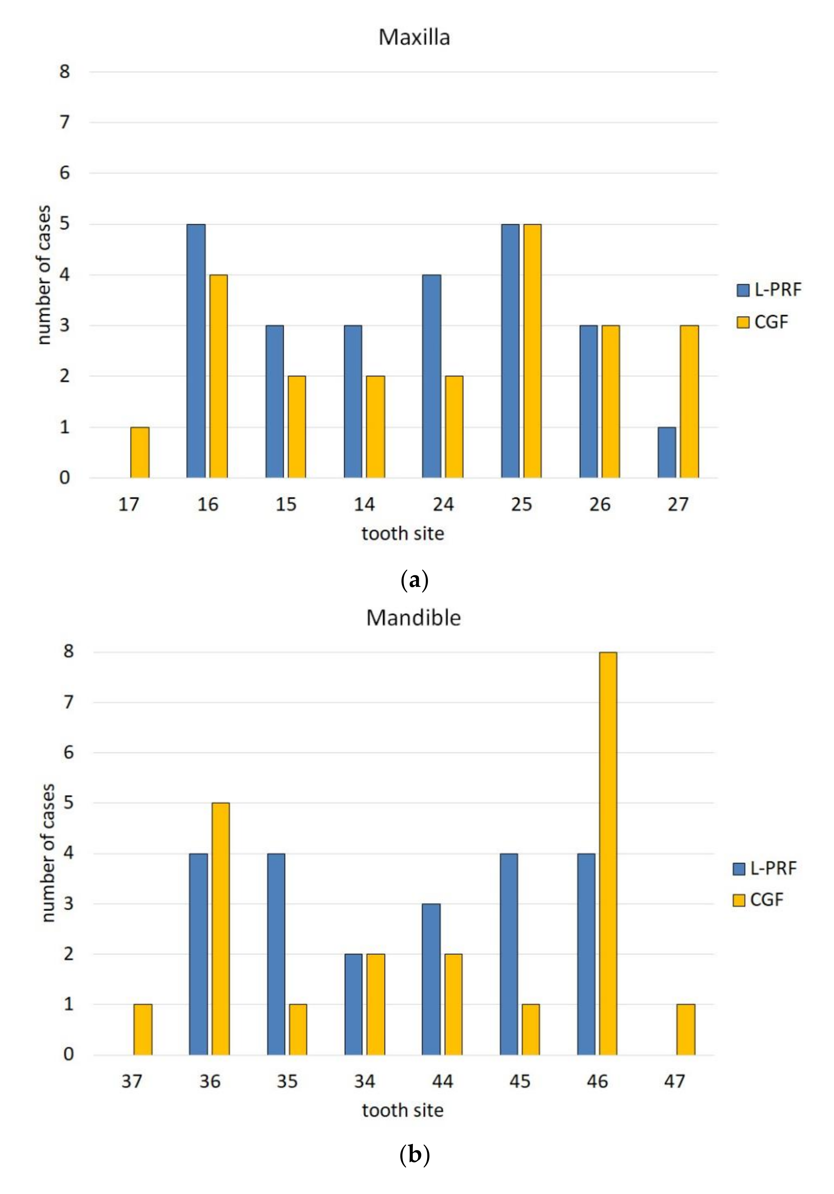
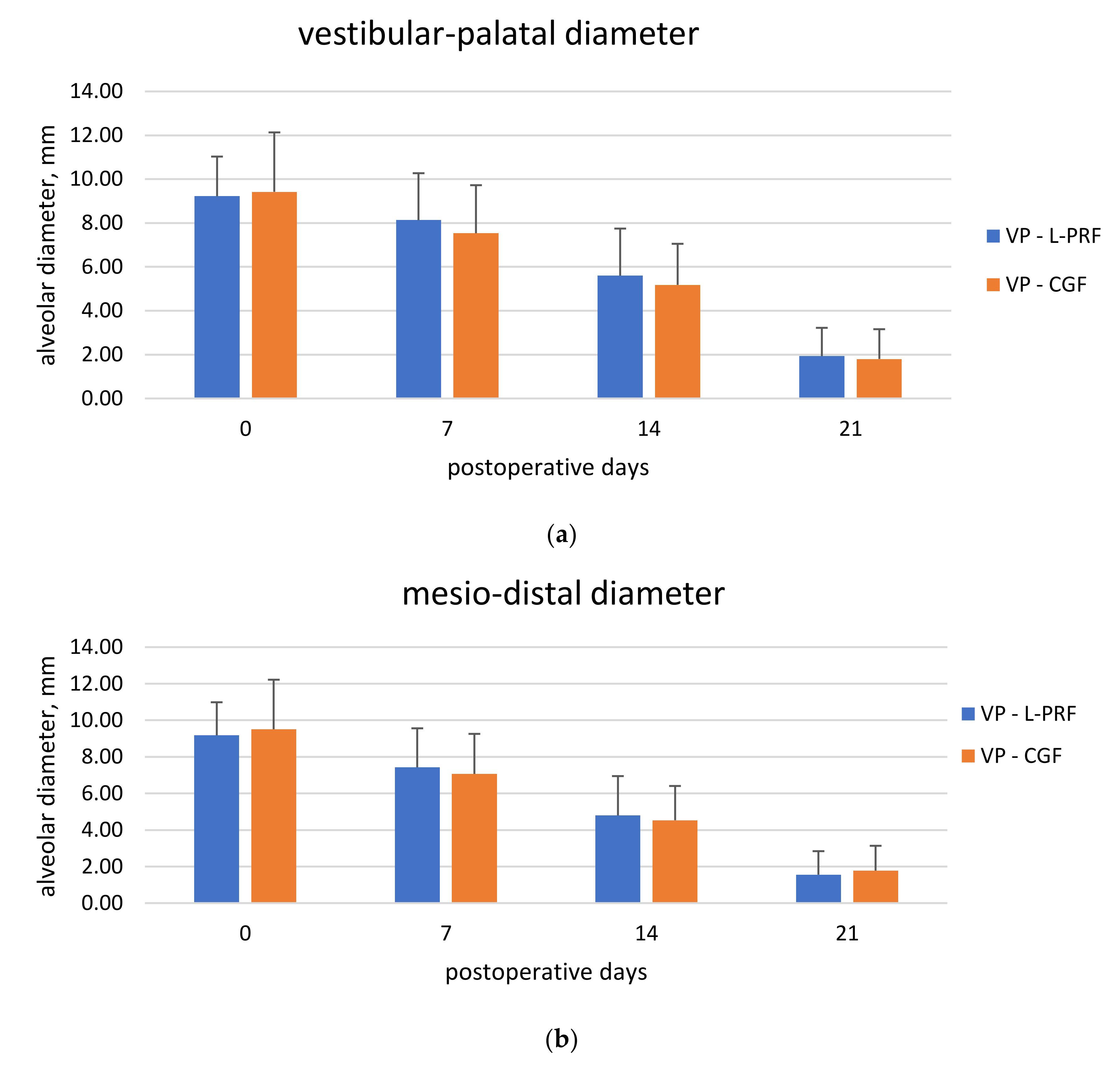
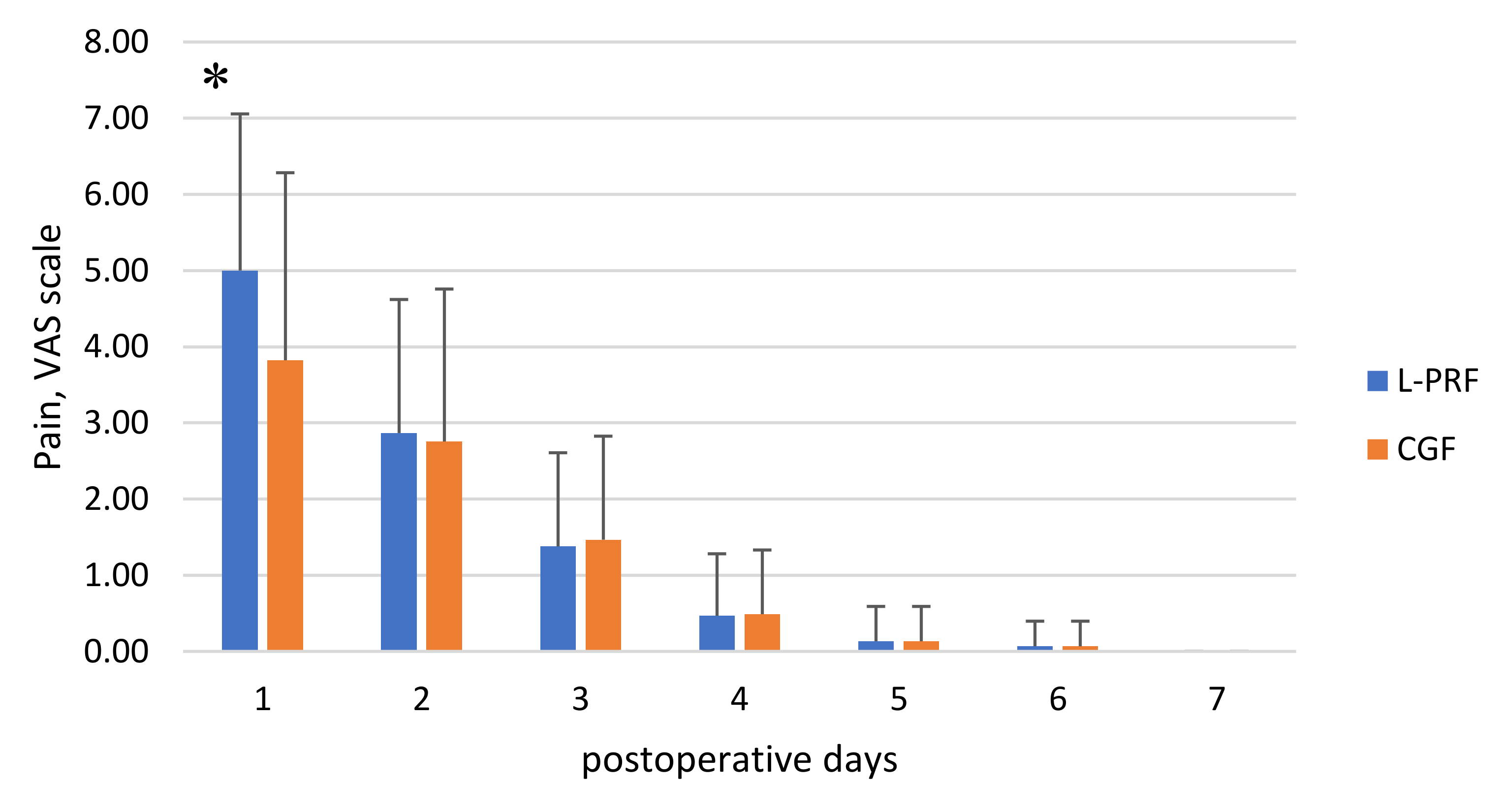
The sample size was estimated based on the equivalence between the two groups regarding the healing index after 7 days. The calculation was made using the online tool at https://www.gigacalculator.com/calculators/power-sample-size-calculator.php. Assuming a type I error of 5% (alpha = 0.05), a power of 80% (1-beta = 0.80), a 50:50 ratio between groups, a mean healing index after 7 days of 5.0, a standard deviation of 1.5, a minimum effect of interest of 0.20, and an equivalence margin of 0.5, 37 samples per group were required. Taking into account the possibility of a 15 to 20% dropout, 45 patients with bilateral defects were recruited.
Descriptive statistics of the data were performed using mean values and standard deviation (SD) for continuous variables normally distributed, and using frequencies and percentages for qualitative variables. The normality of distributions was evaluated through the d’Agostino and Pearson omnibus test. Comparison between groups was performed using paired Student’s t-tests for parametric variables, and Pearson’s chi-square or Fisher’s exact test as appropriate, for qualitative variables. Significance was considered for P-value lower than 0.05.

3. Results

Forty-five patients (24 women and 21 men), ranging in age from 37 to 87 years (mean age 60.52 ± 11.75 years) were enrolled. Full mouth bleeding score (FMBS) and full mouth plaque score (FMPS) at surgery was 20.00 ± 7.67% and 21.76 ± 10.83%. A total of three intra-operative complications (one apex fracture, one fracture of the inter-radicular septum, and one removal of the septum because of a perialveolar cystic lesion) and 4 complications (one apex fracture, one fracture of the inter-radicular septum, and two removals of the septum because of perialveolar cystic lesions) were reported for CGF and PRF group, respectively (P = 0.69). No post-operative complications were reported for both of the study groups.
The location of extraction sites according to the arch in the CGF and PRF group is shown in Figure 1a,b.
The extracted teeth were premolars, first and second molars. The histograms showed a similarity of extraction site distribution between the two treatment groups. The reason for extraction is specified in Table 1.
There was no significant between-group difference in the baseline alveolar size (both in the VP/L and in the MD dimension), as shown in Table 1.
There was a statistically significant difference in vestibular-palatal/lingual (VP/L) diameter reduction at 7 days between CGF and PRF group (p = 0.04) (Table 2). No significant between-group differences of VP/L diameter change were detected at 14 and 21 days (p > 0.05). No statistically significant differences of mesio-distal (MD) diameter reduction was reported at 7, 14 and 21 days between CGF and PRF group (Table 2).
Figure 2a,b show the trend of socket closure in the two treatment groups.
The modified healing index averaged 5.22 ± 1.36 and 5.40 ± 1.29 in the CGF and PRF group, respectively, the values being not significantly different (P = 0.19).
There was significantly lower reported pain in the socket treated with CGF on day 1 after surgery, with respect to PRF group (P < 0.001) (Figure 3). No significant between-group difference was detected for reported pain in the following days.

4. Discussion

Several evidence-based studies and systematic reviews in the last years investigated the efficacy of autologous platelet concentrates used for enhancing alveolar socket healing and reducing postoperative discomfort [6,8,27]. Such reviews demonstrated that different types of platelet concentrates, when compared to spontaneous healing, may produce several beneficial effects. Acceleration and improvement of soft tissue healing (better epithelization, more vascularization, and faster socket closure with respect to control) is the most frequently observed effect. Additionally, better hard tissue healing, evaluated through different techniques (histology and histomorphometry, intraoral radiographs, cone-beam computed tomography, micro-TC, scintigraphy, clinical measurement of ridge width and height changes) was often reported [6,8,27]. In addition, reduced postoperative pain and symptoms, lower incidence of alveolitis and other adverse events respect to control was a common finding in studies that investigated such effects. Nevertheless, almost all clinical studies evaluated a single type of platelet concentrate, used alone or in combination with osteoconductive scaffolds. Therefore, even if most studies emphasize the beneficial effects of the product under investigation, in the absence of direct comparisons it is difficult to know if some platelet concentrate works better than others do, or if all of them produce similar clinical benefits. As highlighted by systematic reviews, there is a lack of clinical studies comparing two or more types of platelet concentrates. In the present study, the effect of two autologous platelet concentrates, on the early healing of post-extraction sites, were compared, with the hypothesis that both products were equally effective. The choice of a split-mouth study allowed eliminating the inter-group variability due to differences in the response to treatment, and in blood characteristics, of different patients. On the other hand, a limitation of the present study is the absence of a control group with sockets left to heal spontaneously. Indeed, there were two reasons for that. Firstly, as said, most of the previous studies used spontaneous healing as the control group, so it seemed useless to repeat once again the same observations made by others. Secondly, the choice of a split-mouth design makes it difficult to add a third group, as the chance of finding a sufficient number of patients in need of at least three comparable tooth extractions, in a reasonable amount of time, is rather low.
Our results confirmed the hypothesis of similarity, though a faster closure at 7 days in the vestibulo-palatal/lingual dimension, and a lower reported pain 1 day postsurgery was recorded in the sites treated with CGF.
The results of the present study, in terms of both healing index and pain control in the first week postoperative, are in line with previous reports on L-PRF for postextraction socket healing. Marenzi et al. in 2015, in a study on L-PRF obtained with Intra-Lock device, used the same modified healing index as in this study, reporting a 7-day healing index of 4.8 ± 0.6, close to excellent healing score and very similar to our findings [28]. Other studies used the original Landry healing index and found scores in the range of excellent [29,30,31]. Regarding pain reduction, other studies using L-PRF reported VAS score and pattern very analogue to that observed in the present study [28,29,30,32,33].
Fewer studies evaluated the effect of CGF on socket healing, due to its more recent introduction in the field of platelet concentrates. Özveri Koyuncu et al. in 2019 reported significant benefits of CGF as compared to spontaneous healing in soft tissue healing, postoperative pain, swelling, and trismus after third molar surgery [34]. Kamal et al. in 2020 reported that CGF is effective in relieving pain and expedite wound healing as compared to conventional treatment alone, represented by socket curettage and saline irrigation, in alveolar osteitis [35]. In another three-arm trial, Kamal et al. compared CGF, low-level laser therapy (LLLT) and conventional treatment (gentle socket curettage and saline irrigation) for the management of dry sockets [36]. They found that the beneficial effects of CGF were superior to those of LLLT with respect to control in healing rate and pain control. It is difficult to compare these studies with the present one, due to different protocols, but the advantages of CGF in both healing and pain relief after tooth extraction were in line with our findings.
In vitro studies showed that, as compared to L-PRP and P-PRP, L-PRF releases greater amounts of TGF-1, shows a longer-term and steady release of growth factors up to at least 10 days, and displays a stronger induction of mesenchymal stem cell migration [20,37,38,39]. Such differences were attributed to the different composition and architecture of PRP and PRF, particularly to the denser and stronger fibrin network of the latter. The fibrin mesh of PRF, which forms through a natural polymerization during centrifugation, in the absence of anticoagulants, may entrap platelets and cells inside the clot, and modulate the growth factors release over time. The degradation of the fibrin mesh of L-PRF occurs more slowly than in PRP, and it is believed to correlate with the longer duration of growth factors released from L-PRF [20,37,38,39].
The Concentrated Growth Factor is the most recently introduced system for producing platelet concentrates [16]. Blood is drawn in tubes without anticoagulant, similar to L-PRF, but CGF is characterized by a peculiar blood centrifugation process, carried out at a constant temperature and at strictly controlled alternating speeds. Additionally, there is a gradual speed increase and decrease at the start and the end of the process, to avoid violent acceleration and deceleration, with the aim of preserving as much as possible the cellular integrity.
The importance of the centrifuge characteristics and the centrifugation protocol have been underlined by recent in vitro studies that compared different commercially available centrifuges for the production of L-PRF [31,40]. These studies pointed out that different centrifugation systems produce clots with different sizes and mechanical consistence, fibrin matrix strength, cellular content, growth factors release profile, and bioactivity. According to these studies, the system producing the best-quality L-PRF clot is the one that was used in the present study, namely the Intra-Spin, manufactured by Intra-Lock [31,40].
Recent in vitro studies compared the features and the biological activity of the advanced PRF clot obtained with the original protocol, and the CGF clot obtained with the Medifuge System, manufactured by Silfradent [21,41]. The study by Isobe et al. found extremely similar composition, mechanical strength, degradation, fibrin fibers thickness and crosslink density of PRF and CGF, in spite of marked differences in the centrifugation process between the two products [21]. The study by Lee et al. found higher tensile strength, higher concentration and amount of PDGF-BB and EGF in CGF, as compared to PRF [41]. In addition, osteoblasts proliferation in cultures enriched with PRF or CGF clots at different % (5%, 10%, and 50%) was comparable to cultures with the medium enriched with fetal bovine serum (FBS, 10%). Osteoblast number, as well as gingival fibroblasts number, independent of the preparation (10% and 50%), was significantly greater with CGF than with PRF [33].
According to the results of these preliminary in-vitro comparative studies, the biological features and activity of CGF were not inferior to those obtained with PRF. Clearly, the result of in vitro investigations need to be confirmed by comparative clinical studies.
In 2019, a case report was published of a 21-year old patient with bilateral multiple gingival recession, treated by coronally advanced flap [42]. PRF and CGF were applied bilaterally during the root coverage procedure, and the outcome of the treatment after three months was compared. Histological analysis of the two clots was also performed. A root coverage of 100% was achieved with both PRF and CGF membranes. Despite this, the side treated with CGF showed less postoperative discomfort and accelerated wound healing (by the 10th postoperative day), with respect to the side treated with PRF [42].
Recently, a three-arm parallel clinical study compared advanced PRF (A-PRF) and CGF as an adjunct to guided tissue regeneration (GTR) procedures in periodontal intrabony defects (IBD) [43]. A group treated with GTR alone was used as control. Standard periodontal parameters (probing pocket depth, clinical attachment level (CAL), intrabony component (IC) depth, radiographic bone level (RBL) and bone defect filling) were measured preoperatively and after 6 months. A-PRF and CGF showed similar effectiveness in improving GTR clinical and radiographic outcomes as compared to control in IBD treatment [43].
In a recent three-arm randomized trial on lower third molar surgery, Torul et al. compared CGF, advanced PRF (A-PRF), and natural healing [44]. They evaluated VAS score, analgesics, edema, trismus in the first post-operative week, and found no significant benefits of CGF or A-PRF over control in any of the variables assessed. This study is hardly comparable to ours, for different reasons. First, they did not evaluate healing index or socket closure, which are our main objective outcomes; second, it was a parallel study, as opposed to ours, and comparing subjective outcomes such as VAS scores between different subjects, should be performed cautiously. Furthermore, A-PRF is obtained with a much different centrifugation system as compared to the one used in the present study. As far as we know, the present split-mouth study is the first reporting clinical results of a comparison between CGF and L-PRF obtained with the Intra-Lock system. The latter was reported to be the best among different centrifugation systems for L-PRF, and its efficacy in improving tissue healing is widely documented in several oral surgery procedures [40].

5. Conclusions

The similar clinical outcomes found in the present study, between the two groups, suggest that the CGF can be considered as an effective alternative to L-PRF for predictable and safe post-extraction socket healing, at least in the early healing phase. The absence of post-operative complications in both groups confirms the effectiveness of CGF and L-PRF not only for enhancing tissue healing, but also for reducing post-operative discomfort (especially with CGF in the first post-op day). More comparative studies with longer follow-up, and possibly with histological and histomorphometric evaluation, are needed to confirm the present results.

Author Contributions

Conceptualization, M.M. and G.G.; methodology, M.M. and M.D.F.; software, G.G. and M.D.F.; validation, G.G., M.T. and M.M.; formal analysis, M.D.F. and M.T.; investigation, M.M. and G.G.; resources, M.M.; data curation, G.G., M.T. and M.D.F.; writing—original draft preparation, G.G., M.T. and M.D.F.; writing—review and editing, M.D.F. and M.T.; supervision, M.M. and M.D.F.; project administration, M.M. All authors have read and agreed to the published version of the manuscript.

Funding

This research received no external funding.

Acknowledgments

The authors declare no acknowledgment.

Conflicts of Interest

The authors declare no conflict of interest.

References

  1. Anitua, E. The use of plasma-rich growth factors (PRGF) in oral surgery. Pract. Proced. Aesthet. Dent. 2001, 13, 487–493. [Google Scholar] [PubMed]
  2. Mihaylova, Z.; Mitev, V.; Stanimirov, P.; Isaeva, A.; Gateva, N.; Ishkitiev, N. Use of platelet concentrates in oral and maxillofacial surgery: An overview. Acta. Odontol. Scand. 2017, 75, 1–11. [Google Scholar] [CrossRef] [PubMed]
  3. Feigin, K.; Shope, B. Use of platelet-rich plasma and platelet-rich fibrin in dentistry and oral surgery: Introduction and review of the literature. J. Vet. Dent. 2019, 36, 109–123. [Google Scholar] [CrossRef] [PubMed]
  4. Anitua, E.; Andia, I.; Ardanza, B.; Nurden, P.; Nurden, A.T. Autologous platelets as a source of proteins for healing and tissue regeneration. Thromb. Haemost. 2004, 91, 4–15. [Google Scholar] [CrossRef] [PubMed]
  5. Mozzati, M.; Martinasso, G.; Pol, R.; Polastri, C.; Cristiano, A.; Muzio, G.; Canuto, R. The impact of plasma rich in growth factors on clinical and biological factors involved in healing processes after third molar extraction. J. Biomed. Mater Res. A 2010, 95, 741–746. [Google Scholar] [CrossRef] [PubMed]
  6. Del Fabbro, M.; Bortolin, M.; Taschieri, S. Is autologous platelet concentrate beneficial for post-extraction socket healing? A systematic review. Int. J. Oral Maxillofac. Surg. 2011, 40, 891–900. [Google Scholar] [CrossRef] [PubMed]
  7. Del Fabbro, M.; Ceresoli, V.; Lolato, A.; Taschieri, S. Effect of platelet concentrate on quality of life after periradicular surgery: A randomized clinical study. J. Endod. 2012, 38, 733–739. [Google Scholar] [CrossRef]
  8. Del Fabbro, M.; Bucchi, C.; Lolato, A.; Corbella, S.; Testori, T.; Taschieri, S. Healing of postextraction sockets preserved with autologous platelet concentrates. A systematic review and meta-analysis. J. Oral Maxillofac. Surg. 2017, 75, 1601–1615. [Google Scholar] [CrossRef]
  9. Srinivas, B.; Das, P.; Rana, M.M.; Qureshi, A.Q.; Vaidya, K.C.; Ahmed Raziuddin, S.J. Wound healing and bone regeneration in postextractcon sockets with and without platelet-rich fibrin. Ann. Maxillofac. Surg. 2018, 8, 28–34. [Google Scholar] [CrossRef]
  10. Soto-Peñaloza, D.; Peñarrocha-Diago, M.; Cervera-Ballester, J.; Peñarrocha-Diago, M.; Tarazona-Alvarez, B.; Peñarrocha-Oltra, D. Pain and quality of life after endodontic surgery with or without advanced platelet-rich fibrin membrane application: A randomized clinical trial. Clin. Oral Investig. 2020, 24, 1727–1738. [Google Scholar] [CrossRef]
  11. Dohan, D.M.; Choukroun, J.; Diss, A.; Dohan, S.L.; Dohan, A.J.J.; Mouhyi, J.; Gogly, B. Platelet-rich fibrin (PRF): A second-generation platelet concentrate. Part I: Technological concepts and evolution. Oral Surg. Oral Med. Oral Pathol. Oral Radiol. Endod. 2006, 101, 37–44. [Google Scholar] [CrossRef] [PubMed]
  12. Choukroun, J.; Diss, A.; Simonpieri, A.; Girard, M.-O.; Schoeffler, C.; Dohan, S.L.; Dohan, A.J.J.; Mouhyi, J.; Dohan, D.M. Platelet-rich fibrin (PRF): A second-generation platelet concentrate. Part IV: Clinical effects on tissue healing. Oral Surg. Oral Med. Oral Pathol. Oral Radiol. Endod. 2006, 101, 56–60. [Google Scholar] [CrossRef] [PubMed]
  13. Miron, R.J.; Fujioka-Kobayashi, M.; Bishara, M.; Zhang, Y.; Hernandez, M.; Choukroun, J. Platelet-rich fibrin and soft tissue wound healing: A systematic review. Tissue Eng. Part B Rev. 2017, 23, 83–99. [Google Scholar] [CrossRef] [PubMed] [Green Version]
  14. Dragonas, P.; Katsaros, T.; Avila-Ortiz, G.; Chambrone, L.; Schiavo, J.H.; Palaiologou, A. Effects of leukocyte-platelet-rich fibrin (L-PRF) in different intraoral bone grafting procedures: A systematic review. Int. J. Oral Maxillofac. Surg. 2019, 48, 250–262. [Google Scholar] [CrossRef] [PubMed]
  15. Pan, J.; Xu, Q.; Hou, J.; Wu, Y.; Liu, Y.; Li, R.; Pan, Y.; Zhang, D. Effect of platelet-rich fibrin on alveolar ridge preservation: A systematic review. J. Am. Dent. Assoc. 2019, 150, 766–778. [Google Scholar] [CrossRef]
  16. Rodella, L.F.; Favero, G.; Boninsegna, R.; Buffoli, B.; Labanca, M.; Scarì, G.; Sacco, L.; Batani, T.; Rezzani, R. Growth factors, CD34 positive cells, and fibrin network analysis in concentrated growth factors fraction. Microsc. Res. Tech. 2011, 74, 772–777. [Google Scholar] [CrossRef]
  17. Sohn, D.-S.; Heo, J.-U.; Kwak, D.-H.; Kim, D.-E.; Kim, J.-M.; Moon, J.-W.; Lee, J.-H.; Park, I.-S. Bone regeneration in the maxillary sinus using an autologous fibrin-rich block with concentrated growth factors alone. Implant Dent. 2011, 20, 389–395. [Google Scholar] [CrossRef] [Green Version]
  18. Durmuşlar, M.C.; Balli, U.; Dede, F.Ö.; Misir, A.F.; Bariş, E.; Kürkçü, M.; Kahraman, S.A. Histological evaluation of the effect of concentrated growth factor on bone healing. J. Craniofac. Surg. 2016, 27, 1494–1497. [Google Scholar] [CrossRef]
  19. Xu, Y.; Qiu, J.; Sun, Q.; Yan, S.; Wang, W.; Yang, P.; Song, A. One-year results evaluating the effects of concentrated growth factors on the healing of intrabony defects treated with or without bone substitute in chronic periodontitis. Med. Sci. Monit. 2019, 25, 4384–4389. [Google Scholar] [CrossRef]
  20. Masuki, H.; Okudera, T.; Watanebe, T.; Suzuki, M.; Nishiyama, K.; Okudera, H.; Nakata, K.; Uematsu, K.; Su, C.-Y.; Kawase, T. Growth factor and pro-inflammatory cytokine contents in platelet-rich plasma (PRP), plasma rich in growth factors (PRGF), advanced platelet-rich fibrin (A-PRF), and concentrated growth factors (CGF). Int. J. Implant Dent. 2016, 2, 19. [Google Scholar] [CrossRef] [Green Version]
  21. Isobe, K.; Watanebe, T.; Kawabata, H.; Kitamura, Y.; Okudera, T.; Okudera, H.; Uematsu, K.; Okuda, K.; Nakata, K.; Tanaka, T.; et al. Mechanical and degradation properties of advanced platelet-rich fibrin (A-PRF), concentrated growth factors (CGF), and platelet-poor plasma-derived fibrin (PPTF). Int. J. Implant Dent. 2017, 3, 17. [Google Scholar] [CrossRef] [PubMed]
  22. Gheno, E.; Palermo, A.; Rodella, L.F.; Buffoli, B. The effectiveness of the use of xenogeneic bone blocks mixed with autologous Concentrated Growth Factors (CGF) in bone regeneration techniques: A case series. J. Osseointegr. 2014, 6, 37–42. [Google Scholar]
  23. Mansour, P.; Kim, P. Use of concentrated growth factor (CGF) in implantology. Australas. Dent. Pract. 2010, 21, 162–176. [Google Scholar]
  24. Anitua, E.; Sánchez, M.; Nurden, A.T.; Nurden, P.; Orive, G.; Andía, I. New insights into and novel applications for platelet-rich fibrin therapies. Trends Biotechnol. 2006, 24, 227–234. [Google Scholar] [CrossRef] [PubMed]
  25. Fabbro, M.D.; Bortolin, M.; Taschieri, S.; Ceci, C.; Weinstein, R.L. Antimicrobial properties of platelet-rich preparations. A systematic review of the current pre-clinical evidence. Platelets 2016, 27, 276–285. [Google Scholar] [CrossRef] [PubMed]
  26. Landry, R.G. Effectiveness of Benzydamine HC1 in the Treatment of Periodontal Post-Surgical Patients. Ph.D. Thesis, University of Toronto, Toronto, ON, Canada, 1985. [Google Scholar]
  27. Del Fabbro, M.; Corbella, S.; Taschieri, S.; Francetti, L.; Weinstein, R. Autologous platelet concentrate for post-extraction socket healing: A systematic review. Eur. J. Oral Implantol. 2014, 7, 333–344. [Google Scholar] [CrossRef] [PubMed]
  28. Marenzi, G.; Riccitiello, F.; Tia, M.; di Lauro, A.; Sammartino, G. Influence of leukocyte- and platelet-rich fibrin (L-PRF) in the healing of simple postextraction sockets: A split-mouth study. Biomed. Res. Int. 2015, 2015, 369273. [Google Scholar] [CrossRef] [Green Version]
  29. Singh, A.; Kohli, M.; Gupta, N. Platelet rich fibrin: A novel approach for osseous regeneration. J. Maxillofac. Oral Surg. 2012, 11, 430–434. [Google Scholar] [CrossRef] [Green Version]
  30. de Almeida Barros Mourão, C.F.; de Mello-Machado, R.C.; Javid, K.; Moraschini, V. The use of leukocyte- and platelet-rich fibrin in the management of soft tissue healing and pain in post-extraction sockets: A randomized clinical trial. J. Cranio Maxillofac. Surg. 2020, 48, 452–457. [Google Scholar] [CrossRef]
  31. Miron, R.J.; Xu, H.; Chai, J.; Wang, J.; Zheng, S.; Feng, M.; Zhang, X.; Wei, Y.; Chen, Y.; de Almeida Barros Mourão, C.F.; et al. Comparison of platelet-rich fibrin (PRF) produced using 3 commercially available centrifuges at both high (~700 g) and low (~200 g) relative centrifugation forces. Clin. Oral Investig. 2020, 24, 1171–1182. [Google Scholar] [CrossRef]
  32. Asmael, H.M.; Jamil, F.A.; Hasan, A.M. Novel application of platelet-rich fibrin as a wound healing enhancement in extraction sockets of patients who smoke. J. Craniofac. Surg. 2018, 29, 794–797. [Google Scholar] [CrossRef] [PubMed]
  33. Ustaoğlu, G.; Göller Bulut, D.; Gümüş, K.Ç. Evaluation of different platelet-rich concentrates effects on early soft tissue healing and socket preservation after tooth extraction. J. Stomatol. Oral Maxillofac. Surg. 2019, in press. [Google Scholar] [CrossRef]
  34. Özveri Koyuncu, B.; Işık, G.; Özden Yüce, M.; Günbay, S.; Günbay, T. Effect of concentrated growth factor (CGF) on short-term clinical outcomes after partially impacted mandibular third molar surgery: A split-mouth randomized clinical study. J. Stomatol. Oral Maxillofac. Surg. 2020, 121, 118–123. [Google Scholar] [CrossRef] [PubMed]
  35. Kamal, A.; Salman, B.; Abdul Razak, N.H.; Qabbani, A.A.; Samsudin, A.R. The Efficacy of concentrated growth factor in the healing of alveolar osteitis: A clinical study. Int. J. Dent. 2020, 2020, 9038629. [Google Scholar] [CrossRef]
  36. Kamal, A.; Salman, B.; Razak, N.H.A.; Samsudin, A.B.R. A Comparative clinical study between concentrated growth factor and low-level laser therapy in the management of dry socket. Eur. J. Dent. 2020, 14, 613–620. [Google Scholar] [CrossRef]
  37. Schär, M.O.; Diaz-Romero, J.; Kohl, S.; Zumstein, M.A.; Nesic, D. Platelet-rich concentrates differentially release growth factors and induce cell migration in vitro. Clin. Orthop. Relat. Res. 2015, 473, 1635–1643. [Google Scholar]
  38. Dohan Ehrenfest, D.M.; Bielecki, T.; Jimbo, R.; Barbe, G.; Del Corso, M.; Inchingolo, F.; Sammartino, G. Do the fibrin architecture and leukocyte content influence the growth factor release of platelet concentrates? An evidence-based answer comparing a pure platelet-rich plasma (P-PRP) gel and a leukocyte-and platelet-rich fibrin (L-PRF). Curr. Pharm. Biotechnol. 2012, 13, 1145–1152. [Google Scholar] [CrossRef]
  39. Kobayashi, E.; Flückiger, L.; Fujioka-Kobayashi, M.; Sawada, K.; Sculean, A.; Schaller, B.; Miron, R.J. Comparative release of growth factors from PRP, PRF, and advanced-PRF. Clin. Oral Investig. 2016, 20, 2353–2360. [Google Scholar] [CrossRef]
  40. Dohan Ehrenfest, D.M.; Pinto, N.R.; Pereda, A.; Jiménez, P.; Corso, M.D.; Kang, B.-S.; Nally, M.; Lanata, N.; Wang, H.-L.; Quirynen, M. The impact of the centrifuge characteristics and centrifugation protocols on the cells, growth factors, and fibrin architecture of a leukocyte-and platelet-rich fibrin (L-PRF) clot and membrane. Platelets 2018, 29, 171–184. [Google Scholar] [CrossRef]
  41. Lee, H.-M.; Shen, E.-C.; Shen, J.T.; Fu, E.; Chiu, H.-C.; Hsia, Y.-J. Tensile strength, growth factor content and proliferation activities for two platelet concentrates of platelet-rich fibrin and concentrated growth factor. J. Dent. Sci. 2020, 15, 141–146. [Google Scholar] [CrossRef]
  42. Krishnakumar, D.; Mahendra, J.; Ari, G.; Perumalsamy, R. A clinical and histological evaluation of platelet-rich fibrin and CGF for root coverage procedure using coronally advanced flap: A split-mouth design. Indian J. Dent. Res. 2019, 30, 970–974. [Google Scholar] [CrossRef] [PubMed]
  43. Lei, L.; Yu, Y.; Han, J.; Shi, D.; Sun, W.; Zhang, D.; Chen, L. Quantification of growth factors in advanced platelet-rich fibrin and concentrated growth factors and their clinical efficacy as adjunctive to the GTR procedure in periodontal intrabony defects. J. Periodontol. 2020, 91, 462–472. [Google Scholar] [CrossRef] [PubMed]
  44. Torul, D.; Omezli, M.M.; Kahveci, K. Evaluation of the effects of concentrated growth factors or advanced platelet rich-fibrin on postoperative pain, edema, and trismus following lower third molar removal: A randomized controlled clinical trial. J. Stomatol. Oral Maxillofac. Surg. 2020, in press. [Google Scholar] [CrossRef] [PubMed]
Figure 1. Tooth distribution in the two groups (a) maxilla; (b) mandible.
Applsci 10 08256 g001
Figure 2. (a) Alveolar diameter in the vestibular-palatal dimension; (b) Alveolar diameter size in the mesio-distal dimension.
Applsci 10 08256 g002
Figure 3. Post-operative pain assessment. Patients of the Concentrated Growth Factors (CGF) group reported significantly less pain in the first day postsurgery (*).
Applsci 10 08256 g003
Table 1. Characteristics of the patients and outcomes in the two groups.
CGFPRFP-Value
Maxilla/mandible (n. teeth)24/2124/21P = 1
Reason for extraction (n.teeth)
- advanced caries
28 26P = 0.89
- Periodontal disease1516
- Tooth fracture23
Baseline alveolar size VP/L, mean ± SD (mm)9.42 ± 2.719.22 ± 1.81P = 0.68
Baseline alveolar size MD, mean ± SD (mm)9.51 ± 4.039.18 ± 2.88P = 0.65
Intra-operative complications (number)34P = 0.69
Post-operative complications (number)00N.A.
Healing index at day 7 (score)5.22 ± 1.365.40 ± 1.29P = 0.53
VP/L = vestibulo-palatal/lingual; MD = mesio-distal; SD = standard deviation; N.A. = not applicable
Table 2. Changes in alveolar size (closure) up to 21 days in the vestibulo-palatal/lingual and mesio-distal dimension in the two groups. Differences with baseline values are expressed in mm as mean values±standard deviation, and in percentage of closure with respect to baseline.
Vestibulo-Palatal/Lingual Change, mm (% Closure Respect to Baseline) Mesio-Distal Change, mm (% Closure Respect to Baseline)
Group0–7 Days0–14 Days0–21 Days0–7 Days0–14 Days0–21 Days
CGF1.89 ± 1.79 (20.0%)4.24 ± 1.90 (45.0%)7.62 ± 2.23 (80.9%)2.44 ± 1.90 (25.7%)4.98 ± 2.50 (52.3%)7.73 ± 3.23 (81.3%)
L-PRF1.09 ± 1.82 (11.8%)3.62 ± 1.95 (39.3%)7.29 ± 2.02 (79.0%)1.76 ± 2.39 (19.1%)4.38 ± 2.60 (47.7%)7.62 ± 3.04 (83.1%)
P-value0.041*0.1080.3490.1490.2100.820
* = statistically significant difference in favor of the CGF group.
Publisher’s Note: MDPI stays neutral with regard to jurisdictional claims in published maps and institutional affiliations.

