Article

Factors Influencing Primary and Secondary Implant Stability—A Retrospective Cohort Study with 582 Implants in 272 Patients

1
Prof. Dr. Dr. Stoll & Partner, 79098 Freiburg, Germany
2
Department of Orthopedics and Trauma Surgery, Medical Centre, Faculty of Medicine, Albert-Ludwigs-University of Freiburg, 79106 Freiburg, Germany
3
Institute of Medical Biometry and Statistics, Faculty of Medicine and Medical Center, University of Freiburg, 79104 Freiburg, Germany
*
Author to whom correspondence should be addressed.
Should be considered joint first author.
Appl. Sci. 2020, 10(22), 8084; https://doi.org/10.3390/app10228084
Submission received: 27 October 2020 / Revised: 6 November 2020 / Accepted: 12 November 2020 / Published: 15 November 2020
(This article belongs to the Special Issue New Trends in Implant-Prosthetic Therapy)

Abstract

:
The success rate of dental implants depends on primary and secondary stability. We investigate predictive factors for future risk stratification models. We retrospectively analyze 272 patients with a total of 582 implants. Implant stability is measured with resonance frequency analysis and evaluated based on the implant stability quotient (ISQ). A linear regression model with regression coefficients (reg. coeff.) and its 95% confidence interval (95% CI) is applied to assess predictive factors for implant stability. Implant diameter (reg. coeff.: 3.28; 95% CI: 1.89–4.66, p < 0.001), implant length (reg. coeff.: 0.67, 95% CI: 0.26–1.08, p < 0.001), and implant localization (maxillary vs. mandibular, reg. coeff.: −7.45, 95% CI: −8.70–(−6.20), p < 0.001) are significant prognostic factors for primary implant stability. An increase in ISQ between insertion and exposure is significantly correlated with healing time (reg. coeff.: 0.11, 95% CI: 0.04–0.19). Patients with maxillary implants have lower ISQ at insertion but show a higher increase in ISQ after insertion than patients with mandibular implants. We observe positive associations between primary implant stability and implant diameter, implant length, and localization (mandibular vs. maxillary). An increase in implant stability between insertion and exposure is significantly correlated with healing time and is higher for maxillary implants. These predictive factors should be further evaluated in prospective cohort studies to develop future preoperative risk-stratification models.

1. Introduction

Implant stability can be defined as an absence of clinical implant mobility and consists of primary and secondary implant stability [1]. Primary stability includes the mechanical attachment of an implant in the surrounding bone at the insertion, whereas secondary implant stability is the tissue response to the implant and subsequent bone remodeling processes. Primary implant stability is known to be a crucial factor for successful osseointegration of dental implants [2,3]. There is sufficient evidence to accept a positive correlation between primary implant stability and implant success, as the success relies on the sustainable integration of the implants into hard and soft tissues [3,4,5,6]. Secondary stability depends on primary stability and has been reported to increase four weeks after implant placement [7,8]. Thus, in the first 2–3 weeks after implant placement, a stability gap with the lowest implant stability is expected [9]. Various methods have been used to examine implant stability at insertion and during the osseointegration period [7]. In 1996, resonance frequency analysis (RFA), as a noninvasive method to investigate the stiffness and stability of the implant-bone interface, was introduced by Meredith et al. [10]. The resonance frequency analysis (RFA) is a measurement of the frequency of a vibrating device. The measurement is performed by a frequency modulator, which exerts a vibration on a sensor (adapter) firmly attached to the implant. The frequency modulator vibrates the adapter at a frequency of 5 to 15 kHz. The vibration of the sensor is positively correlated with implant stability (i.e., stiffness of the bone–implant interface) [1].
The measurement of implant stability is the implant stability quotient (ISQ) and ranges from 1 (lowest implant stability) to 100 (highest implant stability) [1,5]. Reported ISQ values for successful implants range from 57 to 82 [9]. However, ISQ values at implant insertion should be ≥60 to achieve sufficient implant stability [11,12]. It is suggested that implants with high ISQ-values show lower micro-motions and withstand higher forces in the mouth [11,12]. For the immediate loading of dental implants, ISQ values of 60–65 were associated with a good prognosis [13]. Moreover, several studies have confirmed that RFA measurements are able to successfully predict implant failure [14,15,16,17].
Immediate loading protocols are of interest for modern implant therapy since the technique of implant placements immediately following tooth extraction was introduced in the 1970s [18]. Several concerns with two-stage approaches have been stated, such as alveolar bone loss, increased time of edentulism and surgery, need for a second surgical procedure, and psychological factors associated with the aforementioned limitations [19]. In contrast, the implants’ immediate placement in the fresh extraction socket as a one-step procedure reduces the length of surgery as well as the number of interventions, and, consequently, has psychological benefits for patients [19,20,21]. In a recent systematic review conducted by Cosyn et al., immediate placement of a single tooth was associated with a higher risk for early implant loss compared to delayed implant placement [22]. All implant failures were early failures resulting from a lack of osseointegration. As the success of immediate loading approaches directly relies on primary stability, prognostic factors are of interest for preoperative risk-stratification models. Further, innovative surgical techniques, such as ultrasonic site preparation, can be included with the methodology provided to assess other predicting factors in the future [23]. Several recent studies focused on the assessment of implant stability based on radiofrequency analysis [24,25,26,27,28,29]. However, these studies focused on the differences between implant systems and characteristics without considering confounding factors to predict implant stability for future risk stratification models. A large number of implants and appropriate statistical models are needed to adequately predict implant stability.
In the present study, we sought to investigate patient-specific, implant-specific, and surgical-technique-dependent predictive factors for primary and secondary implant stability. We here included the highest number of implants to date regarding this topic to shed light on this field.

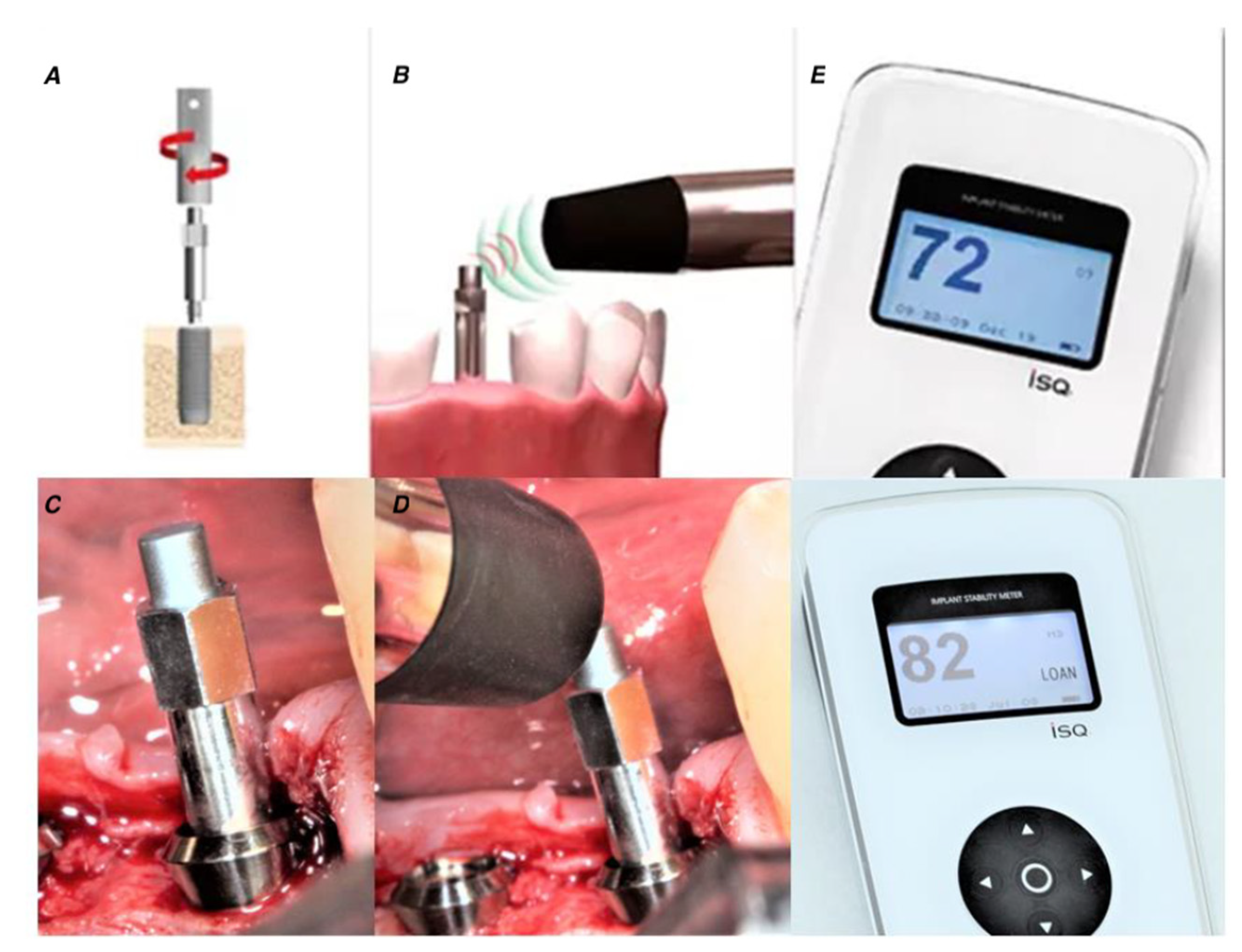
2. Materials and Methods
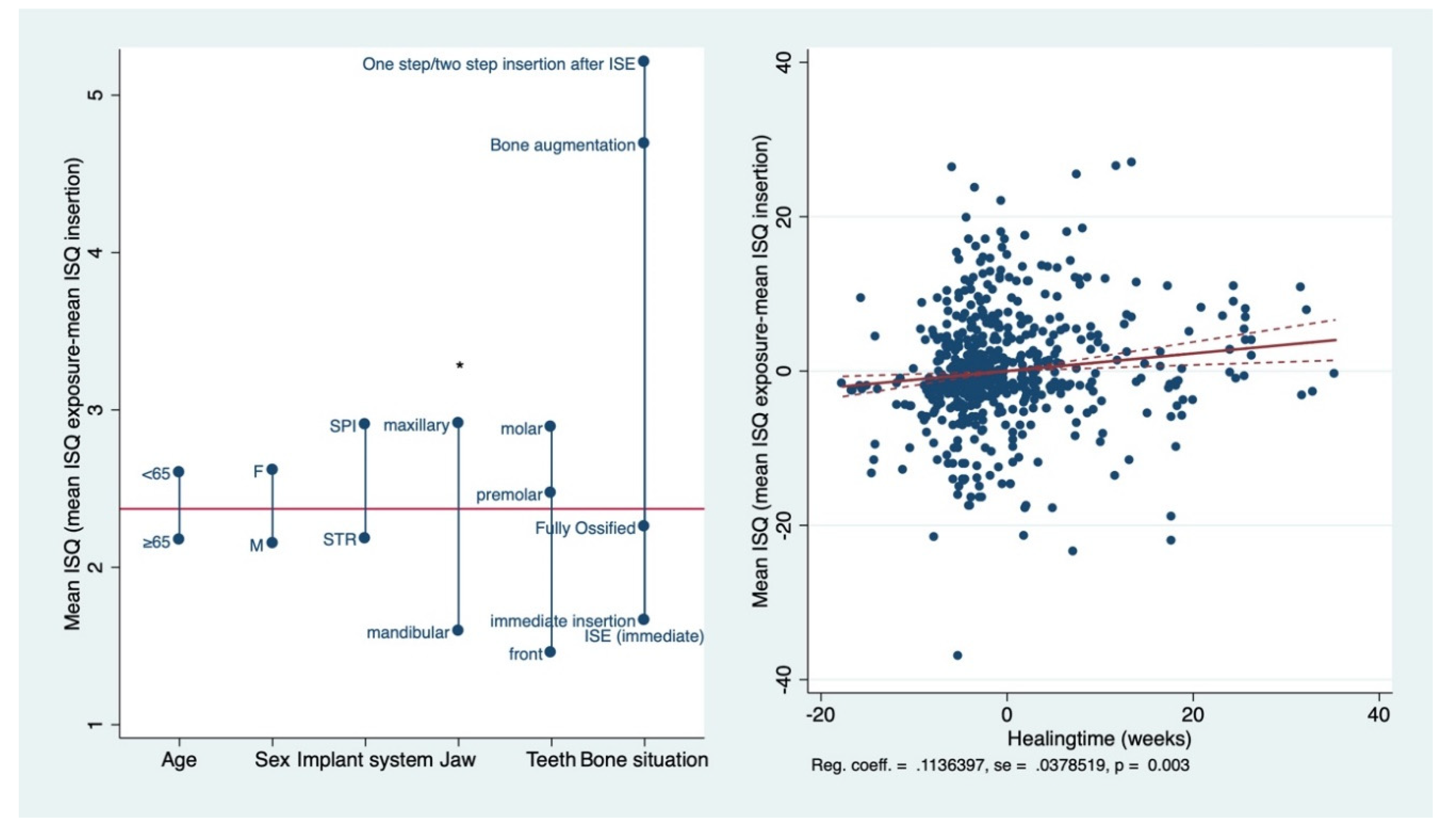
This retrospective study was performed following the revised principles stated in the Declaration of Helsinki and the Good Clinical Practice Guidelines. This retrospective study was exempt from institutional review board approval by the local institutional review board at the University Medical Center in Freiburg, Germany, due to the analysis of fully anonymized data with written confirmation. All patients provided their informed consent prior to their inclusion in the study. The study was performed in compliance with Strengthening the Reporting of Observational studies in Epidemiology (STROBE) guidelines. A retrospective cohort study of 582 implants placed in 282 consecutive patients over a period of 5 years and 7 months was conducted.
Inclusion criteria were: (1) patients treated with at least one SLA® Straumann implant (STR) (Straumann Holding AG, Basel, Switzerland) or Inicell® Thommen implant (SPI)) (Thommen Medical AG, Grenchen, Switzerland); (2) patients age ≥18 years; (3) patients able to understand and sign an informed consent form; (4) measurements conducted with resonance frequency analysis (RFA) at implant insertion and exposure.
Exclusion criteria were: (1) patients with untreated periodontal conditions; (2) patients with diseases or conditions affecting bone metabolism; (3) medication that would affect bone metabolism and implant outcome; (4) any disability or impairment that would impede good oral hygiene.
All patients went through a standardized explanatory meeting, including clinical and radiographic diagnostics. RFA was performed at the time of implant insertion (primary stability = baseline) and before implant loading (secondary stability = exposure) after approximately 120 days. ISQ values were determined using Osstell Mentor™ (Integration Diagnostics Ltd., Goteborg, Sweden) for the palatinal and vestibular sites. Surgeries were performed by two board-certified cranio-maxillofacial surgeons (P.S. and V.G.). Figure 1 illustrates the surgical procedure.

3. Results

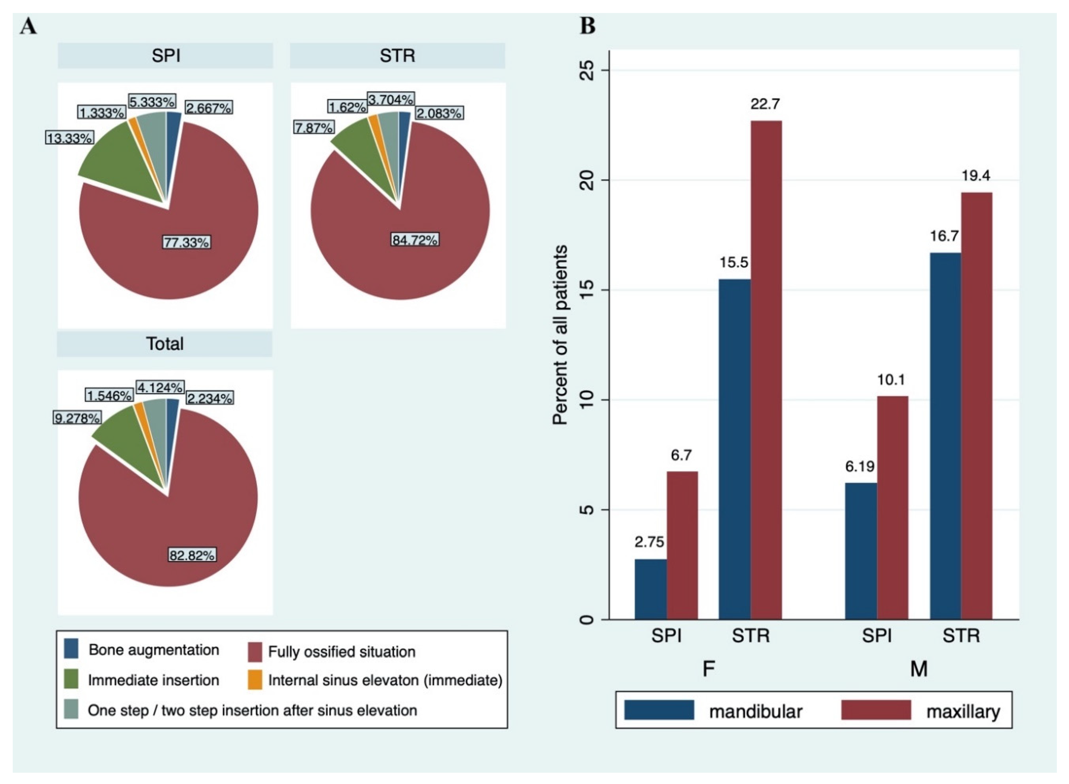
3.1. Basic Characteristics of Patients and Implants

The present cohort consisted of 131 women (48.2%) and 141 mean (51.8%) with a mean age of 65.2 ± 13.9 years (Table 1). Straumann and Thommen implants were used in 206 (75.7%) and 66 (24.3%) patients, respectively. Distribution of men (101/206, 49.0%) and women (105/206, 51.0%) was similar for Straumann implants. In contrast, Thommen implants were predominantly inserted in men (40/66, 60.6%) vs. women (26/66, 39.4%).
A total of 628 implants, 432 (74.2%) Straumann and 150 (25.8%) Thommen, were examined. Median diameter and length for Straumann implants were 4.1 mm (range: 3.3–5.8 mm) and 10 mm (range: 4–14 mm), respectively. The respective values for Thommen implants were 4.5 mm (range 3.5–6 mm) and 11 mm (range: 4.2–14.5 mm). Implants were predominantly inserted in the maxillary premolar region (138/628, 40.2%), followed by the mandibular molar region (127/628, 53.1%) and the maxillary front tooth region (110/628, 32.1%). For Straumann implants, most implants were set in the maxillary premolar (105/432, 24.3%) and mandibular molar tooth region (101/432, 23.4%); for Thommen implants, the maxillary front tooth region (34/150, 22.7%) and maxillary premolar region (33/150, 22.0%) were the most frequent sites. From all included implants, 482/628 (82.8%) were inserted into fully ossified bone, 54/628 (9.3%) were inserted immediately after tooth extraction, 24/628 (4.1%) were inserted in either one or two steps after sinus elevation, 13/628 (2.2%) were inserted after bone augmentation, and 9/628 (1.6%) were inserted immediately after sinus elevation. These bone situations and surgical insertion techniques were equally distributed in STR and SPI groups. However, immediate insertions of implants were more frequently seen in the SPI implant group compared to STR (Figure 2).

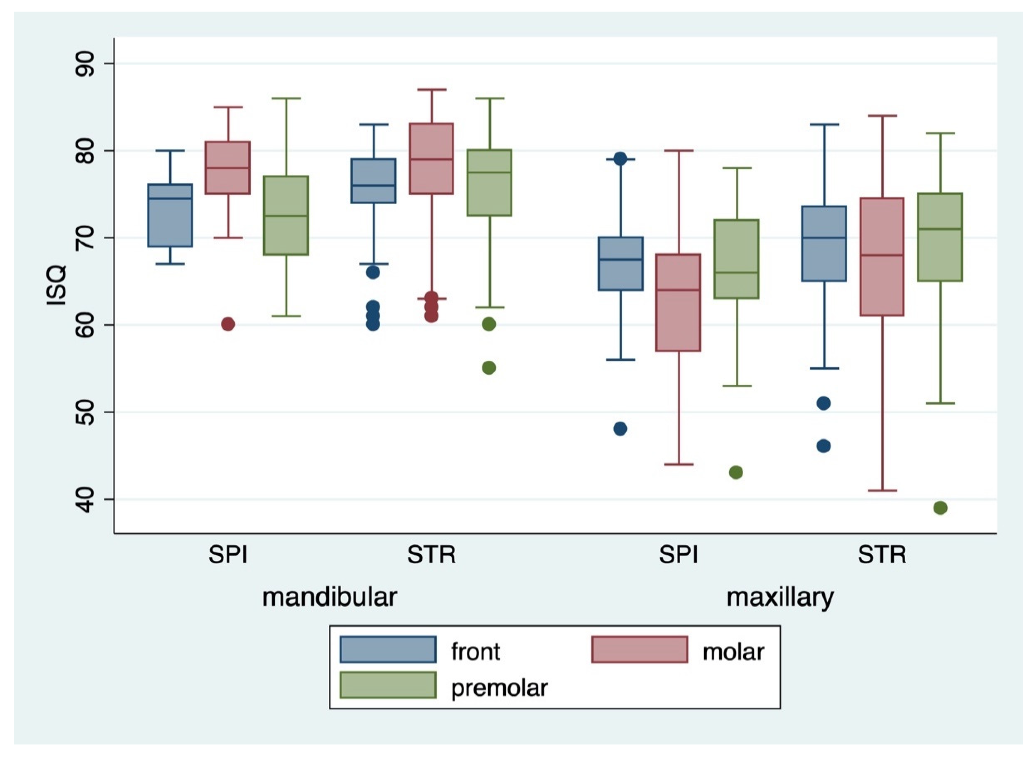
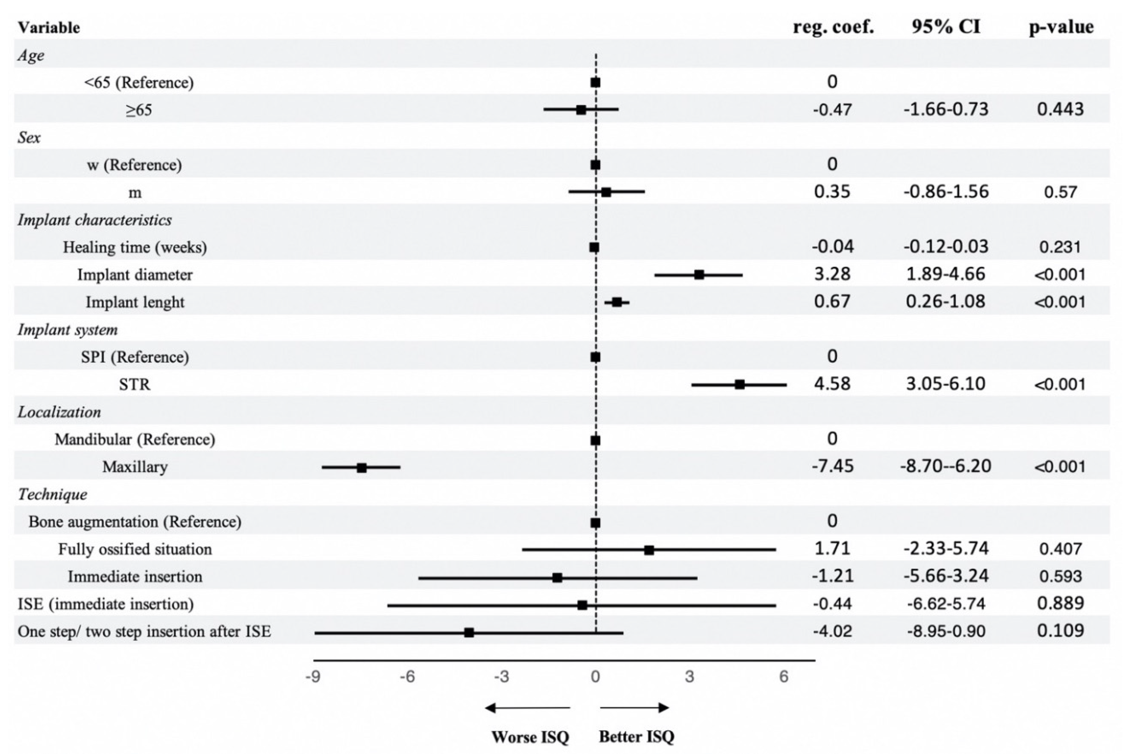
3.2. Primary and Secondary Implant Stability

Mean ISQ at insertion after averaging the values for palatinal and vestibular measurements were 68.59 ± 8.8 (range: 43–86) for SPI and 72.34 ± 8.4 (range: 39–87) for STR. Median ISQ values at insertion were higher for mandibular implants compared to maxillary implants (p < 0.001) and were highest in the mandibular molar tooth region (p < 0.001) (Figure 3). This was observed in both implant groups, STR and SPI. In contrast, implants in the maxillary molar tooth region showed lower median ISQ values than the front or premolar tooth region (p < 0.01). A multivariable linear mixed regression model of the included predictive factors revealed that implant diameter (reg. coeff.: 3.28, 95% CI: 1.89–4.66, p < 0.001), implant length (reg. coeff.: 0.67, 95% CI: 0.26–1.08, p < 0.001), STR vs. SPI implant system (reg. coeff.: 4.58, 95% CI: 3.05–6.10, p < 0.001), and maxillary vs. mandibular (reg. coeff.: −7.45, 95% CI: −8.70–(−6.20), p < 0.001) were significantly associated with primary implant stability (Figure 4). Mean ISQ values at exposure for SPI and STR implants were 71.45 ± 7.9 (range: 49–86) and 74.54 ± 8.1 (range: 36–88), respectively.
Figure 5 shows the mean ISQ at implant insertion and exposure, along with other study variables grouped by implant region (maxillary vs. mandibular) and implant system (STR vs. SPI). Mean healing time was 118.79 ± 59.1 days and was similar in the Straumann (118.03 ± 60.1 days) and Thommen implant group (120.98 ± 56.3 days). Median ISQ values at exposure were higher than the respective ISQ values at insertion, regardless of implant localization and implant system.
In 121/582 (20.8%) implants, ISQ values at exposure were lower than the respective ISQ values at insertion. Furthermore, 36 implants showed a drop of ≥10 in ISQ values between insertion and exposure, from which 29 were STR implants and 7 were SPI implants. The increase in ISQ between insertion and exposure was significantly higher in maxillary implants (2.74 ± 8.8) compared to mandibular implants (1.39 ± 6.5) (p = 0.016), as assessed in the pairwise analysis. Finally, healing time (in weeks) was the only predictive factor associated with an increase in ISQ values between insertion and exposure measurements in the linear regression model (reg. coeff.: 0.11, 95% CI: 0.04–0.19, p = 0.003) (Figure 6).

4. Discussion

The present study aimed to investigate predictive factors of primary and secondary implant stability for future risk stratification models. The present study provided a large dataset and included outcome measures for both primary and secondary implant stability. Furthermore, we stratified our results by two frequently-used implant systems.
We did not observe an influence of the different surgical techniques on implant stability, as already described by other authors [30,31,32,33]. However, we cannot make a conclusion regarding other surgical techniques not included in our study design. In our regression model, implant and bone characteristics also showed no significant prognostic relevance for the increase of ISQ after implant insertion. However, the quality and quantity of jaw bone have been reported to have a significant impact on implant success [34,35,36]. Interestingly, different operation techniques are expected to have a negligible influence on implant survival [7].
The clinical significance of primary stability is expressed by the resistance of the bone during the insertion of an implant [37]. As also seen in our patient cohort, mandible implants are often reported to achieve better primary implant stability [38]. In contrast, maxillary implants resulted in higher increases between primary and secondary measurements in our study cohort, which probably resulted from the lower primary implant stability values of maxillary implants. Additionally, healing time was significantly associated with higher ISQ values in our regression analysis, which is in accordance with the results provided by other authors [39,40,41]. A healing time of 120 days, which considers the initial stability gap in the first weeks after implant insertion, seems to fulfill optimal requirements for successful osseointegration of dental implants [8,42,43,44,45]. Age and sex did not reveal significance for the prediction of primary and secondary implant stability in our study. These findings were also controversially discussed by other authors [46,47,48]. Age is not considered to be a reliable parameter for the success of osseointegration, as shown previously [49,50,51].
Our results further revealed that Straumann implants were significantly associated with higher ISQ compared to Thommen implants. This was seen in jaws as well as all tooth regions, particularly in the premolar region. Regardless, Thommen implants showed a higher increase in ISQ values between implant insertion and exposure. One explanation could be that the already high primary stability values of Straumann implants compared to Thommen implants resulted in lower ISQ increases between primary and secondary measurements, as described before [52]. In contrast, a recent study with 15 implants each (Straumann implants vs. Thommen implants) reported higher ISQ values for Thommen implants for primary stability, whereas no significant differences were found for secondary implant stability [53]. However, the small study size did not allow for an adequate comparison. We also found significant associations between implant length and primary implant stability, though not for secondary implant stability, as confirmed by other authors [54,55,56]. Similar results were found for higher implant diameters, which was also previously confirmed, particularly for primary stability [57,58,59,60]. Primary stability is strongly related to biological (bone density, quantity, and quality) and geometric (implant design, length, and diameter) factors [4,34,54]. All these parameters were reported as non-influential to secondary stability after osseointegration has successfully occurred [54].
Several limitations need to be considered when interpreting the provided results. Retrospective cohort studies are associated with intrinsic limitations, such as selection bias, due to the inclusion of a selected cohort, lack of control, and randomization. Nevertheless, we included important confounding variables in our regression model, allowing us to obtain more precise results than we could have with a matched control group. Furthermore, our predefined inclusion and exclusion criteria could have been stricter, as we did not exclude smokers. However, heavy smokers (>20 cigarettes/day) were not treated with implants in our cohort. Additionally, two surgeons performed all surgeries and measurements independently. This could also have led to bias and should be considered when interpreting the results. Additionally, the RFA measurements have several methodological issues that need to be considered. Micro-movements at the interface can affect tissue differentiation and destroy newly-formed cells and vessels [59]. Upon insertion of the RFA adapter into the implant, forces of up to 10 Ncm can occur [61]. This value corresponds to almost one-third of the force required to insert an implant into the bone and can disturb osseointegration during the early phase [60]. Moreover, different individual measuring adapters for the two implant systems were used, which could impair resulting measurements [62]. A direct comparison would have been difficult, as individual calibration for each implant system is required [63,64,65]. Another limitation of RFA is that the induced oscillation analyzes the entire bone–implant complex and not only the bone–implant interface, which is important for osseointegration [37,66]. The relationship between high ISQ values at the time of implant placement (baseline) and high secondary stability is controversially discussed, and the prognostic value of ISQ values is ambiguous [67]. The lower increase of already high baseline (primary stability) ISQ values compared to secondary stability values was also described previously [52]. Furthermore, even artificially-induced loosening of implants with associated low ISQ values achieved sufficient strength after re-osseointegration [68]. Nevertheless, the RFA proved to be a reliable diagnostic criterion for osseointegration [69].
In contrast to most studies, which mainly considered primary stability, we included secondary stability and the ISQ change during the healing period as an outcome of interest [57,70,71,72]. The osseointegration is dependent on multiple factors and can never be completely reduced to only one parameter. To adequately build predictable risk stratification models, a sufficient number of implants should be examined. As most studies focused on primary implant stability or included fewer implants, the provided results from a large cohort could help to mathematically develop highly predictable preoperative risk stratification models in the future.

5. Conclusions

In conclusion, we found significant associations between primary implant stability and implant diameter, implant length, implant system (STR vs. SPI), and localization (maxillary vs. mandibular). Patients with STR implants, higher implant diameter, higher implant length, and maxillary localization of the implants showed increased implant stability values. An increase in implant stability between insertion and exposure was significantly correlated with healing time and was higher for maxillary implants. These predictive factors should be further evaluated in prospective cohort studies to develop future preoperative risk-stratification models.

Author Contributions

Conceptualization, B.S., A.V. and P.S.; Data curation, B.S., A.V., V.G. and P.S.; Formal analysis, B.S., A.V., D.H. and V.G.; Investigation, B.S., N.A. and V.G.; Methodology, B.S., A.V., G.L., D.H. and P.S.; Project administration, P.S.; Supervision, N.A. and P.S.; Validation, N.A., D.H. and P.S.; Visualization, G.L.; Writing—original draft, B.S. and A.V.; Writing—review & editing, B.S., A.V., G.L., N.A., D.H., V.G. and P.S. All authors have read and agreed to the published version of the manuscript.

Funding

This research received no external funding.

Acknowledgments

Gernot Lang was supported by the Berta-Ottenstein-Programme for Advanced Clinician Scientists, Faculty of Medicine, University of Freiburg. The article processing charge was funded by the Baden-Wuerttemberg Ministry of Science, Research and Art, and the University of Freiburg in the funding program Open Access Publishing.

Conflicts of Interest

The authors declare no conflict of interest. The funders had no role in the design of the study; in the collection, analyses, or interpretation of data; in the writing of the manuscript; or in the decision to publish the results.

References

  1. Sennerby, L.; Meredith, N. Implant stability measurements using resonance frequency analysis: Biological and biomechanical aspects and clinical implications. Periodontol. 2000 2008, 47, 51–66. [Google Scholar] [CrossRef]
  2. Esposito, M.; Hirsch, J.-M.; Lekholm, U.; Thomsen, P. Biological factors contributing to failures of osseointegrated oral implants, (I). Success criteria and epidemiology. Eur. J. Oral Sci. 1998, 106, 527–551. [Google Scholar] [CrossRef]
  3. Lioubavina-Hack, N.; Lang, N.P.; Karring, T. Significance of primary stability for osseointegration of dental implants. Clin. Oral Implants Res. 2006, 17, 244–250. [Google Scholar] [CrossRef]
  4. Javed, F.; Romanos, G.E. The role of primary stability for successful immediate loading of dental implants. A literature review. J. Dent. 2010, 38, 612–620. [Google Scholar] [CrossRef]
  5. Meredith, N. Assessment of implant stability as a prognostic determinant. Int. J. Prosthodont. 1998, 11, 491–501. [Google Scholar]
  6. Saravi, B.E.; Putz, M.; Patzelt, S.; Alkalak, A.; Uelkuemen, S.; Boeker, M. Marginal bone loss around oral implants supporting fixed versus removable prostheses: A systematic review. Int. J. Implant Dent. 2020, 6, 20. [Google Scholar] [CrossRef]
  7. Atsumi, M.; Park, S.-H.; Wang, H.-L. Methods used to assess implant stability: Current status. Int. J. Oral Maxillofac. Implants 2007, 22, 743–754. [Google Scholar] [PubMed]
  8. Raghavendra, S.; Wood, M.C.; Taylor, T.D. Early wound healing around endosseous implants: A review of the literature. Int. J. Oral Maxillofac. Implants 2005, 20, 425–431. [Google Scholar] [PubMed]
  9. Sachdeva, A.; Dhawan, P.; Sindwani, S. Assessment of Implant Stability: Methods and Recent Advances. BJMMR 2016, 12, 1–10. [Google Scholar] [CrossRef]
  10. Meredith, N.; Alleyne, D.; Cawley, P. Quantitative determination of the stability of the implant-tissue interface using resonance frequency analysis. Clin. Oral Implants Res. 1996, 7, 261–267. [Google Scholar] [CrossRef] [PubMed]
  11. Pagliani, L.; Sennerby, L.; Petersson, A.; Verrocchi, D.; Volpe, S.; Andersson, P. The relationship between resonance frequency analysis (RFA) and lateral displacement of dental implants: An in vitro study. J. Oral Rehabil. 2013, 40, 221–227. [Google Scholar] [CrossRef] [PubMed]
  12. Trisi, P.; Carlesi, T.; Colagiovanni, M.; Perfetti, G. Implant stability quotient (ISQ) vs. direct in vitro measurement of primary stability (micromotion): Effect of bone density and insertion torque. J. Osteol. Biomat. 2010, 1, 141–149. [Google Scholar]
  13. Sennerby, L.; Meredith, N. Resonance frequency analysis: Measuring implant stability and osseointegration. Compend. Contin. Educ. Dent. 1998, 19, 493–498, 500, 502; quiz 504. [Google Scholar] [PubMed]
  14. Andersson, P.; Pagliani, L.; Verrocchi, D.; Volpe, S.; Sahlin, H.; Sennerby, L. Factors Influencing Resonance Frequency Analysis (RFA) Measurements and 5-Year Survival of Neoss Dental Implants. Int. J. Dent. 2019, 2019, 3209872. [Google Scholar] [CrossRef] [PubMed] [Green Version]
  15. Rodrigo, D.; Aracil, L.; Martin, C.; Sanz, M. Diagnosis of implant stability and its impact on implant survival: A prospective case series study. Clin. Oral Implants Res. 2010, 21, 255–261. [Google Scholar] [CrossRef] [PubMed]
  16. Sjöström, M.; Sennerby, L.; Nilson, H.; Lundgren, S. Reconstruction of the atrophic edentulous maxilla with free iliac crest grafts and implants: A 3-year report of a prospective clinical study. Clin. Implant Dent. Relat. Res. 2007, 9, 46–59. [Google Scholar] [CrossRef]
  17. Turkyilmaz, I.; McGlumphy, E.A. Influence of bone density on implant stability parameters and implant success: A retrospective clinical study. BMC Oral Health 2008, 8, 32. [Google Scholar] [CrossRef] [Green Version]
  18. Schulte, W.; Kleineikenscheidt, H.; Lindner, K.; Schareyka, R. [The Tübingen immediate implant in clinical studies]. Dtsch. Zahnarztl. Z. 1978, 33, 348–359. [Google Scholar]
  19. Koh, R.U.; Rudek, I.; Wang, H.-L. Immediate Implant Placement: Positives and Negatives. Implant Dent. 2010, 19, 98–108. [Google Scholar] [CrossRef]
  20. Lazzara, R.J. Immediate implant placement into extraction sites: Surgical and restorative advantages. Int. J. Periodontics Restor. Dent. 1989, 9, 332–343. [Google Scholar]
  21. Schwartz-Arad, D.; Chaushu, G. The ways and wherefores of immediate placement of implants into fresh extraction sites: A literature review. J. Periodontol. 1997, 68, 915–923. [Google Scholar] [CrossRef] [PubMed]
  22. Cosyn, J.; De Lat, L.; Seyssens, L.; Doornewaard, R.; Deschepper, E.; Vervaeke, S. The effectiveness of immediate implant placement for single tooth replacement compared to delayed implant placement: A systematic review and meta-analysis. J. Clin. Periodontol. 2019, 46, 224–241. [Google Scholar] [CrossRef] [PubMed] [Green Version]
  23. Schierano, G.; Vercellotti, T.; Modica, F.; Corrias, G.; Russo, C.; Cavagnetto, D.; Baldi, D.; Romano, F.; Carossa, S. A 4-Year Retrospective Radiographic Study of Marginal Bone Loss of 156 Titanium Implants Placed with Ultrasonic Site Preparation. Int. J. Periodontics Restor. Dent. 2019, 39, 115–121. [Google Scholar] [CrossRef] [PubMed]
  24. Ho, D.S.W.; Yeung, S.C.H.; Zee, K.Y.; Curtis, B.; Hell, P.; Tumuluri, V. Clinical and radiographic evaluation of NobelActive TM dental implants. Clin. Oral Impl. Res. 2013, 24, 297–304. [Google Scholar] [CrossRef]
  25. Gultekin, B.A.; Gultekin, P.; Leblebicioglu, B.; Basegmez, C.; Yalcin, S. Clinical Evaluation of Marginal Bone Loss and Stability in Two Types of Submerged Dental Implants. Int. J. Oral Maxillofac. Implants 2013, 28, 815–823. [Google Scholar] [CrossRef] [Green Version]
  26. Nienkemper, M.; Wilmes, B.; Pauls, A.; Drescher, D. Impact of mini-implant length on stability at the initial healing period: A controlled clinical study. Head Face Med. 2013, 9, 30. [Google Scholar] [CrossRef] [Green Version]
  27. Khandelwal, N.; Oates, T.W.; Vargas, A.; Alexander, P.P.; Schoolfield, J.D.; Alex McMahan, C. Conventional SLA and chemically modified SLA implants in patients with poorly controlled type 2 Diabetes mellitus—A randomized controlled trial. Clin. Oral Impl. Res. 2013, 24, 13–19. [Google Scholar] [CrossRef]
  28. Abtahi, J.; Tengvall, P.; Aspenberg, P. A bisphosphonate-coating improves the fixation of metal implants in human bone. A randomized trial of dental implants. Bone 2012, 50, 1148–1151. [Google Scholar] [CrossRef] [Green Version]
  29. Karabuda, Z.C.; Abdel-Haq, J.; Arιsan, V. Stability, marginal bone loss and survival of standard and modified sand-blasted, acid-etched implants in bilateral edentulous spaces: A prospective 15-month evaluation: 15-month prospective evaluation of SLA and modSLA implants. Clin. Oral Implants Res. 2011, 22, 840–849. [Google Scholar] [CrossRef]
  30. Bechara, S.; Kubilius, R.; Veronesi, G.; Pires, J.T.; Shibli, J.A.; Mangano, F.G. Short (6-mm) dental implants versus sinus floor elevation and placement of longer (≥10-mm) dental implants: A randomized controlled trial with a 3-year follow-up. Clin. Oral Impl. Res. 2017, 28, 1097–1107. [Google Scholar] [CrossRef]
  31. Degidi, M.; Daprile, G.; Piattelli, A.; Iezzi, G. Development of a new implant primary stability parameter: Insertion torque revisited. Clin. Implant Dent. Relat. Res. 2013, 15, 637–644. [Google Scholar] [CrossRef] [PubMed]
  32. Patel, S.; Lee, D.; Shiffler, K.; Aghaloo, T.; Moy, P.; Pi-Anfruns, J. Resonance Frequency Analysis of Sinus Augmentation by Osteotome Sinus Floor Elevation and Lateral Window Technique. J. Oral Maxillofac. Surg. 2015, 73, 1920–1925. [Google Scholar] [CrossRef] [PubMed]
  33. Manzano-Moreno, F.J.; Herrera-Briones, F.J.; Bassam, T.; Vallecillo-Capilla, M.F.; Reyes-Botella, C. Factors Affecting Dental Implant Stability Measured Using the Ostell Mentor Device: A Systematic Review. Implant Dent. 2015, 24, 565–577. [Google Scholar] [CrossRef] [PubMed]
  34. Farré-Pagés, N.; Augé-Castro, M.L.; Alaejos-Algarra, F.; Mareque-Bueno, J.; Ferrés-Padró, E.; Hernández-Alfaro, F. Relation between bone density and primary implant stability. Med. Oral Patol. Oral Cir. Bucal. 2011, 16, e62–e67. [Google Scholar] [CrossRef] [PubMed] [Green Version]
  35. Bayarchimeg, D.; Namgoong, H.; Kim, B.K.; Kim, M.D.; Kim, S.; Kim, T.-I.; Seol, Y.J.; Lee, Y.M.; Ku, Y.; Rhyu, I.-C.; et al. Evaluation of the correlation between insertion torque and primary stability of dental implants using a block bone test. J. Periodontal Implant Sci. 2013, 43, 30. [Google Scholar] [CrossRef] [Green Version]
  36. Saravi, B.; Lang, G.; Ülkümen, S.; Burchard, T.; Weihrauch, V.; Patzelt, S.; Boeker, M.; Li, Z.; Woelber, J.P. The tissue renin-angiotensin system (tRAS) and the impact of its inhibition on inflammation and bone loss in the periodontal tissue. Eur. Cells Mater. 2020, 40, 203–226. [Google Scholar] [CrossRef]
  37. Vayron, R.; Nguyen, V.-H.; Lecuelle, B.; Haiat, G. Evaluation of dental implant stability in bone phantoms: Comparison between a quantitative ultrasound technique and resonance frequency analysis. Clin. Implant Dent. Relat. Res. 2018, 20, 470–478. [Google Scholar] [CrossRef] [Green Version]
  38. Gehrke, S.A.; da Silva Neto, U.T. Does the Time of Osseointegration in the Maxilla and Mandible Differ? J. Craniofacial Surg. 2014, 25, 2117–2120. [Google Scholar] [CrossRef]
  39. Esposito, M.; Grusovin, M.G.; Polyzos, I.P.; Felice, P.; Worthington, H.V. Interventions for replacing missing teeth: Dental implants in fresh extraction sockets (immediate, immediate-delayed and delayed implants). Cochrane Database of Syst. Rev. 2010. [Google Scholar] [CrossRef]
  40. Hinkle, R.M.; Rimer, S.R.; Morgan, M.H.; Zeman, P. Loading of Titanium Implants With Hydrophilic Endosteal Surface 3 Weeks After Insertion: Clinical and Radiological Outcome of a 12-Month Prospective Clinical Trial. J. Oral Maxillofac. Surg. 2014, 72, 1495–1502. [Google Scholar] [CrossRef]
  41. Zhang, S.; Wang, S.; Song, Y. Immediate loading for implant restoration compared with early or conventional loading: A meta-analysis. J. Cranio-Maxillofac. Surg. 2017, 45, 793–803. [Google Scholar] [CrossRef] [PubMed]
  42. Huwiler, M.A.; Pjetursson, B.E.; Bosshardt, D.D.; Salvi, G.E.; Lang, N.P. Resonance frequency analysis in relation to jawbone characteristics and during early healing of implant installation. Clin. Oral Implants Res. 2007, 18, 275–280. [Google Scholar] [CrossRef] [PubMed]
  43. Nedir, R.; Bischof, M.; Szmukler-Moncler, S.; Bernard, J.-P.; Samson, J. Predicting osseointegration by means of implant primary stability. A resonance-frequency analysis study with delayed and immediately loaded ITI SLA implants. Clin. Oral Implants Res. 2004, 15, 520–528. [Google Scholar] [CrossRef] [PubMed]
  44. Monje, A.; Suarez, F.; Garaicoa, C.A.; Monje, F.; Galindo-Moreno, P.; García-Nogales, A.; Wang, H.-L. Effect of Location on Primary Stability and Healing of Dental Implants. Implant Dent. 2014, 23, 69–73. [Google Scholar] [CrossRef] [Green Version]
  45. Shokri, M.; Daraeighadikolaei, A. Measurement of Primary and Secondary Stability of Dental Implants by Resonance Frequency Analysis Method in Mandible. Int. J. Dent. 2013, 2013, 1–5. [Google Scholar] [CrossRef] [Green Version]
  46. Brochu, J.-F.; Anderson, J.D.; Zarb, G.A. The influence of early loading on bony crest height and stability: A pilot study. Int. J. Prosthodont. 2005, 18, 506–512. [Google Scholar]
  47. Chrcanovic, B.R.; Albrektsson, T.; Wennerberg, A. Dental implants inserted in fresh extraction sockets versus healed sites: A systematic review and meta-analysis. J. Dent. 2015, 43, 16–41. [Google Scholar] [CrossRef]
  48. Ostman, P.-O.; Hellman, M.; Wendelhag, I.; Sennerby, L. Resonance frequency analysis measurements of implants at placement surgery. Int. J. Prosthodont. 2006, 19, 77–83; discussion 84. [Google Scholar]
  49. Becker, W.; Hujoel, P.; Becker, B.E.; Wohrle, P. Dental Implants in an Aged Population: Evaluation of Periodontal Health, Bone Loss, Implant Survival, and Quality of Life: Dental Implants in Aged Population. Clin. Implant Dent. Relat. Res. 2016, 18, 473–479. [Google Scholar] [CrossRef]
  50. Zix, J.; Hug, S.; Kessler-Liechti, G.; Mericske-Stern, R. Measurement of dental implant stability by resonance frequency analysis and damping capacity assessment: Comparison of both techniques in a clinical trial. Int. J. Oral Maxillofac. Implants 2008, 23, 525–530. [Google Scholar]
  51. Moy, P.K.; Medina, D.; Shetty, V.; Aghaloo, T.L. Dental implant failure rates and associated risk factors. Int. J. Oral Maxillofac. Implants 2005, 20, 569–577. [Google Scholar] [PubMed]
  52. Gómez-Polo, M.; Ortega, R.; Gómez-Polo, C.; Martín, C.; Celemín, A.; del Río, J. Does Length, Diameter, or Bone Quality Affect Primary and Secondary Stability in Self-Tapping Dental Implants? J. Oral Maxillofac. Surg. 2016, 74, 1344–1353. [Google Scholar] [CrossRef] [PubMed] [Green Version]
  53. Makowiecki, A.; Botzenhart, U.; Seeliger, J.; Heinemann, F.; Biocev, P.; Dominiak, M. A comparative study of the effectiveness of early and delayed loading of short tissue-level dental implants with hydrophilic surfaces placed in the posterior section of the mandible—A preliminary study. Ann. Anat. Anat. Anz. 2017, 212, 61–68. [Google Scholar] [CrossRef] [PubMed]
  54. Merheb, J.; Vercruyssen, M.; Coucke, W.; Quirynen, M. Relationship of implant stability and bone density derived from computerized tomography images. Clin. Implant Dent. Relat. Res. 2018, 20, 50–57. [Google Scholar] [CrossRef]
  55. Miyamoto, I.; Tsuboi, Y.; Wada, E.; Suwa, H.; Iizuka, T. Influence of cortical bone thickness and implant length on implant stability at the time of surgery—clinical, prospective, biomechanical, and imaging study. Bone 2005, 37, 776–780. [Google Scholar] [CrossRef]
  56. Möhlhenrich, S.C.; Kniha, K.; Heussen, N.; Hölzle, F.; Modabber, A. Effects on primary stability of three different techniques for implant site preparation in synthetic bone models of different densities. Br. J. Oral Maxillofac. Surg. 2016, 54, 980–986. [Google Scholar] [CrossRef]
  57. Bilhan, H.; Geckili, O.; Mumcu, E.; Bozdag, E.; Sünbüloğlu, E.; Kutay, O. Influence of surgical technique, implant shape and diameter on the primary stability in cancellous bone: What influences primary stability in cancellous bone. J. Oral Rehabil. 2010, 37, 900–907. [Google Scholar] [CrossRef]
  58. Barikani, H.; Rashtak, S.; Akbari, S.; Badri, S.; Daneshparvar, N.; Rokn, A. The effect of implant length and diameter on the primary stability in different bone types. J. Dent. (Tehran) 2013, 10, 449–455. [Google Scholar]
  59. Kim, Y.-T.; Lim, G.-H.; Lee, J.-H.; Jeong, S.-N. Marginal bone level changes in association with different vertical implant positions: A 3-year retrospective study. J. Periodontal Implant Sci. 2017, 47, 231. [Google Scholar] [CrossRef]
  60. Ku, J.-K.; Yi, Y.-J.; Yun, P.-Y.; Kim, Y.-K. Retrospective clinical study of ultrawide implants more than 6 mm in diameter. Maxillofac. Plast. Reconstr. Surg. 2016, 38, 30. [Google Scholar] [CrossRef] [Green Version]
  61. Chang, W.-J.; Lee, S.-Y.; Wu, C.-C.; Lin, C.-T.; Abiko, Y.; Yamamichi, N.; Huang, H.-M. A Newly Designed Resonance Frequency Analysis Device for Dental Implant Stability Detection. Dent. Mater. J. 2007, 26, 665–671. [Google Scholar] [CrossRef] [PubMed] [Green Version]
  62. Coutant, J.-C.; Seguela, V.; Hauret, L.; Caix, P.; Ella, B. Assessment of the Correlation Between Implant Stability and Bone Density by Computed Tomography and Resonance Frequency Analysis in Fresh Cadavers. Int. J. Oral Maxillofac. Implants 2014, 29, 1264–1270. [Google Scholar] [CrossRef] [PubMed] [Green Version]
  63. Al-Nawas, B.; Groetz, K.A.; Goetz, H.; Duschner, H.; Wagner, W. Comparative histomorphometry and resonance frequency analysis of implants with moderately rough surfaces in a loaded animal model. Clin. Oral Implants Res. 2007, 19, 1–8. [Google Scholar] [CrossRef] [PubMed]
  64. Ersanli, S.; Karabuda, C.; Beck, F.; Leblebicioglu, B. Resonance Frequency Analysis of One- Stage Dental Implant Stability During the Osseointegration Period. J. Periodontol. 2005, 76, 1066–1071. [Google Scholar] [CrossRef]
  65. Zanetti, E.M.; Pascoletti, G.; Calì, M.; Bignardi, C.; Franceschini, G. Clinical Assessment of Dental Implant Stability During Follow-Up: What Is Actually Measured, and Perspectives. Biosensors 2018, 8, 68. [Google Scholar] [CrossRef] [Green Version]
  66. Aparicio, C.; Lang, N.P.; Rangert, B. Validity and clinical significance of biomechanical testing of implant/bone interface. Clin. Oral Implants Res. 2006, 17, 2–7. [Google Scholar] [CrossRef]
  67. Krafft, T.; Graef, F.; Karl, M. Osstell Resonance Frequency Measurement Values as a Prognostic Factor in Implant Dentistry. J. Oral Implantol. 2015, 41, e133–e137. [Google Scholar] [CrossRef]
  68. Kim, S.; Lee, J.-S.; Hwang, J.-W.; Kim, M.-S.; Choi, S.-H.; Jung, U.-W. Reosseointegration of mechanically disintegrated implants in dogs: Mechanical and histometric analyses. Clin. Oral Impl. Res. 2014, 25, 729–734. [Google Scholar] [CrossRef]
  69. Huang, H.-M.; Chiu, C.-L.; Yeh, C.-Y.; Lin, C.-T.; Lin, L.-H.; Lee, S.-Y. Early detection of implant healing process using resonance frequency analysis. Clin. Oral Implants Res. 2003, 14, 437–443. [Google Scholar] [CrossRef]
  70. Kim, S.K.; Lee, H.N.; Choi, Y.C.; Heo, S.-J.; Lee, C.W.; Choie, M.K. Effects of anodized oxidation or turned implants on bone healing after using conventional drilling or trabecular compaction technique: Histomorphometric analysis and RFA. Clin. Oral Implants Res. 2006, 17, 644–650. [Google Scholar] [CrossRef]
  71. Yoon, H.-G.; Heo, S.-J.; Koak, J.-Y.; Kim, S.-K.; Lee, S.-Y. Effect of bone quality and implant surgical technique on implant stability quotient (ISQ) value. J. Adv. Prosthodont. 2011, 3, 10–15. [Google Scholar] [CrossRef] [PubMed]
  72. Çehreli, M.C.; Kökat, A.M.; Comert, A.; Akkocaoğlu, M.; Tekdemir, I.; Akça, K. Implant stability and bone density: Assessment of correlation in fresh cadavers using conventional and osteotome implant sockets. Clin. Oral Implants Res. 2009, 20, 1163–1169. [Google Scholar] [CrossRef] [PubMed]
Figure 1. Illustration of the resonance frequency analysis (RFA): (A) insertion of the measurement adapter, (b) resonance frequencies measured with the device, (C) implant with adapter in situ, (D) RFA-measurement from the vestibular side, and (E) implant stability meter with implant stability quotient (ISQ) values for the evaluation.
Applsci 10 08084 g001
Figure 2. Distribution of the bone insertion technique, stratified by implant system (A) and distribution of sex, implant system, and implant location shown as percentage of all patients in our study cohort (B) (n = 582). STR: Straumann implants; SPI: Thommen implants; F: female; M: male.
Applsci 10 08084 g002
Figure 3. Boxplots showing ISQ values at insertion stratified by implant system and localizations. STR: Straumann implants. SPI: Thommen implants. ISQ: implant stability quotient.
Applsci 10 08084 g003
Figure 4. Regression model of factors influencing primary implant stability. STR: Straumann implants. SPI: Thommen implants. ISE: Internal sinus elevation. Reg. coeff.: regression coefficient. 95% CI: 95% confidence interval.
Applsci 10 08084 g004
Figure 5. Distribution of study variables among the study groups. Age, implant diameter, implant length, healing time (in weeks), ISQ values at insertion, and ISQ values at exposure are grouped by implant region (maxillary vs. mandibular) and implant system (Straumann (STR) vs. Thommen (SPI)) (A). The distribution of implant localization (B).
Applsci 10 08084 g005
Figure 6. Illustration of changes in ISQ between primary (implant insertion) and secondary (implant exposure) RFA measurements, grouped by predictive factors (left). Implant localization (maxillary vs. mandibular) showed statistical significance in the pairwise analysis (* p < 0.05). However, healing time (in weeks) was the only predictive factor to show statistical significance in the multivariable linear mixed regression model when adjusted for other study variables (right). STR: Straumann implants. SPI: Thommen implants. ISQ: implant stability quotient. ISE: internal sinus elevation. F: female. M: male. Reg. coeff.: regression coefficient. se: standard error of the mean.
Applsci 10 08084 g006
Table 1. Descriptive characteristics of patients and implants.
VariableTotal Patients
(n = 272; 100%)
n (%)
STR
(n = 206; 75.74%)
n (%)
SPI
(n = 66; 24.26%)
n (%)
Age
Mean ± standard deviation65.15 ± 13.964.21 ± 13.867.86 ± 13.8
<65133 (48.90)110 (53.40)23 (34.85)
≥65139 (51.10)96 (46.60)43 (65.15)
Sex
w131 (48.16)105 (50.97)26 (39.39)
m141 (51.84)101 (49.03)40 (60.61)
VariableTotal implants
(n = 582; 100%)
n(%)
STR
(n = 432; 74.23%)
n(%)
SPI
(n = 150; 25.77%)
n(%)
Region
Mandibular23918752
⇒ Front teeth: tooth 31–33; 41–4340 (16.74)34 (18.18)6 (11.54)
⇒ Premolar tooth 34–35; 44–4572 (30.13)52 (27.81)20 (38.46)
⇒ Molar: tooth 36–38; 46–48127 (53.14)101 (54.01)26 (50.00)
Maxillary343 24598
⇒ Front teeth: tooth 11–13; 21–23110 (32.07)76 (31.02)34 (34.69)
⇒ Premolar: tooth 14–15; 24–25138 (40.23)105 (42.86)33 (33.67)
⇒ Molar: tooth 16–18; 26–2895 (27.70)64 (26.12)31 (31.63)
Implant diameter
Mean ± standard deviation4.22 ± 0.54.11 ± 0.54.53 ± 0.5
Median (range) 4.1 (3.3–6)4.1 (3.3–5.8)4.5 (3.5–6)
3.3 mm7070-
3.5 mm2-2
3.6 mm11-
4 mm15-15
4.1 mm275275-
4.2 mm47-47
4.3 mm1-1
4.5 mm36-36
4.8 mm86851
5 mm43-43
5.8 mm11-
6 mm5-5
Implant length
Mean ± standard deviation10.86 ± 1.610.73 ± 1.511.22 ± 1.7
Median (range)11 (4–14.5)10 (4–14)11 (4.2–14.5)
4 mm11-
4.2 mm1-1
4.5 mm1-1
6 mm14-
8 mm584414
9.5 mm13-13
10 mm181181-
11 mm52151
12 mm20119011
12.5 mm52-52
13 mm22-
14 mm1596
14.5 mm1-1
Healing time (days)
Mean ± standard deviation118.79 ± 59.1118.03 ± 60.1120.98 ± 56.3
Median (range)100 (0–359)99 (0–359)109 (0–348)
Bone situation
Fully ossified situation482 (82.82)366 (84.72)116 (77.33)
Internal sinus elevation (immediate insertion)9 (1.55)7 (1.62)2 (1.33)
Immediate insertion54 (9.28)34 (7.87)20 (13.33)
One step/ two-step insertion after sinus elevation24 (4.12)16 (3.70)8 (5.33)
Bone Augmentation13 (2.23)9 (2.08)4 (2.67)
ISQ § insertion
Vestibular (mean ± standard deviation)71.26 ± 8.972.33 ± 8.668.15 ± 8.9
Vestibular (median and range)72 (37–87)74 (37–87)69 (41–86)
Palatinal (mean ± standard deviation)71.19 ± 8.872.13 ± 8.668.49 ± 8.9
Palatinal (median and range)72 (41–88)73 (41–88)68.5 (43–86)
ISQ § exposure83 (13.22)8 (8.60)21 (14.29)
Vestibular (mean ± standard deviation)73.44 ± 8.374.31 ± 8.170.95 ± 8.4
Vestibular (median and range)75 (36–88)75 (36–88)71 (48–88)
Palatinal (mean ± standard deviation)73.74 ± 8.374.52 ± 8.371.51 ± 7.8
Palatinal (median and range)75 (36–89)75.5 (36–89)72 (49–87)
STR: Straumann implants; SPI: Thommen implants; § ISQ: implant stability quotient.
Publisher’s Note: MDPI stays neutral with regard to jurisdictional claims in published maps and institutional affiliations.

