Article

Insight into the Influence of Grinding on the Extraction Efficiency of Selected Bioactive Compounds from Various Plant Leaves

Department of Chemical and Materials Engineering, University of Auckland, Private Bag 92019, Auckland 1142, New Zealand
*
Author to whom correspondence should be addressed.
Appl. Sci. 2020, 10(18), 6362; https://doi.org/10.3390/app10186362
Submission received: 17 August 2020 / Revised: 5 September 2020 / Accepted: 9 September 2020 / Published: 12 September 2020

Abstract

:
The impact of particle size on bioactive compounds recovery was investigated for two valuable plant matters. This project was to assess the effect of grinding on bioactives extraction of New Zealand Manuka leaves of the Leptospermum scoparium and Stevia rebaudiana plants. Non-grounded and grounded Manuka with particle sizes ranging from 68 μm to 1400 μm were processed using n-hexane extraction for 0–60 min. Moreover, the extraction of two sweeteners from Stevia powder (d ≤ 200 µm) was carried out using hot water extraction and Ultrasound as a non-conventional method. As particle size was reduced, the extraction of bioactives increased, which is expected. However, it was observed that for the small particle size, which was obtained by sever grinding, most of the extraction was achieved as soon as the powder was exposed to the solvent (at zero time) with no further extraction at an extended period. This indicates that short-time exposure appears to be sufficient to recover most of the bioactive compounds, since most of the release of these compounds happened during grinding to fine powder, an issue that has not been addressed in the literature.

Graphical Abstract

1. Introduction

Bioactive compounds from natural flora are an essential constituent of the nutraceuticals pharmaceutical, which signifies the importance of identifying the appropriate method to extract these active components from the source material. These compounds are proved to have antioxidant, anti-diabetic, anti-mutagenic, anticancer, anti-inflammatory, and antimicrobial effects [1]. Extraction is the first step in obtaining these bioactive compounds from various biomaterials. The most traditional methods reported for the bioactive recovery, and still considered as a reference technique, are Soxhlet, cold maceration, boiling, and steam distillation [2].
New technologies are emerging to overcome these challenges, such as microwave-assisted extraction (MAE), supercritical fluid extraction (SFE), and ultrasound-assisted extraction (UAE), as shown in Table 1. These technologies improve the extractability and recovery of bioactive compounds from various biomaterials, with many advantages such as higher extraction yield, shorter extraction time, and being operable at a lower temperature [3]. Moreover, the efficiencies of these methods depend on some critical parameters, such as a good understanding of the chemistry of bioactive and sample pre-treatment.
All steps of extractions, from material pre-treatment (such as grinding, drying) to final extraction, are equally important and can affect the efficiency of the whole extraction process.
A good number of researchers have studied the effect of different extraction parameters, such as extraction temperature, time, biomass percentage, material particle size, and solvent type on the extractability of various bioactives using conventional and non-conventional methods [4,5,6,7,8,9,10,11,12]. It is observed that reducing the material particle size usually has a significant effect on the efficiency of the extraction method used.
The reduction of particle size by milling not only increases the diffusivity of the bioactive compounds, but also ruptures cell walls. Since particle size is one of the critical parameters affecting extraction [4,5,8], this might bring up the question of whether the extraction of bioactive compounds as reported by others was achieved through the extraction process or simply during sample pre-treatment (grinding).
Leptospermum scoparium (Manuka) is a tree that belongs to the family of Myrtaceae. It is a small indigenous tree grown widely throughout most of New Zealand. Maori people have used the bark, leaves, seeds capsules, and flowers of this tree for therapeutic preparation [13].
Manuka oil that is present in the cellular structure of Manuka leaves is high in bioactive compounds. There are three main chemo types of Leptospermum scoparium available in New Zealand: (i) Leptospermum, high in triketones in the North Island “East Cape” and Marlborough Sounds, (ii) Leptospermum, high in monoterpene in North Island and West Coast, (iii) Leptospermum, high in sesquiterpene, grown around New Zealand [13].
Steviol glycoside is a sweetener present in Stevia rebaudiana leaf, an herb cultivated originally in South America (Paraguay and Brazil). It has been used as a sweetener and for medicinal preparations by indigenous Paraguayan people for many centuries [14].
The Stevia plant, which belongs to the family of Asteraceae, is a perennial sub-tropical plant, grown during spring to summer. Today, Stevia is cultivated successfully in many countries around the world, such as Mexico, Canada, Hawaii, Spain, the UK, Italy, Czech Republic, Malaysia, Singapore, South Korea, Japan, Thailand, India, and Indonesia [15], and even in home gardens. Stevia is usually used as a zero-calorie sweetener containing a mixture of diterpene compounds. Commercial sweet glycosides product has about 80% stevioside, 8% rebaudioside A, 0.6% rebaudiosideC, and other glycosides in trace amounts [16].
The concentrations of some of the major bioactive monoterpenes (Pinene<alpha->, Eucalyptol, Terpinene<gamma->) and sesquiterpenes (Caryophyllene<E->, Selinene<beta->, Selinene<alpha->, Calamenene<trans->,Menthol,1’-(butyn-3-one-1-yl)-,(1R,2S,5R)-,Eudesmol<beta->, Eudesmol<alpha->, Eudesmol<gamma->), which are insoluble in water, as products of interest, were used as markers. Moreover, the extraction of two major sweet steviol glycosides, which are water-soluble, was also carried out using conventional hot water extraction (90 °C, 1 h) and ultrasound thermal assisted adiabatic extraction in improving the efficiency of the commonly used ultrasound extraction processing. ‘Zero time,’ or instant extraction, is defined throughout this study to illustrate the measurement when the grounded powder is added to the extractant and immediately removed (within a few seconds).
Due to the increasing demands to obtain bioactive compounds using different methods, there is a need to understand every aspect of the extraction process, such as grinding, and to evaluate the performance of the selected extraction method accurately. Thus, the aim of this paper is to critically investigate the impact of particle size on the extraction yield of bioactive compounds from two valuable plant leaves. In addition, we assessed a hypothesis for whether the extraction of bioactives actually occurs as a result of grinding (prior to any extraction), and the superiority of the extraction method used, an issue that has not been sufficiently addressed.

2. Materials and Methods

2.1. Chemicals

All chemicals and standards were GC-grade. Pinene<alpha->, Eucalyptol, Linalool Caryophyllene<E-> and Eudesmol<beta-> standards were obtained from Sigma–Aldrich, Inc. (St. Louis, MO, USA). Stevioside and Rebaudioside A “HPLC-grade” sweetener standards were obtained from AK Scientific, Inc., USA.

2.2. Manuka Plant Material

Leaves of New Zealand Manuka tree (Leptospermum scoparium) were harvested during October 2018 in controlled plantations at Manuka Bioactives Ltd. (North Island/New Zealand). The materials were left slowly to welt at room temperature for a few weeks and dried using a convection oven at 35 °C for three days. Samples were kept in a plastic bag and stored at 4 °C prior to the process.

2.3. Manuka Leaves Sample Grinding

Dried Manuka leaves were grounded with a blade (food-grade stainless steel) grain miller 28,000/min (Huangcheng, China). Two types of milling were used for particle size reduction. Mild milling (around 1 min) to get a particle size between 250–1400 µm and severe milling (approximately 5 min) to achieve the finest particle size possible, between 68–200 µm Table 2. All samples were prepared immediately before bioactive extraction.

2.4. Manuka Leaves Sample Sieving

Manuka dried and grounded leaves were sieved on a Vibratory Sieve Shaker AS 200, Germany. Mild treatment was sieved through 250, 500, 850, and 1400 µm mesh screens. The finest powder that resulted from severe grinding was sieved through 68, 100, and 200 µm mesh screens. All the fraction samples mentioned before were extracted for bioactive compounds using n-hexane as a solvent. New Zealand Manuka leaves, and their grounded powder with different particle sizes, are shown in Figure 1.

2.5. Preparation of Manuka Extract by Solvent

Dried and grounded Manuka leaves with particular particle size were immersed separately in n-hexane 95%, with a ratio of 1:20 (w/v) at room temperature for 0, 3, 5, 7, 10, 15, 30, and 60 min. After extraction, the solid part was separated by centrifugation at 10,000 rpm for 10 min. All the samples were filtered through a 0.45 µm PTFE filter and then analyzed for terpenes content by GC-MS analysis. All samples were extracted and analyzed in duplicate in this study. The experimental parameters and conditions are presented in Table 3.

2.6. GC-MS Analysis

The essential oil of Manuka primarily consists of terpenes and terpenoids. They have been usually measured by GC-MS [38].
The samples were analyzed by a GC-MS-QP2010 Ultra system (Shimadzu) attached to an AOC-5000 plus autosampler (Shimadzu). A HP-5MS (30 m × 0.25 mm, film thickness 0.25 μm × 0.32 mm ID) fused silica capillary column was used.
Chromatographic conditions were: injector temperature 280.0 °C, injection mode: split, split ratio: 50.0. The column temperature program was 40 to 100 °C at a heating rate of 3 °C/min, then 100 °C to 115 °C at a heating rate of 1 °C/min for 15 min; lastly, from 115 °C to 200 °C at a heating rate of 3 °C/min. The carrier gas was helium. One μL of the hexane extract solution was injected. Mass spectrometry conditions were as follows: acquisition of mass scanning between 50–800, scan time 0.32 s, and ionization voltage of 70 eV Table 4. Oil constituents were identified by comparing the mass spectra of each component with those values stored in GCMS libraries. Moreover, a solution of C8-C20 alkanes was also injected using the same column under the same conditions described for GCMS analysis to calculate the retention indices of the obtained components and compare it with those available authentic standards.

2.7. Stevia Plant Pre-Treatment

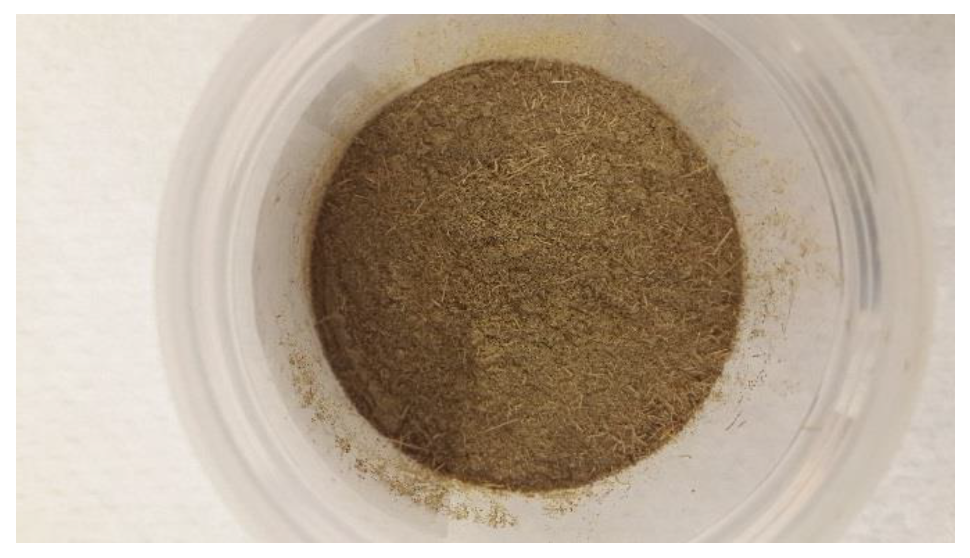
Dry stevia (Stevia rebaudiana Bertoni) leaves were purchased from Eternal Delight Company, Christchurch, New Zealand, which was imported originally from India in a dried form Figure 2. Before each set of experiments, the dried stevia leaves were blended to powder with a food-grade stainless steel grain miller 28,000/min (Huangcheng, China). The powder with ≤200 µm particle size was collected.

2.8. Conventional and Non-Conventional Extraction of Stevia Natural Sweeteners

Dried and grounded stevia leaves were extracted with Milli-Q water in ratio 1:15 (w/v) at 90 °C for 1 h by using a shaking water bath (Acorn Scientific BS-31) at 100 rpm Table 5. After extraction, stevia extract was rapidly cooled down to ambient temperature using an ice water bath. The extracts were centrifuged (10,000 rpm for 10 min) and filtered using Whatman No. 1. The filtrate part was dried in a convection oven at 45 °C overnight.
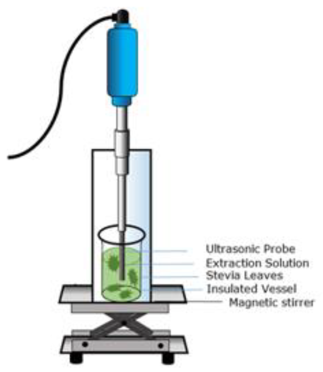
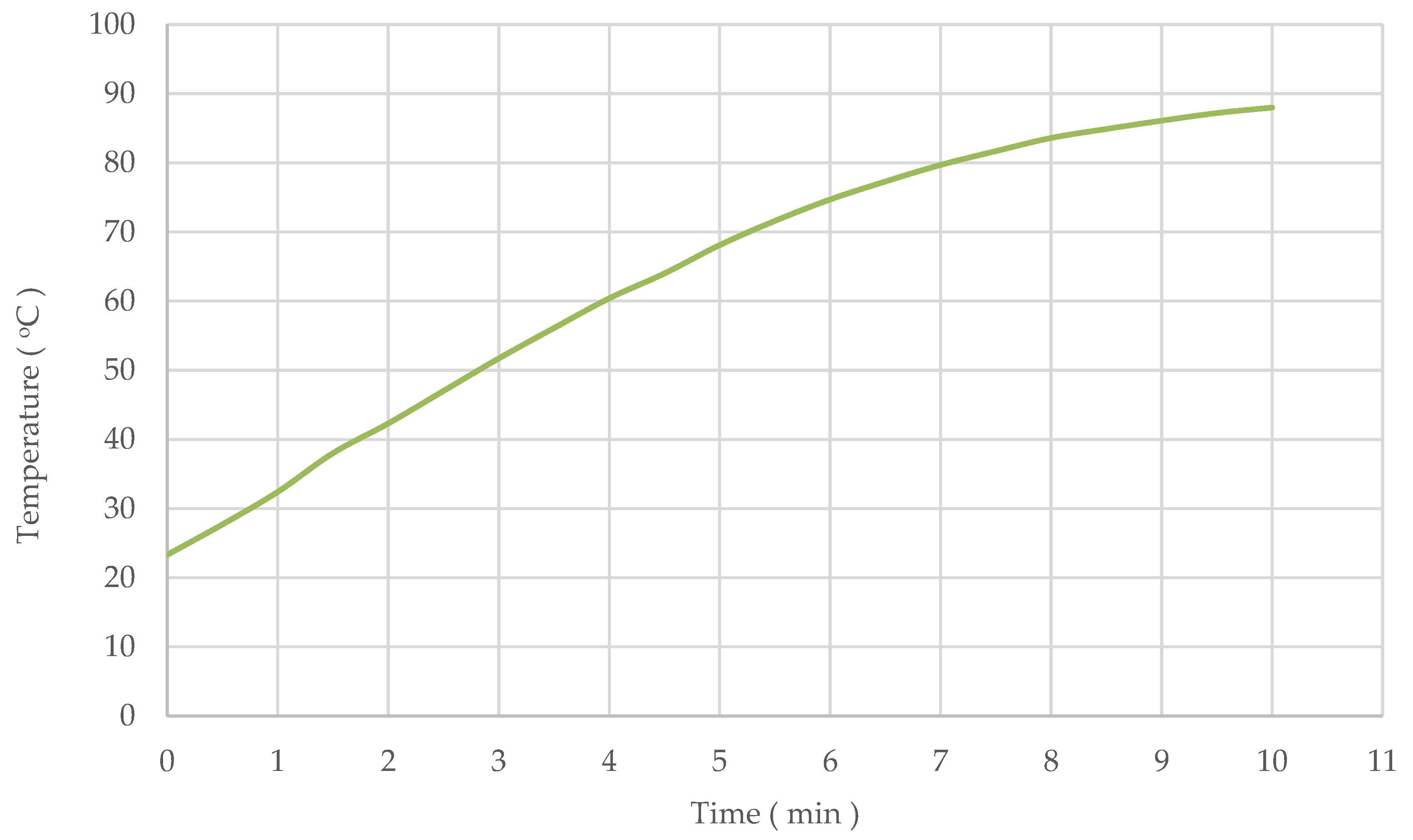
Ultrasound-assisted extraction was also used for stevioside and rebaudioside A. An ultrasonic horn transducer (model 750 W, Vibra-CellTM, USA) with a diameter of 12.75 mm at a frequency of 20 kHz, and a constant amplitude of 100%, was used. The stevia leaf powder was suspended in milli-Q water at ratio (1 g stevia powder: 15 mL Milli-Q water) with continuous stirring. An insulated vessel was used to retain the heat generated during the UAE process. The increments in temperature during ultrasound-assisted extraction were recorded every 30 s, and the temperature has reached a value of 88.00 °C after 10 min. The ultrasound treatment was carried out for 1, 3, 5, 7, and 10 min. After ultrasound treatment, the extracts were centrifuged (10,000× g, 10 min), and vacuum filtered using Whatman No. 1. The filtrate part was dried in a convection oven at 45 °C overnight. All samples were performed in duplicate. The ultrasound-assisted extraction setup is illustrated in Figure 3.

2.9. HPLC Analysis of Stevia Extracts

Typically, steviol glycosides are determined individually by liquid chromatography methods [39]. HPLC system Shimadzu (Tokyo, Japan) was used for stevioside and rebaudioside A analysis together with DGU-20A5 degasser, SPD-20A UV/VIS detector, SIL-20A, and an Agilent Eclipse XDB-C18, 5 µm, 4.6 × 150 mm column. Acetonitrile (A) and 10 mM sodium phosphate buffer (pH = 2.63) (B) with a 32A:68B ratio were used as a mobile phase. The column was purged for 20 min with the mobile phase prior to the sample injection at flowrate 1 mL/min. The detection wavelength, column temperature, and the injection volume were set at 210 nm, 40 °C, and 20 µL, respectively.
HPLC-grade stevioside and rebaudioside standards were obtained from AK Scientific, Inc., USA. The standard stock solution was prepared accurately using 0.01 g of each standard and diluted to 10 mL with the mobile phase. Seven dilutions were prepared at concentrations of 25, 50, 100, 200, 300, 400, and 500 ppm for stevioside and rebaudioside A (Figure S1 in the supplementary file). All the standard solutions were filtered through a 0.45 µm PTFE filter prior to HPLC analysis. About 0.02 g of dried extracts were dissolved in 10 mL Milli-Q water to determine stevioside and rebaudioside A concentrations in the extracts. All the samples were filtered using a 0.45 µm PTFE filter prior to HPLC analysis. HPLC chromatograms of Stevioside, rebaudioside A, and stevia leaves extract are shown in Figure S2 in the supplementary file.

3. Results and Discussion

3.1. The Impact of Particle Size on the Extractability of the Bioactive Compounds

This paper presents the effect of sample pre-treatment (grinding) on the extraction of the bioactive compounds from two selected plants. The relation between several bioactive terpenoids and different particle sizes was demonstrated. Such an effect has been studied before, but in the absence of the possibility of a significant release of bioactive during the process of grinding the leaves, which is the main objective of this paper.
GCMS chromatograms results of un-milled, 500 µm, 850 µm, and 1400 µm Manuka leaves at different times showed no extraction of terpenes for the unmilled and the 1400 µm diameter samples even after 60 min. However, Pinene<alpha-> was the only monoterpenes observed in samples with 850 µm particle size within the defined time. For the diameter of 500 µm, the intensity of four compounds, namely Pinene< alpha-, RT 5.995, Eucalyptol, RT 10.526, Globulol, RT 40.952, and Menthol, 1’-(butyn-3-one-1-yl)-, (1R, 2S, 5R)-, RT 42.84, increased over time, reaching the highest intensity after 60 min. In Figure 4a, the extraction of bioactive terpenoids of Manuka samples with a particle size of d ≥ 250 µm showed almost no extraction at zero time, and the extraction of all bioactives increased with time, which is expected since the sample was only mildly grounded. Figure 4b shows that the extraction happens at zero time, indicating that most of the release of bioactives have occurred during grinding, which supports our hypothesis.
Samples with a diameter of d ≥ 200 µm Figure 4c, a high amount of the terpenes, were again extracted and appeared in the chromatogram at a zero time (in a few seconds). Moreover, the terpenes content in the extracts increased little after 3, 5, and 7 min to remain relatively constant. Furthermore, the total terpenes content after decreasing the particle size (d ≤ 200 µm) is represented in Figure 5a. Indeed, the increase of the terpenes content in the extracts with a sample diameter of d ≤ 200 µm was expected. On a similar approach, Gião et al. studied the effect of particle size on the antioxidant extraction from Agrimonia eupatoria, Salvia sp., and Satureja montana plants [40]. The particle size of 200 µm, which was the smallest, obtained by the coffee grinder, gave the highest rate of extraction [40]. We believe that the 50% increase in the extraction shown in Figure 4c and Figure 5a is due to the release of terpenoids during the grinding process, which supports our hypothesis that the maximum terpenoids concentration occurs as soon as the powder is immersed in the solvent. This is also supported by the results of Figure 5b,c for the fine particles d = 100 µm and 68 µm, respectively. Interestingly, the extraction of terpenes had slightly decreased after 15 min Figure 5c, which could be the degradation of the terpenes. Data comparison of Manuka extracts with the particle size used in this study at Zero time (in a few seconds) is demonstrated in the GCMS chromatogram Figure S3 in the supplementary file.

3.2. Effect of Different Extraction Methods on Stevioside and Rebaudioside A Yield

As mentioned in Section 2.8, the extraction was carried out at a variable temperature in an insulated vessel to retain the heat generated during the ultrasound. The increase in temperature with time is as shown in Figure 6. The temperature has reached values of 35.7 °C, 61.5 °C, 65.5 °C, 81.5 °C, and 88.00 °C during sonication after 1, 3, 5, 7, and 10 min of extraction, respectively. According to the literature, the samples used for extraction are usually placed in a cooling bath to absorb the heat generated by ultrasonication. UAE with a circulating water bath is higher energy consumption compared to UAE under uncontrolled temperature (variable temperature) at the same duration due to (i) the energy required to bring the temperature to a certain degree and (ii) the energy needed to keep the temperature at constant. It was surprising that the energy used during sonification (with a cooling bath) was excessively large, making such treatment highly impractical.
As can be seen from Table 6, the stevioside and rebaudioside A content in the hot water extract was 5.34 ± 0.006 and 2.07 ± 0.015 g/100 g dry leaves, respectively. These values are within the range of steviol glycosides found in different varieties of the stevia plant (stevioside 4–13 g/100 g dry leaves, rebaudioside A 2–4 g/100 g dry leaves) [7,36,39,41].
From the results shown in Table 6, the stevioside content in the extracts obtained by UAE at different times has remained relatively constant (5.02, 5.48, 5.37, 5.33, and 5.47 g/100 g dry leaves with extraction times 1, 3, 5, 7, and 10 min, respectively) compared with hot water extraction (90 °C, 1 h).
Similarly, the yield of rebaudioside A was 2.00 ± 0.004, 2.13 ± 0.009, 2.09 ± 0.039, 2.06 ± 0.001, and 2.15 ± 0.024 g/100 g dry leaves after 1, 3, 5, 7, and 10 min of sonication, respectively. It can be concluded that an exposure extraction time of a few minutes appears to be sufficient to recover most of the bioactives from Stevia rebaudiana fine powder (200 µm) via ultrasound as a novel technology as compared to the classical hot water extraction (90 °C, 1 h) in this study. By decreasing the raw material particle size, rapid extraction could happen.
This, again, supports our hypothesis that most of the bioactive compound is released during grinding since the increase of extraction time did not have any effect.
The extraction of stevioside and rebaudioside A from Stevia rebaudiana fine powder (250 µm) was conducted and compared by[36]. The extraction was performed by microwave–assisted extraction (MAE) and compare it with cold maceration. The results showed that MAE (1 min, 50 °C) was more efficient than cold maceration (12 h, 25 °C). The higher extraction yield of microwave-assisted extraction can be explained by the elevated temperature used in MAE.
In another study, a saponin extraction from Chinese white ginseng (I) and fine root of Panax ginseng C. A. Meyer (II) by microwave-assisted extraction were reported by [19]. The raw materials were milled to 250 µm particle size. The results confirmed that the extraction yield of the target compounds by microwave-assisted extraction at 72.2 °C for two minutes was similar to the conventional reflux (75.1 °C for 12 h). Unfortunately, the authors did not conduct a conventional reflux extraction for a similar period of 2 min as used in microwave extraction, to have a fair comparison. It is unlikely that a microwave could reduce extraction from 12 h to 2 min.
Sumere et al. [42] have studied the influence of different processing factors such as solvent type, temperature, particle size, ultrasound power, and number of cycles on the phenolic compounds extraction from pomegranate peel using a combination of two extraction methods (UAE, PLE) [42]. The results showed that samples with small particle size (680 µm) show higher phenolic compounds extraction as compared to samples with larger particle sizes (1050 µm). However, the authors, due to the constant clogging of the processing line, did not recommend samples with small particle sizes. Moreover, Stamatopoulos et al. found a similar finding [43], showing that the smaller the size of particles, the faster the solvent can travel through it, which will indirectly shorten the extraction time.
Excellent previous work has been done in the literature to obtain a better yield of the target compound/s using different methods. The aim in most of the previous work has been to compare the different extraction methods [34,44,45]. However, none of the researchers conducted the measurement of the bioactive soon after the powder produced by grinding is exposed to the solvent (zero time). The current study suggests doing further investigation to address the effect of grinding on the release of bioactives from plant leaves by conducting zero time extraction, as this has not done in the literature.

4. Conclusions

This work highlights the impacts of particle size on bioactive compounds recoveries from two selected plant leaves. The major bioactive monoterpenes and sesquiterpenes from grounded Manuka leaves were extracted by n-hexane. Moreover, the extraction of two major sweet steviol glycosides from stevia powder was also carried out using conventional hot water extraction (90 °C, 1 h) and ultrasound thermal assisted adiabatic extraction. Previous investigators have not conducted the extraction of a very short period of time. ‘Zero time,’ or instant extraction, was used in this work for the first time by removing the grounded powder from the solvent within a few seconds. Results revealed that the bioactive terpenoids content from Manuka leaves Leptospermum scoparium could be easily extracted by decreasing the particle size to 68–200 µm. The experimental results also revealed that employing fine powder (200 µm) of stevia leaves could shorten the extraction time required to recover most of the sweet steviol glycosides via ultrasound.
The results showed that most of the extraction happened as soon as the fine powder is introduced to the solvent (a few seconds), with no further increase in extraction. This indicated that the release of bioactive compounds occurred during grinding.

Supplementary Materials

The following are available online at https://www.mdpi.com/2076-3417/10/18/6362/s1, Figure S1: title, Table S1: title, Video S1: title.

Author Contributions

Conceptualization, M.F. and N.A.; methodology, N.A.; formal analysis, N.A.; investigation, M.F. and N.A; resources, M.F.; data curation, N.A.; writing—original draft preparation, N.A.; writing—review and editing, M.F.; visualization, M.F. and N.A; supervision, M.F.; project administration, N.A.; funding acquisition, M.F. All authors have read and agreed to the published version of the manuscript.

Funding

This research was carried out as part of the Food Industry Enabling Technologies (FIET) program funded by the New Zealand Ministry of Business, Innovation, and Employment (contract MAUX1402).

Acknowledgments

The authors would like to acknowledge Manuka Bioactives Ltd., located in the East Cape, New Zealand for providing Manuka fresh leaves.

Conflicts of Interest

The authors declare no conflict of interest.

References

  1. Sasidharan, S.; Chen, Y.; Saravanan, D.; Sundram, K.; Latha, L. Extraction, isolation and characterization of bioactive compounds from plants’ extracts. Afr. J. Tradit. Complementary Altern. Med. 2011, 8, 1–10. [Google Scholar] [CrossRef] [Green Version]
  2. Wang, L.; Weller, C.L. Recent advances in extraction of nutraceuticals from plants. Trends Food Sci. Technol. 2006, 17, 300–312. [Google Scholar] [CrossRef]
  3. Azmir, J.; Zaidul, I.S.M.; Rahman, M.M.; Sharif, K.M.; Mohamed, A.; Sahena, F.; Jahurul, M.H.A.; Ghafoor, K.; Norulaini, N.A.N.; Omar, A.K.M. Techniques for extraction of bioactive compounds from plant materials: A review. J. Food Eng. 2013, 117, 426–436. [Google Scholar] [CrossRef]
  4. Syed Jaapar, S.Z.; Morad, N.A.; Iwai, Y.; Nordin, M.F.M. Effects of processing parameters in the sonic assisted water extraction (SAWE) of 6-gingerol. Ultrason. Sonochem. 2017, 38, 62–74. [Google Scholar] [CrossRef]
  5. Yang, B.; Liu, X.; Gao, Y. Extraction optimization of bioactive compounds (crocin, geniposide and total phenolic compounds) from Gardenia (Gardenia jasminoides Ellis) fruits with response surface methodology. Innov. Food Sci. Emerg. Technol. 2009, 10, 610–615. [Google Scholar] [CrossRef]
  6. Gan, C.-Y.; Latiff, A.A. Optimisation of the solvent extraction of bioactive compounds from Parkia speciosa pod using response surface methodology. Food Chem. 2011, 124, 1277–1283. [Google Scholar] [CrossRef]
  7. Šic Žlabur, J.; Voća, S.; Dobričević, N.; Brnčić, M.; Dujmić, F.; Rimac Brnčić, S. Optimization of ultrasound assisted extraction of functional ingredients from Stevia rebaudiana Bertoni leaves. Int. Agrophys. 2015, 29, 231–237. [Google Scholar]
  8. Ćujić, N.; Šavikin, K.; Janković, T.; Pljevljakušić, D.; Zdunić, G.; Ibrić, S. Optimization of polyphenols extraction from dried chokeberry using maceration as traditional technique. Food Chem. 2016, 194, 135–142. [Google Scholar] [CrossRef]
  9. Papoutsis, K.; Pristijono, P.; Golding, J.B.; Stathopoulos, C.E.; Bowyer, M.C.; Scarlett, C.J.; Vuong, Q.V. Optimisation of aqueous extraction conditions for the recovery of phenolic compounds and antioxidants from lemon pomace. Int. J. Food Sci. Technol. 2016, 51, 2009–2018. [Google Scholar] [CrossRef] [Green Version]
  10. Hiranvarachat, B.; Devahastin, S.; Soponronnarit, S. Comparative evaluation of atmospheric and vacuum microwave-assisted extraction of bioactive compounds from fresh and dried Centella asiatica L. leaves. Int. J. Food Sci. Technol. 2015, 50, 750–757. [Google Scholar] [CrossRef]
  11. Palsikowski, P.A.; Besen, L.M.; Klein, E.J.; da Silva, C.; da Silva, E.A. Optimization of ultrasound-assisted extraction of bioactive compounds from B. forficata subsp. Pruinosa. Can. J. Chem. Eng. 2020, 1–13. [Google Scholar] [CrossRef]
  12. Ghafoor, K. Optimized Extraction of Phenolic Compounds from Barley (Hordeum vulgare L.) Seed and Their Radical Scavenging Properties. J. Food Process. Preserv. 2015, 39, 793–799. [Google Scholar] [CrossRef]
  13. Porter, N.; Smale, P.; Nelson, M.; Hay, A.; Van Klink, J.; Dean, C. Variability in essential oil chemistry and plant morphology within a Leptospermum scoparium population. N. Z. J. Bot. 1998, 36, 125–133. [Google Scholar]
  14. Soejarto, D.; Compadre, C.; Medon, P.; Kamath, S.; Kinghorn, A.D. Potential sweetening agents of plant origin. II. Field search for sweet-tastingStevia species. Econ. Bot. 1983, 37, 71–79. [Google Scholar]
  15. Kinghorn, A. Food Ingredient Safety Review: Stevia Rebaudiana Leaves; Herbal Research Foundation: Boulder, CO, USA, 1992. [Google Scholar]
  16. Cardello, H.; Da Silva, M.; Damasio, M. Measurement of the relative sweetness of stevia extract, aspartame and cyclamate/saccharin blend as compared to sucrose at different concentrations. Plant Foods Hum. Nutr. 1999, 54, 119–129. [Google Scholar]
  17. Bruni, R.; Guerrini, A.; Scalia, S.; Romagnoli, C.; Sacchetti, G. Rapid techniques for the extraction of vitamin E isomers from Amaranthus caudatus seeds: Ultrasonic and supercritical fluid extraction. Phytochem. Anal. 2002, 13, 257–261. [Google Scholar] [CrossRef]
  18. András, C.D.; Simándi, B.; Örsi, F.; Lambrou, C.; Missopolinou-Tatala, D.; Panayiotou, C.; Domokos, J.; Doleschall, F. Supercritical carbon dioxide extraction of okra (Hibiscus esculentus L.) seeds. J. Sci. Food Agric. 2005, 85, 1415–1419. [Google Scholar]
  19. Kwon, J.-H.; Bélanger, J.M.R.; Paré, J.R.J.; Yaylayan, V.A. Application of the microwave-assisted process (MAP™☆☆MAP is a Trademark of Her Majesty the Queen in Right of Canada as represented by the Minister of the Environment.) to the fast extraction of ginseng saponins. Food Res. Int. 2003, 36, 491–498. [Google Scholar] [CrossRef]
  20. Sass-Kiss, A.; Simandi, B.; Gao, Y.; Boross, F.; Vamos-Falusi, Z. Study on the pilot-scale extraction of onion oleoresin using supercritical CO2. J. Sci. Food Agric. 1998, 76, 320–326. [Google Scholar] [CrossRef]
  21. Giannuzzo, A.N.; Boggetti, H.J.; Nazareno, M.A.; Mishima, H.T. Supercritical fluid extraction of naringin from the peel of Citrus paradisi. Phytochem. Anal. 2003, 14, 221–223. [Google Scholar]
  22. Chemat, S.; Lagha, A.; AitAmar, H.; Bartels, P.V.; Chemat, F. Comparison of conventional and ultrasound-assisted extraction of carvone and limonene from caraway seeds. Flavour Fragr. J. 2004, 19, 188–195. [Google Scholar] [CrossRef]
  23. Szentmihályi, K.; Vinkler, P.; Lakatos, B.; Illés, V.; Then, M. Rose hip (Rosa canina L.) oil obtained from waste hip seeds by different extraction methods. Bioresour. Technol. 2002, 82, 195–201. [Google Scholar] [CrossRef]
  24. Samaram, S.; Mirhosseini, H.; Tan, C.P.; Ghazali, H.M. Ultrasound-assisted extraction (UAE) and solvent extraction of papaya seed oil: Yield, fatty acid composition and triacylglycerol profile. Molecules 2013, 18, 12474–12487. [Google Scholar]
  25. Zou, T.-B.; Xia, E.-Q.; He, T.-P.; Huang, M.-Y.; Jia, Q.; Li, H.-W. Ultrasound-assisted extraction of mangiferin from mango (Mangifera indica L.) leaves using response surface methodology. Molecules 2014, 19, 1411–1421. [Google Scholar]
  26. Dey, S.; Rathod, V.K. Ultrasound assisted extraction of β-carotene from Spirulina platensis. Ultrason. Sonochem. 2013, 20, 271–276. [Google Scholar] [CrossRef]
  27. Thakker, M.R.; Parikh, J.K.; Desai, M.A. Ultrasound Assisted Hydrotropic Extraction: A Greener Approach for the Isolation of Geraniol from the Leaves of Cymbopogon martinii. ACS Sustain. Chem. Eng. 2018, 6, 3215–3224. [Google Scholar] [CrossRef]
  28. Zhang, G.; He, L.; Hu, M. Optimized ultrasonic-assisted extraction of flavonoids from Prunella vulgaris L. and evaluation of antioxidant activities in vitro. Innov. Food Sci. Emerg. Technol. 2011, 12, 18–25. [Google Scholar]
  29. Pan, G.; Yu, G.; Zhu, C.; Qiao, J. Optimization of ultrasound-assisted extraction (UAE) of flavonoids compounds (FC) from hawthorn seed (HS). Ultrason. Sonochem. 2012, 19, 486–490. [Google Scholar] [CrossRef]
  30. Chen, Y.; Luo, H.; Gao, A.; Zhu, M. Ultrasound-assisted extraction of polysaccharides from litchi (Litchi chinensis Sonn.) seed by response surface methodology and their structural characteristics. Innov. Food Sci. Emerg. Technol. 2011, 12, 305–309. [Google Scholar] [CrossRef]
  31. Kong, Y.; Zu, Y.-G.; Fu, Y.-J.; Liu, W.; Chang, F.-R.; Li, J.; Chen, Y.-H.; Zhang, S.; Gu, C.-B. Optimization of microwave-assisted extraction of cajaninstilbene acid and pinostrobin from pigeonpea leaves followed by RP-HPLC-DAD determination. J. Food Compos. Anal. 2010, 23, 382–388. [Google Scholar] [CrossRef]
  32. Upadhyay, R.; Ramalakshmi, K.; Jagan Mohan Rao, L. Microwave-assisted extraction of chlorogenic acids from green coffee beans. Food Chem. 2012, 130, 184–188. [Google Scholar] [CrossRef]
  33. Rodríguez-Rojo, S.; Visentin, A.; Maestri, D.; Cocero, M.J. Assisted extraction of rosemary antioxidants with green solvents. J. Food Eng. 2012, 109, 98–103. [Google Scholar] [CrossRef] [Green Version]
  34. Li, Y.; Skouroumounis, G.K.; Elsey, G.M.; Taylor, D.K. Microwave-assistance provides very rapid and efficient extraction of grape seed polyphenols. Food Chem. 2011, 129, 570–576. [Google Scholar] [CrossRef]
  35. Trusheva, B.; Trunkova, D.; Bankova, V. Different extraction methods of biologically active components from propolis: A preliminary study. Chem. Cent. J. 2007, 1, 13. [Google Scholar] [CrossRef] [Green Version]
  36. Jaitak, V.; Bandna, B.S.; Kaul, V. An efficient microwave-assisted extraction process of stevioside and rebaudioside-A from Stevia rebaudiana (Bertoni). Phytochem. Anal. 2009, 20, 240–245. [Google Scholar]
  37. Rodríguez-Padrón, D.; Zhao, D.; Garín Ortega, R.N.; Len, C.; Balu, A.M.; García, A.; Luque, R. Characterization and Antioxidant Activity of Microwave-Extracted Phenolic Compounds from Biomass Residues. ACS Sustain. Chem. Eng. 2020, 8, 1513–1519. [Google Scholar] [CrossRef]
  38. Douglas, M.H.; van Klink, J.W.; Smallfield, B.M.; Perry, N.B.; Anderson, R.E.; Johnstone, P.; Weavers, R.T. Essential oils from New Zealand manuka: Triketone and other chemotypes of Leptospermum scoparium. Phytochemistry 2004, 65, 1255–1264. [Google Scholar] [CrossRef]
  39. Jentzer, J.-B.; Alignan, M.; Vaca-Garcia, C.; Rigal, L.; Vilarem, G. Response surface methodology to optimise Accelerated Solvent Extraction of steviol glycosides from Stevia rebaudiana Bertoni leaves. Food Chem. 2015, 166, 561–567. [Google Scholar] [CrossRef] [Green Version]
  40. Gião, M.S.; Pereira, C.I.; Fonseca, S.C.; Pintado, M.E.; Malcata, F.X. Effect of particle size upon the extent of extraction of antioxidant power from the plants Agrimonia eupatoria, Salvia sp. and Satureja montana. Food Chem. 2009, 117, 412–416. [Google Scholar] [CrossRef]
  41. Erkucuk, A.; Akgun, I.; Yesil-Celiktas, O. Supercritical CO2 extraction of glycosides from Stevia rebaudiana leaves: Identification and optimization. J. Supercrit. Fluids 2009, 51, 29–35. [Google Scholar]
  42. Sumere, B.R.; de Souza, M.C.; dos Santos, M.P.; Bezerra, R.M.N.; da Cunha, D.T.; Martinez, J.; Rostagno, M.A. Combining pressurized liquids with ultrasound to improve the extraction of phenolic compounds from pomegranate peel (Punica granatum L.). Ultrason. Sonochem. 2018, 48, 151–162. [Google Scholar] [CrossRef]
  43. Stamatopoulos, K.; Chatzilazarou, A.; Katsoyannos, E. Optimization of Multistage Extraction of Olive Leaves for Recovery of Phenolic Compounds at Moderated Temperatures and Short Extraction Times. Foods 2014, 3, 66–81. [Google Scholar]
  44. Li, C.; Zhang, J.; Zhao, C.; Yang, L.; Zhao, W.; Jiang, H.; Ren, X.; Su, W.; Li, Y.; Guan, J. Separation of the main flavonoids and essential oil from seabuckthorn leaves by ultrasonic/microwave-assisted simultaneous distillation extraction. R. Soc. Open Sci. 2018, 5, 180133. [Google Scholar] [CrossRef] [Green Version]
  45. Khan, M.K.; Abert-Vian, M.; Fabiano-Tixier, A.-S.; Dangles, O.; Chemat, F. Ultrasound-assisted extraction of polyphenols (flavanone glycosides) from orange (Citrus sinensis L.) peel. Food Chem. 2010, 119, 851–858. [Google Scholar]
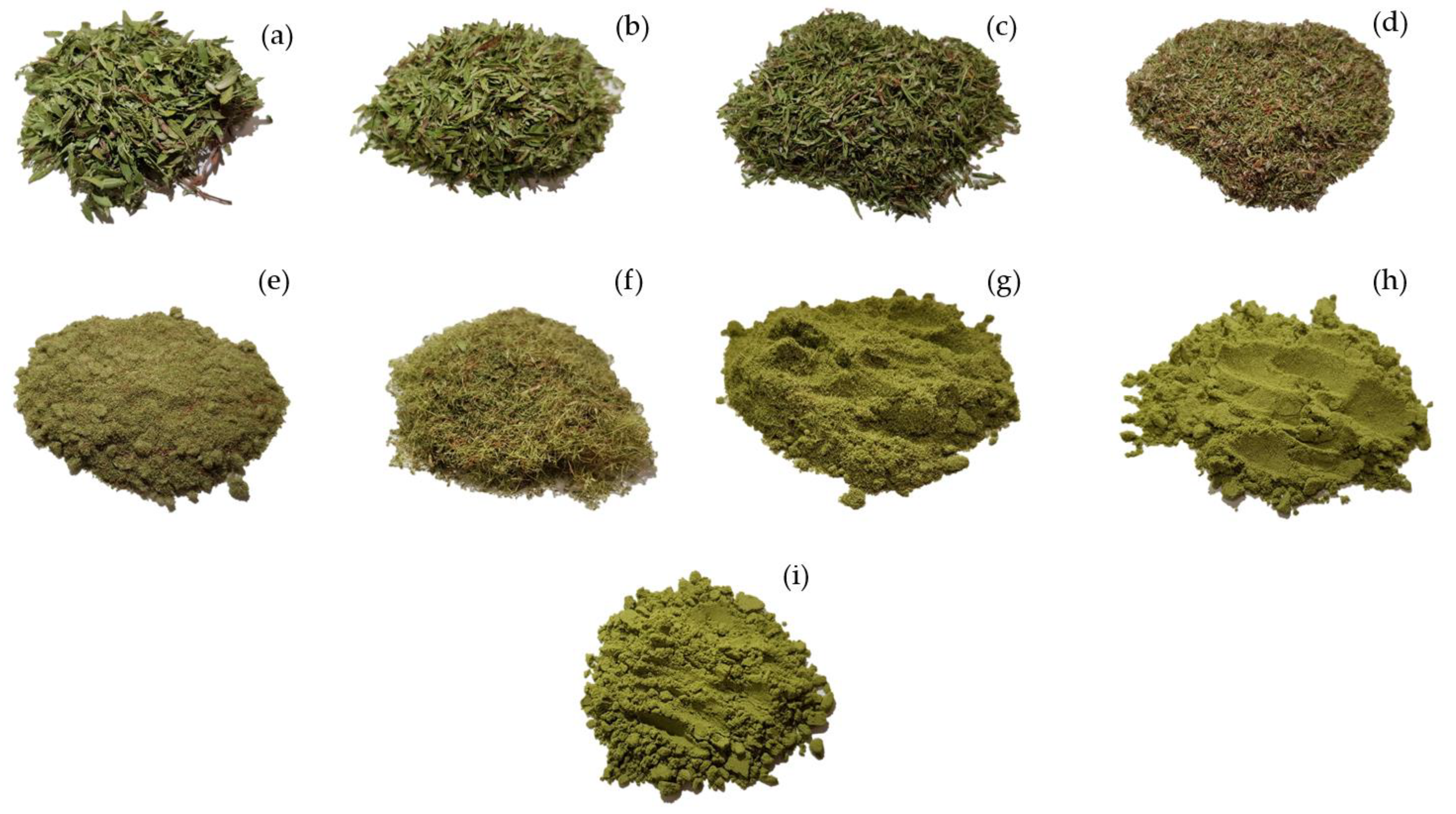
Figure 1. New Zealand Manuka leaves and their corresponding leaves grounded to different particle size: (A) 1400 µm, (B) 850 µm, (C) 500 µm, (D) d ≥ 250 µm, (E) d ≤ 250 µm, (F) d ≥ 200 µm, (G) d ≤ 200 µm, (H) 100 µm, (I) 68 µm.
Applsci 10 06362 g001
Figure 2. Stevia leaves powder.
Applsci 10 06362 g002
Figure 3. Ultrasound-assisted extraction system: without circulating water-bath.
Applsci 10 06362 g003
Figure 4. Total of selected bioactive terpenoids in Manuka samples with particle size of: (a) d ≥ 250, (b) d ≤ 250, (c) d ≥ 200 μm.
Applsci 10 06362 g004
Figure 5. Total of selected bioactive terpenoids in Manuka samples with particle size of: (a) d ≤ 200, (b) d = 100 μm, (c) d = 68 μm.
Applsci 10 06362 g005
Figure 6. Temperature change during UAE for stevia powder suspension (750 W, 12.75 mm, 20 kHz, 100% amplitude).
Applsci 10 06362 g006
Table 1. Studies on the extraction of valuable compounds from various source material.
Raw MaterialBioactive Compound (s)Particle Size (µm)Grinder TypeMethod of ExtractionSolid/Liquid (g/mL)Solvent TypeExtraction Time (min)Yield (mg/g)Ref.
Amaranthus caudatusTocols200Blade grinderSE 1, 25 °C1/20 Methanol14400.07632[17]
200Blade grinderUAE 2, 25 °C1/20 Methanol600.0637
200Blade grinderSLE 3, 25 °C, 400 atm1/30 CO2150.12927
Okra seedβ-Sitosterol/α-Tocopherol/γ-TocopherolgroundedHammer millSoxhlet n-hex-0.00201/0.127/0.38[18]
groundedHammer millSoxhlet EtOH-0.00268/0.129/0.494
groundedHammer millSLE, 50 °C, 450 bar1/24–1/80CO2240–8000.00239/0.148/0.407
GinsengSapoininsd > 250Cutting millSE, 75 °C1/10MeOH 80%72052.4[19]
MAE 4, 75 °C1/10MeOH 80%253.1
OnionSulfur/Oleoresin200–1400-Soxhlet1/20Alcohol2403.78/350[20]
SD 57/120Steam3000.167/0.4
SE, 25 °C1/20n-hex1200.087/11
SE, 25 °C1/20Alcohol1200.895/126
SLE, 65 °C, 300 bar1/14CO21800.208/9
Citrus paradisiNaringinFragmented fresh peelsFood processorSoxhlet1/10EtOH:water (70:30)48015.2[21]
SE, 22–25 °C1/5EtOH:water (70:30)18013.5
SLE, 58.6 °C, 95 bar CO2: EtOH
(85:15)
4514.4
Caraway seedsCarvone/limone-Roller mill (Vector Siever, 930 rpm)Soxhlet1/20n-hex30016.28/15.15[22]
SE, 69 °C1/20n-hex6013.38/12.63
UAE, 69 °C n-hex6014.45/14.27
UAE, 20–38 °C n-hex6017.16/16.16
Rosehip seedsOil360Coffee millSoxhlet1/25n-hex18048.5 [23]
UAE, 69 °C1/25n-hex6032.5
MAE, 40 °C1/3.5n-hex3052.6
SLE, 35 °C, 250 bar-CO28057.2
SLE, 28°C, 100 bar-CO2:propane3566.8
Papaya seedPapaya seed oilpowder-UAE, 50 °C, 40 KHz of 700 W1/8 n-hex30761[24]
SE, shaking water bath 100 rpm, 25 °C 720791
Soxhlet -304
Mango (Mangifera indica L.) LeavesMangiferin (xanthone)Fine powder-UAE, 60 °C, 200 W EtOH 40%19.258.46[25]
Spirulina platensis Algaβ-carotene250-UAE, 30 °C, 167 W/cm21/30n-heptane81.15[26]
Cymbopogon martiniiGeranoil280-UAE,65% amplitude, 60 W, 70% cycle time1/32.51 M sodium cumene sulfonate161.9012[27]
Prunella vulgaris L. PlantFlavonoids250Knife millUAE, 79 °C1/30EtOH 41%30.536.2[28]
Hawthorn seeds by-productFlavonoids297-UAE, 65 °C, 40 W1/18EtOH 72%3716.45[29]
Litchi seeds by-productPolysaccharides250-UAE, 222 W15.0Water453.39[30]
Pigeonpea leaves PlantCajaninstilbene acid(CSA)/Pinostrobin (PI)d < 500-MAE, 65 °C, 300 W1/30EtOH 80%218.00/3.50[31]
Green coffee beans PlantChlorogenic acid, caffeine, and *TPCd < 720Hammer millMAE, 50 °C, at 800 W1/4Water57.25/8.40/10-17mg GAE/g [32]
Rosemary leaves SpiceTPC 6, rosmarinic and carnosic acids200–850-MAE (ON/OFF) cycles of at 250 W1/6Water, EtOH7Higher extraction compared to fresh and non-grinded leaves[33]
Grape seedsTPCPowder-MAE, 60 °C at 150 W-EtOH 47.2%4.696.30[34]
Parkia speciosa
podagro-waste
TPC and flavonoids250-SE, 35–36 °C1/20Ace 50%100–10266,800 and 4960[6]
Raw propolisTPCGround-Maceration, 25 °C1/10EtOH 70%43204300[35]
UAE-bath, 300 W, 25 °C305200
MAE, (ON/OFF) 800 W0.334040
Stevia rebaudiana (Bertoni)Stevioside and rebaudioside 250MortarConventional,25 °C1/10MeOH and EtOH 80%72077.40[36]
UAE, 35 ± 5 °CMeOH and EtOH 80%3061.98
MAE, 50 °CMeOH 80%1109.8
Stevia rebaudiana (Bertoni)Stevioside and rebaudioside -Microfine grinderHot water, 10 °C with shaking Water144096.98[7]
UAE, 81.2 °C10133.40
Walnut shellTPC-Ball millMAE, 100 °C1/20Water/ACN1/13099.09 ± 0.09[37]
SE 1: solvent extraction, UAE 2: ultrasound-assisted extraction, SLE 3: supercritical liquid extraction, MAE 4: microwave-assisted extraction, SD 5: steam distillation, TPC 6: total phenolic compounds.
Table 2. Sample particle size of the mild and severe grinding.
GrindingParticle Size (µm)
No grinding-
Mild1400
d > 850
d > 500
d > 250
d ≤ 250
Severed > 200
d ≤ 200
100
68
Table 3. Experimental parameters of solvent extraction.
Experimental ParametersConditions
Solventn-hexane 95%
Sample: solvent ratio, g/mL1:20
Extraction time (min)0, 3, 5,7, 10, 15, 30, 60 min
Temperature (°C)25
Table 4. Gas Chromatography-Mass Spectrometry Conditions.
GC ConditionsMS Conditions
Injection temperature280 °CIon source temperature250 °C
Injection modeSplitInterface temperature290 °C
Column flow4.81 mL/minSolvent cut time2.5 min
Split ratio50.0Start m/z50.00
End m/z800.00
Oven temperature program
Rate (°C)Temperature (°C)Hold time (min)
-40.05.00
3.00100.00.00
1.00115.00.00
3.00200.05.00
Table 5. Experimental parameters of hot water extraction.
Experimental ParametersConditions
Sample particle sized < 200
SolventMilli-Q water
Sample: solvent (w/v, g/mL)1:15
Extraction time (h)1
Temperature (°C)90
Table 6. Stevioside and rebaudioside A yield using hot water and UAE methods.
MethodExtraction Time (min)Stevioside Yield % (g/100 g Dry Leaves)Rebaudioside A Yield % (g/100 g Dry Leaves)
Hot water, 90 °C605.34 ± 0.0062.07 ± 0.015
UAE15.02 ± 0.0092.00 ± 0.004
35.48 ± 0.0172.13 ± 0.009
55.37 ± 0.0662.09 ± 0.039
75.33 ± 0.0092.06 ± 0.001
105.47 ± 0.0302.15 ± 0.024