Article

Computer-Based 3D Simulation Method in Dental Occlusion Education: Student Response and Learning Effect

1
Department of Prosthodontics, School of Dentistry, Kyungpook National University, Daegu 41940, Korea
2
Institute for Translational Research in Dentistry, Kyungpook National University, Daegu 41940, Korea
*
Author to whom correspondence should be addressed.
Appl. Sci. 2020, 10(17), 6073; https://doi.org/10.3390/app10176073
Submission received: 11 August 2020 / Revised: 25 August 2020 / Accepted: 27 August 2020 / Published: 2 September 2020
(This article belongs to the Special Issue Applied Simulation and Experiment Research in Dentistry)

Abstract

:
Occlusion is a fundamental subject in dental education, and occlusal adjustment is clinically essential in daily dental practices. This study aimed to assess the effects of computer-based 3D simulations on learner responses and learning effect on the principles of occlusal adjustment in undergraduate dental students in comparison with the traditional approach. Two teaching methods, i.e., paper-based 2D presentation and computer-based 3D simulation, were used for teaching the occlusal adjustment concepts. Sixty dental students were divided into two groups using a pair-matching randomization method. In the 2D presentation group, a textbook with 2D illustrations was used. 3D graphic dental models and computer design software were applied in the 3D simulation group. After the course, an attitudinal survey and examination were conducted to evaluate the participants’ feedback and the learning effects resulting from the teaching methods. The independent t test was used to compare the test scores between groups (with α = 0.5). Pearson’s correlation coefficient was calculated to investigate the agreement between the survey data and test scores. Most of the students’ feedback indicated that the 3D simulation method would be effective in acquiring knowledge on occlusion and jaw movement. The examination scores were significantly higher in the 3D simulation group compared with those in the 2D presentation group in the questions for centric relation (P = 0.034). Conversely, the scores were insignificant in the questions for eccentric relation (P = 0.403). There was no correlation observed between the survey data and the actual examination score. Computer-based 3D simulation could increase the participants’ expectations and learning effects in dental occlusion education. Further studies in diversified learning environments are required on the efficacy of digital educational modality.

1. Introduction

Stabilized occlusion is important for maintaining functional equilibrium in the oral cavity [1]. Inappropriate dental occlusion causes clinical complications such as uncomfortable or painful bite, abnormal tooth mobility, restoration fractures, and occlusion-related temporomandibular joint dysfunction [2,3]. It also lowers the long-term success of prosthodontic treatments [2]. Occlusal adjustment is a clinical procedure that removes the substrate of the occlusal surface of teeth or prostheses to fix deflective occlusion [4]. Occlusal treatment is essential and is performed in daily-routine practices. However, it is irreversible and requires an understanding of the coordinated interaction between dentition and neuro-muscular systems [5]. Owing to the clinical importance of occlusion, related knowledge on oral anatomy, jaw movement, and occlusal adjustment has been fundamental subjects in dental education [6].
Learning effects are mainly influenced by the association of teaching modality and education topic [7]. Conventional pedagogics for occlusion education are based on traditional lectures, two-dimensional (2D) anatomical illustrations, and tangible anatomical models [8]. However, because of the dynamic nature of the occlusion and neuromuscular system in the oral cavity, conventional teaching methods have limitations in providing learners with adequate understanding of spatial dentition relations and practical procedures for occlusal adjustment [9]. Concepts of functional occlusion and subsequent occlusal adjustments are difficult to impart using such static methods [10].
With advances in image processing, three-dimensional (3D) visualization technologies using computer modeling and design have improved medical education by making complex knowledge easier to present and teach [10,11]. In dental education, 3D visualization techniques have been applied to multiple training courses such as caries removal, tooth preparation, third molar extraction, gingival pocket probing, and dental calculus removal. [12,13,14,15,16]. Compared with 2D illustrations, 3D visualization allows for the complicated spatial relationships of the dentition to be presented in a 3D polygonal mesh format. Moreover, as the 3D dentition and jaw models can be managed using computer software, learners can perform virtual analysis, planning, and treatment practices on the 3D models [17]. The virtual simulation enhances the spatial memory storage, attention, and perception of learners [18]. This is because the cognitive processes of the human brain can be facilitated during the navigation and exploration of a virtual environment [19].
Education methods assisted by computer software have been reported to be equal or superior to traditional teaching systems for knowledge acquisition in medical fields [10]. Three-dimensional visualization and simulation could improve learning effects in occlusion education by providing students with 3D perception of occlusion and the virtual space for practice. However, the effects of such digital methods have not been thoroughly investigated. The purpose of this study is to evaluate the effects of a computer-based 3D simulation method on learner response and learning effects for the principles of occlusal adjustment in undergraduate dental students. It is then compared with a traditional approach that uses paper-based 2D illustrations. The first null hypothesis was that there is no difference in learning effects between the paper-based 2D presentation and computer-based 3D simulation methods. The second null hypothesis was that learner response on the education method and the actual examination results would not be correlated.

2. Materials and Methods

The workflow for this study is presented in Figure 1. Sixty participants were recruited from a pool of second-year dental students who had the same dental educational background and practical training. The participants were allocated to one of two groups defined by the teaching methods, namely the paper-based 2D presentation method and the computer-based 3D simulation method. For randomized grouping, a pair-matching method was used that is based on the grade point average (GPA) from the students’ first-year dental curriculum [20]. Partitioning into matched pairs by GPA was used to equalize the baseline covariates of learning competency of all attendees. Ethical approval for the study was granted by the institutional review board of Kyungpook National University Dental Hospital (KNUDH-2020-03-06).
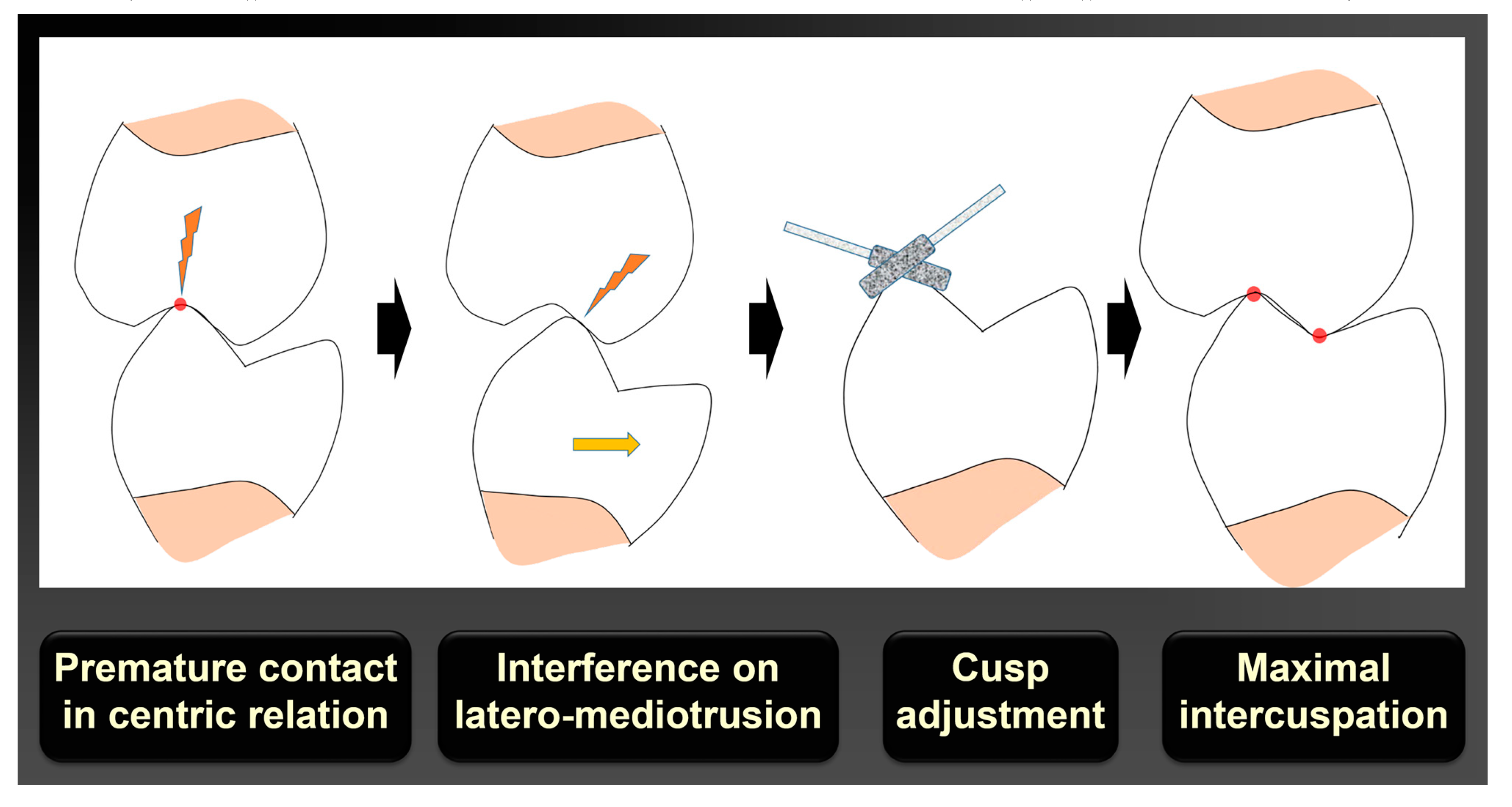
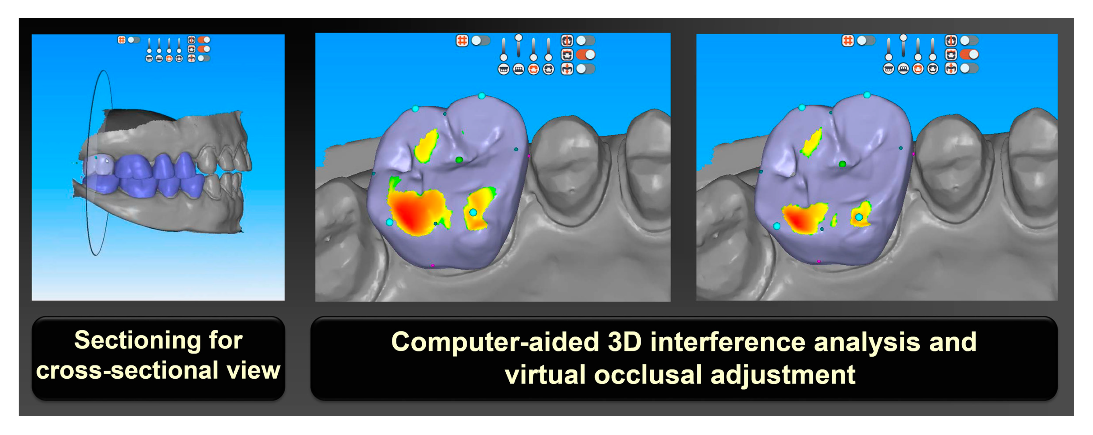
In the paper-based 2D presentation method, 30 students received the occlusion lecture using a textbook [21] and a PowerPoint presentation with 2D illustrations. The lecture consisted of the features of deflective occlusion in the centric and eccentric relation of the jaw. It also included the principles for occlusal adjustments of each interference in the maximal intercuspation and medio-, latero-, and protrusive movement of the jaw (Figure 2). In the computer-based 3D simulation method, the other 30 students received 3D simulated teaching with 3D graphic dental models and computer design software (R2CAD; MegaGen, Daegu, Korea). The 3D models with a deflective occlusal scheme were created in the polygonal mesh format. The occlusion interference of each condition was adjusted in accordance with the principles of occlusal adjustment by using computer design modules under the jaw movement (Figure 3). The characteristics of the teaching systems used in this study are summarized in Table 1. The conventional lecture and 3D simulation teaching methods were performed by an experienced instructor, who was unaware of the purpose of the study.
To assess participants’ responses and feedback regarding the 3D computer-based visualization method, an attitudinal survey was conducted after the training course for the 3D simulation group (Supplementary Materials S1). The survey was comprised of five questions assessing the preference of participants on the method in terms of expected effects and satisfaction [22]. Participants were asked to respond on a scale of 1 to 5. A score toward the high end of the scale indicates a higher preference toward the method.
Afterwards, an examination of the occlusal adjustment was conducted on the same day of class for all students to evaluate the learning effects of different teaching methods. The test consisted of eight questions for the principle of adjustment for deflective occlusion in centric and eccentric relations (Supplementary Materials S2). The questions were formulated following the recommendation by the American sociological review for open- and close-ended questions [23,24]. They were then modified based on the lectures and teaching materials provided to students in the present study. The examination results were first scored in a numeric scale of 0 to 100, and then categorized into five grades [25].
The means and standard deviations of the survey and examination scores were calculated. Statistical comparisons between the two instructional methods were performed by using the independent t test for each question concerning the adjustment strategies for deflective occlusion in centric and eccentric relation. The agreement between the results of the survey and examination was presented using Pearson’s correlation coefficient and a scatterplot. All statistical analyses were carried out using statistical software SPSS (IBM SPSS Statistics, v25.0; IBM Corp., Armonk, NY, USA) at 0.05 significance level.

3. Results

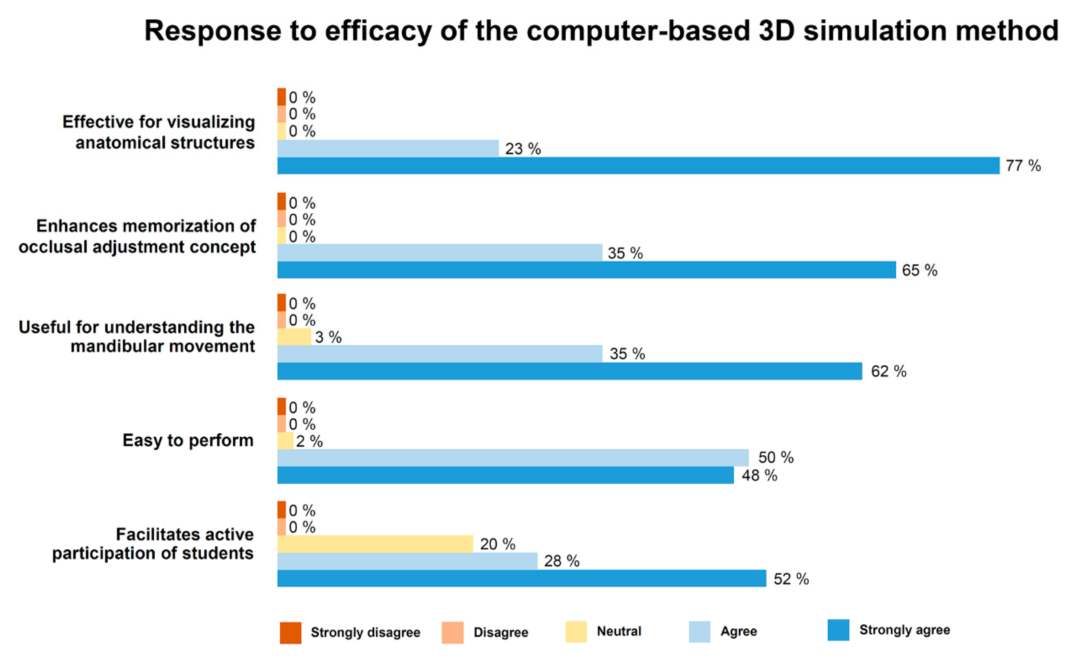
The results of the attitudinal survey are shown in Figure 4 All participants responded that the computer-based 3D simulation method is effective for visualizing anatomical structures and that it enhances memorization of the occlusal adjustment concept. Most of the participants agreed that the 3D simulation is useful for understanding the functional movement of the mandible. They also agreed that the simulation was easy to perform using the computer software. In the class participation question, four out of five participants responded that the 3D simulation facilitated active participation. No student made a negative response to all questions for the efficacy of the 3D simulation method on occlusal education.
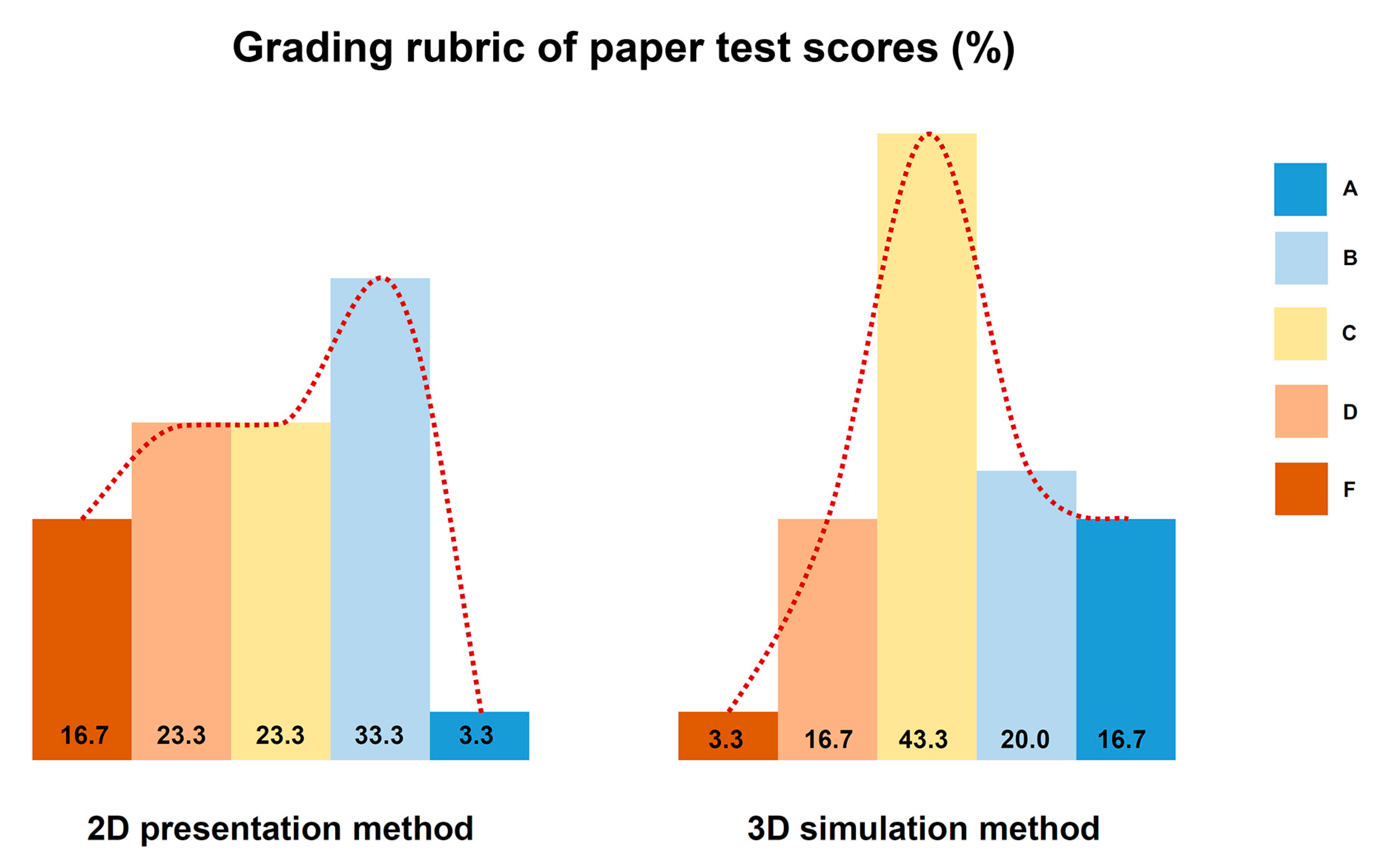
The examination results show that the 3D simulation group produced significantly higher scores than the 2D presentation group in the questions concerning the occlusal adjustment in centric relation (P = 0.034). Conversely, regarding the questions about occlusal adjustment in eccentric relation, there was no statistically significant difference in the test scores between the two teaching groups (P = 0.403). The overall score of the examination was higher in the group with the 3D simulation method than the group with the 2D presentation method (Table 2). The grading of numeric scores in the 2D presentation group showed a skewed distribution to the right, and the portion of grade F was relatively high (Figure 5). The 3D simulation method results produced a normal distribution in the score with a large portion of the A grade.
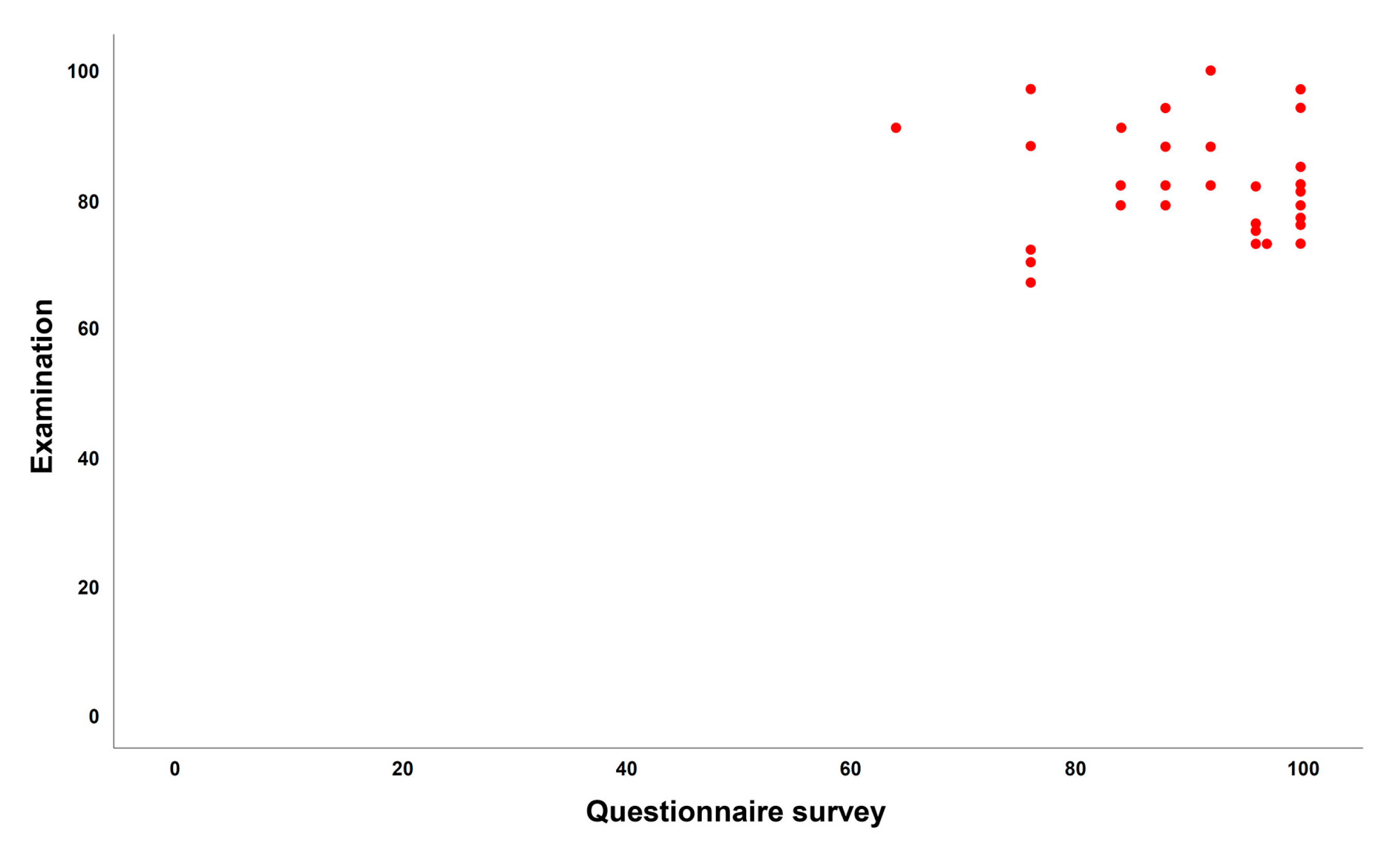
The correlation coefficient r was calculated as −0.48, representing the absence of linear correlation between the survey and examination outcomes. The scatterplot shows a weak and nonlinear association between the two outcomes (Figure 6).

4. Discussion

This study was designed to determine the efficacy of a computer-based 3D visualization and simulation method for knowledge acquisition on the occlusal adjustment compared with a conventional paper-based 2D presentation method. In the questionnaire survey on the expected effects of the 3D simulation method, most of the participants rated this method as highly appropriate for occlusion education, and especially effective for visualizing anatomical structures as well as memorizing occlusal adjustment concepts. Meanwhile, in the examination for evaluating students’ learning effects, the difference in scores between the groups was significant (i.e., in the questions for occlusal adjustment in centric relation). The difference was, however, not significant in the questions for the occlusal adjustment in eccentric relation. The mean score of the test was higher in the 3D simulation group than in the 2D presentation group. Based on the findings, the proposed first null hypothesis (i.e., there is no difference in learning effects between the two methods) was partially rejected. The correlation between the survey and the examination results was also investigated and no statistical association was found. This result implies that the feedback from the students and the actual learning effect on knowledge acquisition could not be directly related. Thus, the second null hypothesis (i.e., learner response on the education method and the actual examination results would not be correlated) was not rejected.
The distribution of examination scores for each student was visualized by classifying the scores in grades. An asymmetry in the score distribution was observed in the 2D presentation group, and it shows a large portion of the scores were in the low ranks. Conversely, in the 3D visualization group, scores were generally higher compared to the 2D presentation group, and a more symmetrical distribution of scores was observed. The bell-shaped curve might be recommended in grading academic achievements of students because this distribution fits well the normal distribution of natural learning ability in humans. Accordingly, based on the score distribution, it is thought that the 3D visualization method would be a more suitable method than the 2D presentation method in conveying the principles of occlusal adjustment in dentistry.
Despite the fact that teacher-oriented lectures with 2D images are still mainstream, this conventional method might be less effective for teaching manual skills and inducing critical reasoning required in higher education, especially in clinical courses in medicine [26,27]. Moreover, student participation would also be low in paper-based education systems [28,29]. Clinical dentistry is largely manual. Hence, conventional demonstrative lectures clearly have limitations as a teaching technique [30,31]. It is a challenge to understand the positional relationship of maxilla and mandibles in dynamic occlusion. It is also difficult to figure out occlusal disharmony that is topographic and its relationship with antagonistic teeth in cusp, fossa, and ridges. Recent studies in dental education revealed that mental training with 3D imagery could improve the in-depth perception for objects and had beneficial effects in acquiring clinical skills [32,33]. Training for manual skills coupled with decision-making can be achieved efficiently in environmental settings mimicking clinical situations. Thus, computer-based 3D visualization and simulation may be relevant in training occlusal adjustment in pre-clinical courses because students are provided spatial and visual aids to estimate distances, sizes, and shapes of anatomical subjects.
In the present study, the use of 3D dental mesh models and computer software providing color-coded occlusal analysis and functions for design modification motivated students to participate in the class. It also assisted with learning the principles of occlusal adjustment. However, because the software program was not developed for occlusion education, there were limitations in representing the jaw movements. Thus, the authors note that the development of a computer software program optimized for representing functional 3D movement of the jaw might be needed. It may also be useful to provide a virtual training environment to practice occlusal treatments.
The participants of this study were students in the same year of the dental curriculum. By using the inclusion criteria and previous knowledge, the experience of learners was controlled. However, these criteria limited the number of candidates that were included. To generalize the findings of this study, multi-center studies need to be conducted in the future. In addition, improvement in learning using computer-based 3D simulation could vary under different factors such as computer software, proficiency in operating software, 3D image model, and the instructor. Thus, further comprehensive studies considering the various influencing factors are necessary.

5. Conclusions

Based on the findings of this study, three main conclusions were drawn as follows. Firstly, the participants who experienced computer-based 3D simulation for occlusion education showed a high expectation of and satisfaction with the method in the accompanying survey. Secondly, the mean examination score on the principle of occlusal adjustment was higher in the group with the 3D simulation method than in the group with the paper-based 2D presentation group. However, the difference varied according the nature of the questions. Finally, there was no correlation observed between the expected effects for the use of 3D simulation and the actual examination score.

Supplementary Materials

The following are available online at https://www.mdpi.com/2076-3417/10/17/6073/s1, S1: Questionnaire sheet for survey on the expected effects of the 3D simulation method, S2: Examination sheet for the test on the principles of occlusal adjustment.

Author Contributions

Conceptualization, H.Y.M. and D.-H.L.; methodology, H.Y.M. and D.-H.L.; data curation, H.Y.M., H.-N.M., and D.-H.L.; formal analysis, H.Y.M. and D.-H.L.; investigation, H.Y.M., H.-N.M., and D.-H.L.; writing—original draft preparation, H.Y.M.; writing—review and editing, D.-H.L.; supervision, D.-H.L. All authors have read and agreed to the published version of the manuscript.

Funding

This research received no external funding.

Conflicts of Interest

The authors declare no conflict of interest.

References

  1. Clark, J.R.; Evans, R.D. Functional occlusion: I. A review. J. Orthod. 2001, 28, 76–81. [Google Scholar] [CrossRef]
  2. Clark, G.T.; Adler, R.C. A critical evaluation of occlusal therapy: Occlusal adjustment procedures. J. Am. Dent. Assoc. 1985, 110, 743–750. [Google Scholar] [CrossRef]
  3. McCullock, A.J. Making occlusion work: I. Terminology, occlusal assessment and recording. Dent. Update 2003, 30, 150–157. [Google Scholar] [CrossRef] [Green Version]
  4. Dawson, P.E. Evaluation, Diagnosis, and Treatment of Occlusal Problems; C.V. Mosby: St. Louis, MO, USA, 1988. [Google Scholar]
  5. Bakke, M. Mandibular elevator muscle: Physiology, action and effect of dental occlusion. Scand. J. Dent. Res. 1993, 101, 314–331. [Google Scholar] [CrossRef]
  6. Bando, E.; Nishigawa, K.; Nakano, M.; Takeuchi, H.; Shigemoto, S.; Okura, K.; Satsuma, T.; Yamamoto, T. Current status of researches on jaw movement and occlusion for clinical application. Jpn Dent. Sci. Rev. 2009, 45, 83–97. [Google Scholar] [CrossRef] [Green Version]
  7. O’Carroll, E. The teaching of occlusion in undergraduate dental schools in the UK and Ireland. Br. Dent. J. 2019, 227, 512–517. [Google Scholar] [CrossRef]
  8. Lee, D.J.; Wiens, J.P.; Ference, J.; Donatelli, D.; Smith, R.M.; Dye, B.D.; Obrez, A.; Lang, L.A. Assessment of occlusion curriculum in predoctoral dental education: Report from ACP Task Force on Occlusion Education. J. Prosthodont. 2012, 21, 578–587. [Google Scholar] [CrossRef]
  9. Truntzer, J.; Lynch, A.; Kruse, D.; Prislin, M. Musculoskeletal education: An assessment of the clinical confidence of medical students. Perspect. Med. Educ. 2014, 3, 238–244. [Google Scholar] [CrossRef] [Green Version]
  10. Jwayyed, S.; Stiffler, K.; Wilber, S.; Southern, A.; Weigand, J.; Bare, R.; Gerson, L. Technology-assisted education in graduate medical education: A review of the literature. J. Emerg. Med. 2011, 4, 1145–1157. [Google Scholar] [CrossRef] [Green Version]
  11. Frizziero, L.; Santi, G.M.; Liverani, A.; Napolitano, F.; Papaleo, P.; Maredi, E.; di gennaro, G.; Zarantonello, P.; Stallone, S.; Stilli, S.; et al. Computer-Aided Surgical Simulation for Correcting Complex Limb Deformities in Children. Appl. Sci. 2020, 10, 5181. [Google Scholar] [CrossRef]
  12. Buchanan, J.A. Experience with virtual reality-based technology in teaching restorative dental procedures. J. Dent. Educ. 2004, 68, 1258–1265. [Google Scholar] [CrossRef] [PubMed]
  13. Eve, E.J.; Koo, S.; Alshihri, A.A.; Cormier, J.; Kozhenikov, M.; Donoff, R.B.; Karimbux, N.Y. Performance of dental students versus prosthodontics residents on a 3D immersive haptic simulator. J. Dent. Educ. 2014, 78, 630–637. [Google Scholar] [CrossRef] [PubMed]
  14. Jasinevicius, T.R.; Landers, M.; Nelson, S.; Urbankova, A. An evaluation of two dental simulation systems: Virtual reality versus contemporary non-computer-assisted. J. Dent. Educ. 2004, 68, 1151–1162. [Google Scholar] [CrossRef] [PubMed]
  15. Pohlenz, P.; Gröbe, A.; Petersik, A.; von Sternberg, N.; Pflesser, B.; Pommert, A.; Höhne, K.H.; Tiede, U.; Springer, I.; Heiland, M. Virtual dental surgery as a new educational tool in dental school. J. Craniomaxillofac. Surg. 2010, 38, 560–564. [Google Scholar] [CrossRef]
  16. Urbankova, A. Impact of computerized dental simulation training on preclinical operative dentistry examination scores. J. Dent. Educ. 2010, 74, 402–409. [Google Scholar] [CrossRef]
  17. Zitzmann, N.; Matthisson, L.; Ohla, H.; Joda, T. Digital Undergraduate Education in Dentistry: A Systematic Review. Int. J. Environ. Res. Public. Health 2020, 17, 3269. [Google Scholar] [CrossRef]
  18. Kwon, C. The Effect of the Degree of Anxiety of Learners during the Use of VR on the Flow and Learning Effect. Appl. Sci. 2020, 10, 4932. [Google Scholar] [CrossRef]
  19. Van Helvoort, D.; Stobbe, E.; Benning, R.; Otgaar, H.; Ven, V. Physical exploration of a virtual reality environment: Effects on spatiotemporal associative recognition of episodic memory. Mem. Cogn. 2020, 48, 691–703. [Google Scholar] [CrossRef] [Green Version]
  20. Balzer, L.B.; Petersen, M.L.; van der Laan, M.J.; Consortium, T.S. Adaptive pair-matching in randomized trials with unbiased and efficient effect estimation. Stat. Med. 2015, 34, 999–1011. [Google Scholar] [CrossRef]
  21. Wiens, J. Fundamentals of Occlusion, 1st ed.; American College of Prosthodontists: Chicago, IL, USA, 2015. [Google Scholar]
  22. Alwin, D.; Krosnick, J. The Reliability of Survey Attitude Measurement. Sociol. Methods Res. 1991, 20, 139–181. [Google Scholar] [CrossRef]
  23. Husain, H.; Bais, B.; Hussain, A.; Samad, S. How to Construct Open Ended Questions. Procedia Soc. Behav. Sci. 2012, 60, 456–462. [Google Scholar] [CrossRef] [Green Version]
  24. Schuman, H.; Presser, S. The Open and Closed Question. Am. Sociol. Rev. 1979, 44, 692–712. [Google Scholar] [CrossRef]
  25. Anderson, L. A critique of grading: Policies, practices, and technical matters. Educ. Policy Anal. Arch. 2018, 26, 49. [Google Scholar] [CrossRef] [Green Version]
  26. Jofré, J.; Fuentes, J.; Conrady, Y.; Michel, M.; Quintana, P.; Asenjo-Lobos, C. Improving Dental Students Fine Motor Skills by Visualization and Mental Imagery: A Pilot Randomized Clinical Trial. Int. J. Odontostomat. 2019, 13, 69–74. [Google Scholar] [CrossRef] [Green Version]
  27. Steinert, Y.; Snell, L.S. Interactive lecturing: Strategies for increasing participation in large group presentations. Med. Teach. 2009, 21, 37–42. [Google Scholar]
  28. Siddiq, F.; Scherer, R.; Tondeur, J. Teachers’ emphasis on developing students’ digital information and communication skills (TEDDICS): A new construct in 21st century education. Comput. Educ. 2016, 92-93, 1–14. [Google Scholar] [CrossRef] [Green Version]
  29. Singh, R.; Gupta, N.; Singh, G. Learning style and teaching methodology preferences of dental students. J. Anat. Soc. India 2017, 65, 179–186. [Google Scholar] [CrossRef]
  30. De Boer, I.R.; Wesselink, P.R.; Vervoorn, J.M. Student performance and appreciation using 3D vs. 2D vision in a virtual learning environment. Eur. J. Dent. Educ. 2016, 20, 142–147. [Google Scholar] [CrossRef]
  31. Perry, S.; Bridges, S.M.; Burrow, M.F. A review of the use of simulation in dental education. Simul. Healthc. 2015, 10, 31–37. [Google Scholar] [CrossRef]
  32. Rao, A.; Tait, I.; Alijani, A. Systematic review and meta-analysis of the role of mental training in the acquisition of technical skills in surgery. Am. J. Sur. 2015, 210, 545–553. [Google Scholar] [CrossRef]
  33. Sobierajewicz, J.; Szarkiewicz, S.; Przekoracka-Krawczyk, A.; Jaśkowski, W.; Lubbe, R. To What Extent Can Motor Imagery Replace Motor Execution While Learning a Fine Motor Skill? Cogn. Psychol. 2016, 12, 179–192. [Google Scholar] [CrossRef] [PubMed]
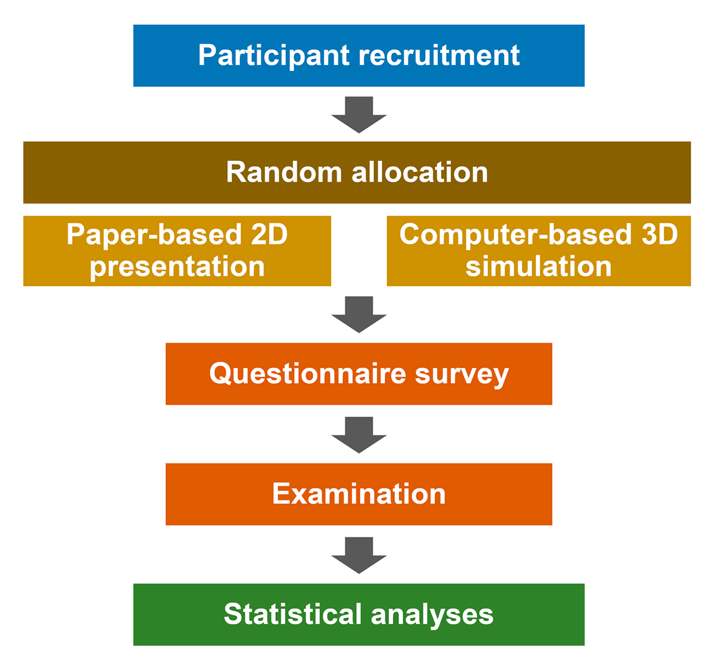
Figure 1. Workflow for this study.
Applsci 10 06073 g001
Figure 2. Two-dimensional presentation method with schematic illustrations.
Applsci 10 06073 g002
Figure 3. Three-dimensional visualization and simulation method with 3D models.
Applsci 10 06073 g003
Figure 4. Attitudinal survey results on the efficacy of the computer-based 3D simulation method.
Applsci 10 06073 g004
Figure 5. Grading of examination scores.
Applsci 10 06073 g005
Figure 6. Scatterplot showing the association between survey and examination.
Applsci 10 06073 g006
Table 1. Characteristics of the teaching system used for occlusal education.
2D Presentation Method3D Simulation Method
Materialbook, paper sheet, 2D figurescomputer, software, 3D graphic models
Contentsdescriptivedescriptive, manipulative, analytic
Visuospatial informationstaticdynamic
Teaching methodteacher-centeredstudent-centered
Cost for facilitylowmoderate to high
Learning methodobservationvirtual practice
Recommend group sizeNSsmall to medium
PreparationNSlearning how to operate software
NS: not specific.
Table 2. Mean and standard deviation of the examination results in groups by different teaching methods.
QuestionTeaching MethodP
2D Presentation3D Simulation
Centric77.0 ± 9.582.6 ± 10.30.034
Eccentric66.0 ± 16.770.3 ± 22.60.403
Whole72.9 ± 10.078.0 ± 11.00.064