Article

Do Waterborne Nanoplastics Affect the Shore Crab Carcinus maenas? A Case Study with Poly(methyl)methacrylate Particles

1
Department of Biology, University of Aveiro, 3810-193 Aveiro, Portugal
2
Centre for Environmental and Marine Studies, Department of Biology, University of Aveiro, 3810-193 Aveiro, Portugal
3
CICECO-Aveiro Institute of Materials, Department of Chemistry, University of Aveiro, 3810-193 Aveiro, Portugal
*
Authors to whom correspondence should be addressed.
Environments 2025, 12(5), 169; https://doi.org/10.3390/environments12050169
Submission received: 27 March 2025 / Revised: 10 May 2025 / Accepted: 15 May 2025 / Published: 20 May 2025
(This article belongs to the Special Issue Ecotoxicity of Microplastics)

Abstract

:
Nanoplastics (NPs) pose a significant environmental threat due to their small sizes, widespread distribution, and bioavailability, enabling interactions with marine organisms from pelagic to benthic species. In this study, the effects of 10 days of exposure to waterborne poly(methyl)methacrylate (PMMA) NPs were evaluated in the crab Carcinus maenas by assessing behavioral and biochemical endpoints (in gills, hepatopancreas, muscle, and hemolymph). Behavioral assessments using an open field test revealed that exposure to PMMA NPs resulted in an increase in distance walked (from 73.662 ± 17.137 cm in control to 248.560 ± 25.462 cm in the highest PMMA NPs concentration) and in random movement patterns. Muscle acetylcholinesterase (AChE) activity decreased from 10.83 ± 0.73 to 6.75 ± 0.45 nmol/min/mg of protein with PMMA NPs concentration increase, which, combined with behavioral responses, suggests neurological incapacities. In the gills and hepatopancreas, defense and detoxification mechanisms were activated, with a significant increase in superoxide dismutase (SOD) activity (at 20 µg/L in gills and 80 µg/L in hepatopancreas) and glutathione S-transferases (GSTs) activity (all PMMA NPs concentrations in gills and 20 and 320 µg/L in hepatopancreas). Despite these activations, oxidative damage was observed, with a significant increase in protein carbonylation (PC) levels (20, 80, and 320 µg/L in gills and 5, 20, and 80 µg/L in hepatopancreas) and lipid peroxidation (LPO) (80 and 320 µg/L in gills and 80 µg/L in hepatopancreas). Effects on hemolymph followed a pattern similar to those reported for gills and hepatopancreas. An increase in SOD hemolymph activity was observed in organisms exposed to 5 and 80 µg/L, and GSTs activity increased in crabs exposed to 80 µg/L. Oxidative damage in hemolymph was only detected through LPO at 5 and 320 µg/L. Overall, this study showed that PMMA NPs induce biochemical alterations and damage in different tissues of C. maenas and affect its behavior with potential impacts at a population level.

1. Introduction

The marine environment accounts for 71% of the Earth’s surface, from coastal to estuarine areas, portraying uniquely different habitats that house a variety of different species. This diversity leads to a variety of ecosystem services, all of which contribute to the general well-being of humans [1]. The constant discharge of plastics into the marine environment can jeopardize these services, with an almost immediate effect on the species that inhabit these habitats and in time threatening human survival [2,3].
Plastics have been present in our daily lives since the 1940s and humans’ dependency on them keeps increasing, as the properties of these cost-effective materials allow widespread use and applications [4,5]. Millions of metric tonnes (Mt) of plastic are produced each year (413.8 Mt in 2023) [6], and the single-use characteristic that society gives to most of these, combined with poor disposal at end-life, leads to their presence in all environments [7,8,9]. Thirty years after their mass production, there were reports of floating plastics in the oceans [10] where, chemical, biological, and physical factors promote their fragmentation into smaller particles such as microplastics (MPs, with sizes lower than 5 mm) [11,12], and nanoplastics (NPs, particles with an upper size limit not consensual, with some authors considering 1000 nm [13,14] and others 100 nm [15]).
Although both types of particles present potential risks to the environment, the smaller sizes may be associated with a higher risk since they have a higher ability to penetrate biological membranes and a higher surface area per unit mass compared to micrometer-sized particles. This allows for different interactions with other contaminants [16,17], and studies have already confirmed the presence of NPs in the environment [18,19,20], with these particles being found in both the water column and sediment, potentially affecting all marine organisms.
The properties of NPs are modulated by their plastic of origin (e.g., chemical composition) and their formation process (e.g., synthetic fabrication or natural weathering) [21]. However, environmental variables, namely pH, organic matter, ionic strength, and microbial activities, are also able to influence their characteristics, bioavailability, and effects on biota. These can affect NPs’ colloidal behavior and stability (e.g., tendency to form aggregates/agglomerates), in turn affecting their dispersibility, deposition, availability, and interaction with other chemicals and contaminants [22].
Poly(methyl)methacrylate (PMMA), an acrylic plastic known for its transparency and undefined crystalline structure [23], is gaining popularity due to its many uses as a glass replacement and in nanotechnology [24]. This polymer has already been detected in the environment [25], and some studies have been evaluating its impact on marine organisms like fish (Dicentrarchus labrax, Sparus aurata) [11,26], polychaetes (Hediste diversicolor) [27,28] and diatoms (Thalassiosira pseudonana, Skeletonema costatum) [29,30].
Despite the dispersion of NPs in the water column, biofouling and their tendency to aggregate and sink led to their accumulation in and near the sediment, becoming available to benthic organisms and causing damage [31]. Benthic communities host a wide array of species, including invertebrates like annelids, bivalves, and crustaceans. These organisms inhabit the bottom substrates and are extremely important for the dynamics of these environments [32], making them important targets for NPs. This is exacerbated by the fact that the mechanisms and species responsible for nutrient resuspension can also resuspend contaminants that were accumulated in the sediment [33].
Carcinus maenas (shore crab), native to Atlantic Europe [34], is considered an ecosystem engineer due to its ability to alter the habitat by modifying the nutrient cycle with sediment remobilization, modifying the food web, and the environment when hiding from predators [35]. These organisms, present in a wide array of habitats, have established populations all over the world, are considered by the IUCN as one of the most invasive species on the planet [36], and are able to adapt aspects of their biology to fit different environments. These organisms may play an important role as a component in aquaculture feed production [37] and have been used as biological indicators of pollution [38,39]. They are also considered a good model species in ecotoxicological studies [40], with published research evaluating their responses to metals [41,42,43], pharmaceuticals [44], salinity disturbances [45], insecticides [46], and MPs [47,48]. However, no studies have focused on NPs. Thus, this work aimed to assess the effects of PMMA NPs on C. maenas, a species that, considering their feeding habits and habitat, may be a target of NPs, studying potential effects on free movement and biochemical alterations in different tissues.

2. Materials and Methods

2.1. Test Organisms

Male C. maenas shore crabs (average cephalothorax size of 53.26 ± 16.84 mm) were collected at a reference site in Ria de Aveiro and allowed to individually acclimate to laboratory conditions for 15 days in glass aquaria containing artificial saltwater (ASW) (salinity 28, pH 8.00), under continuous aeration at 16 °C (±1 °C). Organisms were fed 3 times a week (7% of their body weight) [47] with fresh polychaetes (Hediste diversicolor), raised in the laboratory. The aquaria water was completely renewed every other day.

2.2. Nanoplastic Synthesis and Characterization

The PMMA NPs batch used in this study was previously synthesized and characterized by Neves et al. [27]. Overall, PMMA NPs were synthesized by methyl methacrylate mini-emulsion, stabilized using sodium dodecyl, and then purified for a week [27,49]. Scanning electron microscopy (SEM) demonstrated that NPs presented a spherical shape and an average size of 513 nm in ultrapure water. Zeta potential in ultrapure water (−12 mV) revealed that particles showed a moderately stable nature. Dynamic light scattering (DLS) analysis in ultrapure water at 0 h showed a hydrodynamic size of 1678 ± 138.4 nm that decreased 44.68% at 48 h, and in the test media (artificial saltwater), particles displayed a hydrodynamic size of 5780 ± 178.7 nm that also decreased 12.98% at 48 h [27].

2.3. Experimental Design

Crabs were randomly distributed throughout 5 conditions (0, 5, 20, 80, 320 μg of PMMA NPs/L), each with 7 individuals. PMMA NPs concentrations were defined based on previous NPs quantification studies on water that reported concentrations in the range of 2.7–67 µg/L [50], and previous studies with waterborne PMMA NPs [11,26,51]. Each crab was placed in a glass aquarium with 4 L of test medium (ASW salinity 28, pH 8.00), continuously aerated, in a temperature-controlled room (16 °C ± 1 °C). Test medium was prepared by dispersion of the PMMA NPs in artificial seawater after 15 min sonication. Exposure lasted 10 days, with test medium renewal (every 2 days) 2 h after the animals’ feeding [42].

2.4. Behavioral Assay

After the exposure period, behavioral tests were performed. Crabs were placed in the center of a 60 cm diameter arena, and their movements were recorded for 3 min, based on the methodology of Mesquita et al. [44]. The crabs’ movement was studied by analyzing parameters such as time spent inactive (TSI), total distance walked (TDW), and locomotor velocity (LV), using Tracker v. 6.2.0. These tests were performed in the room of the exposure. After the behavior tests, the crabs were placed in their original aquarium to recover for 2 h. After this period, crabs were placed in ice for 2 min [52], and hemolymph was collected with a 2 mL syringe from the left posterior leg. Then, tissue samples from muscle from both claws, hepatopancreas, and gills were taken, weighed, and kept at −80 °C for endpoint assessments.

2.5. Biochemical Parameters

Hepatopancreas, muscle, and gills were homogenized in 0.1 M Potassium Phosphate Buffer (pH 7.4), with an ultrasonic homogenizer (Sonics Vibra-Cell VCX 130 Ultrasonic Processor, SONICS, Sydney, Australia). Hemolymph and homogenates of hepatopancreas and of gills were divided into two sets of microtubes: one for lipid peroxidation (LPO) level determination and the other for centrifugation (GYROZEN centrifuge 1730R, GYROZEN Co., Ltd., Gimpo, Republic of Korea) at 10,000× g for 20 min, at 4 °C, for post-mitochondrial supernatant (PMS) isolation [53] to determine superoxide dismutase (SOD), glutathione S-transferases (GSTs) activities, and protein carbonylation (PC) levels (only in hepatopancreas and gills). Muscle homogenate was separated into two sets of microtubes: one for determination of glycogen (GLY) content, and the other for centrifugation at 3300× g for 3 min, at 4 °C, to determine acetylcholinesterase (AChE) activity.
The Biuret method [54] was used to quantify the protein levels of all samples, with absorbance read at 450 nm (MOBI UV/Vis Microplate Spectrophotometer, MicroDigital Co., Ltd., Seongnam-si, Republic of Korea).
Acetylcholinesterase (AChE) activity was measured according to Elman’s method [55] adapted for microplate measurements [56]. Results were expressed as μmol of thiocholine produced per mg of protein (molar extinction coefficient Ɛ = 13,600 M 1 c m 1 ). AChE is present at high concentrations in the muscle tissue of the crab C. maenas [57].
Glycogen (GLY) content was evaluated according to Dubois et al. [58], with absorbance read at 492 nm, and the results were expressed as mg/mg of protein.
Superoxide dismutase (SOD) activity was determined according to Beauchamp and Fridovich [59]. Results were expressed in units (U) per mg of protein (U is the measure of the enzyme that halted NBT diformazan formation by 50%).
Glutathione S-transferases (GSTs) activity determination was performed with a modification of the method of Habig et al. [60], and the results were expressed in μmol/min/mg of protein, using Ɛ = 9.6 m M 1 c m 1 .
Protein carbonylation (PC) levels were assessed through the quantification of carbonyl groups using the 2,4-Dinitrophenylhydrazine (DNPH) alkaline method [61], adapted by Udenigwe et al. [62]. Results were expressed as nmol/mg of protein (Ɛ = 22,308 M 1 c m 1 ).
Lipid peroxidation (LPO) levels were verified following Buege and Aust [63], and results were expressed as nmol/mg of protein (Ɛ = 1.56 × 10⁵  M 1 c m 1 ).

2.6. Statistical Analysis

Data collected (behavioral and biochemical assays) were analyzed by hypothesis testing with permutational multivariate analysis of variance by engaging the PERMANOVA+ add-on in PRIMER v7 [64]. A one-way hierarchical design was followed to evaluate the data, with the main fixed factor being the concentrations of PMMA NPs. Data were transformed by square root, and a matrix was constructed using Euclidean distance. Then, a Monte Carlo test was performed with 9999 permutations. In the PERMANOVA main tests, the pseudo-F values were calculated in terms of significance between different concentrations. Main test and pairwise comparisons were performed when significant differences were obtained (p < 0.05). The null hypothesis was tested for each endpoint (behavioral and biochemical), and significance levels (p < 0.05) were presented with an asterisk (*). A correlation analysis, with the Pearson correlation coefficient and a two-tailed test of confidence, was performed between AChE activity results and behavioral parameter results, using IBM SPSS Statistics for Windows, Version 29.0.2.0 (SPSS Inc., Chicago, IL, USA).

3. Results

3.1. Behavioral Responses

An increase in NP concentration resulted in a decreasing trend for time spent inactive (TSI) (Figure 1A), with significant differences relative to control found at the highest concentration tested (23.38% decrease when compared to control). An opposite trend was found for total distance walked (TDW) (Figure 1B), which increased with the PMMA NPs concentration, although only significantly for 80 and 320 μg of PMMA NPs/L. Locomotor velocity (LV) was not affected by PPMA NP exposure (Figure 1C). After the organisms were placed in the center of the arena, two distinct behavioral patterns were observed: a protective behavior (Figure S1A), consisting of either crabs stopping once they reached the edge or continuing to move along the perimeter; and random movement (Figure S1B–E), showing no regard for safety. This random behavior increased with the increase in PMMA NPs concentrations (0%; 57.14%; 57.14%; 71.42%; 100% of organisms from the concentrations 0, 5, 20, 80 and 320 μg of PMMA NPs/L, respectively).

3.2. Biochemical Responses

3.2.1. Muscle

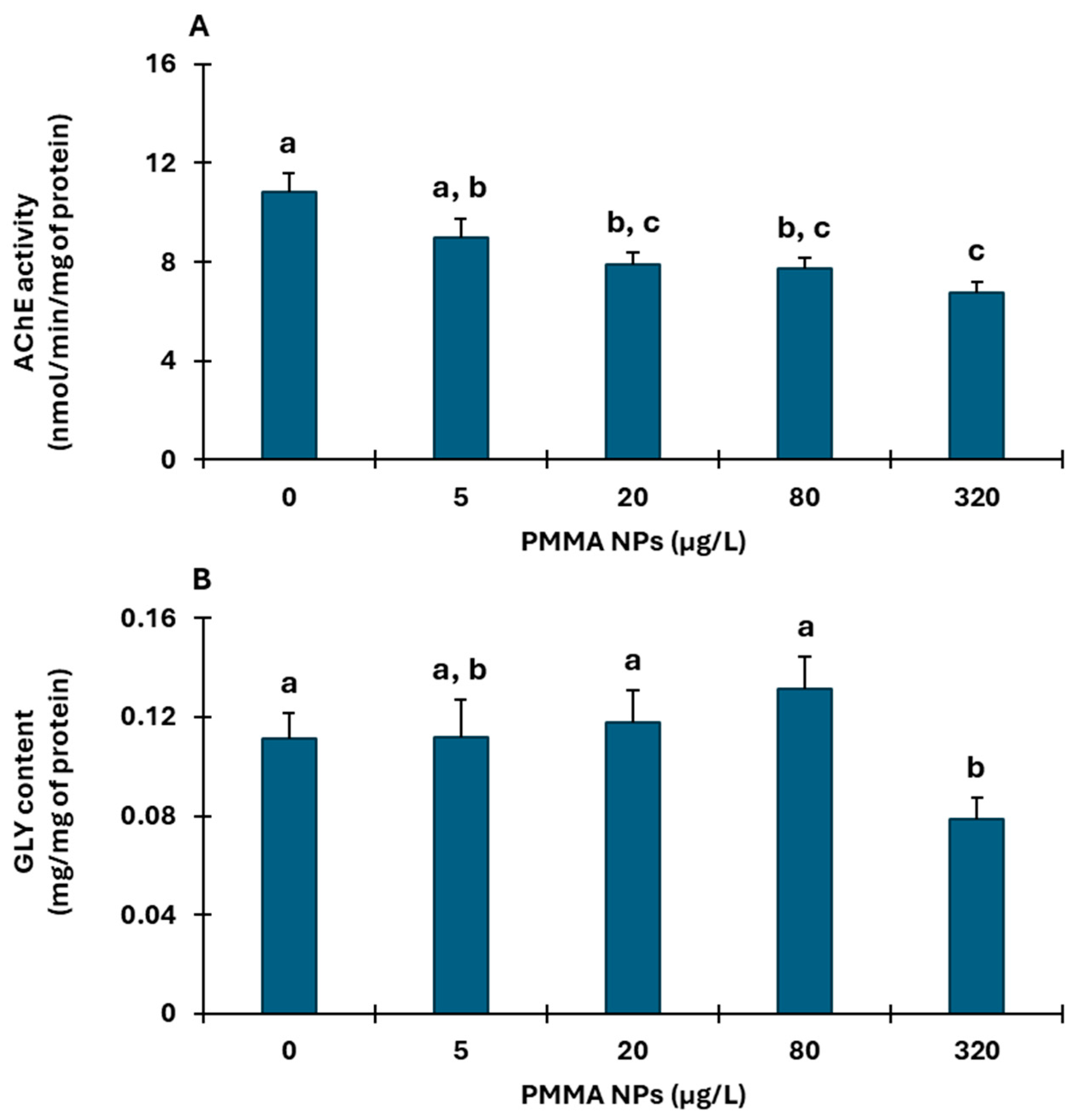
Muscle AChE activity (Figure 2A) was significantly lower compared to control in the organisms exposed to 20, 80, and 320 μg PMMA NPs/L. Correlation analyses between AChE data and behavioral results showed a significant moderate positive correlation between AChE and time spent inactive (r = 0.410; p = 0.014) and a significant moderate negative correlation between AChE and total distance walked (r = −0.642; p < 0.001) and locomotor velocity (r = −0.558; p < 0.001). PMMA NPs only affected the muscle Gly content of the organisms exposed to the highest PMMA NPs concentration (320 μg PMMA NPs/L) (Figure 2B).

3.2.2. Gills

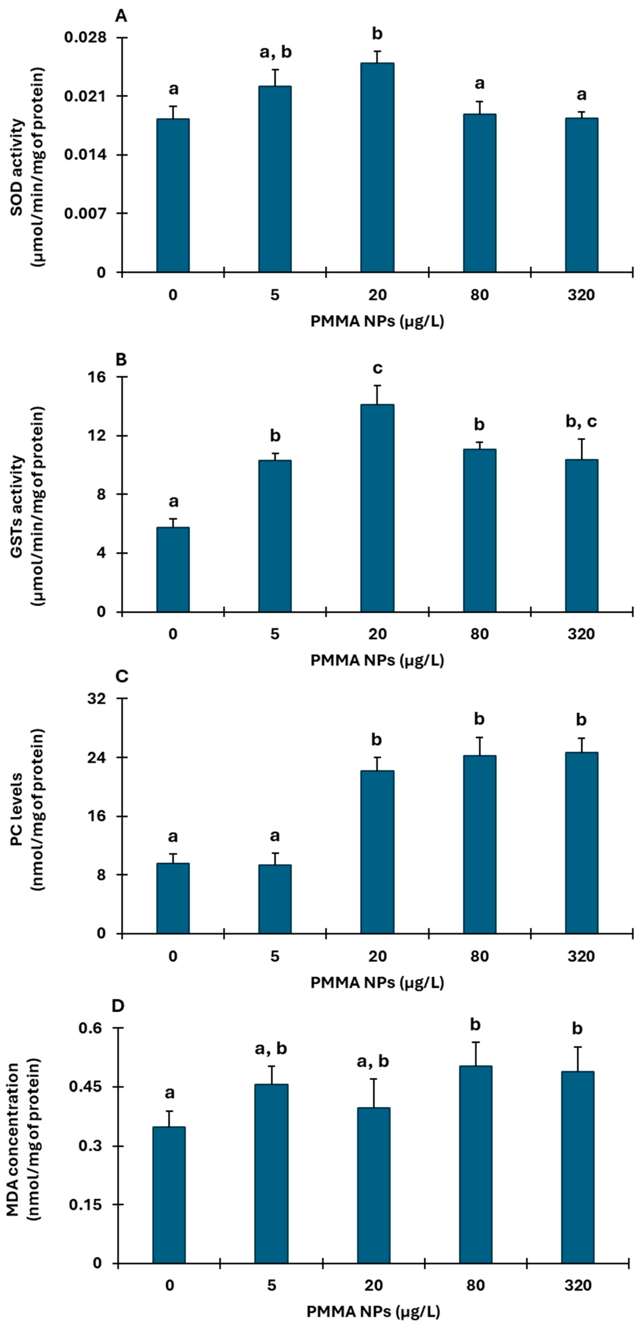
In the gills, an overall activation of antioxidant defenses was observed after PMMA NPs exposure. SOD activity (Figure 3A) was significantly higher relative to control only in organisms exposed to 20 μg PMMA NPs/L, displaying a 26.62% increase, whereas GSTs activity (Figure 3B) was significantly higher relative to control in all tested PMMA NPs concentrations.
Despite the activation of antioxidant defenses in gills, PMMA NPs exposure resulted in oxidative damage. PC levels (Figure 3C) were significantly higher compared to controls in the crabs exposed to 20, 80, and 320 μg PMMA NPs/L, whereas higher LPO levels (Figure 3D), expressed by malonaldehyde (MDA) concentration, were found in the organisms exposed to the two highest concentrations (80 and 320 μg PMMA NPs/L).

3.2.3. Hepatopancreas

PMMA NPs exposure also affected hepatopancreas antioxidant enzymes. SOD activity was significantly higher in the crabs exposed to 80 μg PMMA NPs/L compared to control and remaining exposed organisms (Figure 4A), and GSTs activity (Figure 4B) increased in the crabs exposed to 20 and 320 μg PMMA NPs/L. In terms of damage, an overall increase in PC levels (Figure 4C), was observed in PMMA NPs exposed organisms, though not significant at the highest concentration. Increased MDA levels were also observed in hepatopancreas in organisms exposed to 20 and 80 μg PMMA NPs/L, but only significantly for 80 μg PMMA NPs/L (Figure 4D).

3.2.4. Hemolymph

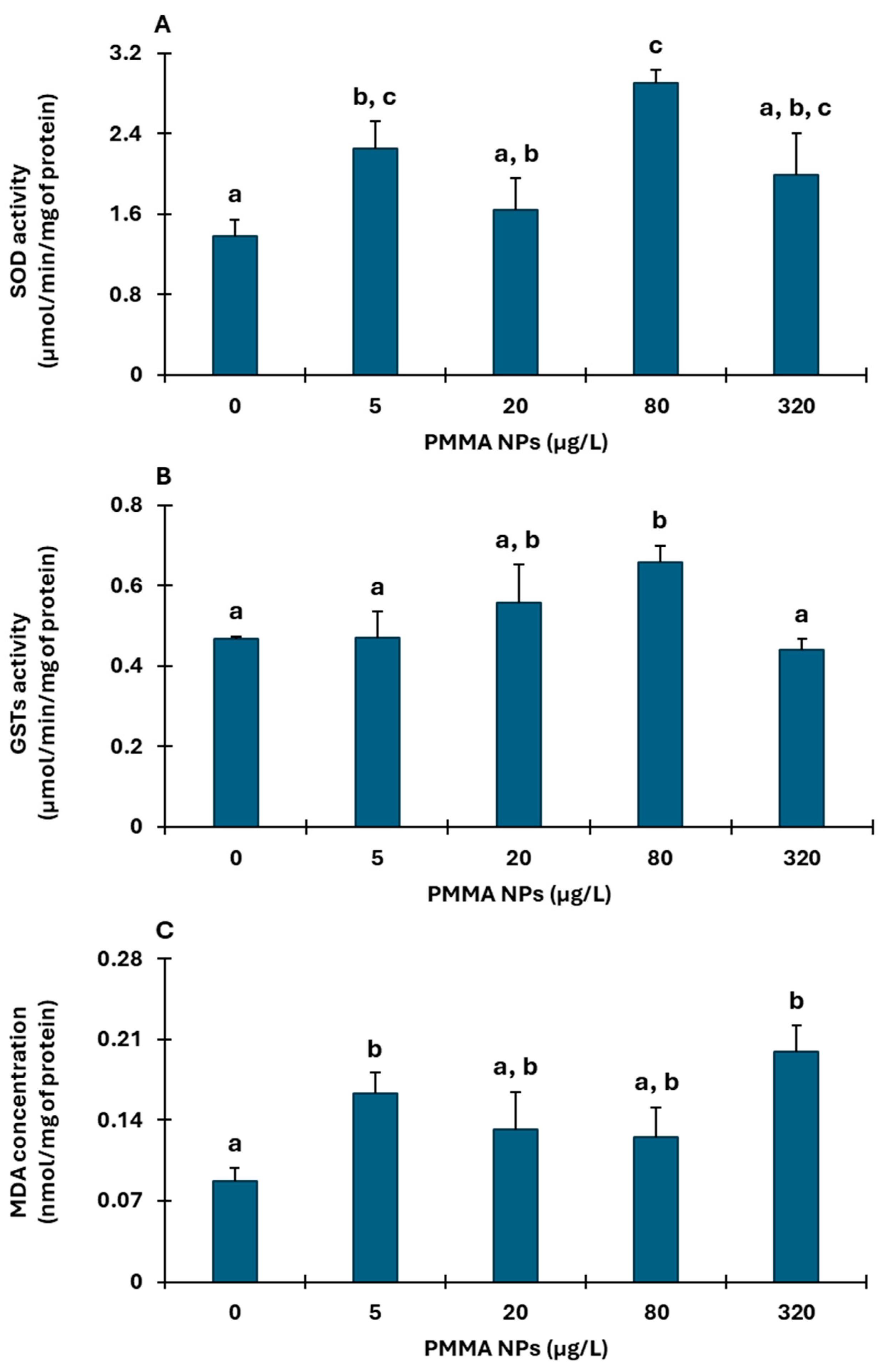
SOD activity showed no clear tendency, with significant differences compared to control observed in organisms exposed to 5 and 80 μg PMMA NPs/L (Figure 5A). GSTs activity showed an increasing trend in organisms exposed up to 80 μg PMMA NPs/L, with significant differences between that concentration and control (Figure 5B) and the lowest and highest concentrations (5 and 320 μg PMMA NPs/L). Cellular damage, evaluated by MDA levels (Figure 5C), demonstrated an increase compared to control; however, the difference was only significant between the control and the concentrations of 5 and 320 μg PMMA NPs/L.

4. Discussion

C. maenas is an important species to the benthic community, assisting in the good functioning of the ecosystem by promoting nutrient resuspension and energy transfer in the food web [33]. Any impediments to their mobility could have negative consequences since their daily movements with the tides allow them to seek shelter and evade predation, hunt for food, and resuspend nutrients that become available for themselves and other species [35]. In the present study, the behavior of C. maenas proved sensitive to PMMA NPs exposure. The observed TSI decrease induced by PMMA NPs suggests that the crabs could be experiencing higher stress levels. In the environment, crabs mostly remain still and hidden, unless stimulated to move due to tides, a need to catch food, or to evade predation. This typical behavior is a protection instinct essential for the maintenance of population levels. Therefore, in this test, organisms should shelter at the edge of the arena, to remain protected, and stop their movement, as there is no stimulus that requires motion. In this sense, the fact that they spent more time moving than controls (TSI) and moved more (TDW), suggests a potentially increased risk (e.g., of being caught) and higher energy consumption. Despite the increasing tendency in TDW, this was not observed in LV. The assay did not reveal relevant differences between control organisms and PMMA NPs-exposed organisms. Several studies carried out with hermit crabs (Pagurus bernhardus) have also shown that exposure to polyethylene MPs altered the normal behavior of this species. The hermit crabs exhibited impaired shell selection, an impaired ability to defend themselves from predators, and reduced strength against prey [65,66,67]. Overall, these studies suggest that MPs have the ability to impair cognition, namely in terms of information gathering and processing. Previous studies [65,66,67] and our study show that, more than behavioral impairments, there seems to be a neurological incapacity that affects overall behavior in organisms exposed to MPs and NPs.
To better understand the changes resulting from the PMMA NPs contamination, behavioral alterations such as movement impairments are usually evaluated alongside biochemical parameters such as AChE activity and GLY content. AChE activity is used as a biomarker of neurotoxicity, and its inhibition in marine organisms is normally associated with potential impairments in movement and natatory behavior [68]. In this study, AChE activity presented a tendency to decrease with increased PMMA NPs concentrations, suggesting neurotoxicity and possible mobility issues. However, in the present study, AChE did not seem to affect C. maenas’ ability to move, given the increase in TDW and the constant LV, suggesting that PMMA NPs may affect other pathways leading to the impaired orientation demonstrated by their altered trajectories in the arena (Figure S1). Nonetheless, the significant correlation between AChE and behavioral endpoints suggests that AChE activity may be involved in these behavioral results. To better understand the crabs’ mobility, Gly content was evaluated to ensure that the organisms had the energy reserves required to be able to move in stressful conditions. The assay results showed no significant differences compared to control at most PMMA NPs concentrations, suggesting that those reserves were either not required or its synthesis activated. However, the highest concentration showed a decrease in GLY content, suggesting its higher consumption. In a study by Sneddon et al. [69] that evaluated muscle GLY content when crabs exercised, data showed that increased exercise led to decreased GLY content. The lower GLY content at the higher PMMA NPs concentration in this study could be due to the increased exercise that the crabs demonstrated by the increase in TDW.
Both gills and hepatopancreas are tissues that may be a target of toxicity from environmental NPs, as they are the first to come into contact with contaminants. The present study data showed not only different basal levels relative to the activities/levels of biomarkers in the controls, but also a relevant difference in the response to PMMA NPs, which may be explained by their different functions and metabolic activity.
Gills are the first organ to come into contact with contaminants, being the entryway into the organism by respiration [70]. The increase in SOD activity, the first line of the defense system against reactive oxygen species (ROS), in some concentrations, as well as the increase in GSTs activity, which plays an important role in the elimination of ROS [71], in all concentrations, suggests that exposure to PMMA NPs triggered defense and detoxification mechanisms against ROS and xenobiotics. However, despite mitigation efforts, irreversible oxidative protein damage and free radical-induced damage to cell membranes were observed, evidenced by an increase in PC and LPO levels, respectively. Studies have reported contrasting responses to MPs, suggesting that the type of polymer, particle size, and exposure duration influence oxidative stress related responses. Litopenaeus vannamei exhibited SOD gene upregulation after 14 days of exposure to polystyrene microspheres (5 μm, 10 μg/L), indicating an adaptive increase in antioxidant defenses [72]. In contrast, Minuca vocator exposed for 5 days to high-density polyethylene MPs (0.1–0.25 mm, 25 mg/L) showed decreased SOD activity but still exhibited oxidative damage, as indicated by increased LPO [73].
The hepatopancreas plays a vital role in digestion and nutrient absorption, as well as in metabolic regulation and protein synthesis. PMMA NPs may be ingested and accumulate in the hepatopancreas, potentially causing harmful effects. In this study, a significant increase in hepatopancreas SOD activity was detected only at 80 μg PMMA NPs/L, while GSTs activity increased at 20 and 320 μg PMMA NPs/L in exposed organisms. These findings suggest a potential impairment in the ability of the hepatopancreas to mitigate damage caused by PMMA NPs, as oxidative damage was observed through increased PC and LPO levels, similarly to what was found in the gills. Other studies highlight similar trends. Consigna et al. [74] evaluated the effects of polyethylene MPs on the crab Xenograspsus testudinatus and observed an activation of antioxidant defenses such as increased SOD and GSTs activity, but damage persisted, as evidenced by an increase in LPO levels. Similarly, La Torre et al. [75] showed that the crab Minuca rapax exposed for 48 h to polyethylene MPs (2 mg/L; 53–63 μm) displayed a slight increase in SOD activity and LPO levels, both in the gills and hepatopancreas. Furthermore, in another study that evaluated the effects of polystyrene NPs (100 nm, 104 particles/L) in whole tissues of the crab Tachypleus tridentatus [76], data demonstrated that after 14 days of exposure, there was an increase in SOD activity, but SOD activity decreased over a 21-day exposure. Moreover, a slight increase in LPO levels was also observed after 14 days of exposure, suggesting damage.
Hemolymph is a circulatory fluid that contains plasma, and antioxidant enzymes, alongside nutrients and waste products that are expelled into the hemolymph once these are in excess in the organs, potentially leading to stress [77,78]. Moreover, the ability of these particles to reach the interstitial space, where the hemolymph is present, depends on their capacity to cross tissue barriers. Previous research showed that polyethylene MPs (10–300 μm) were unable to cross organ tissue barriers in the fish Oncorhynchus mykiss [79], which might explain the lower activation of antioxidant defenses observed in the present study. Specifically, SOD activity only increased at concentrations of 5 and 80 μg PMMA NPs/L, while GSTs activity showed differences only at 80 μg PMMA NPs/L, and oxidative damage remained relatively low. However, when the PMMA NPs can reach the fluid, an inferior number of immune cells might not be enough to counteract PMMA NPs effects even with the increase in antioxidant defenses. Despite the low amount of particles that might be present in the fluid, oxidative damage was evident, as indicated by increased LPO levels in the concentrations of 5 and 320 μg PMMA NPs/L. This suggests that, although antioxidant mechanisms were activated to counter the impact of PMMA NPs, they were unable to prevent damage. Overall, the obtained results show that regardless of the activation of defense mechanisms, exposure to NPs may cause damage in the tissues of crustaceans. Oxidative stress observed may be harmful to their molting cycle, as they need stability and safety during this vulnerable period. In this study, gills appeared to be the most affected organ, showing more damage than the hepatopancreas and hemolymph.
A previous study with 10 days exposure to PMMA NPs exhibiting the same characteristics, assessing effects on two life stages (juveniles and adults) of the polychaete H. diversicolor, has also shown the ability of PMMA NPs to compromise the normal behavior of the polychaete, leading to increased burrowing time. In both life stages, antioxidant defenses were increased after PMMA NPs exposure, but oxidative damage was only observed in juveniles [27]. Comparing the data from both studies, C. maenas appears to be more sensitive, as oxidative damage occurred in the crabs, which was not the case in adult H. diversicolor.

5. Conclusions

Benthic species are essential for ecosystem stability, and the crab C. maenas has a wide array of roles in its habitat. This study showed that PMMA NPs can impact the crab C. maenas. The increase in PMMA NPs concentrations promoted, in addition to increased movements (farther distance moved), increased erratic movements that can have impacts at a population level. These alterations may impact the crabs’ ability to survive, as their instinct to protect themselves and hide is decreased after NP exposure, leaving them more vulnerable to predators. Biochemical analyses showed similar responses in all tissues, with the activation of defense mechanisms that were not able to counter the effects of PMMA NPs, resulting in oxidative damage. Considering that gills and hepatopancreas are responsible for oxygen and nutrient absorption, damage to these tissues can impair the crabs’ ability to function properly and impact their fitness. Overall, data suggest that exposure to PMMA NPs can induce biochemical and behavioral alterations that may lead to increased susceptibility to predators and decreased fitness. Thus, further studies should be performed to assess the long-term effects of PMMA NPs exposure, also considering other sources (e.g., food).

Supplementary Materials

The following supporting information can be downloaded at: https://www.mdpi.com/article/10.3390/environments12050169/s1, Figure S1: Example of C. maenas trajectory in the behavioral assay in control (A) and PMMA NPs concentrations: 5 µg/L (B); 20 µg/L (C); 80 µg/L (D) and 320 µg/L (E); Table S1: PERMANOVA main test results of behavioral (Time Spent Inactive (TSI), Total Distance Walked (TDW) and Locomotor Velocity (LV)) and biochemical endpoints (Acetylcholinesterase activity (AChE), Glycogen content (GLY), Superoxide Dismutase activity (SOD), Glutathione S-Transferases activity (GSTs), Protein Carbonylation levels (PC), and Lipid Peroxidation levels (LPO)) of Carcinus maenas after 10 days exposure to PMMA NPs. Pseudo-F: F value by permutation, P: p-values based on 9999 permutations; Perms: number of permutations. Table S2: Pearson correlation between acetylcholinesterase activity and behavioral results.

Author Contributions

Conceptualization, A.P. and M.O.; methodology, B.N., C.F., M.A. and R.J.B.P.; validation, A.P., M.O., E.F. and R.J.B.P.; formal analysis, A.P. and M.O.; investigation, B.N., M.O., C.F., M.A. and A.P.; resources, A.P., M.O., E.F. and R.J.B.P.; data curation, M.O. and A.P.; writing—original draft preparation, B.N.; writing—review and editing, A.P. and M.O.; visualization, A.P. and M.O.; supervision, A.P. and M.O.; project administration, A.P. and M.O.; funding acquisition, A.P., M.O. and E.F. All authors have read and agreed to the published version of the manuscript.

Funding

This work was funded by the projects GEOPLASTTES (Ref: 2023.00033.RESTART) funded by FCT/MCTES under the program RESTART and NanoPlanet (DOI: 10.54499/2022.02340.PTDC) through national funds. We also acknowledge financial support from FCT/MCTES to UID Centro de Estudos do Ambiente e Mar (CESAM) + LA/P/0094/2020, through national funds.

Data Availability Statement

The data presented in this study are available on request from the corresponding author.

Acknowledgments

B. Neves benefited from a Master’s grant under the project GEOPLASTTES (Ref: BI/UI88/11546/2024) through FCT. A.Pires acknowledges funding from national funds (OE) through the Portuguese Foundation for Science and Technology (FCT) (https://doi.org/10.54499/2022.00500.CEECIND/CP1720/CT0048). C.Frazão benefited from a PhD grant funded by national funds through FCT (2022.11216.BD).

Conflicts of Interest

The authors declare no conflicts of interest.

References

  1. Thushari, G.G.N.; Senevirathna, J.D.M. Plastic pollution in the marine environment. Heliyon 2020, 6, e04709. [Google Scholar] [CrossRef] [PubMed]
  2. Lester, S.E.; McLeod, K.L.; Tallis, H.; Ruckelshaus, M.; Halpern, B.S.; Levin, P.S.; Chavez, F.P.; Pomeroy, C.; McCay, B.J.; Costello, C.; et al. Science in support of ecosystem-based management for the US West Coast and beyond. Biol. Conserv. 2010, 143, 576–587. [Google Scholar] [CrossRef]
  3. Li, Z.; Feng, C.; Wu, Y.; Guo, X. Impacts of nanoplastics on bivalve: Fluorescence tracing of organ accumulation, oxidative stress and damage. J. Hazard. Mater. 2020, 392, 122418. [Google Scholar] [CrossRef] [PubMed]
  4. Tun, T.Z.; Nurlatifah Htwe, A.T.; Than, N.N.; Khine, M.M.; Chavanich, S.; Viyakarn, V.; Isobe, A.; Nakata, H. Polymer types and additive concentrations in single-use plastic products collected from Indonesia, Japan, Myanmar, and Thailand. Sci. Total Environ. 2023, 889, 163983. [Google Scholar] [CrossRef]
  5. Wiesinger, H.; Wang, Z.; Hellweg, S. Deep Dive into Plastic Monomers, Additives, and Processing Aids. Environ. Sci. Technol. 2021, 55, 9339–9351. [Google Scholar] [CrossRef] [PubMed]
  6. Europe, P. Plastics the Fast Facts 2023. 2024. Available online: https://plasticseurope.org/knowledge-hub/plastics-the-fast-facts-2024/ (accessed on 12 January 2025).
  7. Van Emmerik, T.H.M.; González-Fernández, D.; Laufkötter, C.; Blettler, M.; Lusher, A.; Hurley, R.; Ryan, P.G. Focus on plastics from land to aquatic ecosystems. Environ. Res. Lett. 2023, 18, 040401. [Google Scholar] [CrossRef]
  8. Rashid, S.; Majeed, L.R.; Mehta, N.; Radu, T.; Martín-Fabiani, I.; Bhat, M.A. Microplastics in terrestrial ecosystems: Sources, transport, fate, mitigation, and remediation strategies. Euro-Mediterr. J. Environ. Integr. 2025. [Google Scholar] [CrossRef]
  9. Hechmi, S.; Bhat, M.A.; Kallel, A.; Khiari, O.; Louati, Z.; Khelil, M.N.; Zoghlami, R.I.; Cherni, Y.; Melki, S.; Trabelsi, I.; et al. Soil contamination with microplastics (MPs) from treated wastewater and sewage sludge: Risks and sustainable mitigation strategies. Discov. Environ. 2024, 2, 95. [Google Scholar] [CrossRef]
  10. Colton, J.B.; Knapp, F.D.; Burns, B.R. Plastic Particles in Surface Waters of the Northwestern Atlantic. Science 1974, 185, 491–497. [Google Scholar] [CrossRef]
  11. Brandts, I.; Barría, C.; Martins, M.A.; Franco-Martínez, L.; Barreto, A.; Tvarijonaviciute, A.; Tort, L.; Oliveira, M.; Teles, M. Waterborne exposure of gilthead seabream (Sparus aurata) to polymethylmethacrylate nanoplastics causes effects at cellular and molecular levels. J. Hazard. Mater. 2021, 403, 123590. [Google Scholar] [CrossRef]
  12. Isobe, A.; Uchiyama-Matsumoto, K.; Uchida, K.; Tokai, T. Microplastics in the Southern Ocean. Mar. Pollut. Bull. 2017, 114, 623–626. [Google Scholar] [CrossRef] [PubMed]
  13. Cole, M.; Galloway, T.S. Ingestion of Nanoplastics and Microplastics by Pacific Oyster Larvae. Environ. Sci. Technol. 2015, 49, 14625–14632. [Google Scholar] [CrossRef] [PubMed]
  14. Gigault, J.; ter Halle, A.; Baudrimont, M.; Pascal, P.Y.; Gauffre, F.; Phi, T.L.; Hadri, H.E.; Grassl, B.; Reynaud, S. Current opinion: What is a nanoplastic? Environ. Pollut. 2018, 235, 1030–1034. [Google Scholar] [CrossRef] [PubMed]
  15. Bergmann, M.; Gutow, L.; Klages, M. (Eds.) Marine Anthropogenic Litter; Springer International Publishing: Berlin/Heidelberg, Germany, 2015. [Google Scholar] [CrossRef]
  16. Xu, J.; Feng, G.; Yan, Y. Effects of polystyrene nanoplastics and copper on gill tissue structure, metabolism, and immune function of the Chinese mitten crab (Eriocheir sinensis). Front. Mar. Sci. 2025, 12, 1538734. [Google Scholar] [CrossRef]
  17. Worm, B.; Lotze, H.K.; Jubinville, I.; Wilcox, C.; Jambeck, J. Plastic as a Persistent Marine Pollutant. Annu. Rev. Environ. Resour. 2017, 42, 1–26. [Google Scholar] [CrossRef]
  18. Halle, A.T.; Jeanneau, L.; Martignac, M.; Jardé, E.; Pedrono, B.; Brach, L.; Gigault, J. Nanoplastic in the North Atlantic Subtropical Gyre. Environ. Sci. Technol. 2017, 51, 13689–13697. [Google Scholar] [CrossRef] [PubMed]
  19. Kampouris, T.E.; Syranidou, E.; Seridou, P.; Gagoulis, K.; Batjakas, I.E.; Kalogerakis, N. MPs and NPs intake and heavy metals accumulation in tissues of Palinurus elephas (J.C. Fabricius, 1787), from NW Aegean sea, Greece. Environ. Pollut. 2023, 316, 120725. [Google Scholar] [CrossRef]
  20. Materić, D.; Holzinger, R.; Niemann, H. Nanoplastics and ultrafine microplastic in the Dutch Wadden Sea—The hidden plastics debris? Sci. Total Environ. 2022, 846, 157371. [Google Scholar] [CrossRef]
  21. Wang, L.; Wu, W.M.; Bolan, N.S.; Tsang, D.C.W.; Li, Y.; Qin, M.; Hou, D. Environmental fate, toxicity and risk management strategies of nanoplastics in the environment: Current status and future perspectives. J. Hazard. Mater. 2021, 401, 123415. [Google Scholar] [CrossRef]
  22. Dong, Z.; Zhu, L.; Zhang, W.; Huang, R.; Lv, X.W.; Jing, X.; Yang, Z.; Wang, J.; Qiu, Y. Role of surface functionalities of nanoplastics on their transport in seawater-saturated sea sand. Environ. Pollut. 2019, 255, 113177. [Google Scholar] [CrossRef]
  23. Riveiro, A.; Chantada, A.; Soto, R.; Val, J.d.; Arias-González, F.; Comesaña, R.; Boutinguiza, M.; Quintero, F.; Lusquiños, F.; Pou, J. Laser surface texturing of thermoplastics to improve biological performance. In Materials for Biomedical Engineering: Thermoset and Thermoplastic Polymers; Elsevier: Amsterdam, The Netherlands, 2019; pp. 29–56. [Google Scholar] [CrossRef]
  24. Ali, U.; Karim, K.J.B.A.; Buang, N.A. A Review of the Properties and Applications of Poly (Methyl Methacrylate) (PMMA). Polym. Rev. 2015, 55, 678–705. [Google Scholar] [CrossRef]
  25. Hermsen, E.; Pompe, R.; Besseling, E.; Koelmans, A.A. Detection of low numbers of microplastics in North Sea fish using strict quality assurance criteria. Mar. Pollut. Bull. 2017, 122, 253–258. [Google Scholar] [CrossRef] [PubMed]
  26. Brandts, I.; Teles, M.; Tvarijonaviciute, A.; Pereira, M.L.; Martins, M.A.; Tort, L.; Oliveira, M. Effects of polymethylmethacrylate nanoplastics on Dicentrarchus labrax. Genomics 2018, 110, 435–441. [Google Scholar] [CrossRef] [PubMed]
  27. Neves, B.; Oliveira, M.; Frazão, C.; Almeida, M.; Pinto, R.J.B.; Figueira, E.; Pires, A. The Role of Life Stages in the Sensitivity of Hediste diversicolor to Nanoplastics: A Case Study with Poly(Methyl)Methacrylate (PMMA). Toxics 2024, 12, 352. [Google Scholar] [CrossRef]
  28. Silva, M.S.S.; Pires, A.; Vethaak, A.D.; Martínez-Gómez, C.; Almeida, M.; Pinto, R.; Figueira, E.; Oliveira, M. Effects of polymethylmethacrylate nanoplastics on the polychaete Hediste diversicolor: Behavioural, regenerative, and biochemical responses. Aquat. Toxicol. 2023, 265, 106743. [Google Scholar] [CrossRef]
  29. Li, D.; Tang, X.; Xu, X.; Zhao, Y.; Li, L.; Zhang, B.; Zhao, Y. UV-B radiation alleviated detrimental effects of polymethyl methacrylate microplastics on marine diatom Thalassiosira pseudonana. Sci. Total Environ. 2023, 892, 164388. [Google Scholar] [CrossRef]
  30. Xu, T.-T.; Li, Z.-L.; Li, H.-X.; Lin, L.; Hou, R.; Liu, S.; Li, T.; Zeng, E.Y.; Yu, K.-F.; Xu, X.-R. Unraveling the toxicity mechanisms of nanoplastics with various surface modifications on Skeletonema costatum: Cellular and molecular perspectives. Sci. Total Environ. 2024, 953, 176164. [Google Scholar] [CrossRef]
  31. Zhang, Y.; Wang, Y.; Zhu, P.; Jing, S.; Li, J.; Wanger, T.C.; Liu, W.; Liu, K.; Chen, X.; Li, L. Mass concentrations, compositions and burial fluxes of nano- and micro-plastics in a multi-species saltmarsh. Environ. Pollut. 2024, 363, 125181. [Google Scholar] [CrossRef]
  32. Cobelas, Á.; Rojo, C. Bridging community and metacommunity perspectives in benthic photosynthetic organisms (Serranía de Cuenca, Central Spain). Limnetica 2023, 42, 37–54. [Google Scholar] [CrossRef]
  33. Pourabadehei, M.; Mulligan, C.N. Resuspension of sediment, a new approach for remediation of contaminated sediment. Environ. Pollut. 2016, 213, 63–75. [Google Scholar] [CrossRef]
  34. Carlton, J.T.; Cohen, A.N. Episodic global dispersal in shallow water marine organisms: The case history of the European shore crabs Carcinus maenas and C. aestuarii. J. Biogeogr. 2003, 30, 1809–1820. [Google Scholar] [CrossRef]
  35. Young, A.M.; Elliott, J.A. Life history and population dynamics of green crabs (Carcinus maenas). Fishes 2020, 5, 4. [Google Scholar] [CrossRef]
  36. Simberloff, D.; Rejmanek, M. (Eds.) 100 of the World’s Worst Invasive Alien Species: A Selection from The Global Invasive Species Database. In Encyclopedia of Biological Invasions; University of California Press: Oakland, CA, USA, 2019; pp. 715–716. [Google Scholar] [CrossRef]
  37. Cruz, D.R.S. Caracterização da População de Carcinus maenas na Lagoa de Santo André e Potencial de Exploração. Master’s Thesis, Universidade de Lisboa, Lisboa, Portugal, 2021. [Google Scholar]
  38. Ghedira, J.; Jebali, J.; Banni, M.; Chouba, L.; Boussetta, H.; López-Barea, J.; Alhama, J. Use of oxidative stress biomarkers in Carcinus maenas to assess littoral zone contamination in tunisia. Aquat. Biol. 2011, 14, 87–98. [Google Scholar] [CrossRef]
  39. Jebali, J.; Ben-Khedher, S.; Ghedira, J.; Kamel, N.; Boussetta, H. Integrated assessment of biochemical responses in Mediterranean crab (Carcinus maenas) collected from Monastir Bay, Tunisia. J. Environ. Sci. 2011, 23, 1714–1720. [Google Scholar] [CrossRef] [PubMed]
  40. Rodrigues, E.T.; Pardal, M.Â. The crab Carcinus maenas as a suitable experimental model in ecotoxicology. Environ. Int. 2014, 70, 158–182. [Google Scholar] [CrossRef]
  41. Elumalai, M.; Antunes, C.; Guilhermino, L. Effects of Single Metals and Their Mixture on Selected Enzymes of Carcinus maenas. Water Air Soil Pollut. 2002, 141, 273–280. [Google Scholar] [CrossRef]
  42. Elumalai, M.; Antunes, C.; Guilhermino, L. Enzymatic biomarkers in the crab Carcinus maenas from the Minho River estuary (NW Portugal) exposed to zinc and mercury. Chemosphere 2007, 66, 273–280. [Google Scholar] [CrossRef]
  43. Martín-Díaz, M.L.; Bamber, S.; Casado-Martínez, C.; Sales, D.; DelValls, T.A. Toxicokinetics of heavy metals from a mining spill using Carcinus maenas. Mar. Environ. Res. 2004, 58, 833–837. [Google Scholar] [CrossRef]
  44. Mesquita, S.R.; Guilhermino, L.; Guimarães, L. Biochemical and locomotor responses of Carcinus maenas exposed to the serotonin reuptake inhibitor fluoxetine. Chemosphere 2011, 85, 967–976. [Google Scholar] [CrossRef]
  45. Rodrigues, A.P.; Oliveira, P.C.; Guilhermino, L.; Guimarães, L. Effects of salinity stress on neurotransmission, energy metabolism, and anti-oxidant biomarkers of Carcinus maenas from two estuaries of the NW Iberian Peninsula. Mar. Biol. 2012, 159, 2061–2074. [Google Scholar] [CrossRef]
  46. Damasceno, J.M.; Rato, L.D.; Simões, T.; Morão, I.F.C.; Meireles, G.; Novais, S.C.; Lemos, M.F.L. Exposure to the insecticide sulfoxaflor affects behaviour and biomarkers responses of Carcinus maenas (Crustacea: Decapoda). Biology 2021, 10, 1234. [Google Scholar] [CrossRef]
  47. Watts, A.J.R.; Urbina, M.A.; Corr, S.; Lewis, C.; Galloway, T.S. Ingestion of Plastic Microfibers by the Crab Carcinus maenas and Its Effect on Food Consumption and Energy Balance. Environ. Sci. Technol. 2015, 49, 14597–14604. [Google Scholar] [CrossRef]
  48. Watts, A.J.R.; Urbina, M.A.; Goodhead, R.; Moger, J.; Lewis, C.; Galloway, T.S. Effect of Microplastic on the Gills of the Shore Crab Carcinus maenas. Environ. Sci. Technol. 2016, 50, 5364–5369. [Google Scholar] [CrossRef] [PubMed]
  49. Manuel, P.; Almeida, M.; Martins, M.; Oliveira, M. Effects of nanoplastics on zebrafish embryo-larval stages: A case study with polystyrene (PS) and polymethylmethacrylate (PMMA) particles. Environ. Res. 2022, 213, 113584. [Google Scholar] [CrossRef] [PubMed]
  50. Shi, C.; Liu, Z.; Yu, B.; Zhang, Y.; Yang, H.; Han, Y.; Wang, B.; Liu, Z.; Zhang, H. Emergence of nanoplastics in the aquatic environment and possible impacts on aquatic organisms. Sci. Total Environ. 2024, 906, 167404. [Google Scholar] [CrossRef] [PubMed]
  51. Barría, C.; Brandts, I.; Tort, L.; Oliveira, M.; Teles, M. Effect of nanoplastics on fish health and performance: A review. Mar. Pollut. Bull. 2020, 151, 110791. [Google Scholar] [CrossRef]
  52. Premarathna, A.D.; Pathirana, I.; Rajapakse, R.P.V.J.; Pathirana, E. Evaluation of Efficacy of Selected Anesthetic Agents on Blood-Spotted Crab (Portunus sanguinolentus). J. Shellfish Res. 2016, 35, 237–240. [Google Scholar] [CrossRef]
  53. Oliveira, M.; Cardoso, D.N.; Soares, A.M.V.M.; Loureiro, S. Effects of short-term exposure to fluoxetine and carbamazepine to the collembolan Folsomia candida. Chemosphere 2015, 120, 86–91. [Google Scholar] [CrossRef]
  54. Robinson, H.W.; Hogden, C.G. The Biuret Reaction in the Determination of Serum Proteins I. A study of the conditions necessary for the production of a stable color which bears a quantitative relationship to the protein concentration. J. Biol. Chem. 1940, 135, 707–725. [Google Scholar] [CrossRef]
  55. Ellman, G.L.; Courtney, K.D.; Andres, V.; Featherstone, R.M. A new and rapid colorimetric determination of acetylcholinesterase activity. Biochem. Pharmacol. 1961, 7, 88–95. [Google Scholar] [CrossRef]
  56. Guilhermino, L.; Lopes, M.C.; Carvalho, A.P.; Soares, A.M.V.M. Acetylcholinesterase Activity in Juveniles of Daphnia magna Straus. Bull. Environ. Contam. Toxicol. 1996, 57, 979–985. [Google Scholar] [CrossRef] [PubMed]
  57. Walop, J.N.; Boot, L.M. Studies on cholinesterase in Carcinus maenas. Biochim. Biophys. Acta 1950, 4, 566–571. [Google Scholar] [CrossRef]
  58. Dubois, M.; Gilles, K.A.; Hamilton, J.K.; Rebers, P.A.; Smith, F. Colorimetric Method for Determination of Sugars and Related Substances. Anal. Chem. 1956, 28, 350–356. [Google Scholar] [CrossRef]
  59. Beauchamp, C.; Fridovich, I. Superoxide Dismutase: Improved Assays and an Assay Applicable to Acrylamide Gels1. Anal. Biochem. 1971, 44, 276–287. [Google Scholar] [CrossRef]
  60. Habig, W.H.; Pabst, M.J.; Jakoby, W.B. Glutathione S-Transferases: The First Enzymatic Step in Mercapturic Acid Formation. J. Biol. Chem. 1974, 249, 7130–7139. [Google Scholar] [CrossRef]
  61. Mesquita, C.S.; Oliveira, R.; Bento, F.; Geraldo, D.; Rodrigues, J.V.; Marcos, J.C. Simplified 2,4-dinitrophenylhydrazine spectrophotometric assay for quantification of carbonyls in oxidized proteins. Anal. Biochem. 2014, 458, 69–71. [Google Scholar] [CrossRef] [PubMed]
  62. Udenigwe, C.C.; Udechukwu, M.C.; Yiridoe, C.; Gibson, A.; Gong, M. Antioxidant mechanism of potato protein hydrolysates against in vitro oxidation of reduced glutathione. J. Funct. Foods 2016, 20, 195–203. [Google Scholar] [CrossRef]
  63. Buege, J.A.; Aust, S.D. Microsomal Lipid Peroxidation. Methods Enzym. 1978, 52, 302–310. [Google Scholar]
  64. Anderson, M.J.; Gorley, R.N.; Clarke, K.R. Permanova+ for Primer: Guide to Software and Statistical Methods; PRIMER-E: Plymouth, UK, 2008; p. 218. [Google Scholar]
  65. Crump, A.; Mullens, C.; Bethell, E.J.; Cunningham, E.M.; Arnott, G. Microplastics disrupt hermit crab shell selection. Biol. Lett. 2020, 16, 20200030. [Google Scholar] [CrossRef]
  66. Crump, A.; Aiken, C.; Cunningham, E.M.; Arnott, G. Short-Term Microplastic Exposure Impairs Cognition in Hermit Crabs. Animals 2023, 13, 1055. [Google Scholar] [CrossRef]
  67. Cunningham, E.M.; Mundye, A.; Kregting, L.; Dick, J.T.A.; Crump, A.; Riddell, G.; Arnott, G. Animal contests and microplastics: Evidence of disrupted behaviour in hermit crabs Pagurus bernhardus. R. Soc. Open Sci. 2021, 8, 211089. [Google Scholar] [CrossRef]
  68. Oliveira, M.; Ribeiro, A.; Hylland, K.; Guilhermino, L. Single and combined effects of microplastics and pyrene on juveniles (0+ group) of the common goby Pomatoschistus microps (Teleostei, Gobiidae). Ecol. Indic. 2013, 34, 641–647. [Google Scholar] [CrossRef]
  69. Sneddon, L.U.; Taylor, A.C.; Huntingford, F.A. Metabolic consequences of agonistic behaviour: Crab fights in declining oxygen tensions. Anim. Behav. 1999, 57, 353–363. [Google Scholar] [CrossRef]
  70. Maria, V.L.; Santos, M.A.; Bebianno, M.J. Contaminant effects in shore crabs (Carcinus maenas) from Ria Formosa Lagoon. Comp. Biochem. Physiol. Part C Toxicol. Pharmacol. 2009, 150, 196–208. [Google Scholar] [CrossRef] [PubMed]
  71. Park, J.C.; Hagiwara, A.; Park, H.G.; Lee, J.-S. The glutathione S-transferase genes in marine rotifers and copepods: Identification of GSTs and applications for ecotoxicological studies. Mar. Pollut. Bull. 2020, 156, 111080. [Google Scholar] [CrossRef] [PubMed]
  72. Xing, Y.; Zhu, X.; Duan, Y.; Huang, J.; Nan, Y.; Zhang, J. Toxic effects of nitrite and microplastics stress on histology, oxidative stress, and metabolic function in the gills of Pacific white shrimp, Litopenaeus vannamei. Mar. Pollut. Bull. 2023, 187, 114531. [Google Scholar] [CrossRef]
  73. Capparelli, M.V.; Dzul-Caamal, R.; Rodríguez-Cab, E.M.; Borges-Ramírez, M.M.; Osten, J.R.; Beltran, K.; Pichardo-Casales, B.; Ramírez-Olivares, A.I.; Vargas-Abúndez, J.A.; Thurman, C.L.; et al. Synergistic effects of microplastic and lead trigger physiological and biochemical impairment in a mangrove crab. Comp. Biochem. Physiol. Part C Toxicol. Pharmacol. 2024, 276, 109809. [Google Scholar] [CrossRef]
  74. Consigna, M.J.S.; Tseng, L.-C.; Chou, C.; Huang, C.-W.; Shao, Y.-T.; Hwang, J.-S. Pathological and biochemical effects of polyethylene microplastic exposure in hydrothermal vent crab, Xenograpsus testudinatus. Mar. Pollut. Bull. 2025, 212, 117546. [Google Scholar] [CrossRef]
  75. La Torre, K.B.; Vargas-Abúndez, J.A.; Dzul-Caamal, R.; Maraschi, A.C.; Capparelli, M.V. Warming-induced microplastic accumulation and physiological toxicity in fiddler crabs. Comp. Biochem. Physiol. Part C Toxicol. Pharmacol. 2025, 290, 110142. [Google Scholar] [CrossRef]
  76. Arif, I.; Shang, Y.; Zhang, C.; Khan, F.U.; Tan, K.A.; Waiho, K.; Wang, Y.; Kwan, K.Y.; Hu, M. Combined effects of nanoplastics and heavy metal on antioxidant parameters of juvenile tri-spine horseshoe crabs. Front. Mar. Sci. 2022, 9, 1005820. [Google Scholar] [CrossRef]
  77. Fredrick, W.S.; Ravichandran, S. Hemolymph proteins in marine crustaceans. Asian Pac. J. Trop. Biomed. 2012, 2, 496–502. [Google Scholar] [CrossRef] [PubMed]
  78. Gianazza, E.; Eberini, I.; Palazzolo, L.; Miller, I. Hemolymph proteins: An overview across marine arthropods and molluscs. J. Proteom. 2021, 245, 104294. [Google Scholar] [CrossRef] [PubMed]
  79. Kim, J.; Poirier, D.G.; Helm, P.A.; Bayoumi, M.; Rochman, C.M. No evidence of spherical microplastics (10–300 μm) translocation in adult rainbow trout (Oncorhynchus mykiss) after a two-week dietary exposure. PLoS ONE 2020, 15, e0239128. [Google Scholar] [CrossRef]
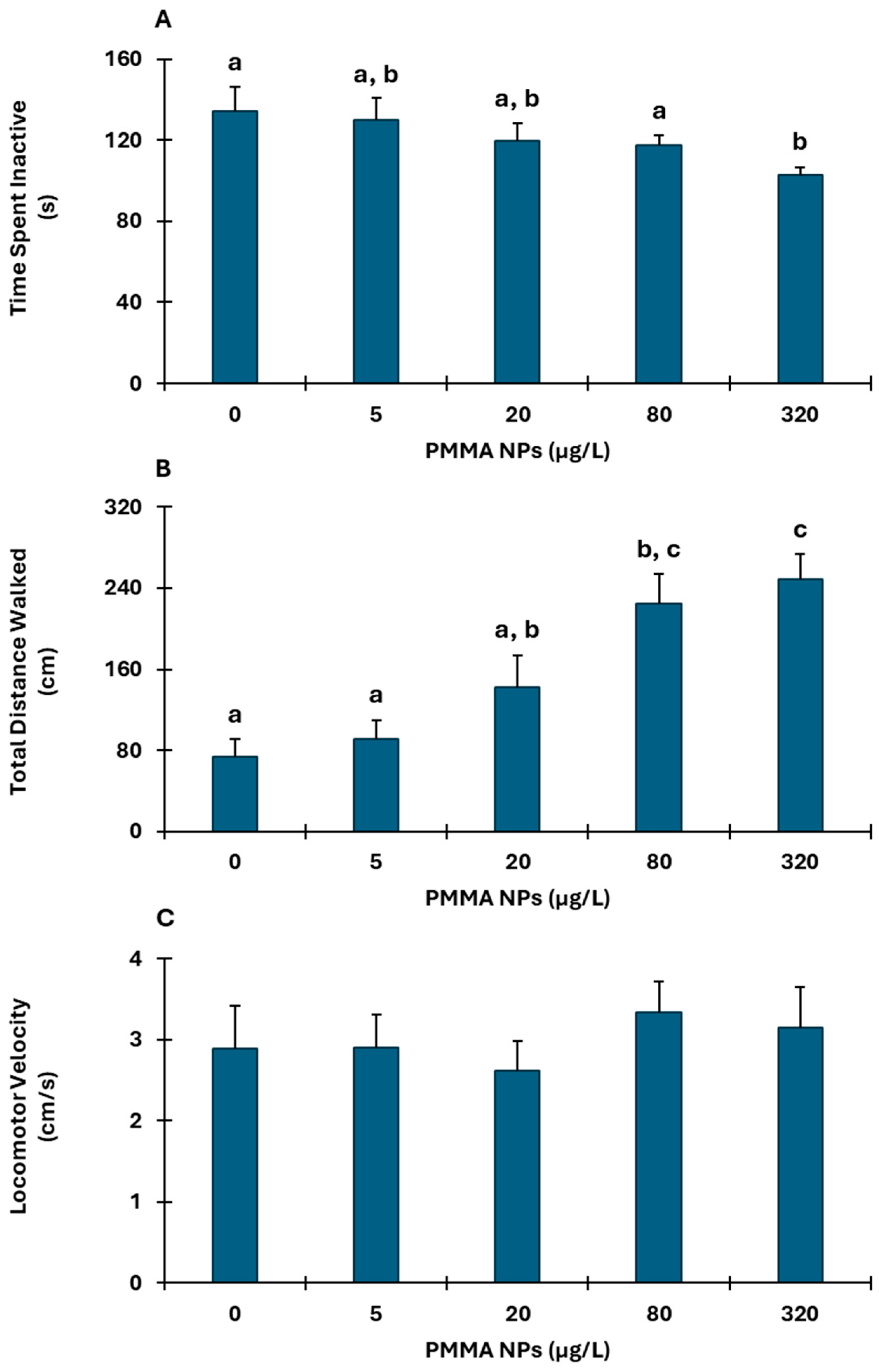
Figure 1. Time Spent Inactive (A); Total Distance Walked (B); and Locomotor Velocity (C) of C. maenas following 10 days exposure to PMMA NPs. Data represented are mean values ± standard error, n = 7. The letters represent significant differences (p < 0.05) among PMMA NPs concentrations.
Environments 12 00169 g001
Figure 2. Acetylcholinesterase (AChE) activity (A) and Glycogen (GLY) content (B) in the muscle of C. maenas following 10 days exposure to PMMA NPs. Data represented are mean values ± standard error, n = 7. The letters represent significant differences (p < 0.05) among PMMA NPs concentrations.
Environments 12 00169 g002
Figure 3. Antioxidant defenses: Superoxide Dismutase (SOD) activity (A); Glutathione-S-Transferases (GSTs) activity (B); Oxidative damage: Protein Carbonylation (PC) levels (C); and Malonaldehyde (MDA) concentration (D) in the gills of C. maenas following 10 days exposure to PMMA NPs. Data represented are mean values ± standard error, n = 7. The letters represent significant differences (p < 0.05) among PMMA NPs concentrations.
Environments 12 00169 g003
Figure 4. Antioxidant defenses: Superoxide Dismutase (SOD) activity (A); Glutathione-S-Transferases (GSTs) activity (B); Oxidative damage: Protein Carbonylation (PC) levels (C); and Malonaldehyde (MDA) concentration (D) in the hepatopancreas of C. maenas following 10 days exposure to PMMA NPs. Data represented are mean values ± standard error, n = 7. The letters represent significant differences (p < 0.05) among PMMA NPs concentrations.
Environments 12 00169 g004
Figure 5. Superoxide dismutase (SOD) activity (A); Glutathione-S-Transferases (GSTs) activity (B); and Malonaldehyde (MDA) concentration (C) in the hemolymph of C. maenas following 10 days exposure to PMMA NPs. Data represented are mean values ± standard error, n = 7. The letters represent significant differences (p < 0.05) among PMMA NPs concentrations.
Environments 12 00169 g005
Disclaimer/Publisher’s Note: The statements, opinions and data contained in all publications are solely those of the individual author(s) and contributor(s) and not of MDPI and/or the editor(s). MDPI and/or the editor(s) disclaim responsibility for any injury to people or property resulting from any ideas, methods, instructions or products referred to in the content.