4. Systematic Palaeontology
Class Mammalia Linnaeus, 1758
Order Carnivora Bowdich, 1821
Suborder Caniformia Kretzoi, 1943
Family Canidae Fischer de Waldheim, 1817
Subfamily Caninae Fischer de Waldheim, 1817
Genus Lycaon Brookes, 1827
Lycaon lycaonoides (Kretzoi, 1938)
Referred material. Południowa Cave: worn, left C1 (JP.1.1), left M2 (JP.1.6), right p1 (JP.1.2), right p2 (JP.1.3), buccal half of the right m1 (JP.1.4), talonid of the right m1 (JP.1.5), right m2 (JP.1.7), proximal half of the right metacarpal 3 (JP.1.8), phalanx 1 (JP.1.9).
Remarks. The species was described in detail in [
9,
13]. The bones of
L.
lycaonoides from Południowa Cave indicate the presence of a particularly robust canid, dimensionally comparable with the largest extant
Canis lupus. They were also characterised by the presence of enlarged and robust canines, high-crowned and vertical premolars, and large and narrow m1 with a long, prominent and vertical trigonid, a narrow and short talonid, as well as an oval-shaped and tricuspid m2 [
9,
13]. The remains of
L.
lycaonoides from Południowa Cave differ so markedly metrically and morphologically from the contemporary bones of
Canis lupus mosbachensis from the same locality that there is no possibility of misunderstanding.
Lycaon appeared in Europe 2.6 M already in a large-sized and robust form, which did not change significantly over the next 2 Ma [
34]. The species dominated Eurasian grasslands and open landscapes for more than 2 Ma, between 2.6 and 0.4 Ma. The last reliable record of
L.
lycaonoides, dated to MIS 11, is from the Polish site Draby 3. The main reasons for this process have been suggested to include climatic changes (especially the extremely massive MIS 12 glaciations), changes in the prey assemblages and strong competition pressure from other carnivores [
9,
13].
Genus Cuon Hodgson, 1838
Species Cuon alpinus (Pallas, 1811)
Cuon alpinus fossilis Adam, 1959
Referred material. Left m1 with broken-off distal root (W.2.1) from Wschodnia Cave.
Remarks. The sole m1 from Wschodnia Cave is the only Sudetes record of
C.
a.
fossilis, and its morphology and occurrence timespan (MIS 8-7) from this locality are well corroborated by the data obtained for this dhole chronosubspecies. The m1 was described as a large individual, whose dimensions have an intermediate position between
Cuon alpinus priscus (Thenius, 1954) and
Cuon alpinus europaeus (Bourguignat, 1868) [
4,
5,
8]. This is visible, among others, in the elongated and robust crown and the still broad but already short talonid.
Cuon alpinus fossilis was described as a large-sized dhole, with strong teeth located in a robust mandible, with moderately loosely arranged premolar row. The p4 is without the paraconid and has weak mesial cingulum broadening before the protoconid. The p4 crown also possesses two small enamel cuspids following the metaconid. The elongated and rather robust m1 has a moderate metaconid and talonid with a prominent hypoconid and rudimentary entoconid. The m2 has a rounded occlusal outline, trigonid with a strong protoconid and weak metaconid, while a low hypoconid occurs on the talonid. Its occurrence timespan was estimated as MIS 10-6 [
4,
5,
8,
88]. The taxonomic position of
C.
a.
fossilis is not well established and fully accepted. Its presence is sometimes linked with
C.
a.
priscus [
36] or is regarded as a primitive form of C. a. europaeus [
89,
90,
91]. Here, we agree with the interpretation of
C. a. fossilis as an independent form [
88], supporting the evolutionary lineage
Cuon alpinus priscus =>
fossilis =>
europaeus [
4,
5,
8].
Genus Canis Linnaeus, 1758
Canis lupus Linnaeus, 1758
Canis lupus spelaeus Goldfuss, 1823
Referred material. Północna Duża Cave: (NISP 10, MNI 2): M1, mandible, humerus, radius, two calcanei, mc 4, mc 5, mt 2, mt 5.
Canis lupus lupus (Linnaeus, 1758)/Canis lupus ssp.
Referred material. Wschodnia/Obok Wschodniej Cave (NISP 166, MNI 8;
Supplementary Materials): five crania fr., seven maxillae fr., 7 C1, 2 P4, 2 M1, 15 mandibles, three c1, p3, 2 p4, 5 m1, scapula, two lumbar vertebrae, caudal vertebrae, three costae, eight humeri, 7 radii, four ulnae, two cuboides, five mc 2, seven mc 3, five mc 4, six mc 5, two pelves, six femora, six tibiae, 13 calcanei, seven talii, six mt 2, six mt 3, four mt 4, seven mt 5, eight phalanges 1, phalanx 2. Naciekowa Cave (NISP 33, MNI 3;
Supplementary Materials): maxilla fr., M1, five mandibles, humerus, radius, ulna, mc 2, mc 3, 2 mc 4, 3 mc 5, four calcanei, talus, four mt 2, two mt 3, two mt 4, three mt 5. Cisowe 1 Shelter (NISP 3, MNI 2;
Supplementary Materials): three C1. Aven w Połomie Cave (NISP 14, MNI 2;
Supplementary Materials): P4, mandible, ulna, radius, mc 2 mc 5, mt 2, 2 mt 3, 2 mt 4, two phalanges 1, phalanx 3. Cisowe 2 Shelter (NISP 6, MNI 2;
Supplementary Materials): maxilla fr., P3, M1, c1, p4, m1. Małgorzaty Shelter (NISP 5, MNI 1;
Supplementary Materials): P4, lumbar vertebrae, radius, mc 4, phalanx 1. Wilcze Shelter (NISP 6, MNI 2;
Supplementary Materials): P4, M1, mandible fr., c1, m1, mt 4. Trwoga Paleontologa Shelter (NISP 4, MNI 1;
Supplementary Materials): M1, mandible fr., phalanx 1, phalanx 3.
Description. The upper tooth row is morphologically diverse. In the mesial part, between C1 and P3, it is almost straight, with teeth separated by broad diastemas. The C1 is especially well separated from the other teeth. The distal part of the upper tooth row between P3 and M2 is strongly arched, and the teeth are closely spaced. The robust and elongated C1 is bilaterally flattened and elongated mesio-distally. A narrow but distinct crest runs from the crown apex to its base on the mesio-lingual side. In lateral view, the crown is arched, with the crown apex strongly curved distally.
The elongated and moderately robust P2 has a strong distal cingulum. The almost centrally located protoconid is moderately high. The buccal and lingual margins are almost straight and are only weakly convex at the level of the protoconid end. The mesial and distal margins are blunt. In the occlusal view, the crown has a similar width, narrowing slightly from the transition between the protoconid and the second distal cuspid (
Figure 3). The overall morphology of the larger P3 is like that of the P2, but the tooth is more robust. The moderately high protoconid is located medially, with its apex oriented slightly mesially. The crown bears an elongated distal cingular projection. A faint mesial crest runs from the apex of the protoconid and ends at the base in the form of a small mesio-buccal prominence of the cingulum. The straight buccal margin bears at the distal part (as the level of the second distal cuspid) a moderately developed, median concavity. On the lingual margin, where a weak cingulum is situated, a moderate convexity is located at the same level. The mesial and distal margins are blunt or rounded (
Figure 3).
The long and robust P4 has blunt or rounded mesial and distal margins. The buccal margin is straight, while both margins, buccal and the lingual of the talon, are curved distally. From the apex of the moderately high paracone runs a strong and well-developed crest, which bifurcates into a fork, with one edge reaching the base of the tubercle, exactly at the border of the paracone and protocone. The other edge connects to the apex of the protocone. The low and short protocone, whose mesial margin is aligned with that of the paracone, is weakly separated from the paracone. A deep valley separates the high and robust metacone from the paracone. Stronger cingulum occurs only on the lingual margin of the metacone.
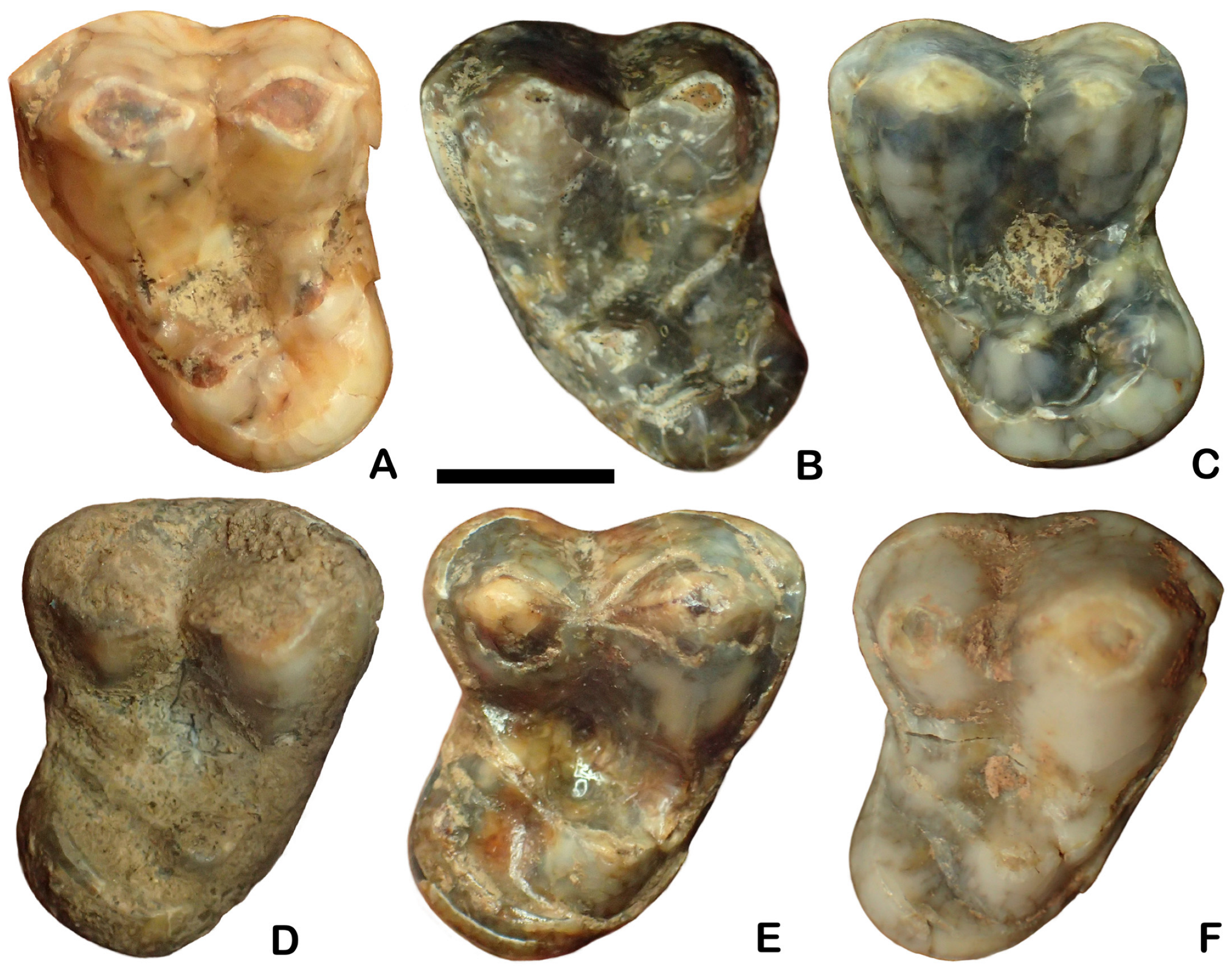
The large and irregularly shaped M1 has a moderately broad and short trigon (
Figure 4). Conical and high paracone and the smaller metacone are separated by a deep, narrow, V-shaped valley. Apexes of both main cusps are connected by a thin, long crest. A broad and deep depression running through the middle part of the crown separates the shorter talon from the trigon. The long and low protocone is divided by a shallow and wide valley into two parts of similar length. Developed as an elongated and low cuspid, the reduced metaconule is situated in the middle part of the crown. A long and thin crest related to the buccal cingulum margin runs in parallel along the whole talon length. The strong lingual cingulum forms a thick crest (
Figure 4).
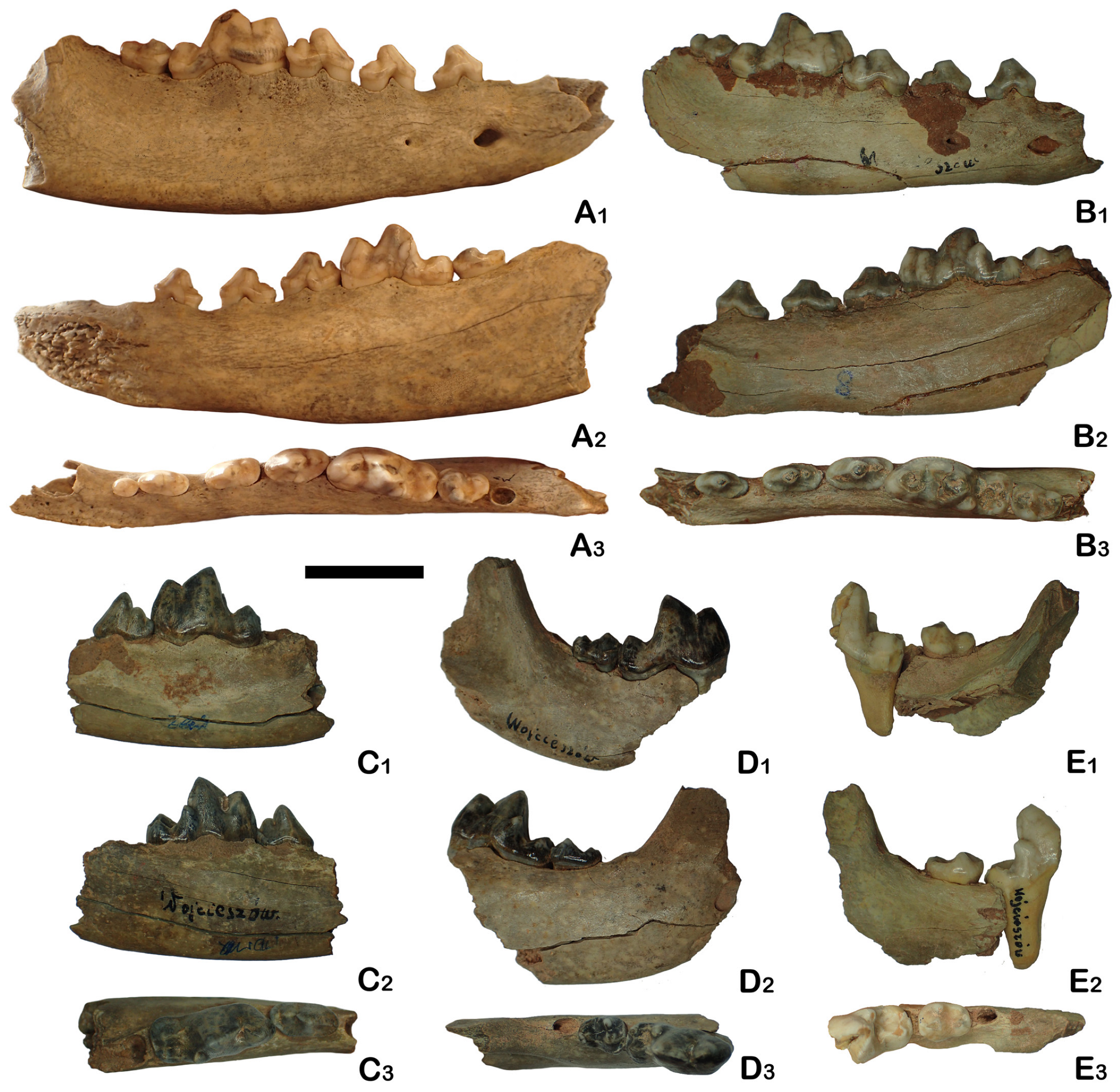
The thickness of the elongated and high mandibular body gradually increases distally, with the maximum under the m1–m2. Its height measured behind the m1 is higher than the m1 length (
Figure 5). The symphysis part is elongated and massive. Two rounded mental foramens are moderately spaced. The larger mesial one is situated under the p1 or the mesial root of the p2. The distal mental foramen is located slightly lower than the mesial one under the mesial or distal root of p3. The rounded mesial edge of the moderately deep masseteric fossa reaches the m2. Between c1 and p3, the lower mandibular body margin is almost straight, while between p4 and m3, it forms a gently curved arch, with the strongest curvature under m1 (
Figure 5).
The loosely arranged and straight premolar row is pushed more buccally in relation to the molars. The molars are tightly spaced, and the axis of the molar row runs from the mesio-buccal to the disto-lingual direction. The c1 has proportionally an elongated and hook-shaped crown (
Figure 5). The low-crowned p2 has the protoconid displaced mesially. Its occlusal outline is almost rectangular, with an elongated distal part. The strong distal cingulum forms a thick ridge, collaring an elongated distal cingular projection. The p3 also has an almost rectangular occlusal outline and the protoconid displaced less mesio-medially than in p2. The tooth possesses relatively strongly developed mesial and distal cingulum. Just after the protoconid, in a straight line, is located a conical, small but well-defined hypoconid. The relatively high-crowned and two-rooted p4 has the prominent protoconid placed almost exactly centrally, with the apex oriented mesially. The crown has a blunt mesial margin, a straight lingual margin and a rounded distal one. It is slightly broadened distally, with a moderate lingual median convexity. The crown has strong mesial and distal cingulum. After the protoconid, a relatively large hypoconid is present, closely associated with the strong distal crest. The crescent-shaped and shallow distal cingular projection is proportionally shorter than that of p2–p3 (
Figure 5).
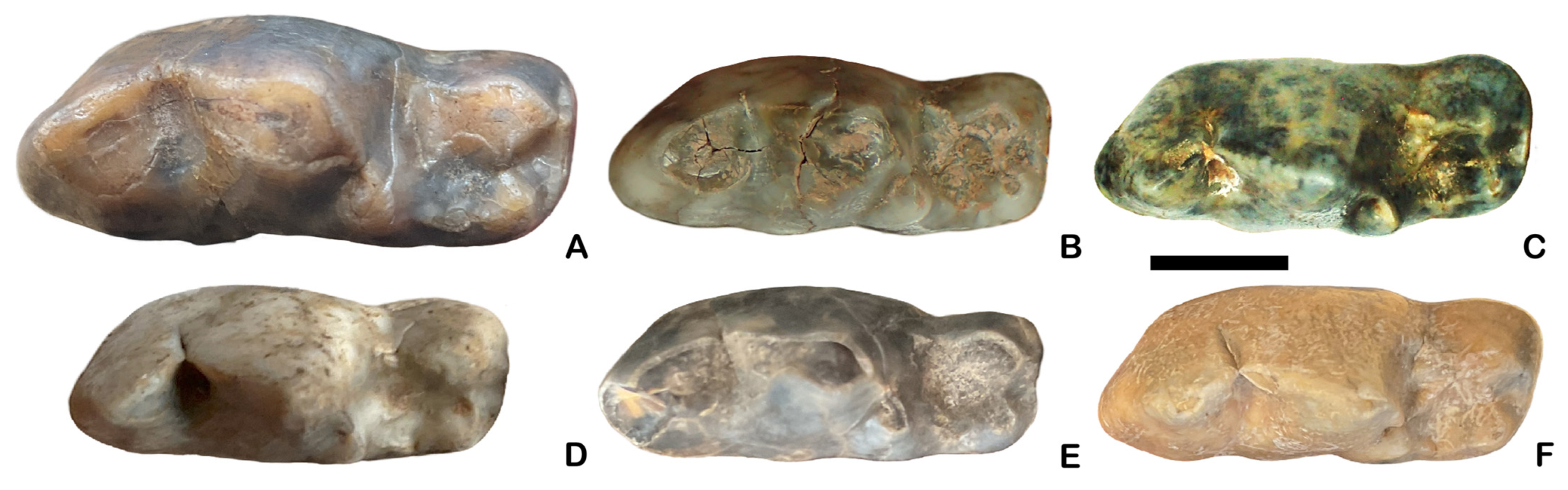
The massive m1 possesses high and robust trigonid and low and short talonid, which is narrower than the trigonid (
Figure 6). The crown has an almost rectangular occlusal outline, with straight buccal margin, blunt distal and rounded mesial margin. Between the trigonid and talonid is located a weak buccal concavity. The lingual margin is also straight and only slightly convex at the end of the talonid. The paraconid edge is weakly developed, while the cingulum is moderately developed. The small metaconid is associated with the protoconid. From the elongated and moderately high hypoconid runs a longitudinal and thin ridge, which surrounds the talonid field and terminates on the metaconid base. The conical hypoconid occupies 70% of the talonid surface, while distinctly lower and smaller, but the still well-defined entoconid is situated disto-lingually. The apexes of both cusps are not connected by any crest or ridge. The occlusal outline of the m2 varies from oval to rounded and mostly has a slightly irregular shape. The elongated, low and distinctly larger paraconid is located mesio-buccally and occupies 70% of the trigonid surface. Conical-shaped, lower and smaller protoconid is situated medially and lingually. The narrower talonid possesses conical and low hypoconid located disto-buccally. Nearby is situated a crescent-shaped and small internal basin, collared by a moderately developed cingulum (
Figure 6).
The lateral and medial epicondylar crests of the humerus are strongly developed, and the second one has a narrow supracondylar foramen. There is no connection between the deep and rounded coronoid fossa and the broad and deep olecranon pit. A rounded and shallow articular surface occurs on the large head of the radius. The laterally widened distal epiphysis possesses a well-marked elliptical glenoid cavity. The elongated and narrow ulna has a short and strong proximal end. The distal epiphysis has a strong protrusion of the ulnar styloid process. The cylindrical and elongated femur has a broad and deep trochanteric fossa. The greater trochanter is obliquely truncated on its lateral side and rises distinctly above the head level. The lesser trochanter is developed in the form of a slight conical prominence. The tibial articulations are nearly equal in size. The elongated and robust tibia has on the lateral side a large tibial fossa. Below the head is situated a narrow and shallow distal surface. The tibial crest is short and strongly developed. The elongated and robust calcaneus has a strong tuber calcanei. The talus has a prominent head and elongated neck.
Comparison. Analysis of
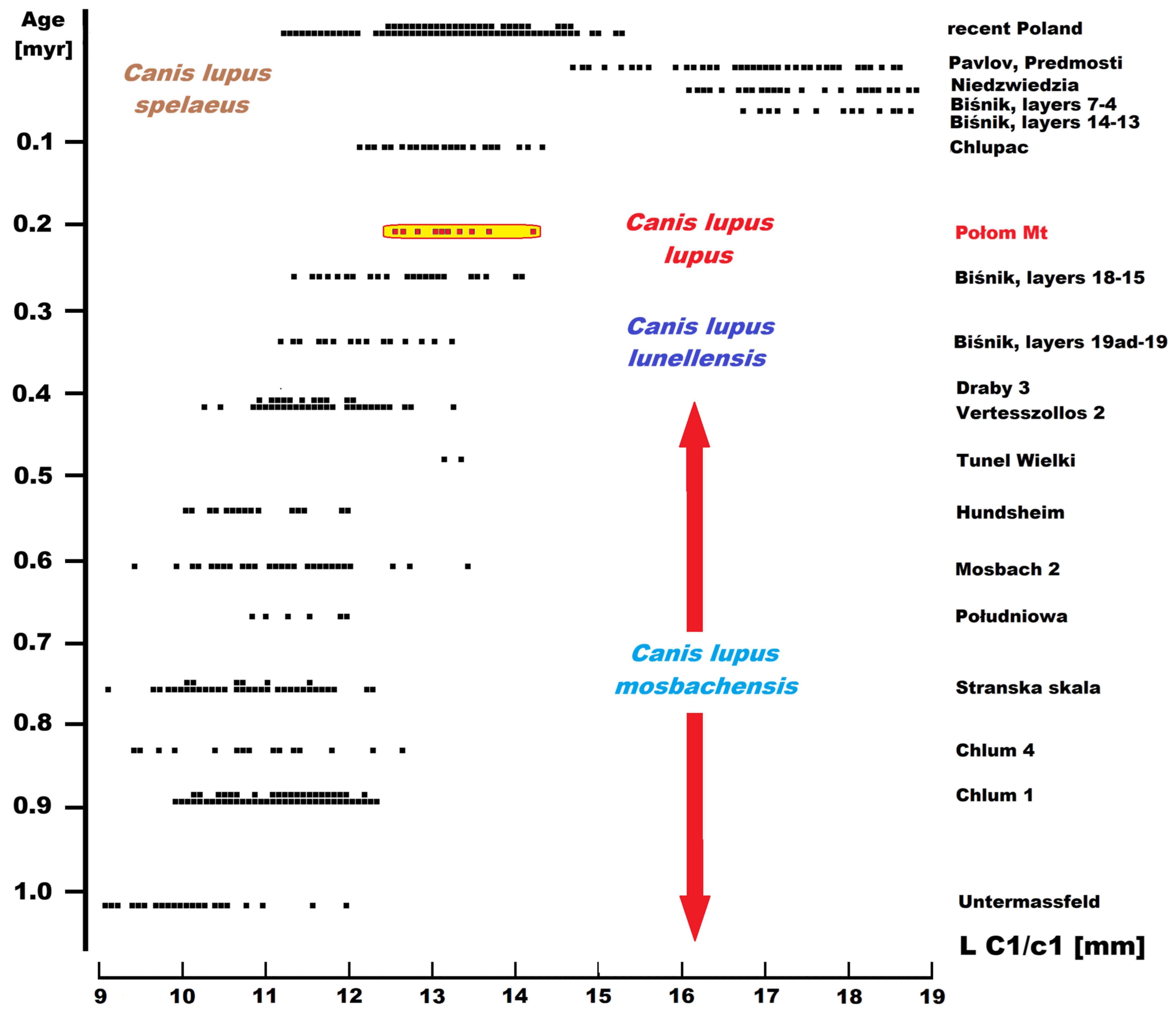
C. lupus remains from three neighbourhoods (Naciekowa, Obok Wschodniej and Wschodnia) revealed that they constituted homogeneous material in terms of metrics, morphology, and state of preservation. Given this, from our perspective, they are treated as a single sample. Because all crania and mandibles are incomplete, we only briefly focus on them. In the studied sample from Połom Mt, no significant morphological differences were found in the structure of C1 (
Figure 7). Some small differences between specimens result from intrapopulation variability rather than significant differences between different populations. Comparison with other populations showed that
C.
lupus from Połom has slightly larger C1 (
Table 1). However, the massiveness ratio (B/L) is generally within the range of variability typical for the species. Essentially, all populations dated between MIS 8 and 5e show similar values. The mean crown length is in the range of 11.8–13.0 mm, its width is 7–8 mm, and the B/L index is 61.0–62.5. The population from Mount Połom also falls within this range but occupies the upper range of variability. A significant change is observed within MIS 5a and MIS 3-2, where all three coefficients reach significantly higher values, the maximum for the species. This is consistent with Bergmann’s rule, climate cooling, and the appearance in the fossil record of the massive
C. l. spelaeus, whose exceptionally large and massive canines are an evolutionary adaptation for hunting large ungulates of the mammoth steppe [
92]. It can also be noted that wolves from central Europe are typically larger than those from contemporaneous sites in western Europe, such as France. Three C1 from Cisowe 1 Shelter showed higher dimensions (L C1 = 14.19 mm, B = 8.83 mm,
n = 3) than those from Połom Mt, but they are similarly robust (62.3,
n = 3). All three parameters are within the range of size variability of the extant Silesian
C. l. lupus. Because the site is dated to MIS 2-1, it is impossible to definitively determine whether the specimens belong to the cold-adapted
C. l. spelaeus or whether they are already large specimens of
C. l. lupus. The limited number (
n = 3) of specimens should also be considered, as they indicate certain trends but do not allow for any broader or far-reaching conclusions (
Figure 7,
Table 1).
Due to the significant degree of unification and minor structural differentiation of P2-P3, these teeth rarely occur in the context of morphological differences between
C. l. mosbachensis,
C. l. lunellensis,
C. l. lupus and
C. l. spelaeus. Only a strongly developed hypoconid on P3 was mentioned [
93]. Analysing the measurements of P2 and P3 and the massiveness of both teeth (B/L), similar properties were observed, which were also found for C1 (
Table 2 and
Table 3). A clear increasing size trend and a progressive increase in crown massiveness are observed from
C. l. lunellensis, by
C. l. lupus up to
C. l. spelaeus. However, this trend is not entirely constant, as the population from Romain-la-Roche (MIS 6) and layers 10–9 of the Biśnik Cave (MIS 5a) stands out for its rather robust P2 compared to other chronosubspecies, including even
C. l. spelaeus. In turn, in P3, a steady upward trend in size and mass is observed, which declines only in MIS 1, along with the disappearance of
C. l. spelaeus. Compared to other populations, the dimensions of P2-P3 of
C.
lupus from Połom Mt fall within the range of variability of populations dated to MIS 9–7. The crown is also characterised by a medium range of variability, typical of the MIS 9–6 period (
Table 2 and
Table 3).
In P4 morphology,
C. l. mosbachensis and
C. l. lunellensis share several morphometric traits [
93,
94] (
Table 4). However, despite their overall similarity, several diagnostic differences have been identified [
92,
93,
94,
95,
96,
97,
98,
99,
100]. Nevertheless, these similarities were regarded as ecological and environmental influences rather than true taxonomic differences [
97,
98,
99,
100]. In
C. l. mosbachensis, the protocone is more prominent and positioned slightly more mesial, with a larger hypocone basin and a more strongly developed lingual cingulum on the metacone than in
C. lupus ssp. [
97].
Progressive evolution has been well documented by the
Canis fossil material from the Spanish Early and Middle Pleistocene sites of Atapuerca, Gran Dolina (TD), Trinchera Galería (TG) and Sima del Elefante (TE) [
98]. Individuals from TD 10, dated to MIS 11–10 (430–350 ka), tend to be larger than those from older stratigraphic levels like TE9–TE14 (1.3–1.1 Ma), TD3-TD5 (1.1–0.9 Ma), TD 6-TD7 (950–770 ka), TD8- TD9 (650–550 ka). They also exhibit morphological features suggesting a gradual transition from
C. l. mosbachensis to
C. l. lunellensis. Individuals of
C. l. mosbachensis from TE9–TE14 to TD3–TD8 show the presence of a slightly more mesial location, prominent and robust protocone and an expanded cingulum adjacent to the metacone.
Canis lupus mosbachensis from TD10 shows a progressive reduction of the lingual cingulum and a smaller protocone compared with
C. l. mosbachensis from earlier chronologies. This indicates a gradual evolutionary shift towards the morphology observed in later populations. Specimens of
C. lupus ssp. from GIII (400–320 ka) exhibit a longer protocone and a weakly developed or absent cingulum on the P4 [
98].
A comparison of wolf P4 morphology from the last 350 ka revealed that the tooth underwent visible changes over time. First and foremost, significant size variation is evident, with the oldest
C. l. lunellensis populations from layers 19ad–19 of the Biśnik Cave and Lunel Viel, dated to MIS 9, being only slightly larger than
C. l. mosbachensis. A distinct change is evident from MIS 8, documented by the analysed specimens from Połom Mt. Their dimensions are like those of the moderately large and slender Eemian
C. lupus, a characteristic faunal element of the last interglacial (MIS 5e). Interestingly, comparing P4 sizes from different European populations once again confirmed that, in addition to the temporal factor, i.e., the age of the site, it is equally important to compare populations of different ages from as similar an area as possible (
Table 4).
Wolves from the cold phases of the last glacial period feature opposite dimensions, being massive and large animals with enlarged carnassials (P4/m1). Their enormous size and robust dentition reflect adaptation to the cold climate (Bergmann’s rule), hunting large prey, and the need to chew through hard, quickly freezing carrion. As a species, C. lupus reached its maximum size in MIS 3–2 in C. l. spelaeus, after which, with the retreat of the ice sheet, body size and mass decreased until they reached the dimensions known today. Extant Silesian wolves are large, but they are far from the giants of the last glacial period. It also turned out that the metacone, and consequently the length of the breaker’s cutting edge, increased, with a maximum noted during MIS 3–2. Overall, the analyses showed that P4 of C. lupus from Połom Mt was characterised by small to medium-sized bodies, a slender crown, and a relatively short metacone. They exhibited a longer protocone and a still relatively strong lingual cingulum on the metacone. Three other P4s from the Holocene strata of Aven w Połomie, Małgorzaty Shelter, and Wilcze Shelter have a similar morphology.
As in the case of P4, several differentiating features were also found in the morphology of M1 between
C. l. mosbachensis and
C.
lupus ssp. In
C. l. mosbachensis, the metacone and paracone are more conical and smaller, the paracone is weakly to moderately higher than the metacone, trigon basin deeper than the talon basin, the hypocone basin is deep and elongated, and the labial cingulum is continuous and strong developed (
Table 5). The M1 of
C.
lupus ssp. is characterised by a larger and more globular metacone and paracone, with the paracone distinctly higher and larger than the metacone, a reduced and shallow hypocone basin, and a subdued labial cingulum, which is often incomplete across the paracone [
90,
91,
92,
93,
94]. Individuals from TD 10, dated on MIS 11–10 (430–350 ka), show an intermediate morphology between
C. l. mosbachensis and
C.
lupus ssp. [
98].
As to the M1 from Połom Mt, its dimensions are relatively small to medium and slightly larger than contemporaneous populations, particularly from Western Europe (
Table 5). Analysis of various dental proportions of the main elements of M1, the trigon and talon, revealed no significant changes in proportions over the last 350 ka. A high degree of differentiation has been observed between different chronosubspecies of
C.
lupus, from which it is difficult to distinguish any consistent trend, apart from, of course, an increase in size. The M1 from Połom is characterised by a proportionally narrow trigon and wide talon, features more typical of
C.
lupus ssp. than
C. l. mosbachensis. In all M1 specimens examined, the paracone is distinctly higher and larger than the metacone. In all specimens, there is a distinct median concave on the buccal margin. In six teeth, the distal edge of the hypocone lies slightly mesially to the distal edge of the metacone, while in two others, it is located slightly distally to the distal rim of the metacone. In all M1 specimens from Połom Mt, the parastyle is reduced and cingulum-like, and the buccal cingulum stops at the paracone and no longer surrounds the paracone completely. The parastyle and preprotocrista are not connected in all M1 specimens. The inner ridge of the paracone at its medial base is present and well-marked. On the contrary, the inner ridge of the metacone is weakly developed. The lingual cingulum around the protocone is disconnected and stronger only at the mesio-lingual side of the cusp.
As with C1, no statistically significant and reliable diagnostic features were identified in c1 that would allow for the distinction between
C. l. mosbachensis and
C.
lupus ssp. In fact, the only determinants are metric features, and a steady increasing trend over time is noticeable (
Table 6). While the sizes of some
C. l. lunellensis from populations dated to MIS 9–8, such as Kudaro, La Fage or Lunel Viel 1, are essentially within the range of
C. l. mosbachensis variability, or slightly exceed it, a noticeable increase in size is observed starting with the MIS 8 specimens from Połom Mt. These are larger, but still not very large, specimens that are far from the massiveness and size of Late Pleistocene specimens. In addition to the increase in body size, there was also a noticeable increase in the massiveness of the crown, culminating in specimens dated to MIS 5a and 3. A clear increasing size trend and a progressive increase in crown massiveness are observed from
C. l. lunellensis, through
C. l. lupus, to
C. l. spelaeus. As with C1, the extremely large and robust canines are an evolutionary adaptation for capturing and hunting large ungulates. Interestingly, c1 specimens from the Eemian interglacial (MIS 5e) are medium-sized and have rather narrow crowns. Extant Silesian wolves, on the other hand, have large and massive canines. While they are not as large as specimens from the Late Pleistocene, their size and massiveness still distinguish them from populations from other Polish regions (
Table 6).
The presence of distal accessory cuspids on the p2–p3 is highly variable among
Canis members, especially in the p3 specimens. In most individuals, p2 is without both accessory cuspids. In the analysed
C.
lupus populations from the last 350 ka, the presence of these cuspids is rather sporadic, and no significant patterns in their occurrence were observed. Even in large samples, records of accessory cuspids are sporadic, and the distal cuspid is much more common, while the mesial cuspid is very rare. When present, it is always small, low, and poorly defined, most often in the form of a low and elongated cingulid. Sometimes it occurs as a rudimentary cuspid, most often associated with a protoconid. All three p2 specimens from Połom Mt lack accessory cuspids, and their dimensions fall within the range of variation for populations dated to MIS 9–6. A significant increase in size is observed from MIS 5e onwards and an increase in mass from MIS 5a onwards. The Late Pleistocene
C. l. spelaeus has the largest and most robust p2, but accessory cuspids occur only very rarely in this chronosubspecies (
Table 7).
Standard p3 of
C.
lupus lacks a mesial accessory cuspid, bears a prominent protoconid, and presents a distal accessory cuspid. In
C. l. mosbachensis, its occurrence is more variable, from lacking both distal accessory cuspids, through displaying only a single cuspid, to some p3 bearing two accessory cuspids located after the protoconid [
32,
92,
97,
101]. This high variation in the presence of the distal accessory cuspids in p3 of
C. l. mosbachensis may reflect intraspecific morphological variability rather than taxonomic differentiation [
98]. As in p2, the mesial accessory cuspid is present only exceptionally in
C. l. mosbachensis and
C.
lupus ssp. However, the distal accessory cuspid is present in most
C. l. mosbachensis and practically in all
C.
lupus. Its absence or poor development in
C.
lupus is only occasionally observed. Its development in
C.
lupus is much more pronounced than that of
C. l. mosbachensis and usually occurs in the form of a clearly defined and distinct cuspid. In addition, a secondary distal cuspulid is present in most specimens, often associated with the distal cingulid. This structure is very rarely found in
C. l. mosbachensis and usually only in larger, more advanced individuals from sites dated to MIS 15–11.
Morphologically, the p3 specimen from Połom Mt has a structure typical of
C.
lupus, although the second cuspid is poorly developed and defined (
Table 8). No mesial accessory cuspid was observed. Metrically, the p3 from Połom Mt exceeds specimens from other populations of the same age and is in this respect closer to specimens dated to MIS 6–5e. Since the last Eemian interglacial, an increase in size has been observed, but the p3 still remains a proportionally narrow tooth. The increase in size is typical for the MIS 5–2 period, where p3
C. l. spelaeus is not only large but clearly more robust than the others. The teeth of individuals from Połom Mt are narrow, like those of other populations dated to MIS 9–5e. Extant Silesian
C. l. lupus have large but narrower teeth than those of the Late Pleistocene
C. l. spelaeus (
Table 8).
Among lower premolars, differences between
C. l. mosbachensis and
C.
lupus ssp. are the most marked in p4, where the first chronosubspecies exhibits a more primitive morphology. In both, the first distal accessory cuspid is present in all specimens. The second distal accessory cuspid–distal cingulid complex is generally much smaller than the first distal cuspid. The main difference between them, together with smaller size and narrower crown, is that in
C. l. mosbachensis, the second distal accessory cuspid is distinctly separated from the distal cingulum. In
C.
lupus ssp., this cuspid is typically fused to it [
32,
92,
97]. The mesial accessory cuspid, if present, always occurs as a rudimentary and poorly defined cuspid or, more often, as an elongated and low ridge; it occurs sporadically in
C. l. mosbachensis and exceptionally in
C. lupus ssp. (
Table 9).
Analysing p4 in different
C.
lupus ssp. populations from the last 350 ka, a clear linear trend of increasing size can be observed, starting from MIS 8. Specimens from Połom Mt are relatively small and have narrow crowns, which is typical of populations from localities dating to MIS 9–7. Individuals from the Eemian interglacial (MIS 5e) are significantly larger and have broader crowns (
Table 9). However, the true increase in body size, and above all, the massiveness of p4, is observed in the Late Pleistocene from MIS 5a onwards to the end of MIS 2. This is obviously related to the presence of
C. l. spelaeus in the fossil record. Extant Silesian wolves have a significantly smaller and narrower p4, comparable to individuals from MIS 5e (
Table 9). Of the 10 p4s from Połom Mt, 7 have typical
C.
lupus morphology, where the second distal accessory cuspid–distal cingulid complex is generally much smaller than the first distal cuspid. In those seven teeth, the second distal accessory cuspid is fused with the distal cingulid. In three others, this fusion is still incomplete, and the second distal accessory cuspid is larger than in standard p4 of
C.
lupus. In all 10 teeth from Połom Mt, the first distal accessory cuspid is present. Only a single p4 showed the presence of what could be considered a mesial accessory cuspid, developed in the form of an elongated and low ridge.
Within the genus
Canis, m1 is traditionally regarded as the best tool in the biochronological and palaeoecological analysis, among others, in body size reconstruction. This tooth is also among the most valuable in taxonomic interpretation;
C. l. mosbachensis and
C.
lupus ssp. differ in many morphological features. The m1 of
C. l. mosbachensis is characterised by a proportionally lower and shorter trigonid and higher and longer talonid; a relatively high paraconid, which is relatively straight along the mesial margin; a distally oriented and sharp protoconid; a prominent and sharp metaconid located at the height of the paraconid; both main cusps of the talonid; the hypoconid and entoconid, which are linked by a well-developed crest; and the presence of two cuspids positioned between the metaconid and the entoconid. The p4 protoconid is situated as high as the m1 paraconid in the Early Pleistocene (1.5–0.8 Ma) or slightly higher in the Middle Pleistocene (0.8–0.4 Ma) specimens. By contrast,
C. lupus displays a proportionally higher and longer trigonid and lower and shorter talonid, low paraconid, more rounded and vertically oriented protoconid, moderately large and rounded metaconid, a larger hypoconid and entoconid in direct contact and not linked by a crest, and the hypoconid distinctly higher and larger than the entoconid. The m1 paraconid is located significantly higher compared to the p4 protoconid, and the mesial margin is distally curved [
92,
95,
97,
100,
102].
Analysis of 11 m1s from Połom Mt showed that they are characterised by a morphology typical of
C.
lupus, although several features are less developed than in Late Pleistocene and extant individuals. Comparative dimensions place
C. l. lupus from Połom Mt closer to sites dated to MIS 6, although it should be remembered that wolves from the French sites with which this population is compared are characterised by smaller average dimensions (
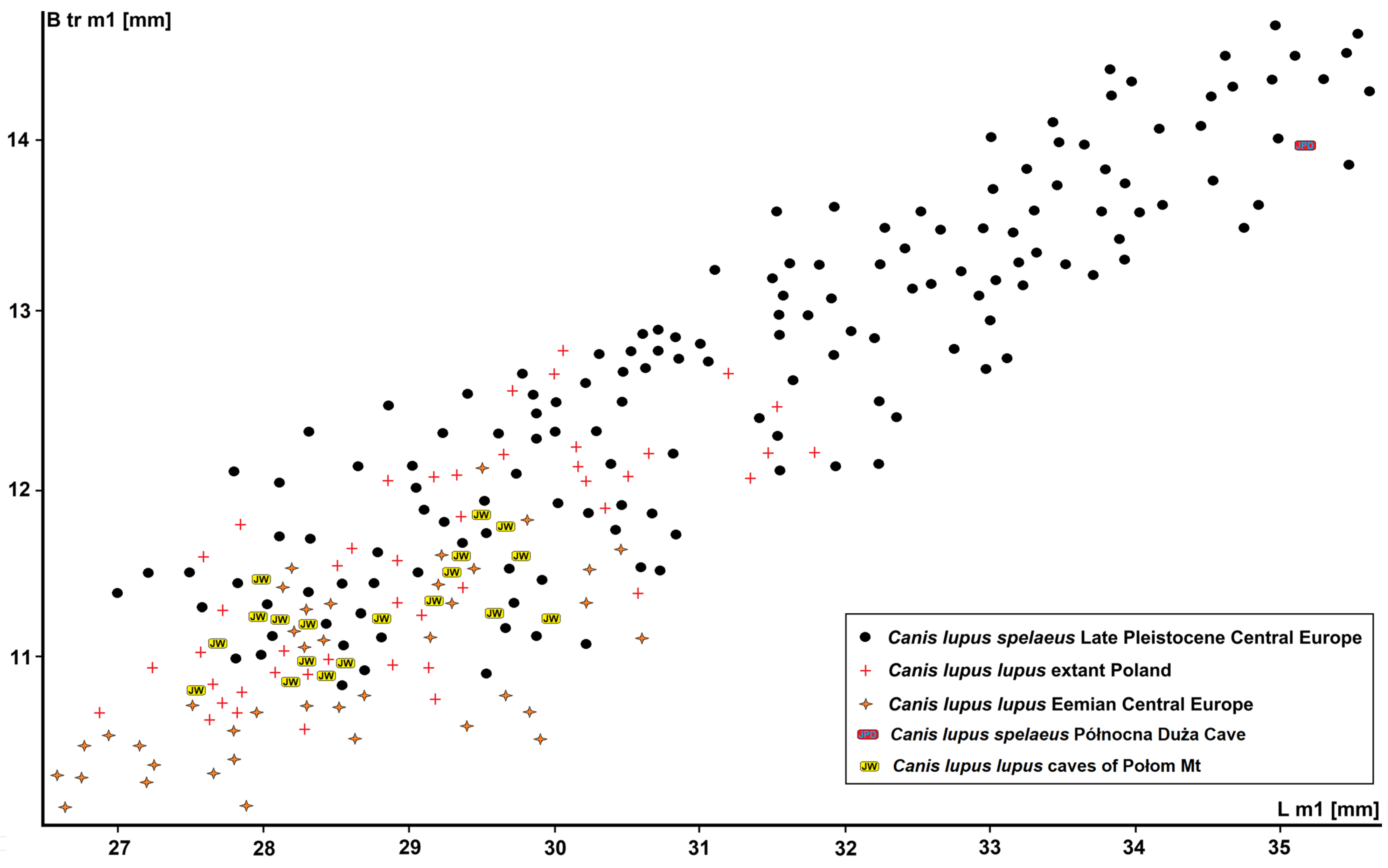
Table 10). The specimens from Połom Mt have dimensions intermediate between
C. l. lunellensis from layers 19ad–19 of Biśnik Cave (L m1 = 26.12 mm) and
C. l. lupus from Eemian sites such as layers 3–4 of Dziadowa Skała (L m1 = 28.27 mm) and layer 13 of the Biśnik Cave (L m1 = 28.78 mm) (
Figure 8). Their dimensions are similar to those from the French site of Romain-la-Roche, dated to MIS 6 (L m1 = 27.62 mm), and layer 14 of Biśnik Cave (L m1 = 27.82 mm). A real increase in m1 size is observed from the cold phases of MIS 5a, when
C. l. spelaeus appears in the fossil record. The m1 reaches a maximum size during MIS 3–2, after which body size decreases to the level known from the Holocene in MIS 1 (
Figure 8;
Table 10).
Extant wolves from Silesia were large, but after their extirpation by the end of the 18th century and their return 200 years later, their dimensions are now about 20% smaller. During the evolution of the last 350 ka, in addition to metric changes, several morphological changes have also been recorded. These include an increase in the length, height, and massiveness of the trigonid and a related, simultaneous trend of shortening and narrowing of the talonid. This is a consistent trend, observed from
C. l. lunellensis, through
C. l. lupus, to
C. l. spelaeus. In both parameters,
C. l. lupus from Połom Mt, with a proportionally narrow trigonid and elongated talonid, ranks among populations dated to MIS 9–7. The chronosubspecies with the most massive and longest trigonid and the shortest talonid is
C. l. spelaeus (
Figure 9;
Table 10). In the m1 of
C. l. lupus from Połom Mt, the mesial edge of the paraconid is distally strongly inclined and has a moderately large and rounded metaconid. The mesio-lingual ridge of the hypoconid is relatively weakly developed and stops at the base of the distal wall of the trigonid. The junction of the two ridges of the hypoconid is located more distally at the mesial slope of the hypoconid. The entoconid is still less reduced than in
C. l. spelaeus and extant
C. l. lupus. However, there are no signs of accessory cuspids on the mesial ridge of the entoconid. In all analysed specimens from Połom Mt, the base of p4 is situated distinctly lower than the base of m1 trigonid.
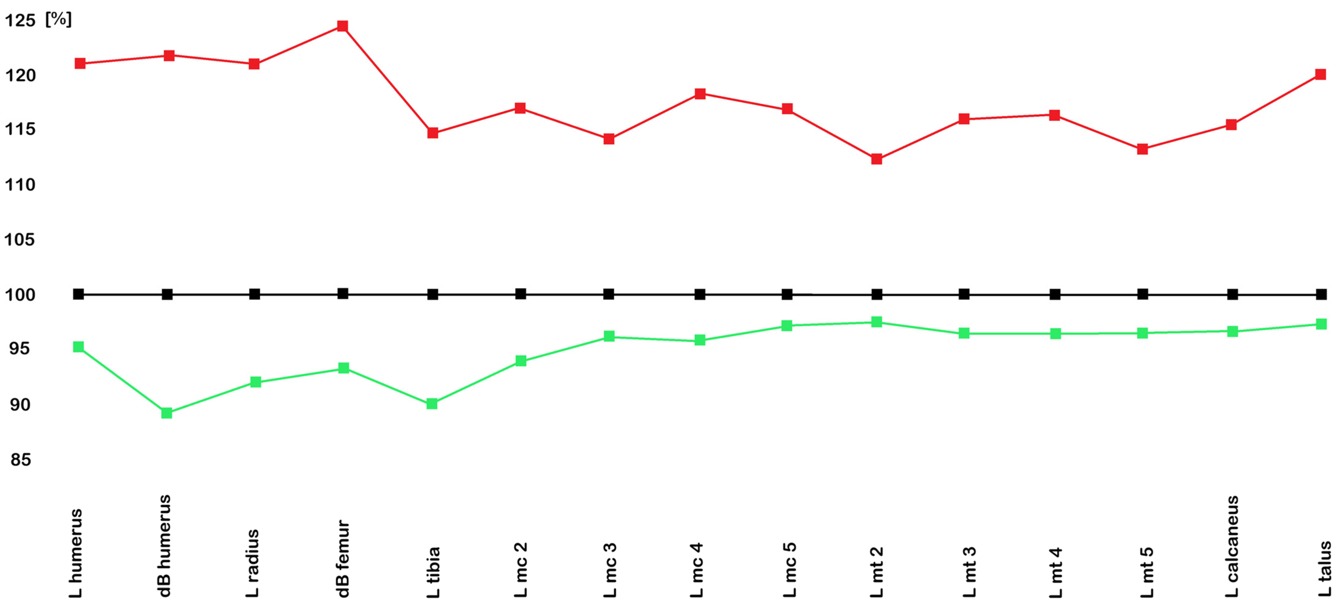
Analysis of the postcranial material revealed no significant differences in the morphology of long bones between the different chronosubspecies of
C.
lupus. In all parameters, both averages and ranges, the postcranial bones of
C. l. lupus from Połom Mt are slightly smaller and more slender than those of the extant Silesian
C. l. lupus (
Figure 9). In fact, these bones do not show specific characteristics, even if the distal articular surfaces are more slender than the homologous structures of the extant
C. l. lupus. The greatest differences in size were recorded for the long bones, while the differences recorded for the metacarpals and metatarsals are relatively small, on the order of 2–4%. All postcranial bones of
C. l. lupus from Połom Mt are characterised by less massive shafts and both epiphyses. This is particularly evident in the metacarpals and metatarsals. This is consistent with the wide trend for Eurasian
C.
lupus of progressive increase in size and robustness within
C. l. lunellensis =>
C. l. lupus =>
C. l. spelaeus evolutionary lineage and applies to elements of the postcranial skeleton. Comparing the dimensions of the long bones, where the differences are greater, on the order of 5–10%, and the metacarpals and metatarsals, where they amount to 2–4%, it can be interpreted that
C. l. lupus from Połom Mt has slightly larger autopodial portions in the forelimbs and hindlimbs. This may suggest better adaptation to locomotion in the uneven and unstable mountainous and upland terrain typical of the Sudetes as well as eastern and southern Silesia (
Figure 9).
Similar conditions have also been observed for
Ursus spelaeus ingressus (Rabeder et al., 2004) and
Panthera spelaea spelaea (Goldfuss, 1810) from Sudetes [
104,
105]. Of course, the difference may be due to differences in individual specimens, as it cannot be unequivocally confirmed that the long bones are identical to the metacarpals and metatarsals and belong to the same individuals. However, the differences between these individual elements of the postcranial skeleton are quite clear and symptomatic and provide cause for reflection. Among all of them,
C. l. spelaeus, of course, has significantly larger and more massive elements of the postcranial skeleton. Similar conditions were observed here, with the significantly larger dimensions of the long bones contrasting with smaller differences in other elements of the postcranial skeleton, such as the metacarpals and metatarsals. Because the postcranial skeleton elements are characterised by a significantly lower number of diagnostic features, and in fact, most of the bones show no significant morphological differences beyond their dimensions, most authors do not devote significant attention to taxonomic analyses. Most authors hypothesise a wide differentiation of the
Canis group, with several species, subspecies, and chronosubspecies. A parsimonious analysis should consider the scarcity of remains and intraspecific variability among some specific forms and populations. The occurrence of wolves, especially from transitional episodes like MIS 12–10 in Europe, is not frequent enough to allow a clear overview of their evolutionary trends. Additionally, sexual dimorphism in
Canis, as in most Canidae species, is distinctly less pronounced than that of other carnivores like ursids, mustelids, or felids, so its impact on the analyses presented above is small and has not been considered. In summary, it can be stated that, in terms of taxonomic determination based on the postcranial skeleton, the main osteological features for identifying skeletal remains of the particular chronosubspecies are dimensions.
Remarks. The population from the Połom Mount caves (Naciekowa, Obok Wschodniej i Wschodnia) was compared with the Middle Pleistocene (MIS 19–11; 800–370 ka) Mosbach wolf
C. l. mosbachensis, Late Pleistocene (MIS 3–2; 50–15 ka) steppe wolf
C. l. spelaeus from Niedźwiedzia Cave, and the extant grey wolf
C. l. lupus population from Silesia. Therefore, the comparison was made in a single area that has undergone numerous changes over time. Given that the wolf from the Połom Mount caves was historically considered one of the first and oldest (MIS 8) occurrences of a large wolf in Europe [
3], a thorough morphological analysis of the material is crucial to determine which chronosubspecies this population actually represents. In this regard, the focus was on a thorough analysis of metric and morphological characteristics and the establishment of criteria that would allow for the characterisation of the Połom Mountains cave population.
Canis lupus mosbachensis is generally considered the ancestor of
C.
lupus, occupying an intermediate position along the wolf evolutionary lineage, namely,
Canis etruscus Forsyth Major, 1877 =>
Canis lupus mosbachensis–
Canis lupus ssp. [
97,
106,
107,
108,
109,
110]. However, because of the lack of clearly defined morphological criteria useful for determination to the species level, general confusion persists in the palaeontological literature. Since the first description by [
106], this problem has heavily affected the taxonomical attributions proposed during the last 100 years. In the past,
C. l. mosbachensis was regarded as a valid and distinct species,
Canis mosbachensis [
106,
107,
108,
109,
110], an evolutionarily advanced form of
C.
etruscus–
Canis etruscus mosbachensis [
106,
107,
108,
109,
110,
111,
112,
113,
114], or an ancestor and primitive form of
Canis lupus–
Canis lupus mosbachensis [
115,
116,
117,
118,
119]. Because of the lack of an established type,
C. l. mosbachensis remains a vaguely defined taxonomic form, whose features were never clearly defined. A good example of how problematic the taxonomic attribution of canids can be is documented by the material from Cueva Victoria [
114]. At the beginning, it was initially assigned to
C. etruscus [
119,
120] and
C. etruscus etruscus [
121], then to
C. mosbachensis [
107], later to
C. arnensis [
122], and finally to
C. mosbachensis [
100].
The taxonomy of Middle Pleistocene Canis lupus in Europe is debatable. This is mainly due to a particular pattern of features shared by all large canids such as the retention of primitive dental characteristics. The absence or presence of some primitive features complicates a clear identification of true diagnostic characters that define interspecific and/or intersubspecific variability [
93,
94,
123]. Additionally, partially complete crania or long bones are exceptionally rare, and the
Canis fossil material is mainly represented by isolated or fragmentary craniodental or postcranial elements (but not long bones). Considering canids, teeth are the most abundant and best-preserved remains considered and used for taxonomical studies [
114]. Among them, the m1 has been considered a valuable parameter and one of the best tools to discriminate
C. l. mosbachensis and
C.
lupus ssp. [
114,
118]. Nevertheless, there is broad overlap in tooth size between
C. l. mosbachensis and early forms of
C.
lupus ssp. [
94,
95,
123]. This emphasises the poor reliability of taxonomic determination exclusively based on dimensional criteria. It is especially crucial for two transitional periods in
Canis evolution, one between 1.6 and 1.4 Ma, when the
C. etruscus–
C. l. mosbachensis transition took place, and a second between MIS 12 and 10, when the
C. l. mosbachensis–
C. lupus ssp. transition occurred [
114].
An important aspect involved in the taxonomy of the genus
Canis is size, with a trend of increasing dimensions depicted along their evolutionary history [
35,
107,
122,
123,
124]. There was general agreement that this trend started with the small Early Pleistocene (2.2–1.6 Ma)
C. etruscus, continued with medium-sized
C. l. mosbachensis from the late Early to mid-Middle Pleistocene (1.1–0.4 Ma), with medium to large-sized
C.
lupus ssp., and terminated with the largest forms from the Late Pleistocene (100–15 ka)
Canis lupus spelaeus [
36]. Nevertheless, considering size as an important tool for taxonomical attribution could be misleading. Representatives of the genus
Canis are adapted to a broad range of habitats and environments and have extremely broad geographical distribution. Extant
Canis lupus shows significant size and posture differences between particular subspecies and a clinal gradient that roughly adheres to Bergmann’s ecogeographical rule [
113,
118,
124,
125,
126,
127,
128,
129,
130]. It should also be noted that
Canis lupus displays considerable morphological polymorphism. Nevertheless, the larger and more robust specimens generally exhibit more robust cusps, more pronounced accessory cusps and cingula, and additional cusps on the molars. It is also likely that these features can be primarily associated with overall tooth size rather than representing specific functional adaptations [
95,
123]. Recent
C. lupus is widespread throughout the northern hemisphere, with populations that differ greatly in several phenotypic features, such as large size and robust build, while southern subspecies are generally characterised by a small size and lesser posture [
113,
118,
124,
125,
126].
Like the extant
C.
lupus,
C. l. mosbachensis had a broad geographical distribution and may show geographical differences. According to this hypothesis, the Early to Middle Pleistocene northern populations, e.g., Mauer, Mosbach 2 or Untermassfeld, include a higher number of larger individuals than those recovered from southern localities such as Escale, Pirro Nord or Petralona [
114]. An extensive comparison of various European populations from European localities dated on 1.1–0.4 Ma, based on size comparison of upper and lower canines and the lower carnassial (m1), showed that
C. l. mosbachensis had roughly the same dimensions between 1.1 and 0.7 Ma [
2,
3,
4,
5,
6,
7,
8,
9]. Those populations formed a biometrically homogeneous group, and the average values are quite similar. Slightly larger dimensions were established for the populations younger than 0.6 Ma, like those of Hundsheim, Mauer or Mosbach 2. However, in general,
C. l. mosbachensis only slightly differs in size up to MIS 11 (~0.4 Ma). A real increase in size started after MIS 11, with the appearance of early forms of
C.
lupus ssp. [
36].
The taxonomy of the late Middle Pleistocene (MIS 10-6, 370–130 ka) history of
C. lupus is the same as that of
C. l. mosbachensis or is even more complicated. This is because it is more widespread and represented by a considerably higher number of localities and fossil materials, which allow more complete morphometrical and statistical analyses. Traditionally,
Canis lupus lunellensis Bonifay, 1971, which appeared ca. 410–400 ka, is regarded as the earliest and first true wolf [
35,
36,
131]. It was determined to be a small-sized and gracile wolf with a straight lower tooth row, small M1 with narrow trigon and the parastyle united with preparacrista, p4 with a second distal cuspid after the hypoconid, m1 possesses a strong buccal cingulum and two talonid cusps, hypoconid and entoconid connected by a thick transversal crest, and bicuspid m3 [
32,
87,
123]. This process occurred either as the evolution of
C.
lupus in situ from
C. l. mosbachensis or as the replacement of
C. l. mosbachensis by
C.
lupus [
114].
The process had probably an irregular character, since, from contemporary European sites, remains are determined either as
C. l. mosbachensis or as
C. lupus [
87,
114,
123,
132,
133,
134,
135,
136,
137,
138,
139]. Some favoured the hypothesis that this was rather due to replacement than the evolution in situ. This is especially supportable when considering the large turnover recorded during and after MIS 12, when glacial conditions may have played a role in triggering the spread of
C.
lupus into Europe. According to this scenario,
C.
lupus may have dispersed into Europe encountering or even interbreeding with
C. l. mosbachensis previously inhabiting this region [
2,
3,
4,
5,
6,
7,
8,
9,
114]. It is also the time (MIS 11–10) when this process is also documented from the Polish territory.
Dimensions of
C. l. mosbachensis from Tunel Wielki Cave (MIS 13–12) and particularly those from Draby 3 (MIS 11) placed them among the largest known specimens. In size and stature, those individuals strongly corroborated with those from Vertesszöllös 2 (Hungary, MIS 11) [
136,
137] or layers 8–5 of Kudaro 1 Cave (Georgia, MIS 11) [
93]. Stratigraphically younger layers 19ad–19 of the Biśnik Cave (MIS 10-9) documented the oldest presence of
C. l. lunellensis in Poland. It still co-occurred with large
C. a.
priscus, but
L.
lycaonoides was already absent. Since MIS 9, a clear increasing size process can be observed, significantly accelerating from MIS 8. This is well documented by the material of
C.
lupus from the Sudetic sites Naciekowa, Obok Wschodniej and Wschodnia [
3].
However, the morphology of wolves from the period dated to MIS 8–6 (240–130 ka) is still variable, and some populations still have some primitive morphological features that differ from those of the extant
C. lupus. Based on this, and the great interspecific variability of
C.
lupus, some authors distinguish different chronosubspecies from this period. Based on the fossil material from the French site of Santenay (MIS 6), Argant [
109] erected
Canis lupus santenaisiensis (Argant, 1991), a form comparable in size with
Canis lupus mediterraneus Patou, in 1984. It was noted that
C. l. santenaisiensis (the L m1 mean 26.5–28.5 mm) did not reach the dimensions of the Late Pleistocene
C. l. spelaeus (the L m1 mean 29.0–30.5 mm) but significantly exceeded those of
C. l. lunellensis (the L m1 mean 25.0–26.0 mm). Later, this form was broadly used in French biochronology, and its occurrence was described and mentioned from sites dated to MIS 6 (Santenay, Romain-la-Roche), MIS 5 (Lazaret, Tarn-et-Garonne), and MIS 4 (Villereversure, Aven de l’Arquet) [
36,
109,
110,
111,
112,
122,
140]. Those from MIS 3 (Igue du Gral, Jaurens, and Maldidier) were assigned to a new chronosubspecies
Canis lupus maximus Boudadi-Maligne, 2012, a large form with robust dentition [
35,
140]. Recently, in a broad European context, this form is regarded as a junior synonym of
C. l. spelaeus.
Excluding
C. l. lunellensis, except for size, no additional morphological features have been identified that distinguish different “subspecies” of
C.
lupus. The evolutionary lineage of
C.
lupus is an excellent biochronological tool, where since MIS 10 and later the size of individuals increased continuously. However, simultaneously, “subspecies” of
C. lupus are still used, effectively complicating and obscuring the picture of the evolution of the species. This blurred vision can be explained by the absence of quantification of individual intra-specific variations. Additionally, spatial and temporal dispersal data are also taken into consideration in that analysis. This does not change the fact that, next to skulls, mandibles and long bones, which are very rarely preserved as complete, it is the length of m1, a very conservative and reliable determinant of the size of canids, that is the best tool in biochronological and palaeoecological analyses [
36,
109,
110,
111,
112,
122,
140].
In summary, a thorough morphometric analysis of
C. l. lupus material from Połom Mt revealed no significant differences between it and the extant
C. l. lupus from Silesia. No features were found that would clearly distinguish the population from the caves of Połom Mt as a distinct chronosubspecies. It represents a slightly smaller and more slender form of
C. l. lupus but otherwise lacks any distinguishing features that would justify its classification as a distinct chronosubspecies. It also differs clearly metrically and morphologically from
C. l. mosbachensis and
C. l. lunellensis. It is also far from the great and robust
C. l. spelaeus, with a significantly smaller stature and noticeably narrower teeth. For this reason, it was assigned to
C. l. lupus, for which the records from the Połom Mt caves are among the oldest. Although no dating conducted so far (AMS, UTh) has provided direct dates, which translates into the lack of a clear age of the remains, it is not precisely known why the material is so poorly dated, but this also applies to ursids, for example, whose dating is also a major challenge. However, the previously proposed MIS 8 dating, or the beginning of MIS 7, is fully validated. The fauna associated with the
C. l. lupus remains from the Połom Mt caves, such as the large and massive
Cuon alpinus fossilis Adam, 1959 [
8],
Ursus savini rossicus Borissiak, 1930 [
141],
Panthera spelaea fossilis (von Reichneau, 1906) and
Panthera spelaea intermedia Argant and Brugal, 2017 [
5,
7,
8,
9], clearly indicates that this horizon dates to MIS 9–8. In this context, it is indeed one of the oldest large
C.
lupus in Europe. The only remaining question is whether the increase in body size and robustness in Central Europe occurred in situ, or whether it is rather an expression of migration to this area from Eastern Europe, Russia, and Asia. This scenario is supported by the fact that these large wolves first appear in Silesia in Central Europe. It is also highly likely that both scenarios, in situ evolution along with changes in climate and prey spectrum and migration, played a role in this process.
Canis arnensis Del Campana, 1913
Canis aureus Linnaeus, 1758
To date, the presence of neither of the two species discussed in this section has been confirmed. However, the presence of sparse skeletal material from the Połom Mt caves provides some reason for this reflection.
Canis arnensis was regarded as an Early Pleistocene Mediterranean endemic and described as a small jackal-like canid with a distinctly smaller stature than
Canis etruscus Forsyth Major, 1877 [
126]. Metrically and morphologically,
C.
arnensis most closely resembles
C. aureus, its likely descendant [
33,
140]. The problem is the large number of species and forms that have been described from the Pleistocene of Europe, often based on fragmentary and sparse material. In the case of a genus with such a uniform morphology as
Canis, the great intra- and inter-population variability is often overlooked. Creating new forms and species based on randomly found, sparse, and often fragmentary remains is always fraught with risk and does not reflect the true variability of the species. The scale of this diversity is brilliantly illustrated today by
C.
lupus. Individuals from the southern ranges, so-called “desert” or “steppe” wolves like Arabian wolf
Canis lupus arabs Pocock, 1934, or Indian wolf
Canis lupus pallipes Sykes, 1831, differ considerably from the largest northern wolves, such as the Interior Alaskan wolf
Canis lupus pambasileus Elliot, 1905, or Eurasian wolf
C. l. lupus. In this respect, these extant wolves represent two metrically and morphologically opposing extremes of the same species. If they were found in fossil material, they would certainly be described as two new species. The situation is no different for extinct members of the genus
Canis, where most of their phylogenetic and systematic relationships have not been resolved due to their similar morphology [
126]. Morphological analysis is very useful and, in fact, the only way to estimate the intraspecific and interspecific relationships and variations that existed between spatially and temporally diverse specimens when it is impossible to obtain genetic material. This method has led to the identification of several canid forms, with some based on material of highly questionable taxonomic status.
Among them is
Canis accitanus Garrido and Arribas, 2008, described from the Spanish site of Fonelas P-1 (1.9–1.8 Ma) [
142]. This new canid was described as the smallest member of the genus
Canis ever recorded from the European Plio-Pleistocene. There is also
Canis senezensis (Martin, 1973), represented by two maxillary fragments from the French site Senèze (2.2–2.1 Ma) [
143,
144]. Studies showed that their morphometric variation was no different from that of extant
C. lupus, with their difference in size related to sexual dimorphism [
36]. There is also
Canis apolloniensis Koufos and Kostopoulos, 1997, from the Greek site Apollonia 1 (1.3–1.2 Ma) [
145,
146]. Some consider
C.
apolloniensis as belonging to the
C.
etruscus–
C. l. mosbachensis evolutionary lineage or as a southern European chronoform of
C. l. mosbachensis [
33,
96,
142,
147]. Others synonymised it with
C. arnensis [
35,
36]. An extensive revision suggests that
C.
apolloniensis can be separated from
C.
etruscus,
C.
arnensis and
C. l. mosbachensis. Its morphology seems to be closer to
C. etruscus and
C. l. mosbachensis and thus could indicate a potential ancestor of the latter taxon [
146]. Recent data indicate that dentognathic features of
C. arnensis are close to those of
C. senezensis and support
C. senezensis as an early form of
C. arnensis. The taxonomic status of
C.
accitanus remains more disputed. Both “species”,
C.
accitanus and
C.
senezensis, are recently regarded as synonyms of
C.
arnensis [
33,
35,
36].
As if this complicated situation was not enough,
C.
arnensis and
C.
etruscus have been compared with each other. Both species are morphologically similar, but differ metrically, and are believed to have spread to Western Europe together during the so-called “
Canis Event” [
33]. The morphology of the dentognathic skeleton of
C.
arnensis and
C.
etruscus shows the presence of intermediate features between
C.
lupus and
C.
aureus. However, it should be noted that of these two,
C. etruscus more closely resembles
C. lupus and
C. arnensis is closer to
C.
aureus, even if, in some cranial characteristics, it is more
C. lupus-like [
126].
Canis etruscus arrived first, followed by
Lycaon falconeri (Forsyth Major, 1877) and
C.
arnensis. These were much better adapted to dry, open landscapes than the two primitive canini
Eucyon Tedford and Qiu, 1996 and
Nyctereutes Temminck, 1838 that they replaced in Europe [
98].
Canis etruscus and
C.
arnensis were replaced by
C. l. mosbachensis 1.6–1.5 Ma [
148,
149].
The history of the evolution and possible occurrence of
C.
aureus in Central Europe is even more complicated and unclear. Fossil and subfossil records of the historical distribution of
C.
aureus in Central Europe are scarce. Even recent main data related to this species, such as its primary habitats, causes and determinants, as well as the main factors responsible for the population explosion and its rapid spread across Europe, remain controversial [
150]. Most of the previous authors concluded that the fossil records of
C.
aureus in Central Europe are extremely rare and uncertain, although some allowed this possibility [
151,
152,
153]. The core population and local populations resulting from their expansion, and the routes of dispersal remain insufficiently studied. Justified from the zoogeographic viewpoint, it has been suggested that
C.
aureus could have reached Central Europe at the end of the Late Pleistocene from the east [
154]. Theoretically, there were two ways in which
C. aureus could penetrate this area during the latest Pleistocene and Holocene. One of them was along the northern Black Sea coast, while the second was through the Bosporus [
155], the opening of which took place just at the beginning of the Holocene, 11.3–11.2 ka [
156].
According to the newest revisions, there are no reliable statements about the Late Pleistocene presence of
C.
aureus in Europe [
144,
151,
152]. The Late Pleistocene climate may have been inappropriate for this species. Factors such as deep snow, extreme frosts, large forest massifs, heavily intersected (steep) reliefs and the presence of coeval wolves might have limited its spread to Central Europe [
153,
154,
155]. Simultaneously, extermination of
C.
lupus in Europe in the 19th–20th century could be the key factor that enabled the expansion of
C.
aureus throughout Europe [
150,
157,
158]. This canid had relatively short legs, and its paws were narrow and small, so it was not well suited for deep snow, and its fur would not have been not adapted for heavy winters [
159,
160,
161,
162].
Canis aureus possibly reached the Transcaucasian region in the Middle Holocene [
161,
162,
163]. Doubts have been expressed about some subfossil records, such as those from Bulgaria and Greece [
164]. These doubts, however, concerned problematic remains, often fragmentary or of little taxonomic value, and turned out to be erroneous [
155]. Revision of most cases showed that they belong to
Canis lupus familiaris (Linnaeus, 1758) [
150,
165,
166]. As an enigmatic and rare faunal element in some rural habitats, without economic value for hunters, it has almost never been hunted. And that might be the main reason why it has not been found yet. The oldest certain European records of
C.
aureus in Europe date to the 14th–15th century [
150,
155,
167].
A similar situation to the one presented above has so far been recorded for Holocene archaeozoological material from Central Europe. Despite the analysis of several hundred sites, including several where the presence of a representative of the genus
Canis other than
C. l. familiaris (presumably
C.
aureus) has been suggested, this species has not yet been detected in the subfossil record from Central Europe. However, several private collections contain rare remains of medium-sized canids, distinctly larger than
Vulpes vulpes but significantly smaller than even the smallest early
C.
lupus. Their morphology is also different, which may indicate the presence of yet another yet unverified representative of the Canidae family in the Sudetes. This applies not only to Late Pleistocene and Holocene materials but also to Early and Middle Pleistocene materials, for example, from the Południowa Cave. These remains are currently being processed, although access to some of them has been difficult. Of course, this does not automatically mean that some jackal-like canid occurred in the Sudetes, but the presence of this material is symptomatic and puzzling. Initially, it can be noted that in some respects these remains somewhat resemble those of such canids, but they require further study and the discovery of more abundant material. Of course, the presence of the extremely large
V.
vulpes cannot be completely ruled out, as we already know that it could reach truly impressive sizes during the cold periods of the Late Pleistocene [
14]. This as-yet-unprocessed and recently discovered material demonstrates how little is still known about the history of canids. Canidae is one of the more deeply studied carnivore families, and its phylogeny is constantly changing and remains a matter of debate. The fossil record of the evolutionary transitions from ancient, primitive forms to more derived ones are still sparse. This does not change the fact that it represents a crucial element that would help to resolve the broad and puzzling history of Canidae evolution [
33]. Although the phylogenetic position of these above-mentioned
Canis remains from the Połom Mt caves is still uncertain, preliminary study indicates that the morphological and morphometric features of these remains considerably differentiate them from
C. lupus and
V. vulpes.
Tribe Vulpini Hemprich and Ehrenberg, 1832
Genus Vulpes Garsault, 1764 93
Vulpes vulpes (Linnaeus, 1758)
Referred material. Wschodnia/Obok Wschodniej Cave (NISP 8, MNI 2;
Supplementary Materials): maxilla fr., P4, three mandibles humerus, femur, tibia. Naciekowa Cave (NISP-6, MNI-1;
Supplementary Materials): C1, P4, c1, m1, ulna, calcaneus. Północna Duża Cave (NISP-6, MNI-1;
Supplementary Materials): maxilla fr., mandible, c1, p4, m1, humerus. Cisowe 1 Shelter (NISP-4, MNI-1;
Supplementary Materials): P4, M1, mandible, c1. Aven w Połomie Cave (NISP-4, MNI-1;
Supplementary Materials): p4, m1, ulna, talus. Cisowe 2 Shelter (NISP-3, MNI-1;
Supplementary Materials): P4, c1, m1. Małgorzaty Shelter (NISP-7, MNI-2;
Supplementary Materials): maxilla fr., two c1, two m1, two talii. Wilcze Shelter (NISP-6, MNI-1;
Supplementary Materials): maxilla fr., C1, M1, c1, humerus, calcaneus. Panna Shelter (NISP-7, MNI-1;
Supplementary Materials): maxilla fr., P4, M1, c1, m1, two talii. Trwoga Paleontologa Shelter (NISP-4, MNI-1;
Supplementary Materials): c1, mandible, m1, calcaneus.
Description. The C1 is flattened laterally and narrow mesio-distally. Two weak crests run from its apex to the base, located mesio-lingually and distally. Elongated and narrow P2–P3 bear a small to moderately large hypoconid situated in the middle of an elongated distal cingular projection. The long and narrow P4 has almost straight buccal and lingual margins alongside the paracone. The prominent paracone bears a crest across its mesial border. It is well separated from the small to moderate protocone. Its mesial margin is aligned with that of the paracone. The metacone is separated from the paracone by a deep valley, and its distal part is curved buccally. The cingulum is strongly developed in the lingual margin of the metacone. The triangular M1 possesses a broad and moderately expanded trigon and a long and narrow talon. The crown has a convex mesial margin and a concave distal one. The prominent paracone is clearly higher and larger than the metacone. The low and small protocone is connected by a sharp and thin crest with the small entocone. The elongated and low hypocone is developed as a crest-like structure. A small protoconule is located on the mesio-lingual part of the talon. Both basins separate the trigon and the talon, and the hypocone and the protocone are shallow. The talon also possesses a strong mesio-buccal cingulum.
The elongated and slender mandibular body has a straight lower margin in the mesial part, which is slightly convex distally under the m1. The body massiveness increases distally, with the highest point at the level of m2–m3. The symphyseal area is elongated and slender. The mesial triangular margin of the shallow masseteric fossa reaches the m3 at most. Two mental foramens are situated at a similar level. The mesial, larger one is located below the p1 or under the mesial root of the p2. The distal, smaller one is located under the mesial root of the p3. Short diastemas separate the premolars, while the molars are set closely together. The crown of the c1 is elongated, narrow, hook-shaped and flattened bucco-lingually. The p2–p3 are elongated and narrow. Both teeth have a crown with a distal margin elevated into a distal cuspid in the middle of a strong distal cingular projection. A crest runs across the mesial and distal margins of the protoconid. Morphologically similar to the p2, the p3 has a stronger mesial cingulum and distal cingular projection. The elongated p4 is more robust than the p2 and p3, with a strong mesial cingulum. The distal cingular projection, distal cuspid and the lingual cingulum are well developed.
The high and stout m1 has a high and robust trigonid and long and low talonid, slightly narrower than the trigonid. The paraconid is low and short. The large and trapezoidal metaconid is distinct from the high and prominent protoconid. The mesial margin is rounded to a triangular shape, while the distal one is blunt. The buccal margin has a strong concavity on the transition between the trigonid and talonid, while the lingual margin is straight. The cingulum is moderately developed. The high and large hypoconid occupies about 70% of the talonid surface, while the entoconid is lower and smaller. The talonid basin is round, smooth and wide, partially enclosed lingually by the entoconulid. Distally, there is a prominent cingulid. The broad m2 has a rounded occlusal outline. On the trigonid, there are two cusps. A higher and larger protoconid is situated mesio-buccally, while a lower and smaller metaconid is located medio-lingually. The narrower talonid bears a conical and low hypoconid, a much lower and smaller entoconid, and a strongly reduced but well-recognised mesoconid. A small proentoconid is located before the entoconid, which has developed an inner edge. Only the distal cingulum is more developed. The postcranial skeleton elements are typical of V. vulpes, relatively large for a fox, slender and elongated, and with a narrow and straight shaft. Both epiphyses are medium-sized and rather slender in proportion to the size of the bones.
Comparison. The analysed material of
V.
vulpes from the Połom Mt caves is abundant and dominated by dentognathic skeletal elements, maxilla and mandible fragments, and isolated teeth. Postcranial material is much less numerous, with autopodial elements such as the calcaneus and talus being the most abundant. Only a few long bones have been preserved completely. Most of them, however, are incomplete and represented by distal epiphyses with fragments of shafts. The material is quite abundant, from many sites, but scattered. No site contained more than 1–2 individuals. It is largely subfossil material and does not differ metrically or morphologically from the extant Silesian
V.
vulpes. This material also well documents the great variability of
V.
vulpes [
168].
Identifying the two fox species, V. vulpes and V. lagopus, commonly found in fossil and subfossil material, is not an easy task. Considerable individual, inter-population, and inter-specific variability; wide geographic range; and local conditions such as climate and environment make both species very similar. Furthermore, the unified morphology of canids complicates matters, with significant differences visible primarily in the dentognathic skeleton. Diagnostic features are much less pronounced in the postcranial skeleton, and this applies almost exclusively to the long bones, calcaneus, and talus. The remaining elements of the postcranial skeleton, as well as some isolated teeth (I1-I2/i1-i2, P1-P3/p1-p3), cannot be reliably distinguished and are usually designated as Vulpes sp. The same applies to most postcranial skeletal elements, except for the smallest, usually classified as V. lagopus, and the largest, determined as V. vulpes. However, metric values are not entirely reliable either, given the significant variation in body size between the two species, especially in V. vulpes.
The main differences between V. vulpes and V. lagopus are expressed in the dentognathic material. Apart from their smaller size, V. lagopus and V. corsac differ from V. vulpes in having more tightly set teeth, distinctly less expanded crowns of I1–I3 with a weak lingual cingulum, more straight and shorter C1s, less reduced and narrower P1s, elongated and high P2s, broad and short P4 with a short protocone, triangular M1s with moderately expanded trigons, M2s with narrow and short trigons, a more compact mandibular body with gently curved lower margin under the m1, narrower and more straight c1s, elongated and narrow p2s–p4s, with moderate developed hypoconids, short and stout m1s with moderate metaconids and without the connection between the entoconid and hypoconid, and narrower m2s.
The morphology of 8 among 10 P4 of
V.
vulpes specimens from the Połom Mt caves has morphology typical for the species, with a narrow mesial margin of the paracone, enlarged and separated protocone, and without cingulum on the mesio-buccal side. The morphology of different structures is highly variable [
168]. In a single P4, the mesial margin of the paracone is reduced, while in another, it is expanded. In two P4s, the prominent protocone is partially associated with the paracone. In nine P4s, the mesial embayment is moderately developed, while in a single one it is quite narrow. The morphology of this structure considerably varies in
V.
vulpes, where it can be absent, broad or narrow. The two P4s of
V. vulpes from the Połom Mt caves have fairly developed cingulums on the mesio-buccal side. In all teeth, these are rather aligned than distinct. The presence of a distinct parastyle is typical for
V.
lagopus, while it is rare in
V.
corsac and
V.
vulpes [
168,
169]. The two P4 have a minute accessory cuspule, located just above the mesial embayment, on the preparacrista. In the remaining eight teeth, this structure is not present. In nine P4s, the medial protocone crest is fused to the preparacrista. Only in a single P4 is this structure curved distally and not reaching it. In nine teeth, this structure is straight, projecting medially toward the lower third of the preparacrista. This type of morphology dominated in
V.
vulpes. Only in one P4, and generally much less commonly in this canid, is there an arched medial protocone crest, with a parabola-like shape. It extends linguo-medially and then ventrally toward the apex of the crown. In all P4s, the protocone is collared by a strong cingulum, which is characteristic for
V.
vulpes. The length of the paracone exceeds the length of the metastyle in nine P4s, while in a single tooth, both cusps are comparable in length. The stage of development of disto-lingual cingulum is highly variable in
V.
vulpes, from nearly absent to distinctly strong. In all analysed specimens, this feature is moderately marked.
The occlusal outline of the M1 is highly variable, particularly the proportion and stage of development between the trigon and talon. All three M1s from the Wschodnia/Obok Wschodniej Cave, Panna and Wilcze Shelter are characterised by an expanded and wide trigon, T-shaped in the occlusal view, with narrow and short talon. The M1 embayment on the buccal cingulum is strongly developed in the first two teeth, while in the M1 from the Wilcze Shelter, this structure is moderate. The morphology of this structure varies in V. vulpes from reduced to strongly developed. In all three teeth, the hypocone lobe connects mesially to the mesial cingulum and possesses an expanded buccal cingulum on the metacone.
Among five p4s from the Aven w Połomie Cave, Cisowe 1 Shelter, Północna Duża Cave and Wschodnia/Obok Wschodniej Cave, the distal portion of the crown is slightly enlarged and elongated distally, with a rounded outline. This morphology is typical for
V.
vulpes, where the occlusal morphology of the distal portion varies, from the mesio-distally elongated and oval shape, through the distally enlarged one, to the strongly enlarged. The distal margin could have a rounded outline, which dominates in
V. vulpes, or a more squared one [
168].
The dimensions and morphology of the m1 specimens from the caves of Połom Mt are highly variable, like those of the extant
V. vulpes. Particularly large and robust individuals are known from the Late Pleistocene horizons of the Naciekowa Cave, Północna Duża Cave, and Wschodnia/Obok Wschodniej Cave. Those from the Cisowe 1 Shelter and Aven w Połomie Cave are bucco-lingually compressed and narrower and smaller, but still large, when compared with the extant Silesian
V.
vulpes. They probably represent the very end of MIS 2 and MIS 1 specimens, and their large size is related to the cold climatic conditions and Bergmann’s rule. Among all 14 m1 specimens from the caves of Połom Mt, in 10 teeth, the inflexion situated on the lingual side of the protoconid is reduced to weakly developed. In these specimens this area is nearly convex. In another four teeth, this structure is more strongly marked. In all 14 teeth, the metaconid is prominent, and it is well separated from the protoconid in 13 m1s. Only a single individual possesses a more reduced and less separated metaconid. Characteristic for the extant
V.
vulpes is the presence of a transverse cristid [
168]. This structure is also present in all 14 teeth from the caves of Połom Mt. In most specimens (
n = 12), this structure is strongly marked, while in 2 others, it is moderately developed. The most variable feature characterising the m1 of the extant
V.
vulpes is the constellation and morphology of accessory lingual cuspulids, situated mesially to the entoconid on the talonid. In 14 teeth, an occurrence of a reduced (
n = 5), moderate (
n = 4) or an enlarged (
n = 5) entoconulid was noted. In five m1s, the entoconulid occurs with the addition of a mesial accessory cuspulid. No m1s from the caves of Połom Mt with the absence of cuspulids mesial to the entoconid have been found. In four individuals, the distal margin is developed as a simple cristid bounding the distal margin. In three others, distal accessory cuspulids are situated in place of a cingulid. In two other teeth, this structure is developed into the form of a distal cristid arising from a hypoconulid. In two m1s, the presence of an enlarged hypoconulid without a distal cristid was noted, while in the next two teeth, a reduced distal cristid occurs. Finally, a single m1 was observed without any cristid.
The occlusal outline and the morphology of the main cusps of the m2 in the extant
V. vulpes are highly variable [
168]. Typical for this species is the presence of two main morphotypes. One has a strongly marked hypoflexid and a more bean-like shape. In contrast, in other specimens, this structure is not present, giving the m2 a more ovoid shape. In most specimens, on the mesio-buccal side of the protoconid, a well-defined buccal cingulid is present. Much less frequently, an enlarged buccal cingulid extends prominently distally on the buccal side of the hypoconid. Occasionally, in some specimens, this structure is reduced in size and limited to the mesial face of the protoconid. Typical of the extant
V. vulpes is the presence of a large entoconid with the addition of a mesial accessory cuspulid, and only some m2 possess the entoconid as a single cuspid or a very reduced one [
168]. In all the above-mentioned features, both m2 specimens from the Wschodnia/Obok Wschodniej Cave possess a morphology typical for the species, with a strongly marked hypoflexid and more bean-like shape, strong buccal cingulid located on the mesio-buccal side of the protoconid, and large entoconid accompanied by a mesial accessory cuspulid. No m3 specimen is present in the analysed material.
The fossil (Aven w Połomie Cave, Cisowe 1 Shelter, Naciekowa Cave, Północna Duża Cave, Wschodnia/Obok Wschodniej Cave) and subfossil (Małgorzaty Shelter, Panna Shelter, Trwoga Paleontologa Shelter, Wilcze Shelter) material of
V.
vulpes from caves of Połom Mt is highly variable, like that of the extant Silesian form. No particular morphological differences distinguishing the dentognathic and postcranial material from caves of Połom Mt and the extant
V.
vulpes have been found. Instead, there are some interesting metric patterns, which are related to Bergman’s rule and the larger size of most carnivores during cooler phases. This scenario is well documented on the m1 length (L m1), the tooth commonly used in the size reconstruction. However, even if individuals from caves of Połom Mt are large and robust, they are still within the size variability range of the extant Silesian population. They are large, but it cannot be said that they reached considerable dimensions, exceeding the mean of the extant Silesian
V.
vulpes, even if their ranges of variation overlap. Comparison of the L m1 of the analysed material from caves of Połom Mt with the extant Silesian
V.
vulpes (16.32 mm, 14.78–18.16 mm,
n = 114 in males and 13.48 mm, 12.66–14.56 mm,
n = 108 in females) allows not only size reconstruction but also separation according to sexual dimorphism. For carnivores, there is a general tendency for canines (C1/c1) to be more dimorphic than carnassials (P4/m1) [
170]. Sexual dimorphism allows a separation of food niches and a consistent trend for males to consume larger prey than females [
171]. This allows them to inhabit the same area and utilise its food resources without significant competition. The usual pattern for most Carnivora is for several (2–5) smaller female territories to form one large male territory. Canids are regarded as carnivores with less specialised killing behaviour than felids and mustelids. This pattern is well visible in the teeth size, with canines being 8–10% larger in males, while the m1 is larger only by 5–6% [
171,
172,
173,
174].
By examining the variability ranges of the extant Silesian V. vulpes, we can determine with a very high degree of probability the proportion of representatives of a given sex at individual sites. In principle, Pleistocene and extant Silesian V. vulpes had the same dimensions and proportions, so the reference rule applies 100%. And so, in the Wschodnia/Obok Wschodniej Cave, two ♂♂ (L m1—17.45 and 16.62 mm) and ♀ (15.58 mm) were found; in the Naciekowa Cave (16.44 mm), Północna Duża Cave (17.64 and 16.69 mm), and Cisowe, one (16.49 mm) and two (17.04 mm) were found in the Shelter; and in the Aven w Połomie Cave (17.32 mm) and Małgorzaty Shelter (16.74 and 17.16 mm), only two ♂♂ have been found. In the Panna Shelter, a single ♀ (15.59 mm) was found, while in the Trwoga Paleontologa Shelter, remains of a small ♂ (16.14 mm) and a large individual (15.54 mm) were found.
Such great variability is characteristic of
V. vulpes [
171,
172,
173,
174,
175,
176,
177,
178,
179]. In other Eurasian foxes, differences between sexes are less expressed [
168,
169,
170,
176,
177,
178,
179,
180,
181]. This great variability in
V.
vulpes results from the combination of different factors like character displacement, interspecific competition, food availability, genetic diversity, habitat productivity, latitude, and population density, with those factors being the most important [
176,
177,
178,
179,
180]. Different
Vulpes species are susceptible to different combinations of these proxies, resulting in different variability and features [
179]. The latitude and longitude as well as the mean annual temperature significantly affect the distribution of morphotypes in
V.
vulpes [
175]. By contrast, those climatic and geographic factors are less important for
V. lagopus. This canid is more influenced by food source accessibility and its interspecific competition with
V.
vulpes [
179,
180]. In terms of habitat conditions and types, the geographic range of the extant
V.
vulpes is enormously larger and more diverse compared to that of
V.
lagopus [
168,
179,
180].
Vulpes vulpes was considered by Palaeolithic hunters as a source of raw material such as fur and teeth. Occasionally, this species was also regarded as a food resource [
181,
182,
183]. A high number of bones of this species were found at many sites from the Polish Jura regarded as hyena dens [
184].
Vulpes vulpes could have been hunted by
Crocuta crocuta spelaea or other carnivores or could have lived as their commensal. Bite and puncture marks and gnawing traces were documented in some, mostly Holocene, subfossil material of
V. vulpes. These are direct evidence that
V. vulpes was hunted or scavenged by other carnivores [
4,
5,
9].
So far, there is almost no direct proof that Palaeolithic hunters were involved in the accumulation of
V. vulpes material in Sudetic sites. Although a few bones bear some delicate signs suggesting that their owners were used as fur providers. However, the true nature of their creation is still the subject of ongoing research and requires additional analyses. Cut marks are often found on small skeletal elements, such as carpals, phalanges, and tarsals. Due to their small size, they are usually only recovered using more detailed methods of excavation such as wet sieving [
183,
185]. As most of the Sudetic localities were previously excavated without any such detailed methods, this may affect the results. During the butchering process carried out by experienced butchers or hunters, not every cut leaves a mark [
185,
186,
187]. Direct evidence of
V. vulpes exploitation by humans is still not well recognised.
Vulpes lagopus (Linnaeus, 1758)
Referred material. Wschodnia/Obok Wschodniej Cave (NISP-3, MNI-1;
Supplementary Materials): maxilla fr., two mandibles. Naciekowa Cave (NISP-3, MNI-1;
Supplementary Materials): C1, M1, calcaneus. Północna Duża Cave (NISP-2, MNI-1;
Supplementary Materials): C1, mandible fr. Cisowe 1 Shelter (NISP-3, MNI-1;
Supplementary Materials): maxilla fr., m1, talus.
Description. The proportionally compact and short P4 has almost straight buccal and lingual margins of the metastyle. The high paracone is well separated from the low and short protocone, which protrudes moderately mesio-lingually. The lingual cingulum alongside the metacone is absent. The triangular M1 has a moderately expanded trigon and broad and long talon. The high and large paracone and the lower and smaller metacone are divided by deep valley. The main basins between the trigon and the talon and between the low and small protocone and hypocone are deep. The strong mesio-buccal cingulum is not connected to the elongated and low hypocone forearm. The protocone is connected by a sharp and thin crest with the small entocone. A minute protoconule is located on the mesio-lingual part of the talon.
The mandibular corpus is low and narrow, and its height gradually increases distally. The rounded mesial edge of the moderately deep masseteric fossa reaches the m3. The lower mandibular body margin is straight, and only occasionally is it gently curved under the m1. The tightly arranged premolars are located more buccally in relation to the molars. The elongated and narrow p2–p3 are high-crowned, with the protoconid strongly displaced mesially. The crowns of both are weakly convex buccally and medially and have an elongated distal cingular projection. The high-crowned p4 is slightly broadened distally. It has a prominent protoconid that is strongly displaced mesio-medially. A distinct hypoconid is present after the protoconid. The elongated and narrow m1 has a moderately high and massive trigonid and a proportionally long and low talonid. The large and trapezoidal metaconid is moderately distinct from the protoconid. The hypoconid is high and large, while the entoconid is lower and smaller. The cingulum is moderately developed.
Comparison. The dentognathic material of
V.
lagopus from the Połom Mt caves is the only material that could be clearly defined as belonging to this species. The problem with properly identifying fox remains from the Połom Mt caves and the associated issues has already been noted above in the discussion of
V.
vulpes. Incomplete postcranial material from foxes, represented by vertebrae and bone fragments, is abundant, but the lack of clearly defined features effectively prevents its correct identification. Regarding postcranial material, most authors stated that there are no substantial morphological differences between
V.
vulpes and
V.
lagopus in this matter [
153,
188]. They concluded that the variability of particular morphological features is so high that it is impossible to establish any reliable characteristics for distinguishing between species. According to this pattern, the only reliable criteria for correctly determining the postcranial elements of
Vulpes species is size. In papers dealing with
Vulpes morphology, a diagnosis of the postcranial skeletal elements is usually not given. It is stated that the great uniformity of canids led either to underestimation or overestimation of differences. Ultimately, this results in the endeavour to concentrate all Late Pleistocene and Holocene Central European foxes into
V.
vulpes and
V.
lagopus [
188,
189]. The morphological features have little value in taxonomic determination between
V.
vulpes and
V.
lagopus, and only metric data are reliable enough to distinguish them [
15,
153,
188,
189,
190,
191,
192,
193,
194,
195,
196,
197,
198,
199,
200]. From the analysed material from the Połom Mt caves, only a single calcaneus from Naciekowa Cave and a sole talus from Cisowe 1 Shelter were assigned to
V. lagopus.
Vulpes lagopus, recorded only from Niedźwiedzia, Radochowska, and Wschodnia caves, is a rare element of the Sudetic fauna in comparison with
Vulpes vulpes [
4,
5,
9]. The large accumulations of
V. lagopus, like those from cave localities in the Polish Jura or open-air sites like Dolní Věstonice, Předmostí, or Kraków Spadzista are due to human exploitation of this canid [
184]. The presence of primarily complete skeletons and many small bones, such as carpals, phalanges, sesamoids, and tarsals is among the most important evidence that hunter-gatherers are responsible for the deposition of
V.
lagopus material [
184]. It is usually combined with the presence of numerous flint artefacts and the remains of campfires. In the Sudetic caves, the accumulation of
V.
lagopus bones was rather incidental and correlated with abiotic factors or carnivore activity rather than human exploitation.
Vulpes corsac Linnaeus, 1768
Among the abundant, and still under-researched, postcranial material of
Vulpes from the Połom Mt caves, a few bones of a very small and slenderly built fox were uncovered. They differ metrically and morphologically from those of
V.
vulpes and
V.
lagopus. These bones could not belong to either of these species, but they might represent
Vulpes corsac, which was occasionally reported from the European Late Pleistocene localities [
15,
153,
188,
189,
190,
191,
192,
193,
194,
195,
196,
197,
198,
199,
200]. According to this pattern, small foxes from the European interglacial and interstadial periods represented
V.
corsac rather than
V.
lagopus [
188]. Owing mostly to the fragmentary state of preservation and representation by postcranial material, most of these records are rather problematic and need revision. This is especially important in view of the recently discovered first Polish record of
V. corsac from the Rogóżka Cave (Sudetes, Silesia) [
15]. Based on this, its presence in other Polish caves is probable.