Article

Evaluation of Oxidative Stress in Dairy Cows with Left Displacement of Abomasum

Department of Veterinary Medicine, University of Sassari, Via Vienna 2, 07100 Sassari, Italy
*
Author to whom correspondence should be addressed.
Authors contributed to this work equally.
Animals 2019, 9(11), 966; https://doi.org/10.3390/ani9110966
Submission received: 14 October 2019 / Revised: 31 October 2019 / Accepted: 9 November 2019 / Published: 13 November 2019
(This article belongs to the Section Cattle)

Abstract

:

Simple Summary

The present study evaluated oxidative and antioxidant status in dairy cows with Left Displacement of the Abomasum (LDA), an economically important postpartum disease in cattle. The oxidant capacity of plasma measured with a test fo reactive oxygen metabolites, the d-ROMs test, was significantly higher and the plasma biological antioxidant potential (BAP), measured with the BAP test was lower in the LDA group compared with the control group. Oxidative status was assessed using an arbitrary index obtained from the ratio between d-ROMs and BAP and the results showed that cows with LDA experience an imbalance between oxidants and antioxidants.

Abstract

Left Displacement of the Abomasum (LDA) is a condition that occurs in high-producing postpartum dairy cows and it causes economic losses. Studies performed in the last decade indicate that adult dairy cows experience oxidative stress. Increasing interest in the role of oxidative status in ruminant medicine has emphasized the need to develop reliable methods to assess it. A few studies have evaluated the relationship between LDA and oxidative status, mostly through the determination of single parameters of oxidation and the determination of antioxidant status separately, with contrasting results. The aim of this study was to assess the oxidative status by the measurement of Reactive Oxygen Metabolites with d-ROMs and Biological Antioxidant Potential BAP and the calculation of the Oxidative Status index in 74 multiparous dairy cows with LDA. Each case was matched with a control herdmate. The amount of free oxygen radicals in plasma samples was determined using the d-ROMs test, the concentration of antioxidants was measured using the BAP test and the Oxidative Status index was also calculated. The concentration of d-ROMs was significantly higher in the study group compared to the control group (179 ± 37.7 U CARR and 158 ± 23.0 U CARR, respectively), while the concentration of BAP was significantly lower in the study group than in the control group (2156 ± 98.1 µmol/L vs. 2558 ± 108.5 µmol/L). The Oxidative Status index value was significantly higher in cows with LDA than in healthy cows (8.3 ± 1.51 vs. 6.2 ± 0.76). The results of this study indicated that an inbalance between oxidants and antioxidants occurred in cattle with LDA.

1. Introduction

Left Displacement of the Abomasum (LDA) is a condition that occurs primarily in high-producing postpartum dairy cows [1]. LDA causes economic losses related to direct (correction, medication, discarded milk, etc.) and indirect (decreased productive and reproductive performances, increased risk of removal from the herd, etc.) costs [2]. It occurs predominantly within the first month of lactation [3], and more than 50% of cases are diagnosed in the first and second week postpartum [4,5,6].
In the postpartum period, cows experience metabolic stress as energy demands overtake energy intake, so animals undergo a state of negative energy balance (NEB) [7]. Increased lipid mobilization as a consequence of NEB may increase the generation of reactive oxygen species (ROS) [8]. Oxidative stress (OS) occurs when there is an imbalance between oxidant production and the neutralizing capacity of antioxidants, which leads to cellular damage and/or dysfunction and has been proposed as the nexus between the metabolic and immune systems of the cows during this stage [9,10]. Studies performed in the last decade indicate that adult dairy cows experience OS around the time of calving [11,12].
Increasing interest in the role of oxidative stress in ruminant medicine has emphasized the need for developing reliable methods to evaluate the systemic redox status. The methods involve the direct or indirect assessment of oxidant and antioxidant concentrations and, given the highly reactive nature of the compounds, specialized equipment is frequently required [13]. Among the commercially available kits, tests for reactive oxygen metabolites (d-ROMs) and biological antioxidant potential (BAP) have been developed to assess oxidant and antioxidant status. The d-ROMs and BAP tests, measured spetrophotometrically, have been indicated as quick, simple, precise and reliable methods for assessing oxidative status in dairy cows [14]. The identification of easy-to-use markers can be of great interest to researchers, and the possibility of assessing oxidant and antioxidant levels directly in blood using simple, reliable and cheap methods provides veterinarians with a useful tool to evaluate oxidative status in clinical situations [15].
Some authors highlighted the importance of not only evaluating concentrations of oxidants and antioxidants separately, but also analysing both components together as a ratio, to accurately detect changes in oxidant status [16]. This ratio, called Oxidative Status index (OSi), has been validated for cattle, and it is used to better assess the redox balance of the animals [17].
A few studies have evaluated the relationship between LDA and oxidative status, mostly through the determination of single parameters of oxidation and the determination of antioxidant status separately, with contrasting results [18,19,20]. Hasanpour et al. [18], Mamak et al. [21] and Aly et al. [19] found significant elevation in oxidation parameters, accompanied with a decrease in the antioxidant capacity in cows with LDA. Maden et al. [22] and Durgut et al. [23] found no significant difference in oxidants and antioxidants between healthy and LDA-affected cows. Above all, the different methods used to estimate oxidative status make it difficult to compare the results.
The objective of our study is therefore to evaluate the oxidative status by the measurement of d-ROMs and BAP with the calculation of OSi in multiparous dairy cows with LDA and in healthy control cows.

2. Materials and Methods

2.1. Animals and Management

This case-control study was carried out between January 2015 and January 2019 on cows with LDA, diagnosed by a veterinarian from the University of Sassari, in a commercial dairy farm in the north of Sardinia (Italy). The farm was composed of 280 lactating Holstein cows, housed in free-stall barns and milked twice a day. The grouping of cows into different pens was based on the stage of lactation. Dry cows were fed a “far off” total mixed ration (TMR) during the first 30–40 days of the dry period, and a “close up” TMR during the last 21 days of gestation. Lactating cows were fed twice a day a TMR that met or exceeded National Research Council requirements for 650 kg lactating cows producing 35 kg/day of milk with 3.5% fat and 3.1% protein [24] (Table 1 and Table 2).
In total, 632 Holstein dairy cows (209 primiparous and 423 multiparous cows) calved in the study period and were monitored twice a week from three weeks prepartum until diagnosis, and were on call if the farmer noticed any health problem. In addition to clinical examination, all the cows were submitted to blood testing for subclinical ketosis and subclinical hypocalcemia twice a week from calving to diagnosis. The diagnosis of LDA was based upon the presence of an acute ping sound on the auscultation and percussion of the left side of the abdomen. The inclusion criteria were: multiparous cows with an LDA independent of the degree of displacement and duration of clinical signs, less than 30 days in milk (DIM) at the time of the diagnosis, absence of concurrent diseases from three weeks prepartum to diagnosis. The concurrent diseases examined were clinical metritis, retained placenta, subclinical hypocalcemia, subclinical ketosis, clinical mastitis and lameness. Clinical metritis was defined as cows having an abnormally enlarged uterus, a fetid, watery, reddish-brown uterine discharge with fever (>39.5 °C) and the presence of signs of systemic illness (decreased milk production, dullness or other signs of toxemia) within 21 days postpartum. Retained placenta was defined as the failure to expel fetal membranes within 24 h after parturition. A blood calcium concentration of ≤2 mmol/L (8 mg/dL) was defined as subclinical hypocalcemia. Cows with Beta-Hydroxybutyrate (BHB) ≥ 1.2 mmol/L, but no clinical signs (off feed, decreased milk yield), were considered to be in a state of subclinical ketosis. Clinical mastitis was defined as the presence of abnormal milk or signs of inflammation in one or more quarters. Lameness was defined as the clinical manifestation of painful disorders, mainly related to the locomotor system, resulting in impaired movement or deviation from normal gait. It was assessed using a five-point locomotion scoring system based on both gait and posture, according to Sprecher et al. [25]. Subjects with a locomotion score ≥2 were classified as lame cows.
During the study period, 117 cows were diagnosed with LDA. Among them, 21/117 (17.9%) were excluded as they were primiparous. Of the remaining 96 multiparous cows, 22/96 (22.9%) were excluded due to concurrent diseases.
After confirmation of eligibility, 74 multiparous cows with LDA were included. Each case was matched with a control herdmate, based on age (±1 month), parity, calving date (±1 week) and 305-d milk yield (±200 kg) in the previous lactation, with an average milk yield of 9578 kg/cow/lactation. The control cows were also selected on the basis of their Body Condition Score (BCS). In particular, BCS was scored using a five-point scale, as proposed by Edmondson et al. [26], from the same trained observer for all the cows at the time of dry-off, at calving and at the time of the diagnosis. Cows were matched if they had the same BCS at dry-off and the same BCS variations from calving to diagnosis (±0.25). The mean BCS for all the animals was 3.00 ± 0.25 at dry-off, 3.25 ± 0.25 at the day of parturition and 3.00 ± 0.25 at the day of diagnosis.
The study protocol was reviewed and approved by the Ethics Committee of the University of Sassari Via Vienna 2, 07100 Sassari, Italy, with the following approval number: 28,539.

2.2. Blood Collection and Analyses

Blood samples were collected at the time of diagnosis from the coccygeal vein into lithium-heparinized vacutainers (10 mL) (BD, Vacutainer, Plymouth, PL6 7BP, UK). Samples were stored in a portable cooler with ice packs (approximately 4 °C) and transported to the laboratory (approximately 30 min), where they were centrifuged at 2000× g for 20 min. Plasma was separated and analysed immediately.
The d-ROMs and BAP concentrations were determined in a dedicated spectrophotometer (Free Radical Analytical System FRAS 4; Evolvo, Langhirano, Italy).
The amount of free oxygen radicals in plasma samples was determined using the d-ROMs test (Diacron, Grosseto, Italy), which determines hydroperoxides (the breakdown products of lipids, as well as of other organic substrates, generated by the oxidative attack of ROS) through their reaction with the chromogen N,N-diethylparaphenylenediamine. Results are expressed in arbitrary Carratelli Units (U CARR), where 1 (U CARR) is equivalent to the oxidizing power of 0.08 mg H2O2/dL.
The concentration of antioxidants was measured using the BAP test (Diacron, Grosseto, Italy). In the BAP test, the plasma samples were mixed with a colored solution obtained by mixing a ferric chloride solution with a thiocyanate derivative solution that causes a discoloration, the intensity of which is measured photometrically at 505 nm and is proportional to the ability of the plasma to reduce ferric ions. The results are expressed as µmol/L of reduced ferric ions.
The degree of oxidative stress is expressed as an Oxidative Status index (OSi) where (d-ROMs/BAP) × 100 = OSi [27].

2.3. Statistical Analysis

The data were analysed using R software (R Software vers. 3.3.1,The R Foundation for Statistical Computing, Vienna, Austria). All data are presented as the means ± standard error of the mean. The normal distribution of data was assessed by the D’Agostino-Pearson normality test and the analysis of statistical differences between groups was carried out using the unpaired two-tailed Student’s t-test. Differences were considered statistically significant when the p-value was <0.05.

3. Results

A total of 74 multiparous cows with LDA and 74 healthy control cows were included in our study.
The median interval between calving and the diagnosis was 11.4 ± 6.8 with a range from 1 to 29 days. Of the 74 cases, 20 were in parity 2 (27.0%), 24 were in parity 3 (32.4%) and 30 were in parity 4 (40.5%) (Table 3).
The interval from calving to LDA diagnosis was ≤7 days in 21 cows (28.4%), between 8 and 14 days in 24 cows (32.4%), between 15 and 21 days in 20 cows (27.0%) and between 22 and 30 days in nine cows (12.2%) (Table 4).
The concentration of d-ROMs was significantly higher in the study group compared to the control group (179 ± 37.7 U CARR and 158 ± 23.0 U CARR, respectively, p < 0.05) (Figure 1).
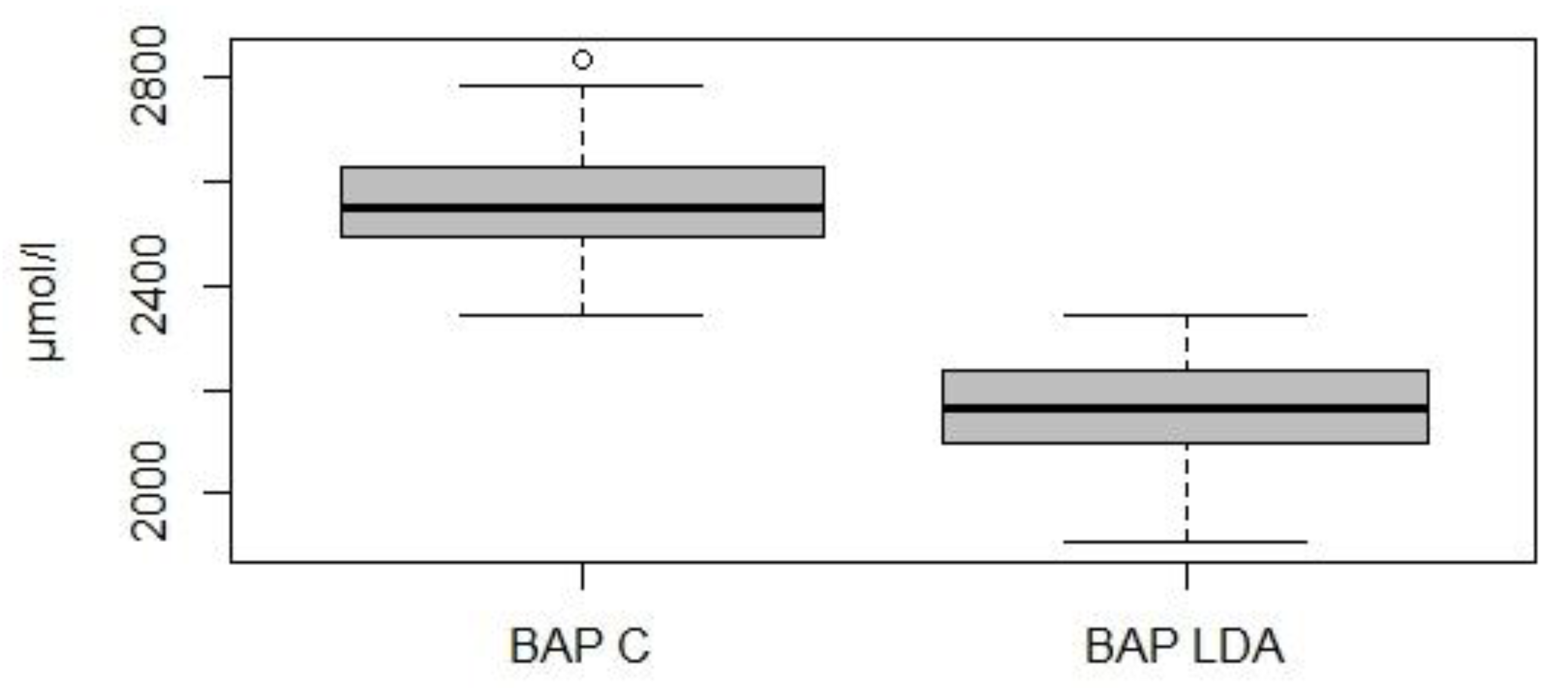
As regards the BAP test, the concentration of antioxidants was significantly lower in the study group than in the control group (2156 ± 98.1 µmol/L vs. 2558 ± 108.5 µmol/L, p < 0.05) (Figure 2).
The OSi value was significantly higher (p < 0.05) in cows with LDA than in healthy cows (8.3 ± 1.51 vs. 6.2 ± 0.76).

4. Discussion

In the present study, LDA occurred most frequently in the first and second week postpartum and in parity 4 cows, which is in accordance with other studies [4,5].
The results showed a significant elevation of d-ROMs and OSi and a significant reduction of BAP in LDA cows, confirming that LDA is linked to shifts in oxidative status [18,19,20,23].
To the best of our knowledge, there is a limited number of studies on the determination of oxidative status using d-ROMs and BAP and the calculation of OSi in ruminants [28,29,30] and, in particular, these tests have never been used in cows with left abomasal displacement. We decided to calculate OSi, in addition to d-ROMs and BAP, because, in accordance with other authors, we consider that it is more important to analyse the relationship between oxidants and antioxidants through a ratio than to evaluate their concentrations separately [31]. Durgut et al. [23] published a paper regarding the changes in oxidative status associated with displaced abomasum (DA) using total antioxidant capacity and total oxidant status (TAC and TOS) to assess oxidant status and OSi as a ratio between TAC and TOS. Other researchers studied the relationship between DA and oxidative status through the determination of the serum or plasma concentrations of different oxidant (malondialdehyde, nitric oxide, etc.) and antioxidant species (superoxide dismutase, glutathione peroxidase, vitamin E, selenium, etc.) separately [18,19,21] or total antioxidant status [32,33]. Contrary to Durgut et al. [23], significantly different d-ROMs and BAP levels were found in cows with LDA and healthy controls in our study. Hasanpour et al. [18] used other methods rather than the ones used in the current work and concluded that the antioxidant status in cows with DA was lower than that of healthy controls, similarly to the findings of our study. Aly et al. [19] observed a significant elevation in the serum levels of H2O2, nitric oxide and malondialdehyde, and a significant reduction in serum catalase and glutathione reductase activities in LDA cows, confirming the induced imbalance in oxidative status linked to LDA. Unfortunately, the fact that most of the reported investigations used different analytical techniques makes it difficult to compare the data.
The results of this study indicated that an imbalance between oxidants and antioxidants occurred in cattle with left displaced abomasum. A shift in redox balance and the acute-phase reaction, as well as the inflammatory response and tissue damage, are present but moderate in LDA, because ischemia/perfusion stimulates the production of free oxygen radicals, generating compounds such as nitric oxide and malondialdehyde [22]. In addition, it was demonstrated that dairy cows with LDA suffered from peripheric fat mobilization due to energy deficits [34] and that increased lipid mobilization may increase the generation of ROS [8], so this may partly explain the significative elevation in plasma d-ROMs in the LDA group. Furthermore, a study showed that cows that developed LDA had 45% lower serum α-tocopherol concentrations than healthy cows at the last blood sampling before LDA diagnosis [20]. This is probably due to a lower Dry Matter Intake before the diagnosis. Even after vitamin E supplementation, it was possible to evidence a decrease in α-tocopherol concentration during the first week postpartum [35,36], which is thought to result from increased lipid peroxidation and the production of reactive oxygen species, increased secretion of α-tocopherol into colostrum and milk, depressed feed intake, inflammation and decreased lipid absorption and transport [37]. A lower DMI might be a probable causative factor for the lower α-tocopherol in LDA cows [20], but most probably factors other than feed intake, such as increased oxidation, play a role in lower α-tocopherol in blood around calving [38].
Given the absence of DMI data and data on the serum α-tocopherol concentrations prior to LDA in our study, we cannot determine whether lower BAP depends on α-tocopherol concentrations, lower DMI or other factors.
The mean concentrations of d-ROMs and BAP in the control group in the present study were in agreement with the findings of other studies on healthy cows in the early postpartum [15,16]. The observed changes in free radical and antioxidant concentrations appear to represent homeorhetic processes that normally occur in early lactation [29].

5. Conclusions

In conclusion, cows with LDA experience an imbalance between prooxidants and antioxidants when compared with healthy control cows. The plasma concentration of ROS and the plasma biological antioxidant potential were measured, using the d-ROMs test and the BAP test, respectively, in cows with LDA in the first month of lactation and in healthy control herdmates. An Oxidative Status index was also calculated. The elevation of the d-ROMs and OSi values, together with the decrease of BAP values in the diseased group, indicated that LDA is linked with a shift in oxidative status. The causes of this shift and its potential role in the pathogenesis of the disease are difficult to determine and need further research. To the authors’ knowledge, this is the first study to use d-ROMs and BAP tests, with the calculation of OSi, to investigate oxidative status in multiparous cows with LDA. This study is small, with a limited number of LDA cows in a single commercial dairy herd, so larger studies are warranted to examine the role of ROS, antioxidants and redox balance in LDA.

Author Contributions

Conceptualization, methodology, data curation and writing, F.F., N.S. and R.C.; laboratory analysis, S.S.; supervision, R.C. All authors read and approved the final manuscript. F.F. and N.S. contributed equally to the work.

Funding

This research was supported by a grant from the Fondazione di Sardegna (Sardinia Foundation, Italy), Grant Number Prot. U949.2013.

Acknowledgments

The authors sincerely thank the owners and farm staff for their assistance during the work.

Conflicts of Interest

The authors declare no conflict of interest.

References

  1. Geishauser, T.; Leslie, K.; Duffield, T. Metabolic aspects in the etiology of displaced abomasum. Vet. Clin. N. Am. Food Anim. Pract. 2000, 16, 255–265. [Google Scholar] [CrossRef]
  2. McArt, J.A.; Nydam, D.V.; Overton, M.W. Hyperketonemia in early lactation dairy cattle: A deterministic estimate of component and total cost per case. J. Dairy Sci. 2015, 98, 2043–2054. [Google Scholar] [CrossRef] [PubMed]
  3. Geishauser, T. Abomasal displacement in the bovine—A review on character, occurrence, aetiology and pathogenesis. Zent. Vet. A 1995, 42, 229–251. [Google Scholar] [CrossRef]
  4. Sexton, M.; Buckley, W.; Ryan, E. A study of 54 cases of left displacement of the abomasum: February to July 2005. Ir. Vet. J. 2007, 60, 605–609. [Google Scholar] [CrossRef] [PubMed]
  5. Jorritsma, R.; Westerlaan, B.; Bierma, M.P.; Frankena, K. Milk yield and survival of Holstein-Friesian dairy cattle after laparoscopic correction of left-displaced abomasum. Vet. Rec. 2008, 162, 743–746. [Google Scholar] [CrossRef] [PubMed]
  6. Fiore, F.; Musina, D.; Cocco, R.; Di Cerbo, A.; Spissu, N. Association between left-displaced abomasum corrected with 2-step laparoscopic abomasopexy and milk production in a commercial dairy farm in Italy. Ir. Vet. J. 2018, 71, 1322. [Google Scholar] [CrossRef]
  7. Ingvartsen, K.L.; Andersen, J.B. Symposium: Dry matter intake of lactating dairy cattle. Integration of metabolism and intake regulation: A review focusing on periparturient animals. J. Dairy Sci. 2000, 83, 1573–1597. [Google Scholar] [CrossRef]
  8. Sordillo, L.M.; Raphael, W. Significance of metabolic stress, lipid mobilization, and inflammation on transition cow disorders. Vet. Clin. N. Am. Food Anim. Pract. 2013, 29, 267–278. [Google Scholar] [CrossRef]
  9. Abuelo, A.; Hernandez, J.; Benedito, J.L.; Castillo, C. The importance of the oxidative status of dairy cattle in the periparturient period: Revisiting antioxidant supplementation. J. Anim. Physiol. Anim. Nutr. 2015, 99, 1003–1016. [Google Scholar] [CrossRef]
  10. Sordillo, L.M.; Mavangira, V. The nexus between nutrient metabolism, oxidative stress and inflammation in transition cows. Anim. Prod. Sci. 2014, 54, 1204–1214. [Google Scholar] [CrossRef]
  11. Castillo, C.; Hernandez, J.; Valverde, I.; Pereira, V.; Sotillo, J.; Alonso, M.L.; Benedito, J.L. Plasma malonaldehyde (MDA) and total antioxidant status (TAS) during lactation in dairy cows. Res. Vet. Sci. 2006, 80, 133–139. [Google Scholar] [CrossRef] [PubMed]
  12. Sordillo, L.M.; Aitken, S.L. Impact of oxidative stress on the health and immune function of dairy cattle. Vet. Immunol. Immunopathol. 2009, 128, 104–109. [Google Scholar] [CrossRef] [PubMed]
  13. Lykkesfeldt, J.; Svendsen, O. Oxidants and antioxidants in disease: Oxidative stress in farm animals. Vet. J. 2007, 173, 502–511. [Google Scholar] [CrossRef] [PubMed]
  14. Merlo, M.; Celi, P.; Barbato, O.; Gabai, G. Relationships between oxidative status and pregnancy outcome in dairy cows. Anim. Prod. Aust. 2008, 27, 84. [Google Scholar] [CrossRef]
  15. Celi, P. Biomarkers of oxidative stress in ruminant medicine. Immunopharmacol. Immunotoxicol. 2011, 33, 233–240. [Google Scholar] [CrossRef]
  16. Abuelo, A.; Hernandez, J.; Benedito, J.L.; Castillo, C. Oxidative stress index (OSi) as a new tool to assess redox status in dairy cattle during the transition period Animal. Anim. Consort. 2013, 7, 1374–1378. [Google Scholar] [CrossRef]
  17. Abuelo, A.; Hernández, J.; Benedito, J.L.; Castillo, C. Redox biology in transition periods of dairy cattle: Role in the health of periparturient and neonatal animals. Antioxidants 2019, 8, 20. [Google Scholar] [CrossRef]
  18. Hasanpour, A.; Saranjam, N.; Amuoghli Tabrizi, B. Antioxidant Concentration Status in the Serum of Cows with Left Displacement Abomasom. Glob. Vet. 2011, 7, 478–481. [Google Scholar]
  19. Aly, M.A.; Saleh, N.S.; Allam, T.S.; Keshta, H.G. Evaluation of Clinical, Serum Biochemical and Oxidantantioxidant Profiles in Dairy Cows with Left Abomasal Displacement. Asian. J. Anim. Vet. Adv. 2016, 11, 242–247. [Google Scholar] [CrossRef]
  20. Qu, Y.; Lytle, K.; Traber, M.G.; Bobe, G. Depleted serum vitamin E concentrations precede left displaced abomasum in early-lactation dairy cows. J. Dairy Sci. 2013, 96, 3012–3022. [Google Scholar] [CrossRef]
  21. Mamak, N.; Devrim, A.K.; Aksit, H.; Aytekin, I.; Yıldız, R. Levels of antioxidant substances, acute phase response and lipid peroxidation in the left and right abomasum displacement in cows. Pol. J. Vet. Sci. 2013, 4, 731–733. [Google Scholar] [CrossRef]
  22. Maden, M.; Ozturk, A.S.; Bulbul, A.; Avci, G.E.; Yazar, E. Acute-Phase Proteins, Oxidative Stress, and Enzyme Activities of Blood Serum and Peritoneal Fluid in Cattle with Abomasal Displacement. J. Vet. Intern. Med. 2012, 26, 1470–1475. [Google Scholar] [CrossRef] [PubMed]
  23. Durgut, R.; Sagkan Ozturk, A.; Ozturk, O.H.; Guzel, M. Evaluation of oxidative stress, antioxidant status and lipid profile in cattle with displacement of the abomasum. Ank. Üniv. Vet. Fak. Derg. 2016, 63, 137–141. [Google Scholar]
  24. National Research Council. Nutrient Requirements of Dairy Cattle, 7th ed.; The National Academies Press: Washington, DC, USA, 2001. [Google Scholar]
  25. Sprecher, D.J.; Hostetler, D.E.; Kaneene, J.B. A lameness scoring system that uses posture and gait to predict dairy cattle reproductive performance. Theriogenology 1997, 47, 1179–1187. [Google Scholar] [CrossRef]
  26. Edmonson, A.J.; Lean, I.J.; Weaver, L.D.; Farver, T.; Webster, G. A body condition scoring chart for Holstein dairy cows. J. Dairy Sci. 1989, 72, 68–78. [Google Scholar] [CrossRef]
  27. Celi, P. Oxidative Stress in Ruminants, in: Studies on Veterinary Medicine; Mandelker, L., Vajdovich, P., Eds.; Humana Press: New York, NY, USA, 2011; Volume 5, pp. 191–231. [Google Scholar]
  28. Celi, P.; Raadsma, H.W. Effects of Yerba Mate (Ilex paraguariensis) supplementation on the productive performance of dairy cows during mid-lactation. Anim. Sci. 2010, 50, 339–344. [Google Scholar] [CrossRef]
  29. Pedernera, M.; Celi, P.; García, S.C.; Salvin, H.E.; Barchia, I.; Fulkerson, W.J. Effect of diet, energy balance and milk production on oxidative stress in early-lactating dairy cows grazing pasture. Vet. J. 2010, 186, 352–357. [Google Scholar] [CrossRef]
  30. Mirzad, A.N.; Tada, T.; Ano, H.; Kobayashi, I.; Yamauchi, T.; Katamoto, H. Seasonal changes in serum oxidative stress biomarkers in dairy and beef cows in a daytime grazing system. J. Vet. Med. Sci. 2018, 80, 20–27. [Google Scholar] [CrossRef]
  31. Castillo, C.; Hernandez, J.; Bravo, A.; Lopez-Alonso, M.; Pereira, V.; Benedito, J.L. Oxidative status during late pregnancy and early lactation in dairy cows. Vet. J. 2005, 169, 286–292. [Google Scholar] [CrossRef]
  32. Mudron, P.; Rehage, J.; Sallmann, H.P.; Mertens, M.; Scholz, H.; Kovac, G. Plasma and liver α-tocoferol in dairy cows with left abomasal displacement and fatty liver. J. Vet. Med. A 1997, 44, 91–97. [Google Scholar] [CrossRef]
  33. Ismael, M.M.; Elshahawy, I.I.; Abdullaziz, I.A. New Insights on Left Displaced Abomasum in Dairy Cows. Alex. J. Vet. Sci. 2018, 56, 127–136. [Google Scholar] [CrossRef]
  34. Mudron, P.; Herzog, K.; Höltershinken, M.; Rehage, J. Effects of Abdominal Surgery on Thiobarbituric acid Reactive Substances and Plasma Anti-oxidative Capacity in Dairy Cows. J. Vet. Med. 2007, 54, 441–444. [Google Scholar] [CrossRef] [PubMed]
  35. Meglia, G.E.; Jensen, S.K.; Lauridsen, C.; Persson Waller, K. α-Tocopherol concentration and stereoisomer composition in plasma and milk from dairy cows fed natural or synthetic vitamin E around calving. J. Dairy Res. 2006, 73, 227–234. [Google Scholar] [CrossRef] [PubMed]
  36. Weiss, W.P.; Hogan, J.S.; Wyatt, D.J. Relative bioavailability of all-rac and RRR vitamin E based on neutrophil function and total α-tocopherol and isomer concentrations in periparturient dairy cows and their calves. J. Dairy Sci. 2009, 92, 720–731. [Google Scholar] [CrossRef] [Green Version]
  37. Baldi, A. Vitamin E in dairy cows. Livest. Prod. Sci. 2005, 98, 117–122. [Google Scholar] [CrossRef]
  38. Goff, J.P.; Kimura, K.; Horst, R.L. Effect of mastectomy on milk fever, energy, and vitamins A, E, and β-carotene. J. Dairy Sci. 2002, 85, 1427–1436. [Google Scholar] [CrossRef]
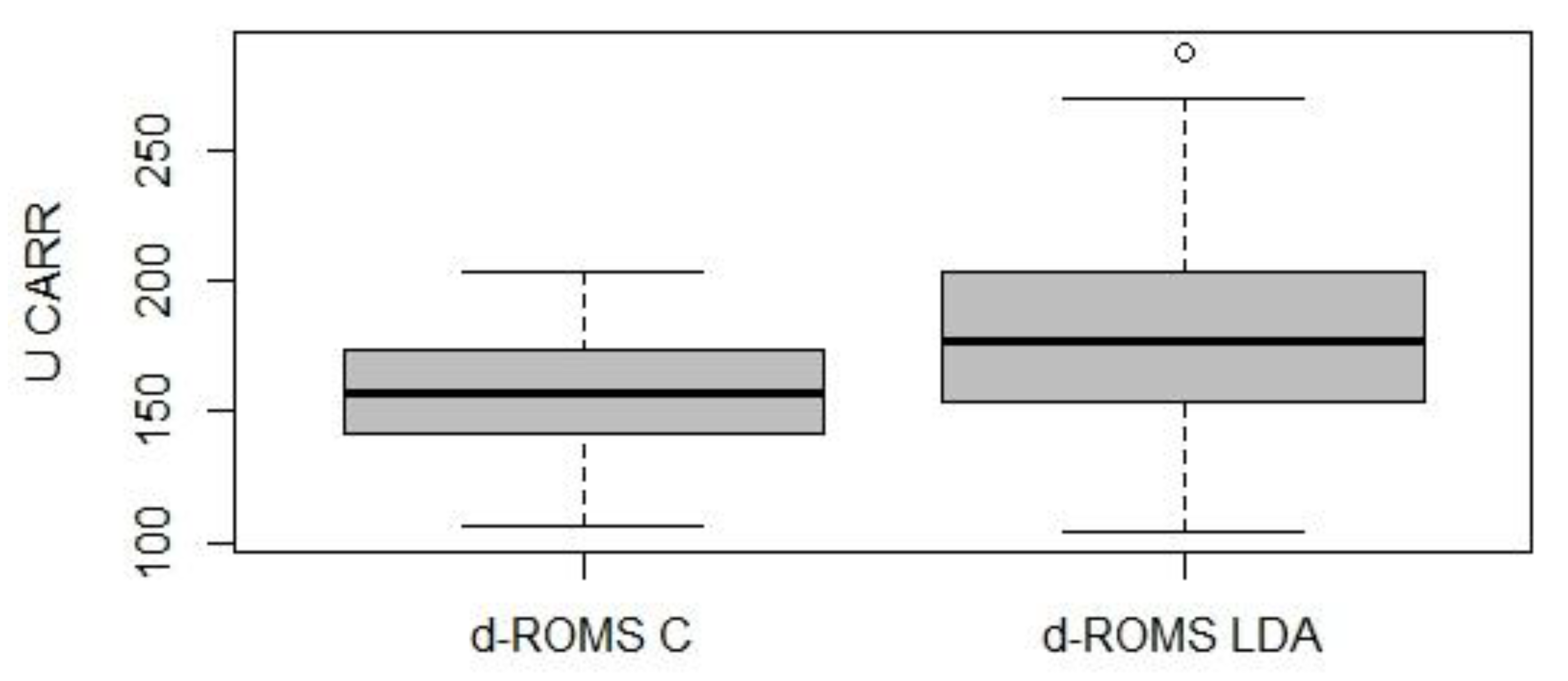
Figure 1. Graphical representation of the d-ROMs test’s values in the control group (n = 74; d-ROMS C) and in the study group (n = 74; d-ROMS LDA).
Animals 09 00966 g001
Figure 2. Graphical representation of the BAP test’s values in the control group (n = 74; BAP C) and in the study group (n = 74; BAP LDA).
Animals 09 00966 g002
Table 1. Ingredients of the Total Mixed Ration for lactating cows.
Ingredients
Grass hay34.7%
Steam flaked corn19.0%
Cane-beet molasses blend3.8%
Grain mix:42.5%
- Wheat bran29.6%
- Sorghum grain29.4%
- Soybean meal21.6%
- Flaked soybean14.7%
- Calcium carbonate2.2%
- Sodium chloride1%
- Magnesium oxide0.4%
- Sodium bentonite0.9%
- Vitamin and premix:0.3%
Vitamin A40,000 IU
Vitamin D34000 IU
Vitamin E (α-tocopherol 92%)30 mg
Vitamin B15 mg
Vitamin B23 mg
Vitamin B61.5 mg
Vitamin B120.06 mg
Vitamin K5 mg
Para-aminobenzoic acid5 mg
Vitamin PP (niacin)150 mg
Choline chloride50 mg
Fe100 mg
Co1 mg
I5 mg
Mn120 mg
Cu10 mg
Zn130 mg
Table 2. Composition of the TMR diet and the hay (mean ± Standard Deviation).
ItemTMRHay
DM, %85.77 ± 0.7188.51 ± 2.89
Ether extract, % of DM2.48 ± 0.34 1.70 ± 0.18
Ash, % of DM 9.80 ± 0.37 8.97 ± 0.66
ADF, % of DM 20.73 ± 4.18 42.21 ± 1.71
ADL, % of DM 2.89 ± 0.63 6.74 ± 0.76
Starch, % of DM 23.60 ± 4.74 1.89 ± 2.17
CP, % of DM 14.31 ± 0.88 8.63 ± 1.17
Soluble protein, % of DM 3.89 ± 0.41 3.21 ± 0.42
DM, Dry Matter; AFD, Acid Detergent Fiber; ADL, Acid Detergent Lignin; CP, Crude Protein.
Table 3. Distribution of 74 cows with Left Displacement of Abomasum (LDA) as defined by lactation number.
Lactation NumberNo. of CasesPercentage of Cases (%)
22027.0
32432.4
43040.5
Table 4. Time interval from calving to LDA diagnosis in 74 cows.
Time Interval (days)No. of CasesPercentage of Cases (%)
≤72128.4
8–142432.4
15–212027.0
22–30912.2