Multicentric Round Cell Neoplasia with Plasmacytic Differentiation in a Cat with Systemic Progression: Multimodal Imaging and Treatment Response


1
Department of Veterinary Medical Imaging, College of Veterinary Medicine, Konkuk University, Seoul 05029, Republic of Korea
2
Royal Animal Medical Center, Seoul 02140, Republic of Korea
*
Author to whom correspondence should be addressed.
Animals 2026, 16(7), 1089; https://doi.org/10.3390/ani16071089
Submission received: 6 March 2026
/
Revised: 27 March 2026
/
Accepted: 1 April 2026
/
Published: 2 April 2026
(This article belongs to the Special Issue Abdominal Imaging in Small Animals: New Insights)
Simple Summary
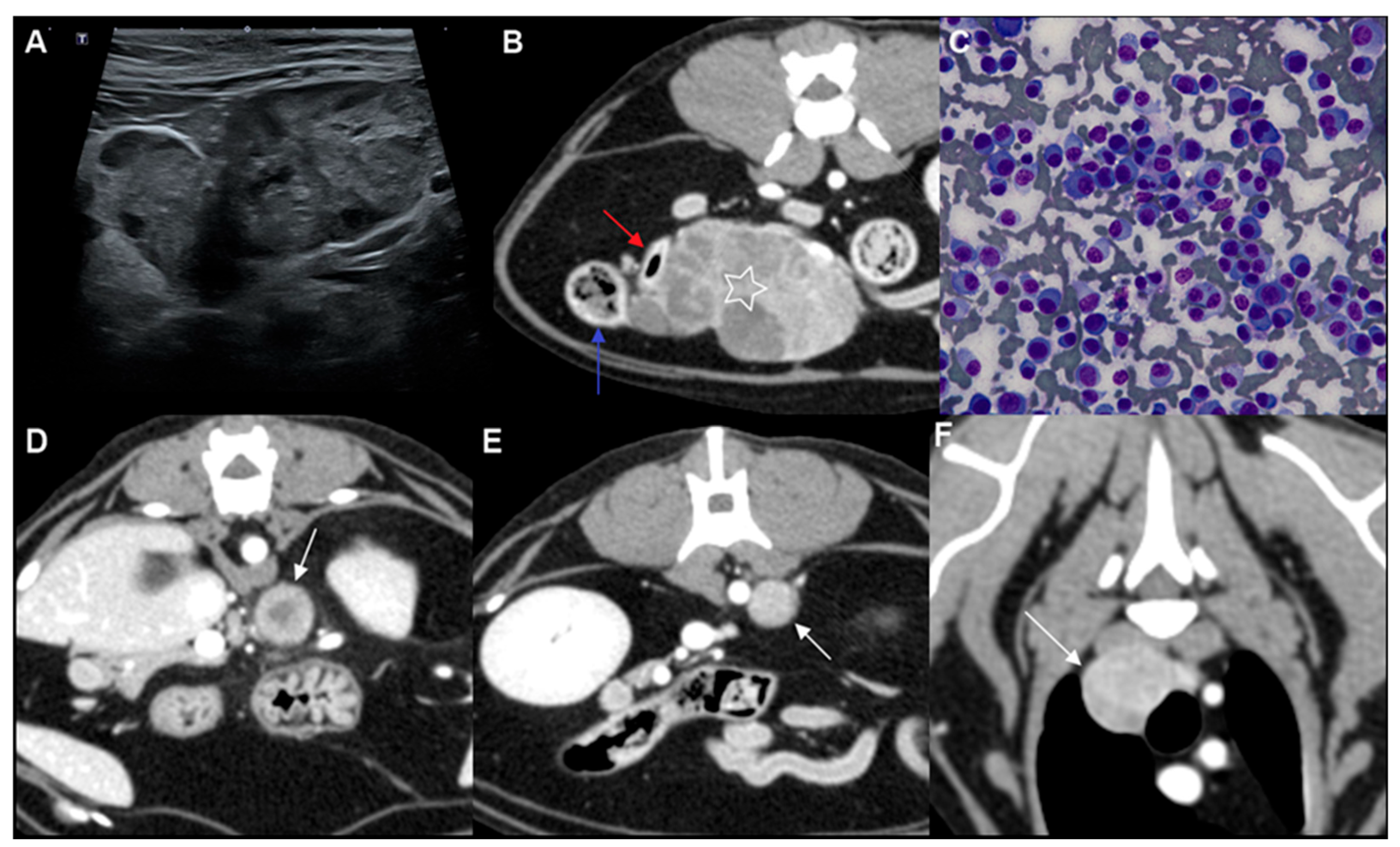
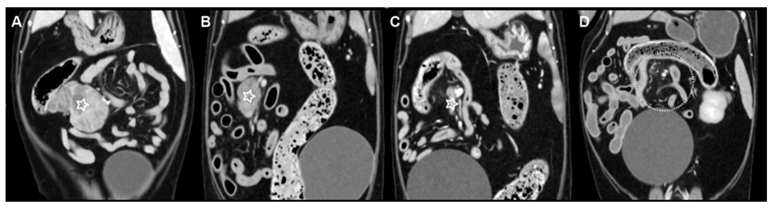
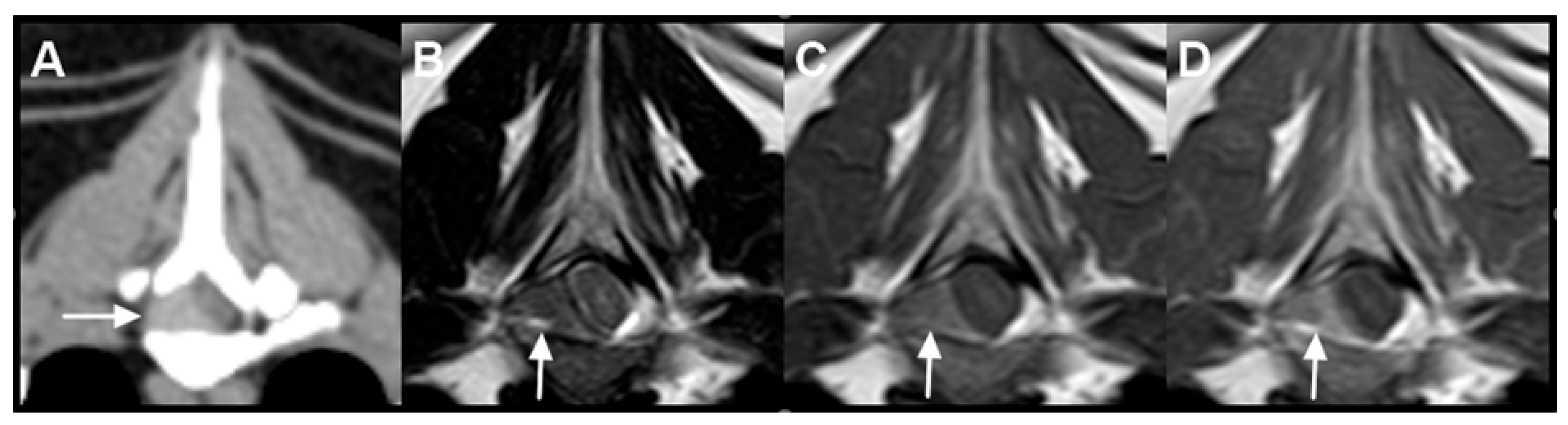
Plasma cell neoplasms are uncommon in cats, and their imaging appearance and treatment response are not well documented, particularly when disease is distributed predominantly at lymph node sites. This report describes an older cat with multiple masses in the abdomen and chest that were considered most consistent with enlarged lymph nodes on imaging. Ultrasound, computed tomography, and magnetic resonance imaging were used to determine disease extent and monitor treatment response over time. Examination of cells collected from the largest abdominal mass supported plasmacytic differentiation and raised plasma cell neoplasia as an important diagnostic consideration in addition to the more common diagnosis of lymphoma. The cat was treated with radiation therapy directed at the largest mass together with chemotherapy. Follow-up imaging showed a rapid and marked decrease in the size of the treated mass, which later became undetectable on imaging. In contrast, other presumed nodal lesions progressed, and a new mass later developed near the spinal cord. This case shows that round cell neoplasia with plasmacytic differentiation may closely resemble lymphoma on imaging studies, and that repeated imaging is valuable for evaluating treatment response and disease progression.
Abstract
Plasma cell neoplasia is uncommon in cats, and multicentric nodal-predominant involvement has not been well characterized. This report describes a multicentric round cell neoplasia with plasmacytic differentiation in a 14-year-old Domestic Shorthair cat, emphasizing multimodal imaging features and treatment response. Contrast-enhanced computed tomography and magnetic resonance imaging were performed for staging and longitudinal assessment. Cytomorphology supported plasmacytic differentiation, and flow cytometry did not demonstrate an immunophenotype consistent with conventional B- or T-cell lymphoma. Because histopathology, immunohistochemistry, bone marrow evaluation, and assessment for monoclonal gammopathy were not performed, definitive classification was not possible; however, cytomorphology supported plasmacytic differentiation, with plasma cell neoplasia remaining an important diagnostic consideration. A hypofractionated radiotherapy protocol (36 Gy in six fractions) combined with systemic chemotherapy was administered. Serial imaging demonstrated complete radiologic resolution of the irradiated mass, whereas non-irradiated presumed nodal lesions progressed and an extradural spinal lesion subsequently developed. These findings highlight the capacity of round cell neoplasia with plasmacytic differentiation to mimic lymphoma on imaging and illustrate the dissociation between effective local control and ongoing systemic progression.