Regional Microarchitecture of the Skin of the Arabian Carpetshark (Chiloscyllium arabicum): A Histological Study Using Polarised Light Microscopy


 ,
,  and
and
Simple Summary
Abstract
1. Introduction
2. Materials and Methods
2.1. Animals
2.2. Ethical Statement
2.3. Histological Analysis
2.4. Analysis of Collagen Fibre Organisation Under Polarised Light
3. Results
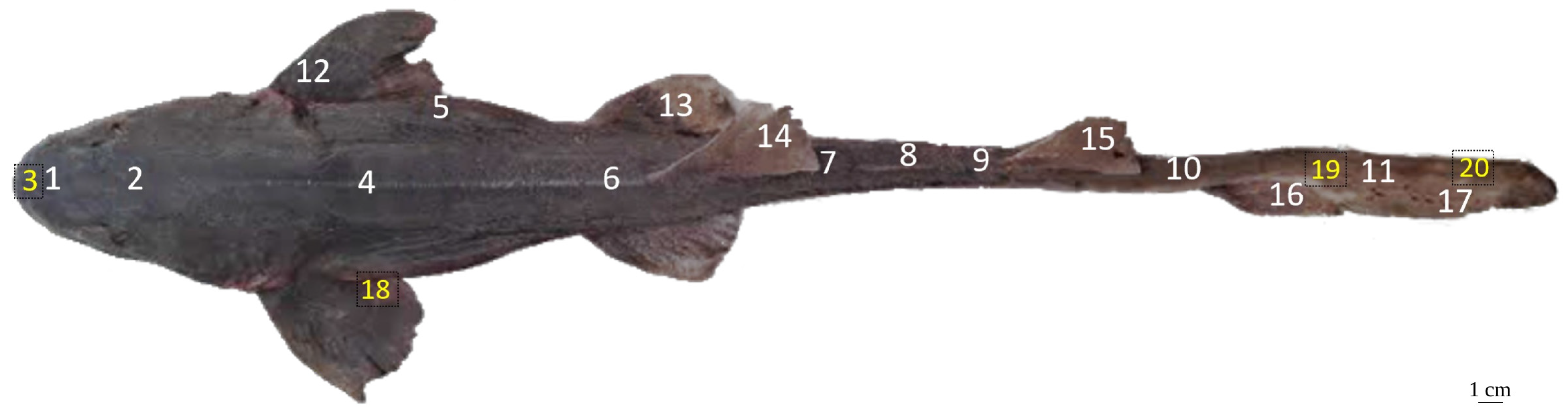
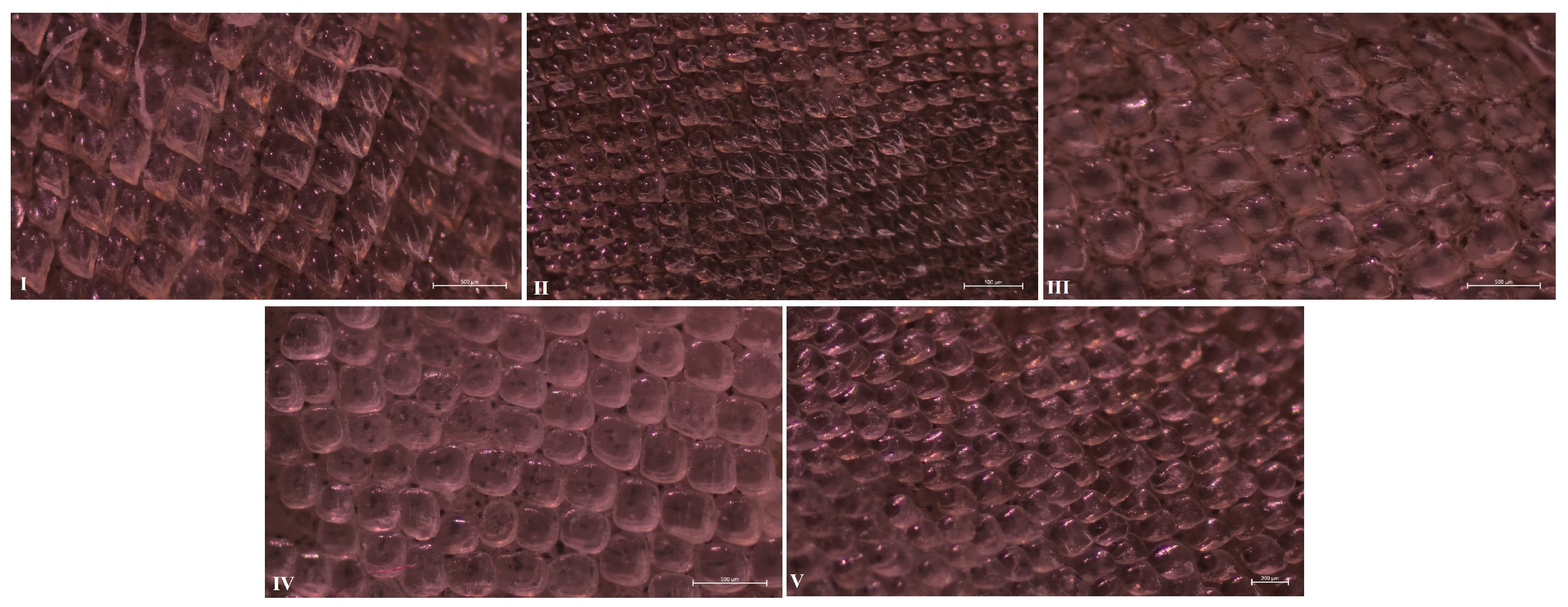
3.1. Dermal Denticle Morphotypes and Morphometrics
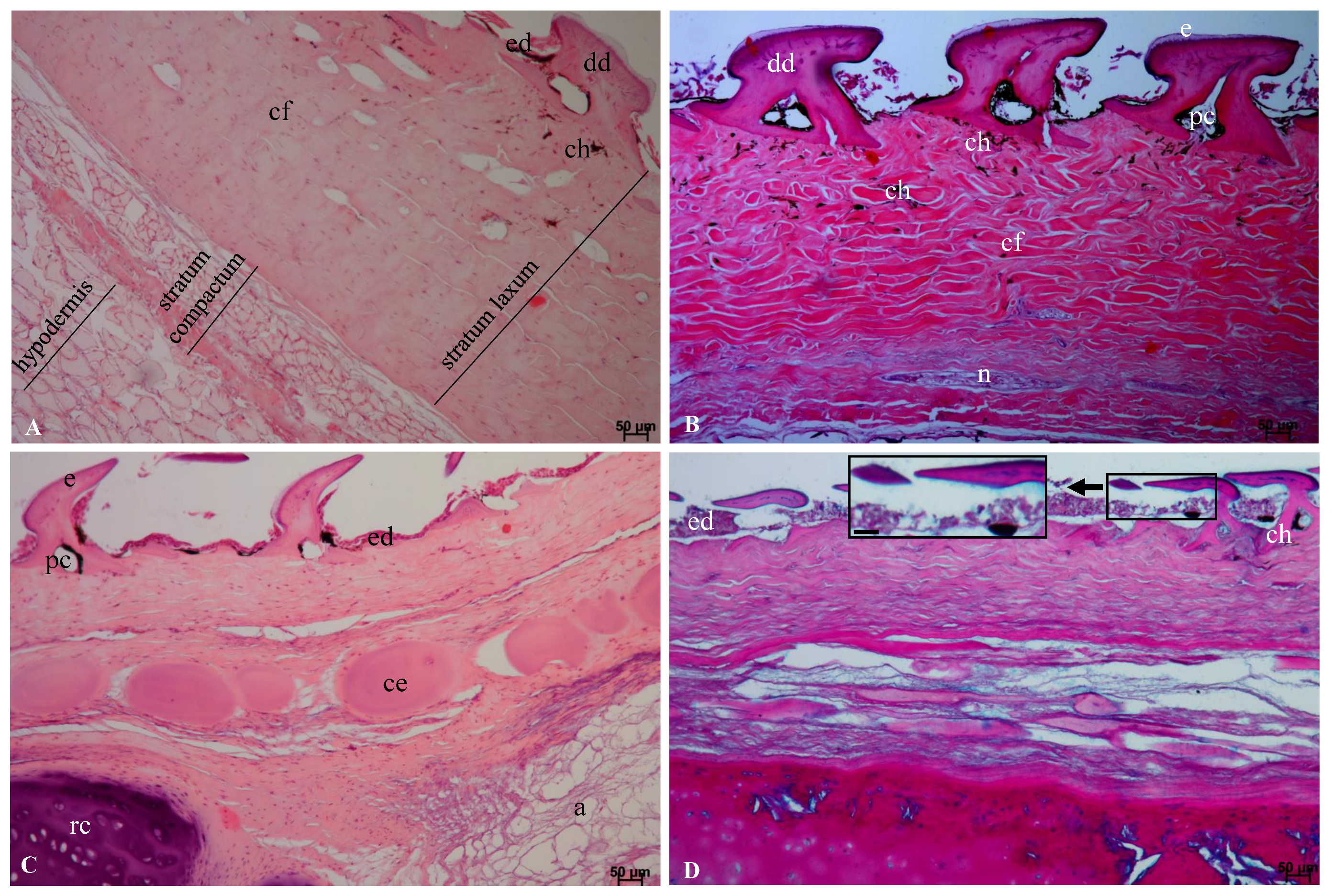
3.2. General Histological Structure of the Integument
3.2.1. Epidermis
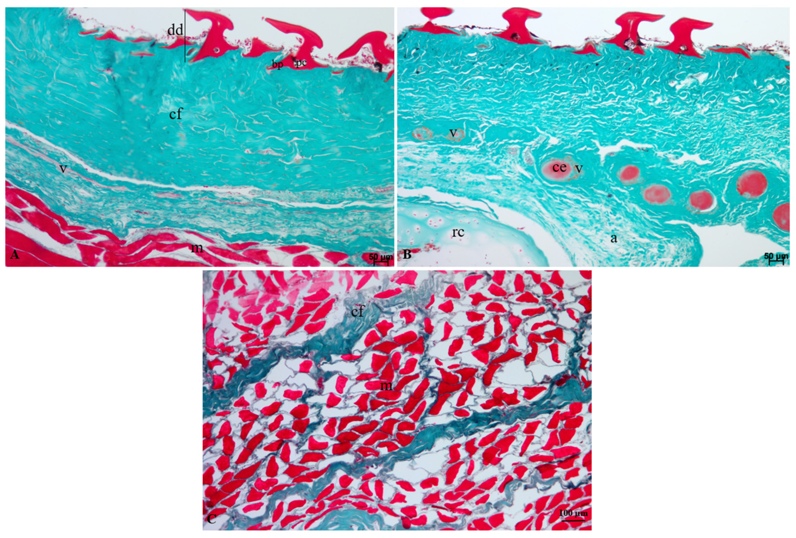
3.2.2. Dermis and Subcutaneous Layer
3.3. Organisation of Collagen Fibres
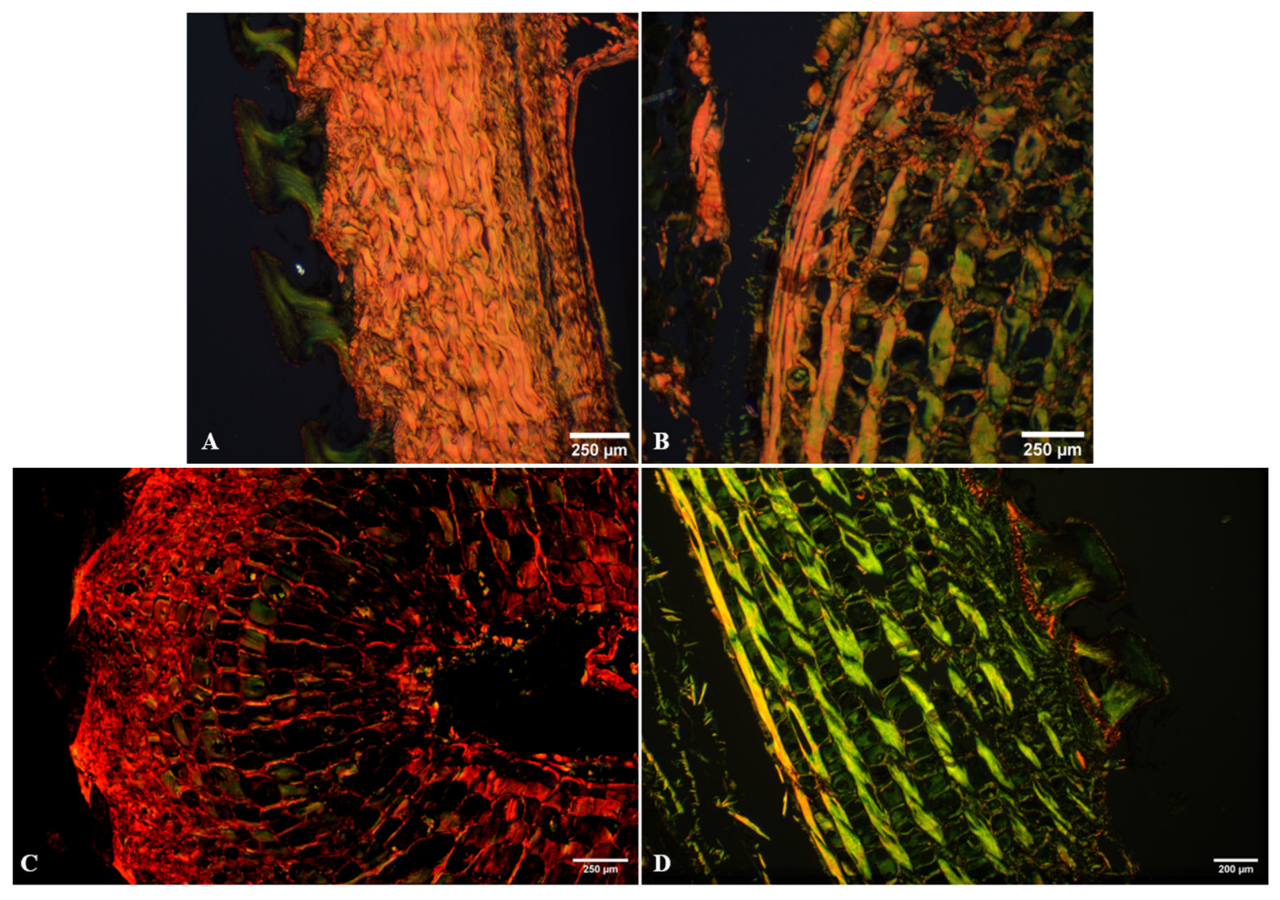
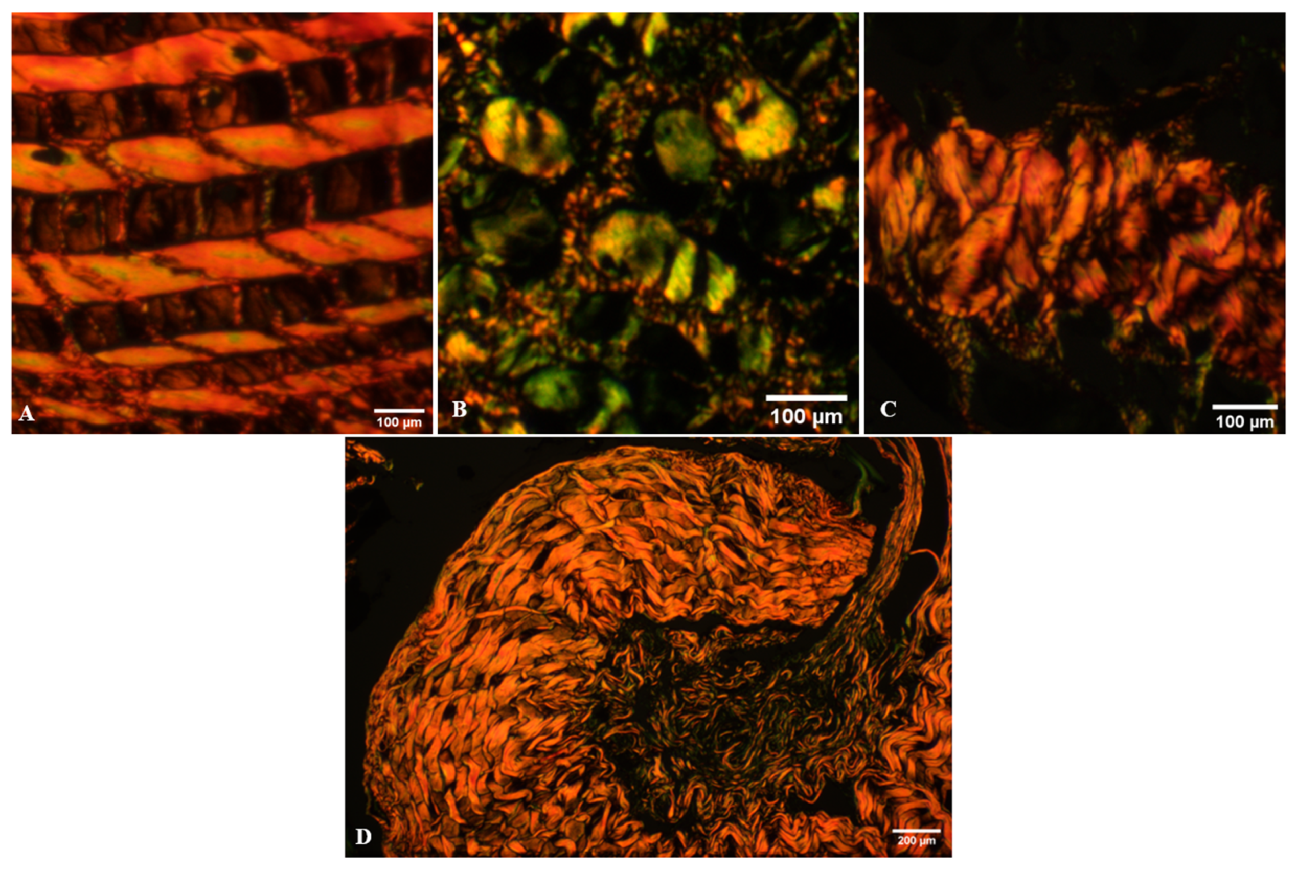
3.3.1. Regions Exhibiting Periodic Fibre Organisation
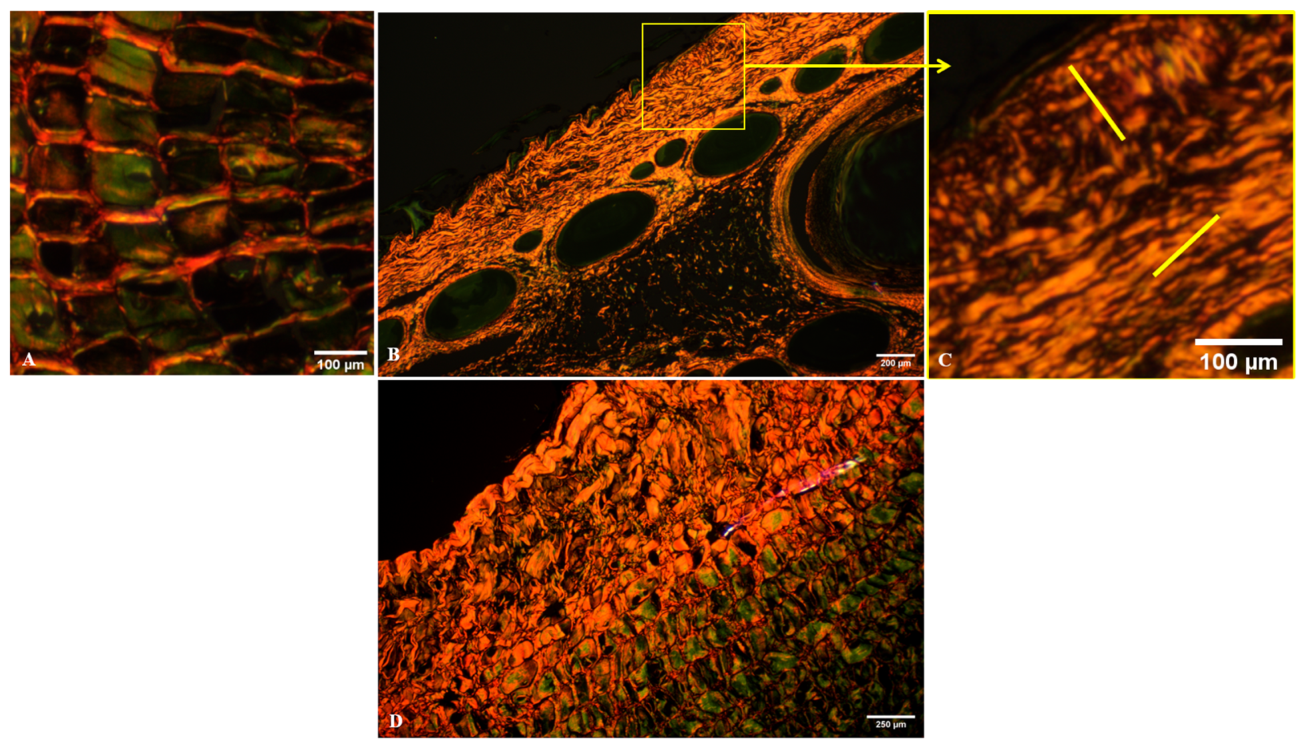
3.3.2. Regions with Interwoven Fibre Organisation
3.3.3. Collagen Organisation Associated with Cells and Cartilage
3.3.4. Layered Organisation of the Skin
3.4. Regional Co-Occurrence of Dermal Denticle Morphotypes and Collagen Organisation
4. Discussion
5. Conclusions
Author Contributions
Funding
Institutional Review Board Statement
Informed Consent Statement
Data Availability Statement
Conflicts of Interest
References
- Reif, W.-E. Squamation and Ecology of Sharks; Courier Forschungsinstitut Senckenberg; Senckenberg Gesellschaft für Naturforschung: Frankfurt am Main, Germany, 1985; Volume 78, pp. 1–5. [Google Scholar]
- Kemp, N.E. Integumentary system and teeth. In Sharks, Skates, and Rays: The Biology of Elasmobranch Fishes; Hamlett, W.C., Ed.; The Johns Hopkins University Press: Baltimore, MD, USA, 1999; pp. 43–68. [Google Scholar]
- Motta, P.J. Anatomy and functional morphology of dermal collagen fibers in sharks. Copeia 1977, 1977, 454–464. [Google Scholar] [CrossRef]
- Motta, P.J.; Habegger, M.L.; Lang, A.; Hueter, R.E.; Davis, R.L. Scale morphology and flexibility in the shortfin mako Isurus oxyrinchus and the blacktip shark Carcharhinus limbatus. J. Morphol. 2012, 273, 1096–1110. [Google Scholar] [CrossRef] [PubMed]
- Dahl, R.B.; Ghimire, A.; Iizuka, Y.; Huang, J.-P.; Møller, P.R.; Mai, G.-S.; Lin, W.-C.; Yang, C.-C.; Chou, Y.-C.; Huang, T.-H.; et al. Diverse mechanical properties, composition, and performance of shark dermal denticles. Nat. Commun. 2026, 17, 3668. [Google Scholar] [CrossRef] [PubMed]
- Ghimire, A.; Dahl, R.B.; Shen, S.-F.; Chen, P.-Y. Shark skin denticles: From morphological diversity to multi-functional adaptations and applications. Adv. Funct. Mater. 2024, 34, 2307121. [Google Scholar] [CrossRef]
- Hagood, M.E.; Alexander, J.R.S.; Porter, M.E. Relationships in shark skin: Mechanical and morphological properties vary between sexes and among species. Integr. Comp. Biol. 2023, 63, 1154–1167. [Google Scholar] [CrossRef] [PubMed]
- Hagood, M.E.; Alexander, J.R.S.; Passerotti, M.; Porter, M.E. Ecomorphology and ontogeny modulate the mechanical properties of shark skin. Acta Biomater. 2025, 204, 470–486. [Google Scholar] [CrossRef] [PubMed]
- Schuitema, O.; Motta, P.J.; Gelsleichter, J.; Horton, M.; Habegger, M.L. Histological comparison of shark dermis across various ecomorphologies. Anat. Rec. 2025, 308, 1463–1479. [Google Scholar] [CrossRef] [PubMed]
- Creager, S.B.; Porter, M.E. Stiff and tough: A comparative study on the tensile properties of shark skin. Zoology 2018, 126, 154–163. [Google Scholar] [CrossRef] [PubMed]
- Raschi, W.G.; Musick, J.A. Hydrodynamic Aspects of Shark Scales; NASA Contractor Report 3963; National Aeronautics and Space Administration: Washington, DC, USA, 1986; pp. 1–110.
- Oeffner, J.; Lauder, G.V. The hydrodynamic function of shark skin and two biomimetic applications. J. Exp. Biol. 2012, 215, 785–795. [Google Scholar] [CrossRef] [PubMed]
- Bechert, D.W.; Bruse, M.; Hage, W.; Meyer, R. Fluid mechanics of biological surfaces and their technological application. Naturwissenschaften 2000, 87, 157–171. [Google Scholar] [CrossRef] [PubMed]
- Wen, L.; Weaver, J.C.; Lauder, G.V. Biomimetic shark skin: Design, fabrication and hydrodynamic function. J. Exp. Biol. 2014, 217, 1656–1666. [Google Scholar] [CrossRef] [PubMed]
- Popp, M.; White, C.F.; Bernal, D.; Wainwright, D.K.; Lauder, G.V. The denticle surface of thresher shark tails: Three-dimensional structure and comparison to other pelagic species. J. Morphol. 2020, 281, 938–955. [Google Scholar] [CrossRef] [PubMed]
- Gabler-Smith, M.K.; Lauder, G.V. Ridges and riblets: Shark skin surfaces versus biomimetic models. Front. Mar. Sci. 2022, 9, 975062. [Google Scholar] [CrossRef]
- Domel, A.G.; Saadat, M.; Weaver, J.C.; Haj-Hariri, H.; Bertoldi, K.; Lauder, G.V. Shark skin-inspired designs that improve aerodynamic performance. J. R. Soc. Interface 2018, 15, 20170828. [Google Scholar] [CrossRef] [PubMed]
- Long, J.H., Jr.; Nipper, K.S. The importance of body stiffness in undulatory propulsion. Am. Zool. 1996, 36, 678–694. [Google Scholar] [CrossRef]
- Kelly, D.A.; Wainwright, S.A.; Gosline, J.M. Mechanical properties of fish skin. J. Exp. Biol. 1981, 90, 27–38. [Google Scholar]
- Ankhelyi, M.V.; Wainwright, D.K.; Lauder, G.V. Diversity of dermal denticle structure in sharks: Skin surface roughness and three-dimensional morphology. J. Morphol. 2018, 279, 1132–1154. [Google Scholar] [CrossRef] [PubMed]
- Gabler-Smith, M.K.; Wainwright, D.K.; Wong, G.A.; Lauder, G.V. Dermal denticle diversity in sharks: Novel patterns on the interbranchial skin. Integr. Org. Biol. 2021, 3, obab034. [Google Scholar] [CrossRef] [PubMed]
- Raschi, W.; Tabit, C. Functional aspects of placoid scales: A review and update. Mar. Freshw. Res. 1992, 43, 123–147. [Google Scholar] [CrossRef]
- Compagno, L.J.V. Sharks of the World. In An Annotated and Illustrated Catalogue of Shark Species Known to Date. Volume 2. Bullhead, Mackerel and Carpet Sharks; FAO Species Catalogue for Fishery Purposes No. 1, Vol. 2; FAO: Rome, Italy, 2001. [Google Scholar]
- Alhajji, A.H.; Hsu, H.H.; Alkhamis, Y.A.; Alsaqufi, A.S.; Khan, S.A.; Nazeer, Z. Maturity and reproduction in the Arabian carpet shark, Chiloscyllium arabicum, from the Saudi Arabian waters of the Arabian Gulf. Mar. Biol. Res. 2022, 18, 361–371. [Google Scholar] [CrossRef]
- Wainwright, S.A.; Biggs, W.D.; Currey, J.D.; Gosline, J.M. Mechanical Design in Organisms; Princeton University Press: Princeton, NJ, USA, 1976. [Google Scholar]
- Elliott, D.G. The Skin: The Many Functions of Fish Integument. In Encyclopedia of Fish Physiology: From Genome to Environment; Farrell, A.P., Ed.; Academic Press: San Diego, CA, USA, 2011; Volume 1, pp. 471–475. [Google Scholar] [CrossRef]
- Moore, A. Chiloscyllium arabicum; The IUCN Red List of Threatened Species; IUCN: Gland, Switzerland, 2017; p. T161426A109902537. [Google Scholar] [CrossRef]
- Suvarna, S.K.; Layton, C.; Bancroft, J.D. (Eds.) Bancroft’s Theory and Practice of Histological Techniques, 8th ed.; Elsevier: Amsterdam, The Netherlands, 2018. [Google Scholar]
- Savi, F.M.; Brierly, G.I.; Baldwin, J.; Theodoropoulos, C.; Woodruff, M.A. Comparison of different decalcification methods using rat mandibles as a model. J. Histochem. Cytochem. 2017, 65, 705–722. [Google Scholar] [CrossRef] [PubMed]
- Kiernan, J.A. Histological and Histochemical Methods: Theory and Practice, 5th ed.; Scion Publishing: Banbury, UK, 2015. [Google Scholar]
- Rittié, L. Method for Picrosirius red–polarization detection of collagen fibers in tissue sections. Methods Mol. Biol. 2017, 1627, 395–407. [Google Scholar] [CrossRef] [PubMed]
- Junqueira, L.C.U.; Bignolas, G.; Brentani, R.R. Picrosirius staining plus polarization microscopy, a specific method for collagen detection in tissue sections. Histochem. J. 1979, 11, 447–455. [Google Scholar] [CrossRef] [PubMed]
- Montes, G.S.; Junqueira, L.C.U. The use of the Picrosirius-polarization method for the study of the biopathology of collagen. Mem. Inst. Oswaldo Cruz 1991, 86, 1–11. [Google Scholar] [CrossRef] [PubMed]
- Schneider, C.A.; Rasband, W.S.; Eliceiri, K.W. NIH Image to ImageJ: 25 years of image analysis. Nat. Methods 2012, 9, 671–675. [Google Scholar] [CrossRef] [PubMed]
- Kittiphattanabawon, P.; Benjakul, S.; Visessanguan, W.; Kishimura, H.; Shahidi, F. Isolation and characterisation of collagen from the skin of brownbanded bamboo shark (Chiloscyllium punctatum). Food Chem. 2010, 119, 1519–1526. [Google Scholar] [CrossRef]
- Lloyd, C.J.; Peakall, J.; Burns, A.; Keevil, G.; Dorrell, R.; Wignall, P.; Fletcher, T.M. Hydrodynamic efficiency in sharks: The combined role of riblets and denticles. Bioinspir. Biomim. 2021, 16, 046008. [Google Scholar] [CrossRef] [PubMed]
- Sayama, S.; Natsuhara, M.; Shinohara, G.; Maeda, M.; Tanaka, H. Three-dimensional shape of natural riblets in the white shark: Relationship between the denticle morphology and swimming speed of sharks. J. R. Soc. Interface 2024, 21, 20240063. [Google Scholar] [CrossRef] [PubMed]
- Sullivan, T.; Regan, F.; Clarke, C.; Kelleher, B. The characterization, replication and testing of dermal denticles of Scyliorhinus canicula for physical mechanisms of biofouling prevention. Bioinspir. Biomim. 2011, 6, 046001. [Google Scholar] [CrossRef] [PubMed]
- Díez, G.; Soto, M.; Blanco, J.M. Biological characterization of the skin of shortfin mako shark Isurus oxyrinchus and preliminary study of the hydrodynamic behaviour through computational fluid dynamics. J. Fish Biol. 2015, 87, 123–137. [Google Scholar] [CrossRef] [PubMed]
- Zhang, C.; Gao, M.; Liu, G.; Zheng, Y.; Xue, C.; Shen, C. Relationship between skin scales and the main flow field around the shortfin mako shark Isurus oxyrinchus. Front. Bioeng. Biotechnol. 2022, 10, 742437. [Google Scholar] [CrossRef] [PubMed]
- Patricia, F.W.; Guzman, D.; Inigo, B.; Urtzi, I.; Maria, B.J.; Manu, S. Morphological characterization and hydrodynamic behavior of shortfin mako shark (Isurus oxyrinchus) dorsal fin denticles. J. Bionic Eng. 2019, 16, 730–741. [Google Scholar] [CrossRef]



| Morphotype | Body Regions | The Geometry of the Crown | Cusp | Surface Ornamentation | Differentiated Crown–Neck–Base | Arrangement of Denticles | Description of the Denticles |
|---|---|---|---|---|---|---|---|
| I (elongated ridge -bearing) | 1, 2, 4, 6–10 (dorsal) | Elongated, anisotropic, laterally flattened | Present, directed caudally | Prominent longitudinal ridges (median and lateral) inter-ridge grooves, and surface micro-ornamentation | Well-defined | Imbricated | Pronounced anteroposterior asymmetry, with directional organisation of the crown |
| II (flattened tile-like) | 5 (lateral) | Low-profile, isotropic, rhomboidal to sub-rectangular | None | Smooth or very subtle microtexture | Reduced (lacking a distinct neck) | Tessellated (mosaic-like) with edge-to-edge contact | Flattened crown, lacking directional structures |
| III (dome-shaped fin type) | 11, 12, 13–15 (fins) | Low-profile to moderately domed, rounded, rounded-quadrangular | None | Smooth or very subtle microtexture | Limited; neck short or undifferentiated | Regular rows, parallel to the body axis | Domed crown; regional variability (more rounded vs. more regular) |
| IV (reduced plate-like) | 3, 18–20 (ventral) | Very low-profile, low-domed, isotropic | None | Smooth, lacking ornamentation | Highly reduced (fused crown–base, neck absent) | Variable: ranging from densely packed to dispersed | Morphological reduction |
| V (transitional reduced) | 16, 17 (transitional) | Low-profile, irregular and frequently asymmetrical | None | Smooth | Poorly defined | Irregular, with variable orientation | Intermediate form |
| Anatomical Region | Organisation Type | Main Features | Parameters (Selected) |
|---|---|---|---|
| Anterior to mid dorsal region | Anisotropic | Parallel bundles | ~80.85° ± 8.26°; ~250 μm |
| Dorsal region beneath the denticles | Multi-layered cross-ply | Oblique interweaving | ~45°; ~70 μm |
| Dorsal region | Periodic (2 populations) | Thick + thin fibres | ~75/~15 μm; ~100/~12 μm |
| Transitional dorsal region | Periodic (mixed) | Interweaving scales | ~80/~15 μm; ~45 μm |
| Caudodorsal region | Interwoven (multiscale) | Multiscale network | 7–100 μm |
| Ventral region | Interwoven (intertwined) | Network + spaces | ~15 μm; ~90 μm; ~40° |
| Nasal region | Periodic (crimped/undulated) | Fibre undulation | ~−73.5°; 6–8 μm; ~140 μm |
| Fins regions | Orthogonal | Layers 0°/90° | ~200 + ~200 μm |
| Deep trunk tissues | Plywood-like | Layered arrangement | ~950 + ~1200 μm |
Disclaimer/Publisher’s Note: The statements, opinions and data contained in all publications are solely those of the individual author(s) and contributor(s) and not of MDPI and/or the editor(s). MDPI and/or the editor(s) disclaim responsibility for any injury to people or property resulting from any ideas, methods, instructions or products referred to in the content. |
© 2026 by the authors. Licensee MDPI, Basel, Switzerland. This article is an open access article distributed under the terms and conditions of the Creative Commons Attribution (CC BY) license.
Share and Cite
Lipińska, A.; Tarnowska, M.; Cyprych, K.; Bednarska, M.; Kordas, J.; Janeczek, M.; Kuropka, P. Regional Microarchitecture of the Skin of the Arabian Carpetshark (Chiloscyllium arabicum): A Histological Study Using Polarised Light Microscopy. Animals 2026, 16, 2118. https://doi.org/10.3390/ani16142118
Lipińska A, Tarnowska M, Cyprych K, Bednarska M, Kordas J, Janeczek M, Kuropka P. Regional Microarchitecture of the Skin of the Arabian Carpetshark (Chiloscyllium arabicum): A Histological Study Using Polarised Light Microscopy. Animals. 2026; 16(14):2118. https://doi.org/10.3390/ani16142118
Chicago/Turabian StyleLipińska, Anna, Małgorzata Tarnowska, Konrad Cyprych, Małgorzata Bednarska, Jakub Kordas, Maciej Janeczek, and Piotr Kuropka. 2026. "Regional Microarchitecture of the Skin of the Arabian Carpetshark (Chiloscyllium arabicum): A Histological Study Using Polarised Light Microscopy" Animals 16, no. 14: 2118. https://doi.org/10.3390/ani16142118
APA StyleLipińska, A., Tarnowska, M., Cyprych, K., Bednarska, M., Kordas, J., Janeczek, M., & Kuropka, P. (2026). Regional Microarchitecture of the Skin of the Arabian Carpetshark (Chiloscyllium arabicum): A Histological Study Using Polarised Light Microscopy. Animals, 16(14), 2118. https://doi.org/10.3390/ani16142118