3. Discussion
Scientific reports of glaucoma in non-domestic large felids are scarce. In tigers (
Panthera tigris), two cases have been documented: unilateral secondary glaucoma with anterior staphyloma in a 22-year-old circus-rescued male tiger with bilateral intraocular pressures of 90 and 88 mmHg [
17], and unilateral congenital glaucoma with goniodysgenesis in an 8-month-old female tiger with a rebound-tonometry value of 70 mmHg in the affected eye [
18]. In lions (
Panthera leo), secondary glaucoma has been reported in association with anterior uveal neoplasia, including a buphthalmic globe caused by anterior uveal melanoma in a 19-year-old lioness [
19], and diffuse iris melanoma with buphthalmos, corneal edema, and an intraocular mass occupying the ciliary cleft in a 12-year-old male African lion [
20]. Scientific case reports of glaucoma in the snow leopard (
Panthera uncia), leopard (
Panthera pardus), and puma (
Puma concolor) could not be identified in the indexed veterinary literature available to the authors.
Normative intraocular-pressure data for
Panthera tigris are limited to the study of Owens et al. [
14], in which 33 eyes of 17 clinically normal adult tigers, with subspecies not specified, under general anesthesia yielded a mean ± SD applanation-tonometry IOP of 14.7 ± 2.69 mmHg. Subspecies-specific ophthalmic reference data for the Siberian tiger (
Panthera tigris altaica) are not available in the scientific literature to the authors’ knowledge; therefore, comparison between the present case and the published adult-tiger data must be interpreted as a cross-subspecies comparison.
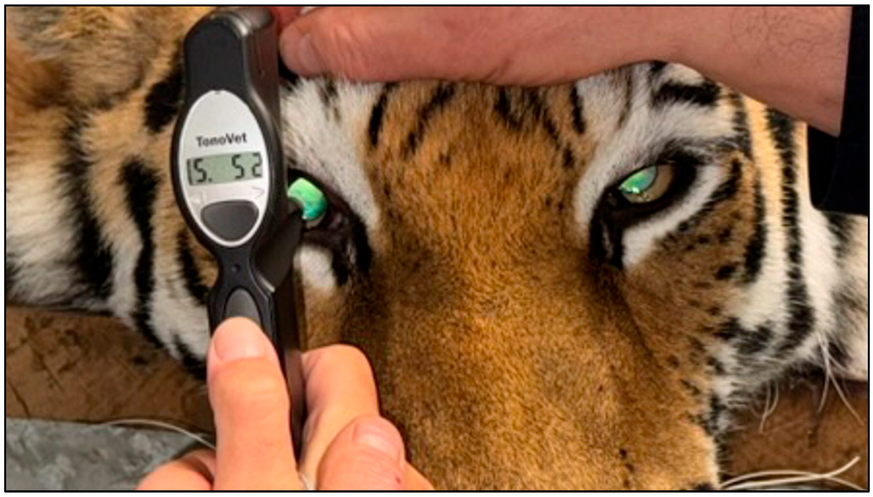
To contextualize the rebound-tonometry values of 48 mmHg and 52 mmHg recorded in the present Siberian tiger, comparison with published feline intraocular-pressure reference data obtained with the same class of instrument is informative. In clinically normal domestic cats (
Felis catus), Rusanen et al. [
21] established a mean IOP of 20.74 mmHg using the TonoVet
® rebound tonometer in a cohort of 100 cats, with a concurrent applanation-tonometer mean of 18.4 mmHg; the authors explicitly reported that rebound-tonometry values exceeded applanation values by 2–3 mmHg, a systematic offset within clinically acceptable limits. Kovalcuka et al. [
22] subsequently reported a TonoVet
® mean IOP of 18.88 ± 3.98 mmHg in 20 clinically healthy cats. The TonoVet
® has also been validated against direct manometry in a cannulation study performed in normal and glaucomatous cats across the clinically relevant range of 5–70 mmHg, with readings generally 2–5 mmHg higher than true manometric pressure [
23], a finding consistent with the newer TONOVET Plus
® data reported for normal and glaucomatous cats [
24].
Even after accounting for the typical 2–5 mmHg positive offset of rebound tonometry in felids, the IOP values recorded in the present case exceeded all published normal feline and adult-tiger reference ranges by more than twofold and were therefore consistent with severe glaucoma. Terminally elevated glaucomatous IOPs in the range of 70–90 mmHg have been reported in the two published tiger cases [
17,
18], and buphthalmic, blind end-stage globes with secondary glaucoma have been reported in lions [
19,
20]. The physiological IOP range of the Siberian tiger measured by rebound tonometry remains to be established.
In veterinary ophthalmology, glaucoma is classified as primary or secondary based on the underlying cause of impaired aqueous humor outflow, and as open-angle or closed-angle based on the morphology of the iridocorneal angle [
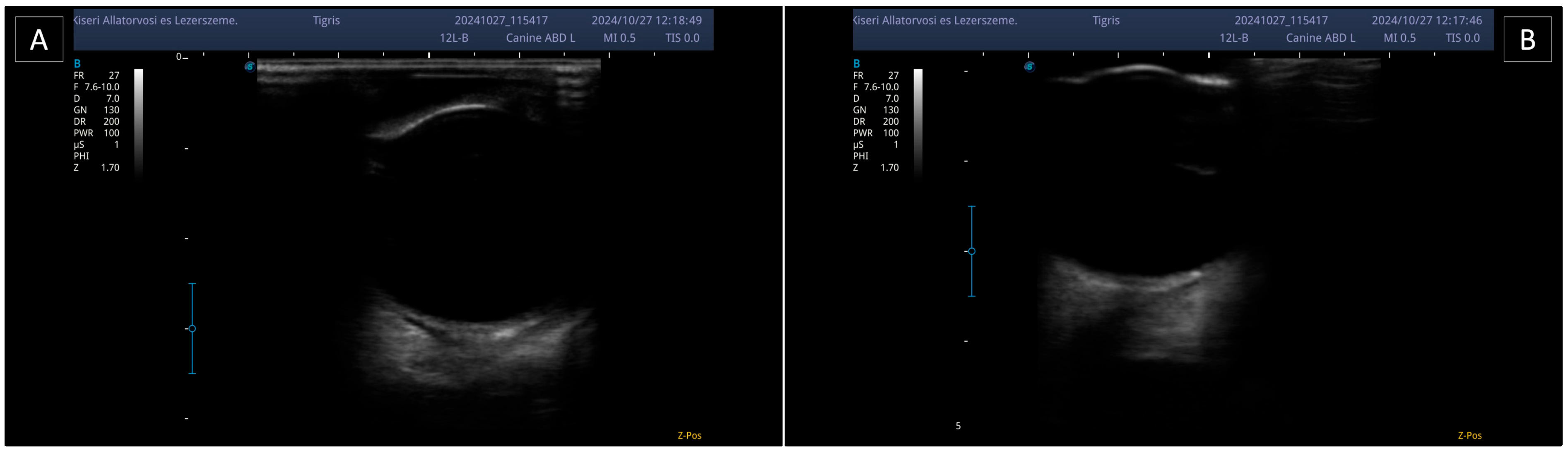
2]. In the present case, the absence of prior ocular disease, such as chronic uveitis, intumescent cataract, or intraocular neoplasia, supported a primary etiology. Biomicroscopy performed with a portable slit lamp and B-mode ultrasonography suggested that the entry to the ciliary cleft remained open. In the absence of direct gonioscopic evaluation, this finding, together with the elevated intraocular pressure and fundic changes, supported the diagnosis of open-angle glaucoma.
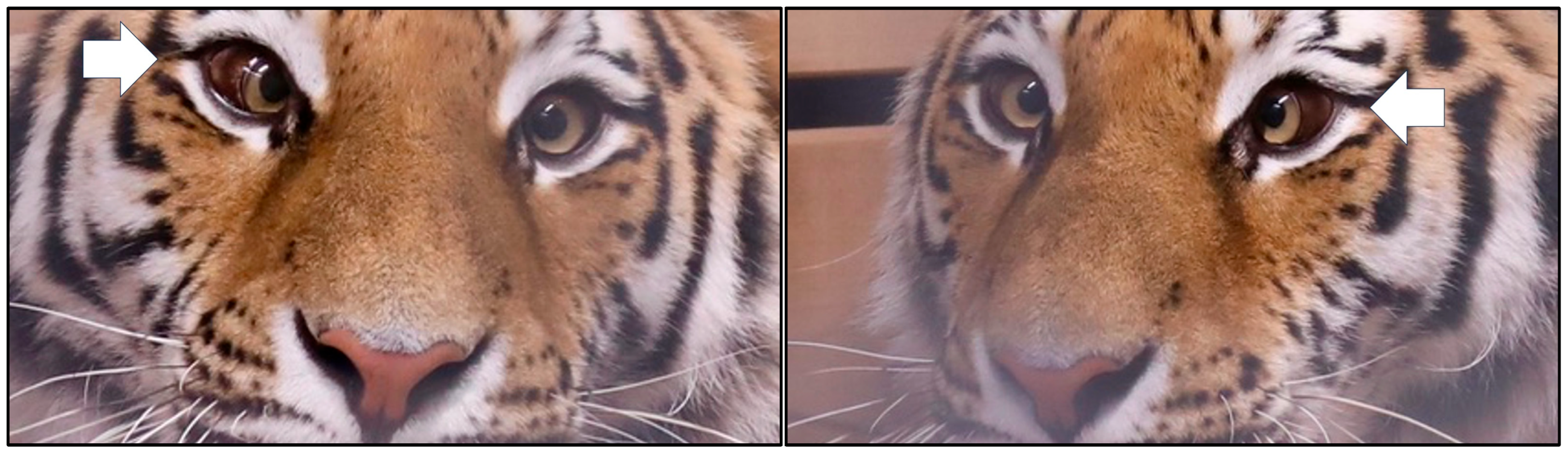
The clinical diagnosis was well supported by the markedly elevated intraocular pressure, irreversible bilateral blindness, bilateral buphthalmos, and extensive retinopathy, all consistent with advanced glaucomatous disease inferred from functional and indirect structural evidence; however, in vivo determination of the exact morphological subtype of the glaucoma was subject to methodological limitations. In the absence of ultrasound biomicroscopy (UBM), analysis of the microstructure of the iridocorneal angle (e.g., subtle changes in the trabecular meshwork or the pectinate ligament) was not feasible. High-frequency ultrasound and UBM can provide detailed visualization of the iridocorneal angle and have become established non-invasive modalities in domestic species [
16]; their application in non-domestic felids, however, is largely limited by the need for specialized equipment. The pathogenesis of primary open-angle glaucoma typically involves a gradual increase in aqueous humor outflow resistance within the trabecular meshwork, often attributed to biochemical or ultrastructural changes in the extracellular matrix. In tigers maintained under human care, genetic predisposition cannot be excluded, as the process is often bilateral and slowly progressive prior to reaching the advanced stage observed in this individual.
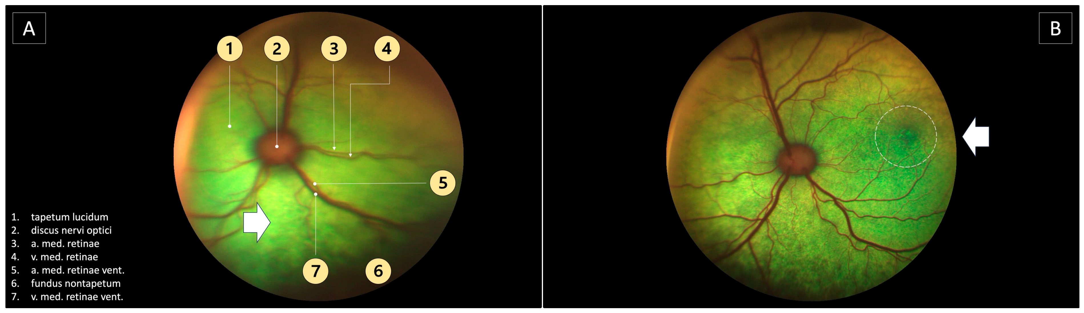
The mild lens opacities observed during ophthalmoscopy were not judged to be of significant visual consequence. The bilateral extensive retinopathy with a map-like pattern, together with the focal degenerative lesion in the left eye, are most consistent with advanced glaucomatous retinal damage. Chronically elevated intraocular pressure produces RGC loss and secondary retinal vascular and pigmentary changes, as reviewed in the feline glaucoma literature [
2]. Oral examination revealed no pathological findings that would have supported an infectious or inflammatory systemic differential; this finding, together with the absence of ocular signs suggestive of uveitis, such as aqueous flare, keratic precipitates, posterior synechiae, or ocular hypotony, is consistent with the interpretation of the disease as primary, rather than secondary, glaucoma.
Glaucoma is increasingly recognized as a neurodegenerative disease characterized by primary damage to the RGC axons at the level of the optic nerve head, followed by retrograde axonal degeneration and subsequent apoptotic loss of the RGCs, together resulting in irreversible optic neuropathy and visual loss [
25,
26]. Although elevated intraocular pressure remains the most significant modifiable risk factor, the cellular pathways implicated in disease progression are multifactorial and include impaired axonal transport, cellular hypoxia, disturbances of local blood supply, and excitotoxicity associated with glutamate accumulation [
26]. In principle, early structural detection of RGC loss or dysfunction is possible by imaging of the optic disk and retinal nerve fiber layer, as discussed in the general glaucoma literature [
25], and by electroretinography (ERG), which provides an objective record of retinal function and is an established clinical modality in veterinary ophthalmology [
27]. Spectral-domain optical coherence tomography (OCT) would have permitted non-invasive in vivo quantification of the peripapillary retinal nerve fiber layer and optic nerve head morphology [
25,
26]. However, none of these modalities were available under the cage-side field conditions of the present case, because of the distance between the examination site and the nearest equipped veterinary facility.
Recognizing glaucomatous optic nerve head cupping by ophthalmoscopy was particularly difficult in this case owing to the distinctive anatomy of the felid optic nerve head. In domestic cats—and, by extension, in other felids—myelination of the RGC axons does not begin until posterior to the lamina cribrosa, and the disk normally sits at, or slightly below, the level of the peripapillary retina, so that a physiological excavation comparable to that of the human or canine optic disk is absent. The recognition of early glaucomatous excavation is therefore considerably more difficult than in dogs, in which the prelaminar myelin gives the disk a raised appearance [
2]. The subtle ophthalmoscopic signs that may indicate advanced feline glaucomatous optic neuropathy—notably a peripapillary pigmented or hyper-reflective halo and increased prominence of the laminar pores, the so-called “laminar dot sign”—were not clearly discernible on the fundus photographs recorded in the present case, most probably because of the extensive retinopathy and mild media opacities already described, as well as the species-specific optic nerve head anatomy of felids outlined above [
2]. Nevertheless, the chronically and markedly elevated intraocular pressure documented in both eyes, measuring 48–52 mmHg at examination and consistently above the feline reference range in previous measurements, together with bilateral buphthalmos, irreversible blindness, fixed dilated pupils, and absent menace responses, strongly supports the diagnosis of advanced glaucomatous optic neuropathy [
2,
21,
22,
23,
24].
The therapeutic evidence base for glaucoma in large felids is limited, but scientific case reports indicate that both medical and surgical management have been attempted. Unilateral secondary glaucoma in a circus-rescued male tiger, attributed to training-related ocular trauma, has been documented under sedation [
17], and unilateral congenital glaucoma in an 8-month-old female tiger was successfully managed by transconjunctival enucleation [
18]; diffuse iris melanoma with secondary glaucoma in an adult African lion was likewise treated by enucleation [
20]. The broader therapeutic framework for feline glaucoma, comprehensively reviewed for the domestic cat [
2], is largely applicable by extension to non-domestic felids. Medical management relies primarily on topical carbonic anhydrase inhibitors (e.g., dorzolamide) and β-adrenergic antagonists (e.g., timolol) to reduce aqueous-humor production; prostaglandin F
2α analogs are typically ineffective or ocularly irritating in cats [
2]. In non-domestic felids under human care, however, the frequent topical administration required is often impracticable without intensive operant-conditioning training, and repeated close handling carries a substantial occupational-safety risk [
7]. Among vision-preserving surgical procedures, laser cyclophotocoagulation and anterior-chamber shunts are less extensively reported in cats than in dogs and appear less effective in felids than in canids, a difference that may in part reflect species-related variation in ciliary-body pigmentation and architecture [
2]. In irreversibly blind or painful eyes, enucleation and evisceration with an intrascleral silicone prosthesis are the two established advanced-stage options. In cats, enucleation is generally preferred because of the possibility of occult intraocular neoplasia and the comparatively higher failure rate of intrascleral prostheses [
2], and both enucleation and evisceration have been successfully performed in tigers and other large felids [
17,
18,
20].
The complete absence of direct and consensual pupillary light reflexes observed in the examined individual is consistent with severe damage to the visual pathways, including the retina and optic nerve, resulting from advanced glaucoma. Although functional blindness can be associated with persistent mydriasis, the fixed pupillary dilation recorded during the anesthesia was probably also influenced by the pharmacological action of ketamine, a non-competitive N-methyl-D-aspartate (NMDA)-receptor antagonist that activates central sympathetic outflow and the limbic system, thereby modulating pupillary tone in felids. Ketamine may also contribute to mydriasis through inhibition of muscarinic acetylcholine receptors [
7,
12]. In felids, the effect of the α
2-adrenergic agonist medetomidine to pupillary size is variable, and mydriasis has been reported in cats after α
2-agonist administration [
8]. The effects of the medetomidine–ketamine–butorphanol combination on intraocular pressure and physiological parameters were carefully considered throughout the immobilization protocol [
10,
11].
Beyond the glaucomatous ocular pathology, the clinical picture in this case included neurological features that warrant separate consideration. The severe bilateral convergent strabismus (esotropia) recorded during remote observation is of considerable diagnostic significance. This sign is not a direct consequence of glaucomatous ocular hypertension: in veterinary neurology it is classically attributed to dysfunction of the abducens nerve (cranial nerve VI), whose lateral-rectus motor output normally antagonizes the medial rectus. Bilateral abducens nerve dysfunction may therefore produce bilateral esotropia and is a recognized non-localizing sign of raised intracranial pressure because of the long subarachnoid course of the nerve [
9]. In combination with disorientation and the seizure activity documented in the present case, this finding raises the differential diagnoses of a space-occupying intracranial process, meningoencephalitis, or generalized intracranial hypertension; however, without advanced cross-sectional neuroimaging, none of these conditions can be confirmed or excluded.
The coexistence of bilateral convergent strabismus and the recorded epileptic episodes suggest possible involvement of the central nervous system, whose exact etiology cannot be clarified without advanced cross-sectional imaging. Magnetic resonance imaging (MRI) is generally considered a key diagnostic modality for the differentiation of inflammatory, neoplastic, vascular, and degenerative intracranial disease in both domestic and non-domestic felids. Hecht et al. [
28] reported the largest MRI case series to date in non-domestic
Felidae under human care, comprising 50 animals, including 18 tigers, 11 lions, and 4 leopards, and documenting Chiari-like malformation, meningoencephalitis, pituitary lesions, leukoencephalopathy, and vascular events among the most frequent intracranial diagnoses. That same series also illustrates the considerable logistical and anesthetic demands of performing MRI in large non-domestic felids and the limited availability of such examinations outside specialized academic institutions [
28]. In the absence of CT or MRI in the present case, a causal relationship between the glaucomatous blindness and the neurological signs—for example, a lesion involving the optic chiasm, leukoencephalopathy, or another space-occupying process—remains speculative, and other congenital or acquired intracranial anomalies cannot be formally excluded.
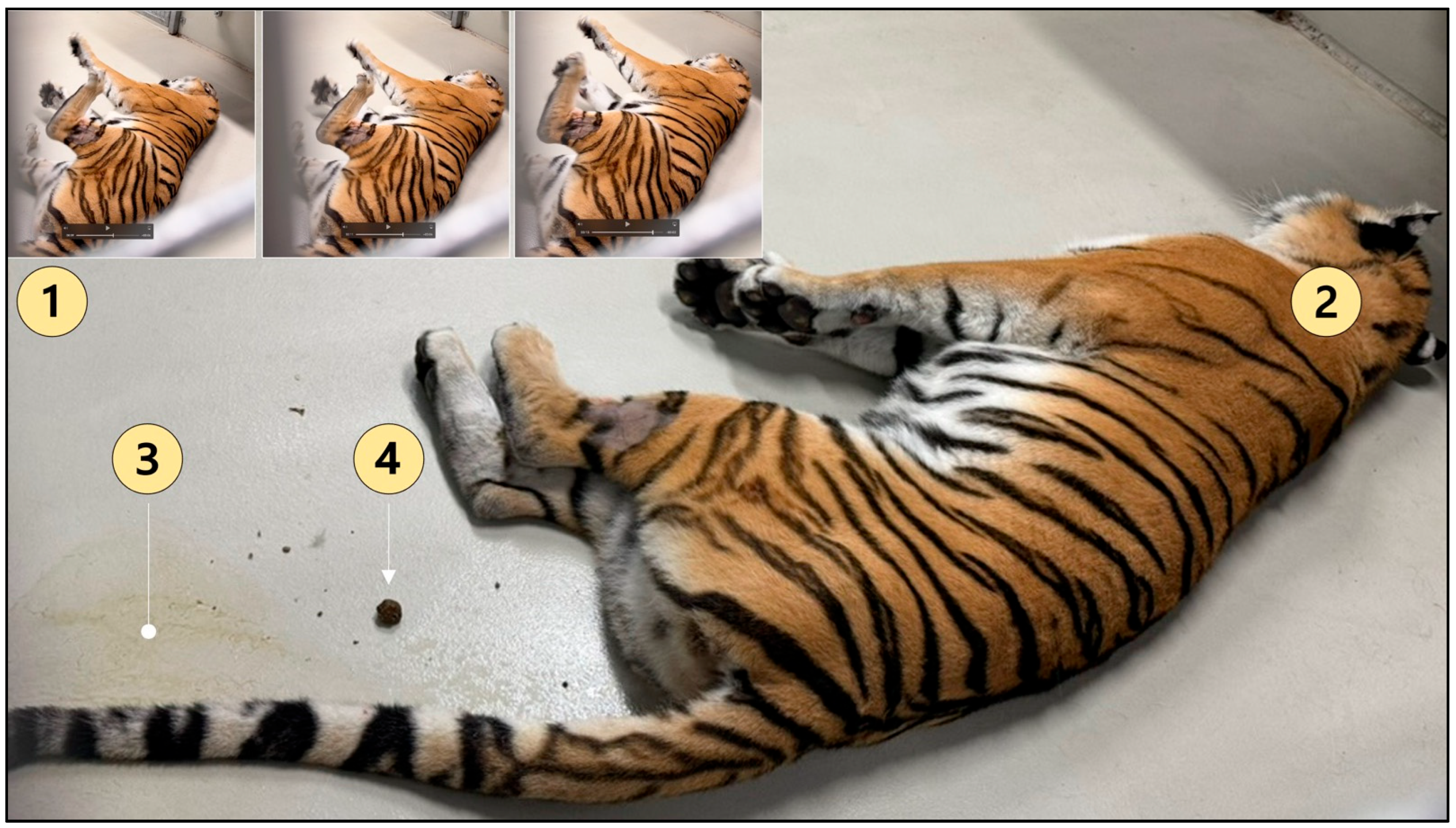
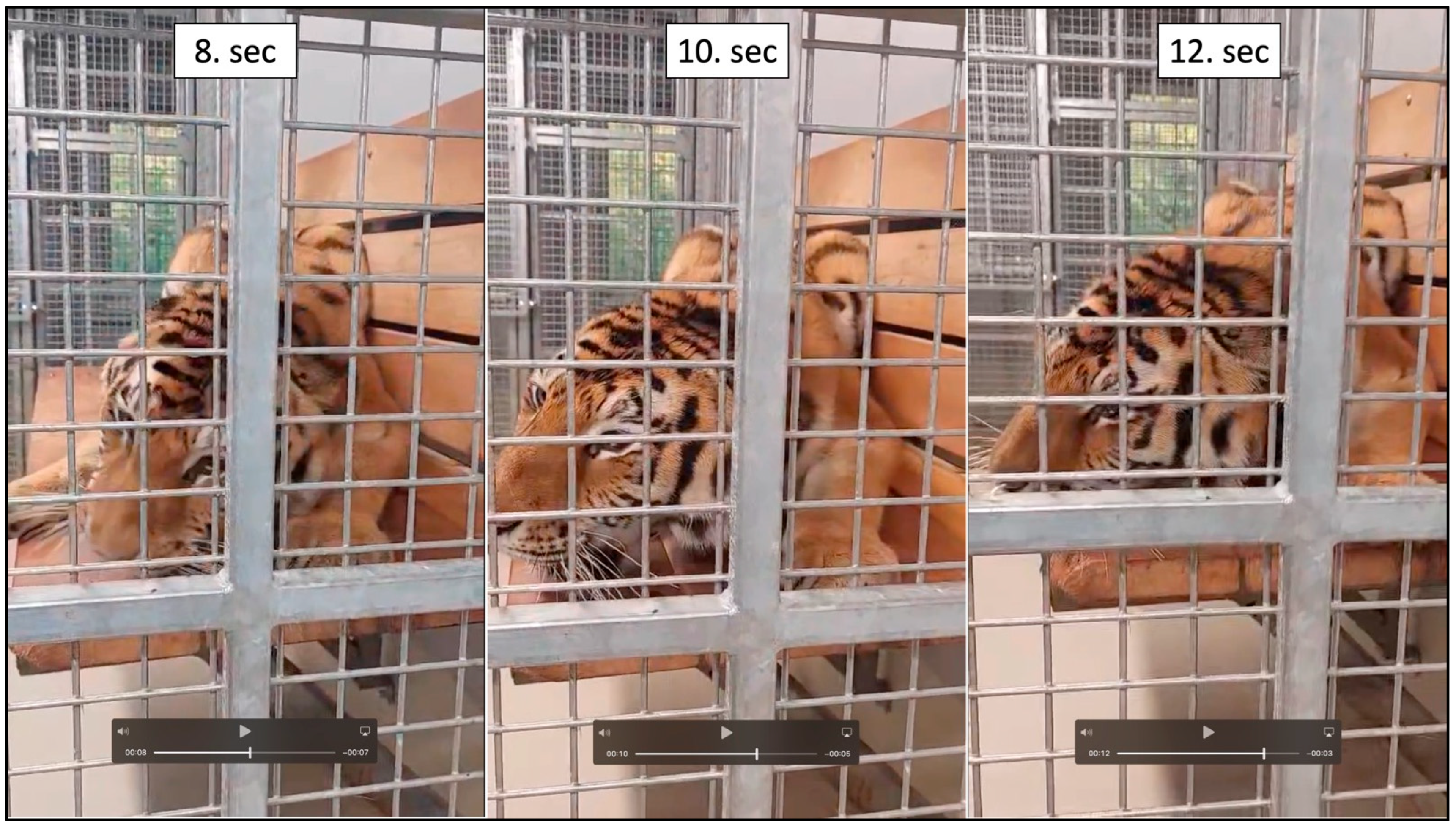
Two distinct types of seizure-like episodes were observed: brief, stereotyped focal episodes reported by the keepers since the animal’s arrival at the zoo four months before anesthesia, and generalized tonic–clonic seizures observed only during anesthetic recovery (
Figure 5). A representative focal episode also documented on the day after the examination (
Figure 6). These two event types are not mutually exclusive in their interpretation: the recovery-phase generalized seizures may have represented secondary generalization of an underlying chronic focal epileptogenic process, facilitated by the combined effects of residual ketamine and premature α
2-adrenoceptor antagonism. The interval between the final ketamine dose (T = 32 min) and atipamezole administration (T = 59 min) was 27 min; in the established veterinary literature on the medetomidine–ketamine–atipamezole protocol in non-domestic mammals, premature antagonism of the α
2-adrenoceptor agonist before residual ketamine has been substantially metabolized is recognized as a cause of rough recoveries with muscle rigidity, tremor, and, less commonly, excitatory phenomena [
11,
29]. Experimental evidence in the domestic cat further indicates that ketamine activates the limbic system and can induce clinical seizure activity closely resembling an animal’s habitual spontaneous seizure pattern when a pre-existing epileptogenic substrate is present, a phenomenon potentially relevant to this case [
13].
Qualitatively, ketamine-related myoclonic or tremor-like movements described during recovery from dissociative anesthesia are typically brief, self-limiting, and clinically distinguishable from both the generalized tonic–clonic seizures and the stereotyped focal episodes observed here [
7,
11,
13,
16]. The decision to withhold emergency anti-seizure treatment during the recovery phase was a deliberate clinical choice based on the short, self-limiting duration of the episodes, the continued presence of residual sedative effect, the practical constraints of operating within an outdoor enclosure shared with other large felids, and the substantial occupational-safety risk of invasive drug administration in this setting; neither the recovery-phase episodes nor the focal seizure observed on the following day met the accepted clinical criteria for status epilepticus or cluster seizures [
30]. A chronic, active inflammatory central nervous system disease—for which published imaging series in non-domestic felids report characteristic findings typically accompanied by progressive weight loss, hyporexia, and deteriorating body condition [
28]—was considered unlikely in this animal, which maintained excellent body condition, normal appetite, and stable activity throughout the four-month observation period before anesthesia. For future immobilization of large felids with suspected or documented neurological comorbidity, a longer interval (ideally ≥45–60 min) between the final ketamine dose and α
2-adrenoceptor antagonist administration, lower incremental ketamine doses where clinically acceptable, or alternative protocols such as medetomidine–midazolam–butorphanol or butorphanol–azaperone–medetomidine with low-dose ketamine should be considered [
2,
6,
10,
11].
In the present case, the combination of bilateral, irreversible blindness, the absence of clinically evident ocular pain or self-directed behavior, and the logistical and safety constraints of frequent topical therapy or repeated general anesthesia in an adult tiger held in a small private zoo led the attending team to shift the therapeutic goal from vision preservation to long-term pain surveillance and welfare maintenance. The veterinary recommendations issued after the examination were therefore as follows: (i) adaptation of the housing environment to a simplified, enclosed indoor enclosure with a consistent layout, familiar olfactory cues, and protected feeding and watering stations, in order to minimize injury risk associated with blindness; (ii) referral to a facility equipped with advanced cross-sectional neuroimaging, including CT and MRI, electroencephalography, and specialist ophthalmic surgery, to permit both etiological characterization of the focal-seizure phenomena and definitive surgical management of the bilateral advanced-stage glaucoma; (iii) consideration of long-term systemic analgesia pending definitive surgery or if referral was not feasible, with explicit account taken of the occupational-safety implications of repeated handling; and (iv) ongoing monitoring of neurological signs and welfare indicators by the holding-zoo veterinary team, with a clearly defined threshold for escalation to humane euthanasia should the welfare balance deteriorate [
2,
7].



 ,
,