Article

Effects of Copper Citrate and Copper Sulfate on Intestinal Health, Muscle Fiber Traits, and Antioxidant Capacity in Weaned Pigs

1
College of Animal & Veterinary Sciences, Southwest Minzu University, Chengdu 610041, China
2
Key Laboratory of Qinghai-Tibetan Plateau Animal Genetic Resource Reservation and Utilization, Southwest Minzu University, Ministry of Education, Chengdu 610041, China
3
Key Laboratory of Animal Science of National Ethnic Affairs Commission of China, Southwest Minzu University, Chengdu 610041, China
4
Animal Breeding and Genetics Key Laboratory of Sichuan Province, Sichuan Animal Science Academy, Chengdu 610066, China
5
College of Life Sciences and Agri-Forestry, Southwest University of Science and Technology, Mianyang 621010, China
*
Author to whom correspondence should be addressed.
These authors contributed equally to this work.
Animals 2026, 16(11), 1615; https://doi.org/10.3390/ani16111615
Submission received: 7 April 2026 / Revised: 19 May 2026 / Accepted: 20 May 2026 / Published: 26 May 2026

Simple Summary

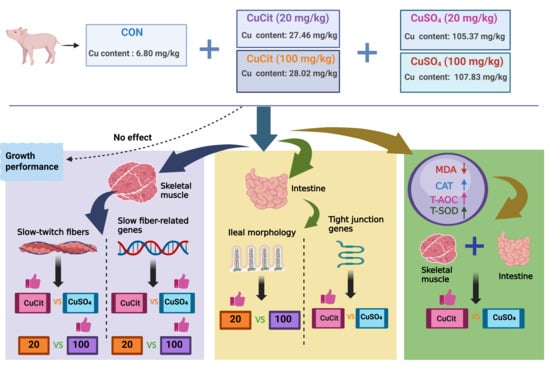
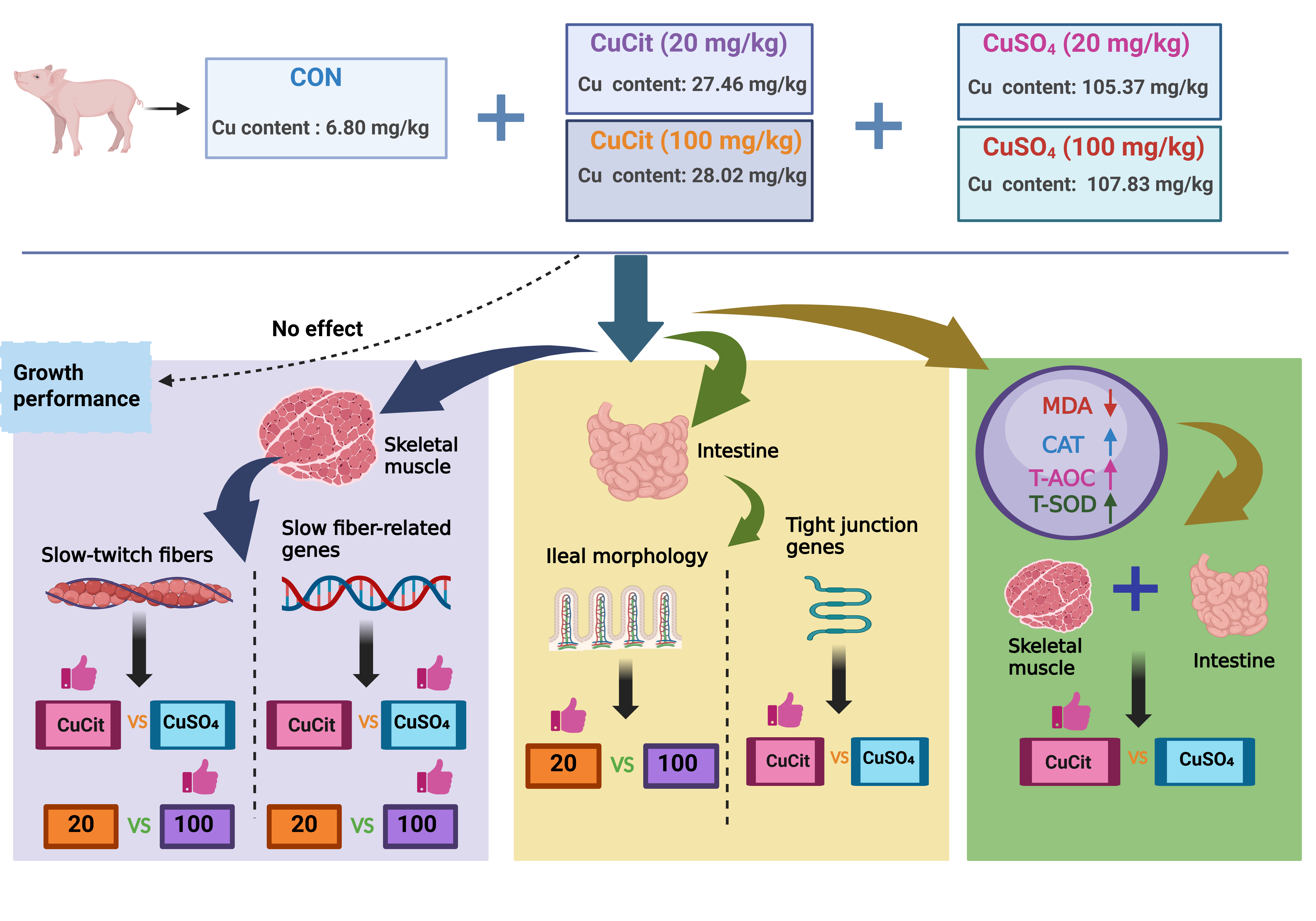
Copper is vital for piglet health, and different supplement forms vary in effectiveness. This study aims to compare the effects of copper citrate and copper sulfate at different supplementation levels on muscle fiber traits, intestinal health, and antioxidant status in weaned pigs. Ninety pigs were randomly assigned to five treatments for 28 d: a basal diet, and the basal diet supplemented with copper citrate or copper sulfate at 20 or 100 mg/kg. At a lower dose, copper citrate strengthened the gut barrier, enhanced antioxidant defenses, and affected skeletal muscle fiber traits. In contrast, copper sulfate was needed at a higher dose to achieve similar effects. This suggests that copper citrate may achieve comparable health benefits with lower copper input, thereby helping to reduce feed costs. This work provides science-based guidance for precise, sustainable copper use in piglet nutrition.

Abstract

Copper (Cu) is an essential trace mineral additive in weaned pig diets. Copper citrate (CuCit) and copper sulfate (CuSO4) are common feed additives, yet their tissue-specific effects at different inclusion levels remain unclear. In this study, ninety pigs (six pens per treatment, three pigs per pen) with an initial body weight of 7.71 ± 0.15 kg were randomly assigned to five treatments for 28 d: a basal diet, and the basal diet supplemented with CuCit or CuSO4 at 20 or 100 mg/kg. The results show that CuCit increased slow-twitch fiber proportion in longissimus dorsi muscle (p = 0.009), whereas CuSO4 more markedly upregulated the expression of MyHC I (p = 0.005) and downregulated MyHC IIx (p = 0.006) and MyHC IIb (p = 0.033). Compared with the control group, CuCit and CuSO4 supplementation increased villus height and decreased crypt depth in ileum (p < 0.05). CuCit at 20 mg/kg showed higher ZO-1 expression than CuSO4 at 100 mg/kg (p < 0.05). Furthermore, CuCit showed greater T-SOD (p = 0.018) and CAT activities (p = 0.002) than CuSO4 in muscle, as well as greater T-SOD activity (p = 0.011) and lower MDA content (p = 0.001) in ileal mucosa. These results suggest that CuCit and CuSO4 exhibit different tissue-specific effects in weaned pigs, providing novel insights for precision Cu supplementation.

Graphical Abstract

1. Introduction

In modern intensive livestock production, feed cost represents one of the most significant expenses in the industry, making the enhancement of feed efficiency and the maintenance of animal health central to sustainable development [1]. Feed additives enhance profitability, welfare, and reduce environmental emissions by optimizing nutritional structure, digestion, absorption, intestinal health, and immune-antioxidant functions. Among them, trace minerals, despite low inclusion levels, are indispensable for growth, development, metabolic regulation, and overall health [2]. Copper (Cu) is an essential trace mineral that serves as a catalytic cofactor for a wide array of metalloenzymes involved in fundamental biological processes, including iron metabolism, neuropeptide synthesis, connective tissue formation, and cellular antioxidant defense systems [3]. Cu is widely used as a feed additive to enhance growth performance in animal nutrition, particularly in swine production [4]. The dosage and source of Cu affect animal performance, as an appropriate amount promotes growth and enhances production, while excessive levels can induce toxicity in animals and pose environmental risks [5]. Dietary Cu supplementation at levels exceeding nutritional requirements remains an effective strategy to enhance growth performance and feed efficiency in weaned pigs, particularly during the immediate post-weaning phase when pigs face multifaceted stress factors [6]. The efficacy of Cu supplementation is profoundly influenced by its chemical form and bioavailability. Inorganic copper sulfate (CuSO4) has been conventionally utilized due to its cost-effectiveness and documented benefits in modulating oxidative stress and microbiota composition, and organic Cu complexes such as copper citrate (CuCit) have garnered increasing interest due to their proposed higher absorption efficiency and metabolic stability [7,8]. Beyond these functions, Cu is essential for muscle development, intestinal integrity, and immune responses, all of which are key determinants of swine productivity [9,10].
The post-weaning period presents substantial challenges to intestinal integrity and systemic homeostasis. Weaned pigs undergo a critical transition from suckling on a sow’s milk to consuming solid feed, during which precise control of Cu supplementation in the diet is required [11]. The intestinal tract is not only the primary site for nutrient absorption but also a critical barrier against pathogens and toxins [12]. Weaning stress frequently disrupts gut architecture, impairs barrier function through dysregulation of tight junction proteins, and induces localized inflammation and oxidative stress [13]. Recent research has expanded the understanding of Cu’s biological roles beyond growth promotion. Cu contributes to intestinal health by promoting villus development, modulating immune responses, and enhancing mucosal antioxidant capacity through Cu-dependent enzymes such as SOD [9]. Concurrently, skeletal muscle development, a primary determinant of growth performance, can be influenced by trace mineral status [14]. Cu participates in muscle energy metabolism and modulates muscle fiber type composition by altering the metabolic phenotype of myofibers, thereby influencing the overall growth rate and post-fattening meat quality [15].
However, a systematic comparison of CuCit and CuSO4 as dietary Cu sources under practical production conditions remains limited. Previous studies have primarily focused on individual parameters such as growth performance or hematological indices, with insufficient research systematically evaluating the effects of different Cu sources on multiple physiological functions in weaned pigs within a unified experimental framework. Therefore, this study aims to systematically compare the effects of CuCit and CuSO4 at different supplementation levels on growth performance, muscle fiber type composition, intestinal morphology, and antioxidant status in weaned pigs. Based on the differences in absorption and metabolism between inorganic and organic Cu, we hypothesize that CuCit and CuSO4 differ in their effects on weaned pigs and may exert tissue-specific actions. This study provides evidence-based guidance for scientific Cu source selection and precision supplementation.

2. Materials and Methods

2.1. Animals, Design, and Diets

Animal procedures were conducted in compliance with the guiding principles of the Animal Care and Ethics Committee of Southwest Minzu University (Approval No. SMU-202307145). Ninety 28-day-old Duroc × Landrace × Yorkshire (DLY) crossbred male piglets, derived from thirty multiparous sows (parities 2 to 3), with an average body weight of 7.71 ± 0.15 kg, were divided into blocks according to initial body weight and randomly assigned to five dietary treatments, with each group comprising six replicates (pens) and three piglets per pen. The experiment began with a 3-day adaptation period, during which all pigs were fed the basal diet, which was analyzed to contain 6.8 mg/kg of Cu from intrinsic dietary sources. Following adaptation, the trial entered a 28-day formal period. Throughout the 28-day trial, the control group (CON) continued to receive the basal diet (Supplementary Materials, Table S1), and the other four groups were fed the basal diet supplemented with CuCit or CuSO4 at 20 or 100 mg of Cu per kg of diet. The Cu content in the diet was determined by flame atomic absorption spectrometry (Table S2). The basal diet was formulated in accordance with the recommendations of the Nutrient Requirements of Swine (2012). Feed and water were available ad libitum. All pigs were housed in an environmentally controlled room maintained at 28 ± 1 °C and 65–75% relative humidity. The CuCit (≥98.5% purity, containing ≥34.0% Cu) and CuSO4 (≥98.5% purity, containing ≥25.0% Cu) were provided by Sichuan Livestock Science Group Co., Ltd. (Chengdu, China).

2.2. Performance Measurements and Sample Collection

Pigs were weighed individually on days 0 and 28 to calculate average daily gain (ADG). Daily feed intake was recorded to determine average daily feed intake (ADFI), and feed-to-gain ratio (F/G) was derived as ADFI/ADG. On day 28, one pig per pen with body weight closest to the pen average was selected and euthanized after a 12 h feed restriction. Pigs were intramuscularly injected with Zoletil (25 mg/kg), and after confirming that they had entered a state of deep anesthesia, they were euthanized by rapid exsanguination via carotid artery incision. Tissue samples were collected immediately. The longissimus dorsi (LD) muscle was dissected from the right side between the 10th and 12th ribs, cut into cubes, and frozen in liquid nitrogen. A segment of the middle ileum was excised after ligation, flushed with ice-cold saline, and approximately 2 cm was fixed in 4% paraformaldehyde for histological assessment. Mucosa from the remaining segment was harvested with a sterile glass slide and snap-frozen in liquid nitrogen. All frozen samples were stored at −80 °C for subsequent analyses.

2.3. Total RNA Extraction and Real-Time Quantitative PCR

Total RNA was extracted using the RNAiso Plus reagent (TaKaRa, Dalian, China), and reverse transcription was performed according to the instructions of the PrimeScript RT reagent Kit with gDNA Eraser (TaKaRa). The integrity of the RNA was examined by agarose gel electrophoresis. Real-time quantitative PCR detection was performed with TB Green® Premix Ex Taq™ II (TaKaRa) following the manufacturer’s instructions. Primer sequences were designed based on the known gene sequences available in GenBank to specifically amplify the target gene mRNA for analysis (Table S3). The amplification efficiency for the primer pair was determined from standard curves, and melting curve analysis confirmed a single product for each amplicon. Glyceraldehyde-3-phosphate dehydrogenase (GAPDH) was found to be relatively stable in this experiment and was used as an internal reference gene to normalize expression levels. Relative mRNA abundance was calculated using the 2–ΔΔCT method.

2.4. Immunofluorescence Homology Double-Labeling Assay

Longissimus dorsi samples were fixed, dehydrated, cleared, and embedded in paraffin, then sectioned at 10 μm. Following dewaxing, rehydration, and microwave-assisted antigen retrieval in EDTA buffer (pH 8.0), sections were rinsed in PBS and treated with 3% H2O2 to block endogenous peroxidase. After blocking with 3% BSA, double immunofluorescence labeling was performed: sections were incubated overnight at 4 °C with anti-slow MyHC primary antibody, followed by a secondary antibody for 1 h at room temperature. After washing with TBST and repeating antigen retrieval to remove the first antibody complex, sections were incubated overnight at 4 °C with anti-fast MyHC primary antibody, followed by its corresponding secondary antibody. Nuclei were counterstained with DAPI. After applying autofluorescence quencher and washing, sections were mounted with anti-fade medium and examined under fluorescence microscopy. Slow-twitch fibers exhibited green fluorescence, fast-twitch fibers red fluorescence. Fields of view were systematically selected from the central region of each section to avoid edge artifacts. For each sample, three random fields of view were captured at 100× magnification. Percentage of slow-twitch fibers = (number of slow fibers/total number of fibers) × 100. All observations and counts were performed by observers who were blinded to the treatment groups.

2.5. Antioxidant Properties

Tissue samples from the LD muscle and ileal mucosa were processed for biochemical analysis. The LD muscle and ileal mucosa were directly homogenized. Both tissue types were then homogenized in ice-cold PBS at a 1:9 (w/v) ratio using a tissue homogenizer. The homogenates were centrifuged at 2500× g for 15 min at 4 °C. Protein concentration in tissue homogenate supernatants was determined using a bicinchoninic acid assay kit (Jiancheng, Nanjing, China) according to the manufacturer’s instructions. Bovine serum albumin standards were used to generate a standard curve. All antioxidant parameters, including MDA content and the activities of T-SOD, CAT, and T-AOC, were examined in accordance with the manufacturer’s protocols provided with the respective commercial assay kits (Jiancheng), and normalized to protein content. MDA content is expressed as nmol/mg protein, and enzyme activities (T-SOD, CAT, and T-AOC) are expressed as U/mg protein.

2.6. Histological Analysis

Ileal tissue segments were processed for histological examination. Following fixation, the samples were dehydrated, cleared, and embedded in paraffin wax. Serial sections of 10 μm thickness were prepared and stained with hematoxylin and eosin (H&E). Morphological evaluation was performed using an optical microscope (Nikon Eclipse E100, Tokyo, Japan). The measurements on villus height (V) and crypt depth (C) were done on at least 10 villus and 10 crypts that were well oriented for each sample with 100× magnification using the Image-Pro Plus software (version 6.0; Media Cybernetics, Bethesda, MD, USA). These were then averaged per pig for all further analyses. From these measurements, the ratio of villus height to crypt depth (V/C) was calculated. All histological measurements were averaged from multiple sections per animal, and all morphological measurements were performed by an observer blinded to the treatment groups.

2.7. Statistical Analysis

Data were analyzed as a randomized complete block design using the MIXED procedure of SAS (version 9.4; SAS Inst. Inc., Cary, NC, USA), with the pen and the individual pig designated as the experimental unit for growth performance and sample-based analyses, respectively. The statistical model evaluated the interaction effect and the main effect of Cu source within the four Cu-supplemented groups. The control group was included in the evaluation of individual treatment effects and the main effect of Cu level. A MIXED model was used to compare the five treatment groups, followed by Tukey’s test to assess differences between groups. Significant differences were considered at p < 0.05, and tendencies were considered at 0.05 < p ≤ 0.10.

3. Results

3.1. Growth Performance

The effects of dietary Cu sources and levels on the growth performance of weaned pigs are presented in Table 1. There were no significant interactions or main effects of Cu source and level on final body weight, ADG, and F/G (p > 0.05). ADFI tended to be greater at the 20 mg/kg Cu level than at 0 mg/kg (p = 0.081).

3.2. Percentage of Slow-Twitch Muscle Fibers

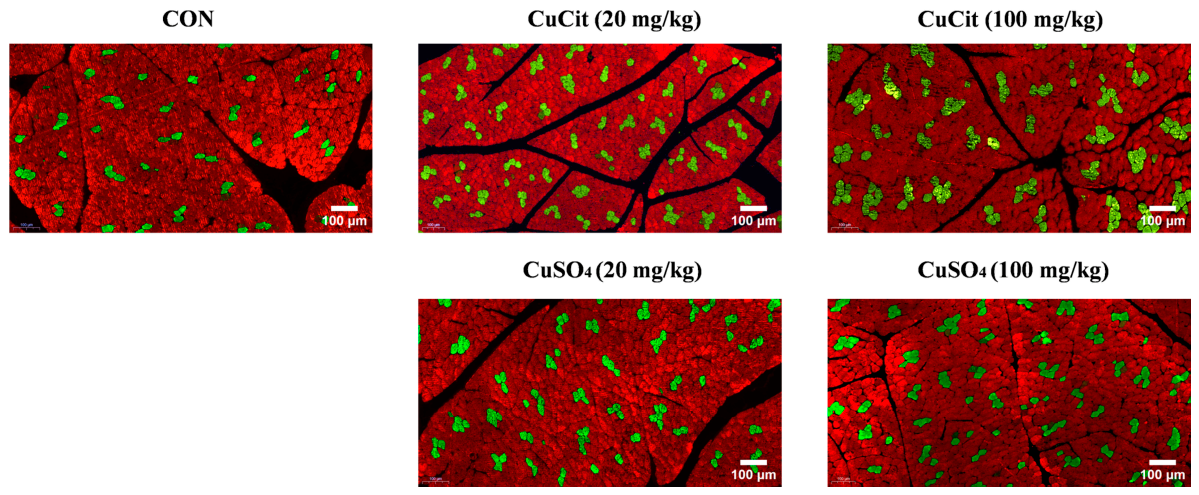
The data presented in Figure 1 and Table 2 indicate no significant interaction between Cu source and level in affecting muscle fiber type composition in the LD muscle of weaned pigs (p = 0.157). Weaned pigs fed CuCit had a higher percentage of slow-twitch muscle fibers than those fed CuSO4 (p = 0.009). Regarding the main effect of dietary Cu level, the 100 mg/kg treatment was higher than the 0 and 20 mg/kg treatments (p = 0.002).

3.3. The Expression of Genes Related to Muscle Fiber Types

As shown in Table 3, dietary Cu supplementation influenced the mRNA expression of genes related to muscle fiber type in LD muscle. A significant interaction between Cu source and level was detected for the expression of MyHC I, MyHC IIa, Tnni1, Myoglobin, AMPKα1, and AMPKα2 (p < 0.05). At 20 mg/kg, the expressions of MyHC I, Tnni1, Myoglobin, and AMPKα2 in CuSO4 groups were higher than in CuCit and CON. For MyHC IIb at 20 mg/kg, CuCit and CuSO4 maintained expression levels higher than CON, and CuCit upregulated its expression compared to CuSO4. At 20 mg/kg and 100 mg/kg supplementation levels, the expression of MyHC IIx in the CuCit group was higher than in CuSO4 and CON. At 100 mg/kg, CuCit and CuSO4 maintained the expression of MyHC I, MyHC IIb, MyHC IIa, and Tnni1 levels higher than CON, while CuSO4 resulted in higher MyHC IIa expression relative to CuCit. For AMPKα1 at 100 mg/kg, CuCit groups were higher than in CuSO4 and CON. Regarding the main effect of Cu source, pigs fed CuSO4 demonstrated upregulation of MyHC I (p = 0.005) and downregulation of MyHC IIx (p = 0.006) and MyHC IIb (p = 0.033) compared with the CuCit group. Compared with CuCit, CuSO4 tended to have greater Tnni1 expression (p = 0.074). PGC-1α expression tended to be higher in the CuSO4 at 100 mg/kg than in the CON (p = 0.056). Regarding the main effect of dietary Cu level, 100 mg/kg increased the expression of MyHC I compared with 0 mg/kg and 20 mg/kg (p < 0.05), and 20 mg/kg and 100 mg/kg up-regulated MyHC IIa, MyHC IIb, Tnni1, and AMPKα2 compared with 0 mg/kg (p < 0.05). The expression of MyHC IIx, AMPKα1 and PGC-1α showed a tendency to be greater at 100 mg/kg than at 0 mg/kg (p < 0.10).

3.4. Muscle Antioxidant Status

As shown in Table 4, significant source and level interactions were observed for MDA content and the activities of CAT (p < 0.05). At 20 mg/kg, CuCit showed a lower MDA content than CON and CuSO4, whereas CuSO4 showed higher CAT activity than CON and CuCit. At 100 mg/kg, CuCit and CuSO4 decreased MDA content relative to the CON, with CuSO4 achieving the lowest values, and CuCit resulted in higher CAT activity than the CON and CuSO4 groups. For the main effect of source, weaned pigs fed CuCit had a higher MDA content (p < 0.001) and higher activities of T-SOD (p = 0.018) and CAT (p = 0.002) compared to those fed CuSO4. Regarding the main effect of level, pigs fed the 100 mg/kg Cu exhibited a reduction in MDA content (p = 0.002) and an increase in CAT activity (p = 0.040) compared with those fed the 0 or 20 mg/kg diets.

3.5. Ileal Morphology

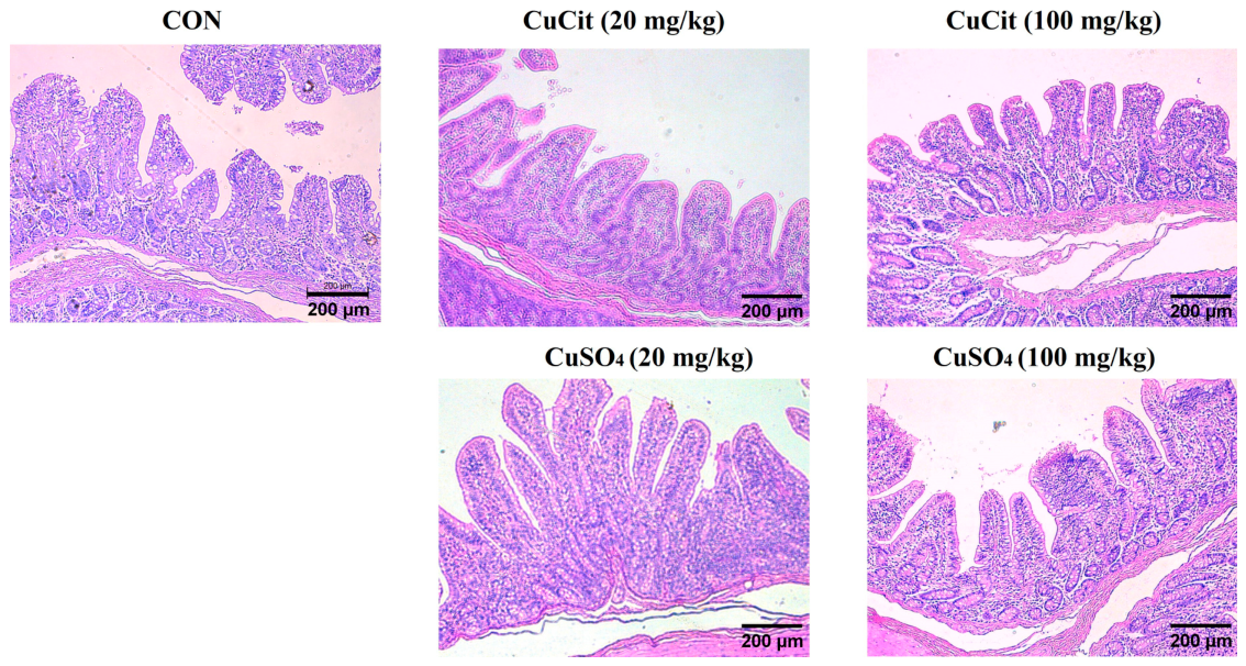
The data presented in Figure 2 and Table 5 indicate no significant source and level interactions for villus height, crypt depth, and the ratio of villus height to crypt depth (p > 0.05). Dietary Cu level influenced ileal morphology. Villus height was greater in pigs fed 20 mg/kg Cu than in those fed either 0 or 100 mg/kg (p = 0.005). Compared with the 0 mg/kg, supplementation at 20 and 100 mg/kg resulted in a reduced crypt depth (p = 0.003) and an increased ratio of villus height to crypt depth (p < 0.001).

3.6. Ileal Tight Junction Gene Expression

The relative mRNA expression of tight junction genes in the ileal mucosa is presented in Table 6. Significant source and level interactions were observed for ZO-1 and occludin expression (p < 0.05). At 20 mg/kg, CuCit upregulated ZO-1 expression compared to the CON and CuSO4 groups. At 100 mg/kg, CuSO4 enhanced ZO-1 expression relative to CON and CuCit groups. Notably, compared with the CuCit group at 20 mg/kg, the CuSO4 group at 100 mg/kg exhibited lower ZO-1 expression.

3.7. Ileal Inflammatory Gene Expression

As presented in Table 7, the expression of inflammation genes in ileal mucosa was not significantly altered by dietary Cu treatments. There were no significant interactions or main effects of source and level detected for IL-1β and TNF-α expression (p > 0.05). Compared with CuSO4, CuCit tended to have greater IL-10 expression (p = 0.093).

3.8. Ileal Antioxidant Status

As shown in Table 8, the antioxidant status in the ileal mucosa was affected by dietary Cu supplementation. Significant source and level interactions were detected for MDA content (p < 0.001). At 20 and 100 mg/kg supplementation levels, MDA content was reduced in the CuCit and CuSO4 groups compared to the CON, with the CuCit groups showing a further reduction relative to the CuSO4 groups. Analysis of the main effects revealed that pigs fed CuCit had lower MDA content (p = 0.001) and higher T-SOD activity (p = 0.011) in the ileal mucosa compared to those fed CuSO4. Regarding the main effect of level, the results revealed that 20 and 100 mg/kg supplementation reduced MDA content and enhanced T-AOC compared to the 0 mg/kg level (p < 0.001).

4. Discussion

CuCit and CuSO4 are widely utilized Cu supplements in livestock feed, recognized for their efficacy in enhancing growth performance and promoting overall health [6]. Dietary Cu supplementation did not significantly improve the final body weight, ADG or F/G, but ADFI tended to be greater at 20 mg/kg than at 0 mg/kg. This suggests that weaned pigs may prefer Cu-supplemented diets, consistent with previous findings [16]. The basal diet in this study already contained 6.8 mg/kg Cu from the mineral premix, which is close to the NRC (2012) requirement estimate for weaned pigs. It has been suggested that dietary Cu levels of 100–250 mg/kg in weaned pigs are typically required to produce significant improvements in growth performance [17]. In the present study, the supplemented levels of 20 and 100 mg/kg may not have attained the requisite threshold to elicit a significant growth-promoting effect.
Muscle development is a key determinant of the economic returns in swine production, with feed nutrition serving as a central approach to regulating these processes [18]. Skeletal muscle development and fiber type composition serve as a fundamental physiological basis influencing livestock growth efficiency and meat production performance [19]. The proportion of different muscle fiber types, which are defined by their specific MyHC isoforms, dictates the contractile and metabolic properties of the muscle and influences broader physiological processes during growth [20]. Slow-twitch muscle fibers are more conducive to efficient energy utilization and sustained protein synthesis, attributable to their higher myoglobin content, greater oxidative metabolic capacity, lower glycogen levels, and finer diameter [21]. Muscle fiber type composition is regulated by the AMPK signaling pathway [22]. As critical subunits mediating AMPK activation, AMPKα1 and AMPKα2 support the slow-twitch phenotype by enhancing oxidative metabolic genes and activating PGC-1α, which is a key regulator of mitochondrial biogenesis and slow fiber gene expression [22,23]. Studies have shown that Cu as a feed additive can promote muscle growth and development in livestock and poultry, potentially by mediating the AMPK signaling pathway and thereby promoting mitochondrial fission and PGC-1α expression [24]. Our results suggest that CuCit and CuSO4 increased the expression of AMPKα1 and AMPKα2 in muscle. CuCit significantly increased the proportion of slow-twitch fibers relative to CuSO4, whereas CuSO4 upregulated slow-twitch muscle gene expression compared with CuCit. And at the 20 mg/kg supplementation level, CuSO4 significantly increased the expression of slow-twitch-related genes, including MyHC I, Tnni1, Myoglobin, and AMPKα2, compared with CuCit. At these two Cu supplementation levels, CuCit increased the expression of MyHC IIx and MyHC IIb compared with CuSO4. However, the difference in the regulation of slow-twitch muscle genes between CuCit and CuSO4 diminished at 100 mg/kg. These differences may be due to the temporal and hierarchical regulation of muscle fibers: transcriptional changes may precede phenotypic remodeling, and changes in mRNA may precede or lag behind changes in proteins [25,26]. Cu plays an important regulatory role in skeletal muscle development [27]. In this study, Cu supplementation enhanced the expression of the fast-twitch fiber genes MyHC IIa and MyHC IIb, as well as the slow-twitch fiber genes MyHC I, Tnni1, and AMPKα2. Although there is currently limited research directly exploring the correlation between Cu supplementation and muscle fiber types, previous studies have confirmed that Cu is essential for myoblast proliferation and differentiation [28]. Furthermore, dietary supplementation with organic Cu has been shown to significantly affect the number of type I, IIa, and IIb muscle fibers in finishing pigs [29]. These results suggest that inorganic and organic Cu may participate in the expression of muscle fiber type genes through different regulatory mechanisms, and further investigation is needed to clarify the differences in the mechanisms.
Early weaning can lead to a reduction in villus height, an increase in crypt depth, and a compromise in intestinal barrier function [30]. Dietary Cu supplementation improves intestinal morphology in weaned pigs, notably by increasing villus height and reducing crypt depth, which supports enhanced digestion, absorption, and nutrient utilization following weaning [31]. This study found that ileal morphology was primarily influenced by Cu level rather than Cu source. Intestinal absorption of Cu is primarily mediated by transporters such as copper transporter 1 (CTR1) in intestinal epithelial cells, a process characterized by saturability and carrier-mediated kinetics [32,33]. Cu level directly regulates the proliferation rate of intestinal epithelial cells, affecting Cu absorption, whereas the differences between Cu sources may become apparent at specific doses [34,35,36]. However, when Cu levels exceed their absorption and transport capacity, it may interfere with the physiological functions of Cu by affecting intestinal barrier function and cell viability [34,35]. In the present study, villus height was greater at 20 mg/kg than at 100 mg/kg. Owing to the differential intestinal absorption efficiency between organic and inorganic Cu, the dose thresholds at which they exert beneficial effects are also distinct [34,37]. Organic Cu upregulates peptide transporter 1 and the amino acid transporter in the intestine, resulting in higher intestinal absorption efficiency compared with inorganic Cu within a certain threshold [38]. Meanwhile, CuCit at 20 mg/kg showed higher ZO-1 expression than CuSO4 at 100 mg/kg. The results of this trial are consistent with those of previous similar studies, indicating that although organic and inorganic Cu can improve intestinal morphology in weaned pigs, the effective doses may differ [39,40]. The cytokine IL-10 is a key anti-inflammatory cytokine that plays an important role in suppressing excessive intestinal inflammatory responses and maintaining mucosal immune homeostasis [41]. The trend toward higher IL-10 expression in the CuCit group suggests that CuCit may have greater potential than CuSO4 in enhancing the intestinal anti-inflammatory capacity of weaned pigs [42].
Cu is established as a pivotal micronutrient in antioxidant defense [43]. As a cofactor for antioxidant enzymes such as Cu/Zn-SOD, Cu at appropriate levels reduces lipid peroxidation by enhancing the activity of these enzymes [6]. The antioxidant status is typically assessed by biomarkers such as MDA for lipid peroxidation, and T-SOD, CAT, and T-AOC to evaluate the antioxidant defense system [44]. In this study, CuCit and CuSO4 enhanced the antioxidant capacity of the LD muscle compared to the CON group, among which the CuCit group exhibited higher T-SOD and CAT activities but relatively higher MDA content compared to the CuSO4 group. This observation may be attributable to differential temporal kinetics in the antioxidant responses induced by distinct Cu sources, meaning that although CuCit exhibits increased activities of T-SOD and CAT, its effect on MDA may exhibit a time delay [45]. Furthermore, the number of animal replicates used for tissue-level analysis in this study was relatively limited, and sampling was performed at a single time point, which may have restricted the observation of potential differences in MDA or enzyme activities between CuCit and CuSO4. Although our data indicates the two Cu sources effectively alleviated oxidative stress in muscle compared with the CON, more refined comparisons between CuCit and CuSO4 require further investigation. In ileal mucosa, CuCit demonstrated a distinct advantage over CuSO4 and showed lower MDA content and higher T-SOD activity than CuSO4. These results further indicate that the effects of CuCit and CuSO4 are tissue-specific. Organic and inorganic Cu have different solubility in the gastrointestinal tract, which may lead to differences in their absorption and utilization by intestinal cells [34]. Dietary Cu is primarily taken up by enterocytes via the high-affinity CTR1, processed intracellularly through chaperone proteins like antioxidant 1 copper chaperone, and then exported into the portal circulation by the Cu-transporting ATPase 1 [46]. Studies have found that, compared to inorganic CuSO4, organic Cu sources can differentially regulate these transporters and enhance SOD1 expression [34]. In this study, CuCit significantly reduced MDA content and enhanced T-SOD activity compared with the CuSO4 group in ileum. These results suggest that CuCit may be associated with greater intestinal antioxidant capacity and higher intestinal utilization efficiency [8]. This interpretation is in line with previous observations in piglets and other livestock species [6,47]. Previous studies also indicate that organic Cu sources, such as Cu methionine hydroxy analog chelate, may be superior to inorganic Cu sources in terms of functional efficacy at lower supplementation levels [48].

5. Conclusions

Cu sources did not affect growth performance but exhibited tissue-specific effects. CuCit increased the proportion of slow-twitch muscle fibers, whereas CuSO4 showed stronger upregulation of slow-twitch muscle genes. Compared with CuSO4, CuCit exhibited higher T-SOD and CAT activities in muscle as well as higher T-SOD activity and lower MDA content in ileum. CuCit and CuSO4 improved intestinal morphology. And CuCit at 20 mg/kg resulted in higher ZO-1 expression than CuSO4 at 100 mg/kg. These tissue-specific effects indicate that CuCit and CuSO4 differ in their impacts on muscle fiber traits, intestinal parameters, and antioxidant status. However, the limited sample size necessitates further studies to confirm their bioavailability and long-term effects.

Supplementary Materials

The following supporting information can be downloaded at: https://www.mdpi.com/article/10.3390/ani16111615/s1, Table S1: Composition of experimental diets for weaned pigs (as-fed basis). Table S2: Analyzed Cu concentrations (mg/kg) of complete diets. Table S3: Primers used for the gene expression analysis.

Author Contributions

Conceptualization, M.X.; Formal analysis, Z.C., W.W. and Q.L.; Investigation, Z.C., W.W. and Q.L.; Data curation, Z.C., Y.G. and Q.L.; Writing—original draft, Z.C.; Writing—review and editing, Z.C., H.D. and M.X.; Supervision, H.D., Y.Z. and M.X.; Resources, M.X., H.D. and Y.Z.; Visualization, W.W. and Y.Z.; Funding acquisition, Y.G. and M.X. All authors have read and agreed to the published version of the manuscript.

Funding

This study was supported by the Natural Science Foundation of Sichuan Province (2022NSFSC1613), the Program for Pig Industry Technology System Innovation Team of Sichuan Province (sccxtd-2025-08) and the Fundamental Research Funds for the Central Universities/Southwest Minzu University (ZYN2026025).

Institutional Review Board Statement

Animal procedures were conducted in compliance with the guiding principles of the Animals Care and Ethics Committee of Southwest Minzu University (Approval No. SMU-202307145).

Informed Consent Statement

Not applicable.

Data Availability Statement

Data supporting this study are available from the corresponding author upon request.

Acknowledgments

We are grateful to Wenting An at Chongqing Three Gorges Vocational College for the help and support during the experimental work, including optimizing the figures and providing software support.

Conflicts of Interest

The authors declare no conflicts of interest.

Abbreviations

The following abbreviations are used in this manuscript:
CuCopper
CuCitCopper citrate
CuSO4Copper sulfate
ADGAverage daily gain
ADFIAverage daily feed intake
F/GFeed-to-gain ratio
LD muscleLongissimus dorsi muscle
MyHCMyosin heavy chain
ZO-1Zonula occludens-1
IL-1βInterleukin-1 beta
IL-10Interleukin-10
TNF-αTumor necrosis factor-alpha
GAPDHGlyceraldehyde-3-phosphate dehydrogenase
MDAMalondialdehyde
T-SODTotal superoxide dismutase
CATCatalase
T-AOCTotal antioxidant capacity
H&EHematoxylin and eosin
SEMStandard error of the mean

References

  1. Saeed, M.; Hassan, F.; Al-Khalaifah, H.; Islam, R.; Kamboh, A.A.; Liu, G. Fermented Banana Feed and Nanoparticles: A New Eco-Friendly, Cost-Effective Potential Green Approach for Poultry Industry. Poult. Sci. 2025, 104, 105171. [Google Scholar] [CrossRef] [PubMed]
  2. Rao, Z.-X.; Tokach, M.D.; Woodworth, J.C.; DeRouchey, J.M.; Goodband, R.D.; Gebhardt, J.T. Effects of Various Feed Additives on Finishing Pig Growth Performance and Carcass Characteristics: A Review. Animals 2023, 13, 200. [Google Scholar] [CrossRef]
  3. Zhou, B.; Cao, Y.; Zhang, Y.; Xing, M.; Wang, Y. Unveiling Excessive Feed-Sources Copper-Induced Ileitis in Chickens: Insights into Tight Junction Damage and ROS/NLRP3/Pyroptosis Axis. Comp. Biochem. Physiol. Part C Toxicol. Pharmacol. 2026, 299, 110357. [Google Scholar] [CrossRef]
  4. Fernandez, M.; Thompson, J.; Calle, A. Novel Feed Additive Delivers Antimicrobial Copper and Influences Fecal Microbiota in Pigs. Microbiol. Spectrum 2024, 12, e04281-23. [Google Scholar] [CrossRef]
  5. Wang, Y.; Yan, Q.; Shi, Y.; Long, M. Copper Toxicity in Animals: A Review. Biol. Trace Elem. Res. 2024, 203, 2675–2686. [Google Scholar] [CrossRef] [PubMed]
  6. Peng, C.C.; Yan, J.Y.; Dong, B.; Zhu, L.; Tian, Y.Y.; Gong, L.M. Effects of Graded Levels of Cupric Citrate on Growth Performance, Antioxidant Status, Serum Lipid Metabolites and Immunity, and Tissue Residues of Trace Elements in Weaned Pigs. Asian-Australas. J. Anim. Sci. 2017, 30, 538–545. [Google Scholar] [CrossRef] [PubMed]
  7. Wu, X.; Zhou, Y.; Lu, Z.; Zhang, Y.; Zhang, T.; Jiang, Q. Cupric Citrate Supplementation Improves Growth Performance, Nutrient Utilization, Antioxidant Capacity, and Intestinal Microbiota of Broilers. Anim. Biosci. 2024, 38, 530–538. [Google Scholar] [CrossRef]
  8. Du, Y.; Tu, Y.; Zhou, Z.; Hong, R.; Yan, J.; Zhang, G.-W. Effects of Organic and Inorganic Copper on Cecal Microbiota and Short-Chain Fatty Acids in Growing Rabbits. Front. Vet. Sci. 2023, 10, 1179374. [Google Scholar] [CrossRef]
  9. Liu, B.; Yan, J.; Hao, H.; Yong, F.; Yang, L.; Yang, W.; Che, D. Effects of Dietary Fiber and Copper on the Performance and Gut Microbiota of Finishing Pigs. Animals 2024, 14, 3168. [Google Scholar] [CrossRef]
  10. Ma, R.; Feng, L.; Wu, P.; Liu, Y.; Ren, H.-M.; Jin, X.-W.; Li, S.-W.; Tang, L.; Zhou, X.-Q.; Jiang, W.-D. Dietary Copper Improves Intestinal Structural Integrity in Juvenile Grass Carp (Ctenopharyngodon idella) Probably Related to Its Increased Intestinal Antioxidant Capacity and Apical Junction Complex. Anim. Nutr. 2024, 18, 96–106. [Google Scholar] [CrossRef]
  11. Di Giancamillo, A.; Rossi, R.; Martino, P.A.; Aidos, L.; Maghin, F.; Domeneghini, C.; Corino, C. Copper Sulphate Forms in Piglet Diets: Microbiota, Intestinal Morphology and Enteric Nervous System Glial Cells. Anim. Sci. J. 2018, 89, 616–624. [Google Scholar] [CrossRef]
  12. Tang, X.; Liu, X.; Zhong, J.; Fang, R. Potential Application of Lonicera Japonica Extracts in Animal Production: From the Perspective of Intestinal Health. Front. Microbiol. 2021, 12, 719877. [Google Scholar] [CrossRef] [PubMed]
  13. Tang, X.; Xiong, K.; Fang, R.; Li, M. Weaning Stress and Intestinal Health of Piglets: A Review. Front. Immunol. 2022, 13, 1042778. [Google Scholar] [CrossRef]
  14. Cho, K.H.; Kang, S.W.; Yoo, J.S.; Song, D.K.; Chung, Y.H.; Kwon, G.T.; Kim, Y.Y. Effects of Mealworm (Tenebrio molitor) Larvae Hydrolysate on Nutrient Ileal Digestibility in Growing Pigs Compared to Those of Defatted Mealworm Larvae Meal, Fermented Poultry by-Product, and Hydrolyzed Fish Soluble. Asian-Australas. J. Anim. Sci. 2019, 33, 490–500. [Google Scholar] [CrossRef]
  15. Abdullah, S.S.; Masood, S.; Zaneb, H.; Rabbani, I.; Akbar, J.; Kuthu, Z.H.; Masood, A.; Vargas-Bello-Pérez, E. Effects of Copper Nanoparticles on Performance, Muscle and Bone Characteristics and Serum Metabolites in Broilers. Braz. J. Biol. 2022, 84, e261578. [Google Scholar] [CrossRef]
  16. Villagómez-Estrada, S.; Pérez, J.F.; Van Kuijk, S.; Melo-Durán, D.; Karimirad, R.; Solà-Oriol, D. Dietary Preference of Newly Weaned Pigs and Nutrient Interactions According to Copper Levels and Sources with Different Solubility Characteristics. Animals 2020, 10, 1133. [Google Scholar] [CrossRef]
  17. Espinosa, C.D.; Stein, H.H. Digestibility and Metabolism of Copper in Diets for Pigs and Influence of Dietary Copper on Growth Performance, Intestinal Health, and Overall Immune Status: A Review. J. Anim. Sci. Biotechnol. 2021, 12, 13. [Google Scholar] [CrossRef] [PubMed]
  18. Al-Soufi, S.; García, J.; Nicodemus, N.; Lorenzo, J.M.; Cegarra, E.; Muíños, A.; Losada, A.P.; Miranda, M.; López-Alonso, M. Marine Macroalgae in Rabbit Feed—Effects on Meat Quality. Meat Sci. 2024, 216, 109584. [Google Scholar] [CrossRef]
  19. Ma, W.; Ma, Z.; Zhang, X.; Mao, P.; Gao, M.; Zhao, L.; Wu, Q. Effects of Astragalus Polysaccharide Supplementation on Growth Performance, Carcass Quality, Muscle Fiber Characteristics, Meat Quality, and Development of Finishing Pigs. Front. Nutr. 2025, 12, 1654720. [Google Scholar] [CrossRef] [PubMed]
  20. Park, J.; Moon, S.S.; Song, S.; Cheng, H.; Im, C.; Du, L.; Kim, G.-D. Comparative Review of Muscle Fiber Characteristics between Porcine Skeletal Muscles. J. Anim. Sci. Technol. 2024, 66, 251–265. [Google Scholar] [CrossRef]
  21. Hou, X.; Liu, Q.; Meng, Q.; Wang, L.; Yan, H.; Zhang, L.; Wang, L. TMT-Based Quantitative Proteomic Analysis of Porcine Muscle Associated with Postmortem Meat Quality. Food Chem. 2020, 328, 127133. [Google Scholar] [CrossRef]
  22. Okamoto, S.; Asgar, N.F.; Yokota, S.; Saito, K.; Minokoshi, Y. Role of the A2 Subunit of AMP-Activated Protein Kinase and Its Nuclear Localization in Mitochondria and Energy Metabolism-Related Gene Expressions in C2C12 Cells. Metab. Clin. Exp. 2019, 90, 52–68. [Google Scholar] [CrossRef]
  23. Röckl, K.S.C.; Hirshman, M.F.; Brandauer, J.; Fujii, N.; Witters, L.A.; Goodyear, L.J. Skeletal Muscle Adaptation to Exercise Training: AMP-Activated Protein Kinase Mediates Muscle Fiber Type Shift. Diabetes 2007, 56, 2062–2069. [Google Scholar] [CrossRef]
  24. Mendonça, M.V.; Nakasone, D.H.; Martinez, C.H.G.; Gemelli, J.L.; Pereira, A.S.C.; Pugine, S.M.P.; De Melo, M.P.; De Andrade, A.F.C.; Araújo, L.F.; Augusto, K.V.Z.; et al. Copper and Zinc Hydroxychloride Cosupplementation Improve Growth Performance and Carcass and Reduce Diarrhea Frequency in Grower-Finisher Pigs. Transl. Anim. Sci. 2021, 5, txab202. [Google Scholar] [CrossRef] [PubMed]
  25. Mauger, D.M.; Siegfried, N.A.; Weeks, K.M. The Genetic Code as Expressed through Relationships between mRNA Structure and Protein Function. FEBS Lett. 2013, 587, 1180–1188. [Google Scholar] [CrossRef] [PubMed]
  26. Buccitelli, C.; Selbach, M. mRNAs, Proteins and the Emerging Principles of Gene Expression Control. Nat. Rev. Genet. 2020, 21, 631–644. [Google Scholar] [CrossRef] [PubMed]
  27. Whitlow, T.J.; Zhang, Y.; Ferguson, N.; Perez, A.M.; Patel, H.; Link-Kemp, J.A.; Larson, E.M.; Mezzell, A.T.; Shanbhag, V.C.; Petris, M.J.; et al. Regulation of Atp7a RNA Contributes to Differentiation-Dependent Cu Redistribution in Skeletal Muscle Cells. Metallomics 2023, 15, mfad042. [Google Scholar] [CrossRef]
  28. Vest, K.E.; Paskavitz, A.L.; Lee, J.B.; Padilla-Benavides, T. Dynamic Changes in Copper Homeostasis and Post-Transcriptional Regulation of Atp7a during Myogenic Differentiation. Metallomics 2018, 10, 309–322. [Google Scholar] [CrossRef]
  29. Colin-Alvarez, B.M.; Dominguez-Vara, I.A.D.; Trujillo-Gutierrez, D.; Bojorquez-Gastelum, J.L.; Sanchez-Torres, J.E.; Morales-Almaraz, E.; Olivan, M.C.; Velazquez-Garduño, G.; Pinos-Rodriguez, J.M.; Grageola-Nuñez, F. Growth Performance, Carcass Traits, Meat Quality and Histochemical Characteristics of Muscle Fibers in Mexican Hairless Finishing Pigs Supplemented with Organic Copper. Trop. Subtrop. Agroecosyt. 2024, 27, 40. [Google Scholar] [CrossRef]
  30. Wu, J.; Wang, J.; Lin, Z.; Liu, C.; Zhang, Y.; Zhang, S.; Zhou, M.; Zhao, J.; Liu, H.; Ma, X. Clostridium Butyricum Alleviates Weaned Stress of Piglets by Improving Intestinal Immune Function and Gut Microbiota. Food Chem. 2023, 405, 135014. [Google Scholar] [CrossRef]
  31. Nguyen, H.T.T.; Kheravii, S.K.; Wu, S.; Roberts, J.R.; Swick, R.A.; Toghyani, M. Sources and Levels of Copper Affect Liver Copper Profile, Intestinal Morphology and Cecal Microbiota Population of Broiler Chickens Fed Wheat-Soybean Meal Diets. Sci. Rep. 2022, 12, 2249. [Google Scholar] [CrossRef]
  32. Lönnerdal, B. Intestinal Regulation of Copper Homeostasis: A Developmental Perspective. Am. J. Clin. Nutr. 2008, 88, 846S–850S. [Google Scholar] [CrossRef] [PubMed]
  33. Zhang, H.; Wu, X.; Mehmood, K.; Chang, Z.; Li, K.; Jiang, X.; Nabi, F.; Ijaz, M.; Rehman, M.U.; Javed, M.T.; et al. Intestinal Epithelial Cell Injury Induced by Copper Containing Nanoparticles in Piglets. Environ. Toxicol. Pharmacol. 2017, 56, 151–156. [Google Scholar] [CrossRef]
  34. Li, R.; Wen, Y.; Lin, G.; Meng, C.; He, P.; Wang, F. Different Sources of Copper Effect on Intestinal Epithelial Cell: Toxicity, Oxidative Stress, and Metabolism. Metabolites 2020, 10, 11. [Google Scholar] [CrossRef] [PubMed]
  35. Yin, L.; Yang, Q.; Zhang, Y.; Wan, D.; Yin, Y.; Wang, Q.; Huang, J.; Li, J.; Yang, H.; Yin, Y. Dietary Copper Improves Intestinal Morphology via Modulating Intestinal Stem Cell Activity in Pigs. Animals 2021, 11, 2513. [Google Scholar] [CrossRef]
  36. Fry, R.S.; Ashwell, M.S.; Lloyd, K.E.; O’Nan, A.T.; Flowers, W.L.; Stewart, K.R.; Spears, J.W. Amount and Source of Dietary Copper Affects Small Intestine Morphology, Duodenal Lipid Peroxidation, Hepatic Oxidative Stress, and mRNA Expression of Hepatic Copper Regulatory Proteins in Weanling Pigs1,2. J. Anim. Sci. 2012, 90, 3112–3119. [Google Scholar] [CrossRef]
  37. Armstrong, T.A.; Cook, D.R.; Ward, M.M.; Williams, C.M.; Spears, J.W. Effect of Dietary Copper Source (Cupric Citrate and Cupric Sulfate) and Concentration on Growth Performance and Fecal Copper Excretion in Weanling Pigs12. J. Anim. Sci. 2004, 82, 1234–1240. [Google Scholar] [CrossRef] [PubMed]
  38. Wen, Y.; Li, R.; Piao, X.; Lin, G.; He, P. Different Copper Sources and Levels Affect Growth Performance, Copper Content, Carcass Characteristics, Intestinal Microorganism and Metabolism of Finishing Pigs. Anim. Nutr. 2022, 8, 321–330. [Google Scholar] [CrossRef]
  39. Tomaszewska, E.; Dobrowolski, P.; Kwiecień, M. Alterations in Intestinal and Liver Histomorphology and Basal Hematological and Biochemical Parameters in Relation to Different Sources of Dietary Copper in Adult Rats. Ann. Anim. Sci. 2017, 17, 477–490. [Google Scholar] [CrossRef]
  40. Kim, B.; Jeong, J.Y.; Park, S.H.; Jung, H.; Kim, M. Effects of Dietary Copper Sources and Levels on Growth Performance, Copper Digestibility, Fecal and Serum Mineral Characteristics in Growing Pigs. J. Anim. Sci. Technol. 2022, 64, 885–896. [Google Scholar] [CrossRef]
  41. Branchett, W.J.; Saraiva, M.; O’Garra, A. Regulation of Inflammation by Interleukin-10 in the Intestinal and Respiratory Mucosa. Curr. Opin. Immunol. 2024, 91, 102495. [Google Scholar] [CrossRef]
  42. Forouzandeh, A.; Blavi, L.; Pérez, J.F.; D’Angelo, M.; González-Solé, F.; Monteiro, A.; Stein, H.H.; Solà-Oriol, D. How Copper Can Impact Pig Growth: Comparing the Effect of Copper Sulfate and Monovalent Copper Oxide on Oxidative Status, Inflammation, Gene Abundance, and Microbial Modulation as Potential Mechanisms of Action. J. Anim. Sci. 2022, 100, skac224. [Google Scholar] [CrossRef] [PubMed]
  43. Xiong, X.; Li, Q.; Zhang, Y.; Liu, P.; Pan, Y.; Song, Q. Exploring Copper Metabolism and Cuproptosis, and Their Implications in Ocular Diseases. Eur. J. Pharmacol. 2026, 1011, 178472. [Google Scholar] [CrossRef] [PubMed]
  44. Zhou, Z.; Jiang, Q.; Han, M.; Zhu, C.; Zhu, T.; Tay, Y.J.; Liu, G. Quercetin Enhances Antioxidant Defense and Modulates Immune Homeostasis in Mandarin Fish (Siniperca chuatsi): Insights from Biochemical and Transcriptomic Analyses. Comp. Biochem. Physiol. Part D Genom. Proteom. 2026, 57, 101690. [Google Scholar] [CrossRef] [PubMed]
  45. Zhao, H.; Liu, J.; Pan, S.; Sun, Y.; Li, Q.; Li, F.; Ma, L.; Guo, Q. SOD mRNA and MDA Expression in Rectus Femoris Muscle of Rats with Different Eccentric Exercise Programs and Time Points. PLoS ONE 2013, 8, e73634. [Google Scholar] [CrossRef] [PubMed]
  46. Prohaska, J.R.; Gybina, A.A. Intracellular Copper Transport in Mammals. J. Nutr. 2004, 134, 1003–1006. [Google Scholar] [CrossRef]
  47. Zhou, Y.-H.; Liu, X.-P.; Gu, X.-M.; Lv, H.-X.; Yang, Y.; Cai, Z.-X.; Di, B.; Wang, C.-K.; Gao, Y.-Y.; Jin, L. Effects of Dietary Nano-Composite of Copper and Carbon on Antioxidant Capacity, Immunity, and Cecal Microbiota of Weaned Ira White Rabbits. Animals 2025, 15, 184. [Google Scholar] [CrossRef]
  48. Paganin, A.C.L.; Monzani, P.S.; Carazzolle, M.F.; Araujo, R.B.; Gonzalez-Esquerra, R.; Haese, D.; Kill, J.L.; Rezende, G.S.; De Lima, C.G.; Malavazi, I.; et al. Assessment of Cecal Microbiota Modulation from Piglet Dietary Supplementation with Copper. BMC Microbiol. 2023, 23, 92. [Google Scholar] [CrossRef]
Figure 1. Immunofluorescence of fast (red) and slow-twitch (green) muscle fibers in LD muscle of weaned pigs (scale bar = 100 μm) (n = 6).
Animals 16 01615 g001
Figure 2. H&E staining of the ileum morphology of weaned pigs (scale bar = 200 μm) (n = 6).
Animals 16 01615 g002
Table 1. Effect of dietary Cu sources and levels on growth performance of weaned pigs (n = 6).
Cu SourceAdded Cu Level (mg/kg)Starting Weight (kg)Final Weight (kg)ADG
(g)
ADFI
(g)
F/G
CON07.7115.34272.60456.801.68
CuCit207.6715.52280.43499.881.79
1007.7415.86290.07503.791.75
CuSO4207.7015.44276.29490.621.79
1007.7015.76287.71480.981.67
SEM 0.1860.42410.68314.2470.078
Main effect of source
CuCit 7.7015.69285.25501.841.77
CuSO4 7.7015.60282.00485.801.73
SEM 0.1710.4149.20610.2430.063
Main effect of level
07.7115.34272.64456.80 *1.68
207.6915.48278.36495.00 *1.79
1007.7215.81288.89492.001.71
SEM 0.1850.41710.34814.1130.076
p-value for individual treatments 0.6160.9700.9240.6290.589
p-value for source 0.9890.6690.6320.2730.537
p-value for level 0.8660.2510.2810.0810.109
p-value for source × level 0.6450.9610.8930.6580.626
* Mean values in the same column with a significant difference tendency (0.05 < p ≤ 0.10).
Table 2. Effect of dietary Cu sources and levels on percentage of slow-twitch muscle fibers in LD muscle of weaned pigs (n = 6).
Cu SourceAdded Cu Level (mg/kg)Slow-Twitch Muscle Fibers (%)
CON013.06
CuCit2016.40
10018.59
CuSO42013.39
10016.24
SEM 0.206
Main effect of source
CuCit 17.49
CuSO4 14.81
SEM 0.582
Main effect of level
013.06 b
2014.89 b
10017.41 a
SEM 0.802
p-value for individual treatments 0.155
p-value for source 0.009
p-value for level 0.002
p-value for source × level 0.157
a,b Values with different superscripts in the same column are significantly different (p < 0.05).
Table 3. Effects of dietary Cu sources and levels on the expression of genes related to muscle fiber types in LD muscle of weaned pigs (n = 6).
Cu SourceAdded Cu Level (mg/kg)MyHC IMyHC IIaMyHC IIxMyHC IIbTnni1Tnni2MyoglobinAMPKα1AMPKα2PGC-1α
CON01.00 c1.00 d1.00 c1.00 c1.00 c1.001.00 b1.00 c1.00 c1.00 *
CuCit201.42 c7.98 ab2.16 a4.33 a1.42 bc1.060.84 b1.28 bc1.89 bc1.43
1004.77 a4.55 c2.13 a4.69 a2.62 a1.411.32 ab2.28 a2.36 ab1.46
CuSO4204.35 ab5.43 bc1.05 bc3.11 b2.59 a1.381.54 a1.87 ab3.16 a1.51
1004.81 a8.99 a1.71 c4.08 a2.21 ab1.131.08 ab1.35 bc1.82 bc2.15 *
SEM 0.3431.1200.1880.3390.1710.0960.1140.1360.1880.225
Main effect of source
CuCit 3.106.272.154.522.02 *1.241.081.782.131.44
CuSO4 4.587.211.383.602.40 *1.261.311.612.491.83
SEM 0.2690.4860.1450.2940.1320.0680.0900.1250.1460.171
Main effect of level
01.01 b1.00 b1.00 *1.00 b1.00 b1.001.001.00 *1.00 b1.00 *
202.89 b6.71 a1.603.72 a2.00 a1.221.191.582.53 a1.47
1004.79 a6.77 a1.92 *4.39 a2.41 a1.271.201.82 *2.09 a1.80 *
SEM 0.6750.4140.2970.4170.2980.1230.1830.2570.3300.253
p-value for individual treatments 0.002<0.0010.003<0.001<0.0010.0440.011<0.001<0.0010.056
p-value for source 0.0050.2060.0060.0330.0740.8450.1150.1900.1210.134
p-value for level 0.0020.0040.079<0.0010.0080.2480.6490.0680.0090.083
p-value for source × level 0.005<0.0010.1290.4450.0030.0150.0060.0010.0040.280
a–d Values with different superscripts in the same column are significantly different (p < 0.05). * Mean values in the same column with a significant difference tendency (0.05 < p ≤ 0.10).
Table 4. Effect of dietary Cu sources and levels on antioxidant indexes in LD muscle of weaned pigs (n = 6).
Cu SourceAdded Cu Level (mg/kg)MDA
(nmol/mg Prot)
T-SOD
(U/mg Prot)
CAT
(U/mg Prot)
T-AOC
(U/mg Prot)
CON01.17 a9.23 ab2.28 d0.02
CuCit200.37 d8.86 ab2.24 d0.02
1000.87 b10.46 a5.36 a0.11
CuSO4200.61 c8.57 b3.52 b0.08
1000.33 d8.96 ab2.92 c0.14
SEM 0.0220.3010.1150.068
Main effect of source
CuCit 0.639.663.800.07
CuSO4 0.478.773.220.11
SEM 0.0150.2130.0870.062
Main effect of level
01.17 a9.232.28 b0.02
200.49 b8.72 *2.88 b0.05
1000.61 b9.71 *4.14 a0.13
SEM 0.1250.4160.5740.018
p-value for individual treatments <0.0010.010<0.0010.469
p-value for source <0.0010.0180.0020.461
p-value for level 0.0020.0950.0400.216
p-value for source × level <0.0010.077<0.0010.822
a–d Values with different superscripts in the same column are significantly different (p < 0.05). * Mean values in the same column with a significant difference tendency (0.05 < p ≤ 0.10).
Table 5. Effect of dietary Cu sources and levels on the ileum morphology of weaned pigs (n = 6).
Cu SourceAdded Cu Level (mg/kg)Villus HeightCrypt DepthVillus Height: Crypt Depth
CON0376.02313.711.22
CuCit20473.88226.982.12
100433.11218.321.98
CuSO420463.38240.121.92
100369.12191.711.93
SEM 23.35320.5480.124
Main effect of source
CuCit 453.49222.652.05
CuSO4 416.25215.911.93
SEM 22.78711.7500.079
Main effect of level
0376.02 b313.71 a1.22 b
20468.63 a233.55 b2.02 a
100401.11 b205.01 b1.95 a
SEM 26.08019.7110.120
p-value for individual treatments 0.1010.3560.578
p-value for source 0.2310.6940.310
p-value for level 0.0050.003<0.001
p-value for source × level 0.5780.1910.571
a,b Values with different superscripts in the same column are significantly different (p < 0.05).
Table 6. Effect of dietary Cu sources and levels on the expression of tight-junction genes in ileal mucosa of weaned pigs (n = 6).
Cu SourceAdded Cu Level (mg/kg)ZO-1Occludin
CON01.00 c1.00
CuCit202.12 a1.25
1001.05 c0.92
CuSO4201.04 c0.86
1001.82 b1.28
SEM 0.1150.106
Main effect of source
CuCit 1.591.08
CuSO4 1.431.07
SEM 0.2160.100
Main effect of level
01.001.00
201.581.06
1001.441.10
SEM 0.2810.146
p-value for individual treatments <0.0010.003
p-value for source 0.6210.915
p-value for level 0.2830.916
p-value for source × level <0.0010.002
a–c Values with different superscripts in the same column are significantly different (p < 0.05).
Table 7. Effect of dietary Cu sources and levels on the expression of inflammation-related genes in ileal mucosa of weaned pigs (n = 6).
Cu SourceAdded Cu Level (mg/kg)IL-1βIL-10TNF-α
CON01.001.001.00
CuCit200.851.521.06
1001.061.340.85
CuSO4201.121.141.06
1001.071.210.95
SEM 0.1800.1300.093
Main effect of source
CuCit 0.951.43 *0.95
CuSO4 1.091.18 *1.01
SEM 0.120.0950.068
Main effect of level
01.001.001.00
200.981.331.06
1001.061.280.89
SEM 0.1740.1450.087
p-value for individual treatments 0.4790.3340.625
p-value for source 0.3880.0930.577
p-value for level 0.9000.2160.201
p-value for source × level 0.4610.3010.732
* Mean values in the same column with a significant difference tendency (0.05 < p ≤ 0.10).
Table 8. Effect of dietary Cu sources and levels on antioxidant indexes of the ileal mucosa of weaned pigs (n = 6).
Cu SourceAdded Cu Level (mg/kg)MDA
(nmol/mg Prot)
T-SOD
(U/mg Prot)
T-AOC
(U/mg Prot)
CON01.03 a482.140.02
CuCit200.32 d526.050.03
1000.33 d512.170.04
CuSO4200.65 b485.250.03
1000.45 c483.460.04
SEM 0.01210.5350.002
Main effect of source
CuCit 0.32519.110.03
CuSO4 0.55484.360.04
SEM 0.0338.1670.001
Main effect of level
01.03 a482.140.02 c
200.49 b505.650.03 b
1000.39 b497.820.04 a
SEM 0.07414.0060.002
p-value for individual treatments <0.0010.5710.623
p-value for source 0.0010.0110.844
p-value for level <0.0010.418<0.001
p-value for source × level <0.0010.6190.638
a–d Values with different superscripts in the same column are significantly different (p < 0.05).
Disclaimer/Publisher’s Note: The statements, opinions and data contained in all publications are solely those of the individual author(s) and contributor(s) and not of MDPI and/or the editor(s). MDPI and/or the editor(s) disclaim responsibility for any injury to people or property resulting from any ideas, methods, instructions or products referred to in the content.