Article

Molecular Characterization and Antibiotic Resistance of Avian Pathogenic Escherichia coli (APEC) Isolates from Broiler Chickens in Algeria

by
Ismail Boulbair
1,2,*,
Jiangang Hu
3,
Abdelhamid Hammoudi
1,2,
Beibei Zhang
3,
Saad Aissat
1,4,
Xinyu Wang
3,
Mohammed Foudil
1,2,5 and
Shaohui Wang
3,*
1
Institute of Veterinary Sciences, University of Tiaret, Tiaret 14000, Algeria
2
Laboratory of Hygiene and Animal Pathology, University of Tiaret, Tiaret 14000, Algeria
3
Shanghai Veterinary Research Institute, Chinese Academy of Agricultural Sciences, Shanghai 200241, China
4
Laboratory of Research on Local Animal Products, University of Tiaret, Tiaret 14000, Algeria
5
Laboratory of Nutrition, Biodiversity and Environment, University of Medea, Medea 26000, Algeria
*
Authors to whom correspondence should be addressed.
Animals 2025, 15(22), 3324; https://doi.org/10.3390/ani15223324
Submission received: 23 October 2025 / Revised: 14 November 2025 / Accepted: 15 November 2025 / Published: 18 November 2025
(This article belongs to the Special Issue Bacterial Disease Research in Livestock and Poultry)

Simple Summary

Avian pathogenic Escherichia coli (APEC) is a significant pathogen that affects commercial poultry and leads to substantial financial losses worldwide. These bacteria utilize various strategies to infect avian hosts and can pose a potential public health threat. In this study, APEC isolates collected from broiler chickens in Algeria were analyzed for their serotypes, virulence genes, phylogenetic groups, and resistance to antibiotics. Notably, this study is the first to report that the O157 serotype, typically associated with human diseases, may represent an emerging serotype within the APEC population in Algeria. The study also revealed a variable distribution of virulence genes and phylogenetic groups among the isolates. Furthermore, most isolates showed resistance to antimicrobials commonly used in avian medicine, with a high proportion exhibiting multi-antibiotic resistance. These findings emphasize the necessity for regular monitoring of these harmful, antibiotic-resistant bacteria, which allows the early detection and implementation of control measures to limit their spread in poultry and prevent transfer to humans.

Abstract

Avian pathogenic Escherichia coli (APEC) causes avian colibacillosis, a disease responsible for high morbidity and mortality in commercial poultry flocks, leading to devastating economic losses to the poultry industry worldwide. APEC may also act as a source of virulence and antibiotic resistance genes that can be transferred to other Escherichia coli pathotypes. Therefore, this study aimed to determine the serotypes, phylogenetic background, virulence genes, and antibiotic resistance profiles of APEC in Algeria. A total of 98 APEC strains were isolated from chicken samples with characteristic colibacillosis signs between 2019 and 2020. O-serotyping identified O157 (20.41%) and O78 (11.22%) as the predominant serotypes. The isolates were classified into groups B1 (43.87%), C (29.59%), A (12.24%), E (7.14%), F (5.10%), and B2 (2.04%). Virulence gene analysis revealed that among the 31 genes investigated, a high occurrence of mat, crlA (100% each), yijP (98.98%), fimC, ibeB, ompA (97.96% each), iucD (89.80%), iroN (81.63%), iss (80.61%), and eae (79.59%) was observed. The highest resistance rates were found for ampicillin (97.96%), amoxicillin–clavulanic acid (96.94%), nalidixic acid (94.90%), tetracycline (90.82%), and ciprofloxacin (79.59%). Additionally, 92.86% of APEC isolates were resistant to three or more antibiotics, reflecting extensive antimicrobial use in Algerian poultry farms and highlighting a major challenge for both animal health management and potential zoonotic transmission. Our data provide valuable insights into the characteristics of the APEC populations in broiler chickens in Algeria. This may assist in understanding APEC pathogenesis and in developing effective control strategies.

1. Introduction

Avian pathogenic Escherichia coli (APEC), a subgroup within the pathovar of extraintestinal pathogenic Escherichia coli (ExPEC), is the causative organism of both localized and systemic infections affecting a wide range of avian species, including chickens, turkeys, and ducks [1]. APEC infections can manifest in various forms, such as airsacculitis (chronic respiratory disease), colisepticemia, and omphalitis/yolk sac infection, collectively termed avian colibacillosis [2]. This disease causes enormous economic losses to the avian industry worldwide, primarily due to high mortality rates and reduced productivity in terms of growth, hatching, and egg-laying, as well as carcass seizures at slaughterhouses and expenses associated with treatment and prophylactic measures [3].
APEC strains possess a range of genes encoding virulence factors that enable them to establish infection in avian hosts, including adhesins, invasins, iron uptake systems, protectins, and toxins [1]. These factors contribute to bacterial adhesion, invasion, colonization, replication, and host cell damage, as well as evasion of the host’s immune defenses [4]. Several studies have identified various combinations of virulence genes to define the APEC pathotype and distinguish it from commensal avian fecal Escherichia coli (AFEC). However, this remains complex, and characterizing the APEC pathotype continues to be challenging [5,6,7,8]. It is widely accepted that APEC represents a group of Escherichia coli (E. coli) with multiple distinct genotypes, capable of acting as both primary and opportunistic pathogens [3,9].
Phylogenetic typing remains a valuable method not only to characterize E. coli populations but also to highlight the link between phylotypes and virulence potential, as well as diseases caused by this organism. Clermont and colleagues described a phylogenetic approach to categorize E. coli into eight phylogenetic groups, including E. coli sensu stricto A, B1, B2, C, D, E, F, and E. coli cryptic clade I [10]. Logue et al. [11] reported that the majority of human ExPEC strains belonged to phylogroups B2 and D, whereas most APEC were classified in groups C, F, and B1.
Although APEC is one of the major bacterial pathogens affecting poultry, only a limited number of serotypes, such as O1, O2, and O78, have been commonly associated with the disease outbreaks [1].
For several decades, antibiotics have been widely used as prophylactic and therapeutic agents to combat infections caused by APEC [12]. The widespread irrational use of antibiotics in poultry farming has led to the selection and dissemination of resistant and/or multidrug-resistant (MDR) E. coli [13]. In addition to treatment failures and the resulting economic losses in poultry production, these resistant pathogens can be transmitted to humans through the food chain, contaminated environments, or direct contact with infected birds, posing significant challenges to public health [14,15,16]. It has been demonstrated that certain APEC strains share genetic similarities and a comparable virulence gene content with human ExPEC, particularly uropathogenic E. coli (UPEC) and neonatal meningitis E. coli (NMEC) [1]. This overlap and evidence of experimental cross-infections involving avian hosts and animal models of human infections suggest that APEC may act as zoonotic agents [9].
Poultry production in Algeria has expanded rapidly over the past decades to meet growing protein demand, particularly in urban areas, with intensive farming systems becoming increasingly common [17]. However, poor management practices in these intensive systems have increased the risk of diseases such as colibacillosis [18], resulting in antibiotic misuse and, thereby, facilitating the emergence of antimicrobial resistance among poultry-associated bacteria, including APEC, as recently reported by Kamel et al. [19] and Chenouf et al. [20]. Despite the importance of APEC and the risk of antimicrobial resistance in Algerian poultry, data on the characteristics as well as the antibiotic resistance of APEC remain limited [20,21]. Therefore, the objective of the present study was to investigate the serotypes, virulence genes, phylogenetic groups, and antimicrobial susceptibility patterns of APEC isolates from diseased broiler chickens.

2. Materials and Methods

2.1. Sampling, E. coli Isolation, and Identification

A total of 120 organ (heart, liver, spleen, and yolk sac) samples were collected between January 2019 and August 2020 from diseased and dead broiler chickens with characteristic clinical signs and pathological lesions of colibacillosis. These broilers originated from different commercial poultry farms located in Algeria.
Swabs from organs were aseptically streaked on MacConkey agar and incubated aerobically at 37 °C for 18–24 h. Suspect bacterial colonies were sub-cultured onto methylene blue (EMB) agar and incubated overnight aerobically at 37 °C, and the green metallic sheen suspected colony was picked for further characterization.
The isolates with typical morphological characteristics were subsequently subjected to the Gram stain, the oxidase test, and biochemical identification with API® 20E commercial kits (bioMérieux, Marcy-l’Étoile, France) according to the manufacturer’s instructions. E. coli isolates were also confirmed by polymerase chain reaction (PCR) identification for the E. coli alkaline phosphatase phoA gene [22]. All E. coli isolates were stored in Luria–Bertani (LB) broth containing 50% glycerol at −80 °C until further analysis.

2.2. DNA Extraction

For the PCR assays, bacterial DNA from each isolate was extracted using the boiling method as previously reported [20]. In brief, bacterial cultures were collected by centrifugation and resuspended in nuclease-free water. The bacterial suspensions were then boiled at 100 °C for 10 min. After centrifuging the bacterial lysates, the supernatants were collected and used as DNA templates.

2.3. Serotyping

Conventional agglutination technique and O-genotyping PCR were used to identify the O-serotypes as previously described [23]. The serotyping was performed by targeting genes involved in O-antigen biosynthesis using primers as described previously, with details provided in Table S1 [24,25]. The serotypes of APEC isolates were confirmed through a serum agglutination test using monovalent O antisera (Statens Serum Institute, Copenhagen, Denmark) according to the protocol provided by the manufacturer.

2.4. Phylogenetic Analysis

The phylogenetic classification of the APEC isolates was determined using a quadruplex PCR protocol for simultaneously detecting the following genes: chuA, yjaA, arpA, and the DNA fragment TspE4.C2, as previously reported [10]. E. coli isolates were categorized into eight distinct phylogroups (A, B1, B2, C, D, E, F, and clade I) according to the PCR assay results.

2.5. PCR-Based Screening of Virulence Genes

APEC isolates were investigated for the presence of virulence genes by multiplex PCR as described previously [26]. The multiplex PCR assays were designed to simultaneously detect the following virulence genes: 1 (fimC, papC, tsh, mat, aatA); 2 (ibeA, ibeB, yijp, vat); 3 (iss, cva/cvi, ompA, neuC); 4 (chuA, fyuA, irp2, iucD, iroN); 5 (iha, cnf1/2); 6 (stx1, F17b); 7 (afa/draB, crlA); 8 (hlyA, STb); 9 (LT, STa); 10 (stx2, eae, F17c).
All primers used in the amplification procedures were synthesized by Sangon Biotech (Shanghai, China), and their details are listed in Table S1 [26,27,28,29,30,31,32]. PCR amplifications were performed in a 20 μL reaction system as per the manufacturer’s instructions (Vazyme Biotech Co., Ltd., Nanjing, China). APEC strains O18, O78, DE719, and E. coli O157 were employed as positive controls in PCR experiments [24,30,33], whereas sterile distilled water was included as the negative control. The amplified PCR products were analyzed by electrophoresis on a 1% agarose gel, visualized under UV light, and compared with the DL2000 molecular size marker (Vazyme Biotech Co., Ltd., Nanjing, China).

2.6. Antimicrobial Susceptibility Assessment

The Kirby–Bauer disk diffusion method on Mueller–Hinton agar was performed to test the antibiotic sensitivity of the isolates using the following commercially available antibiotics: ampicillin (10 μg), amoxicillin–clavulanic acid (20/10 μg), cefotaxime (30 μg), gentamicin (10 μg), kanamycin (30 μg), tetracycline (30 μg), ciprofloxacin (5 μg), nalidixic acid (30 μg), trimethoprim–sulphamethoxazole (1.25/23.75 μg), and chloramphenicol (30 μg). These antibiotics belonged to six different classes and were chosen based on their common use in clinical practice or their importance in epidemiological surveillance. All inhibition zone diameter results were recorded, and isolates were classified as susceptible (S), intermediate (I), and resistant (R) according to the Clinical and Laboratory Standards Institute (CLSI) guidelines [34]. E. coli ATCC 25922 served as the quality control strain.

2.7. Statistical Analysis

Data analysis was performed using Microsoft Excel 2021, and graphs were generated using GraphPad Prism (version 10.5.0) for Windows (GraphPad Software, San Diego, CA, USA).

3. Results

3.1. Isolation and Confirmation of E. coli

Out of 120 organ samples collected for bacteriological examination, 98 isolates (81.66%) were identified as E. coli using the API® 20E system and PCR.

3.2. Serotype Determination

O-genotyping PCR and serum agglutination techniques showed that a large proportion of the APEC isolates (65.31%, 64/98) were unidentified serotypes, whereas (34.69%, 34/98) were typeable.
Among the typeable APEC isolates, four O-serotypes were identified, with O157 being the most frequently observed (20.41%, 20/98), followed by O78 (11.22%, 11/98). Other serotypes, such as O8 and O9, were uncommon, accounting for (2.04%, 2/98) and (1.02%, 1/98), respectively. None of the isolates belonged to the O1, O2, O18, or O149 serotype (Figure 1 and Table S2).

3.3. Identification of APEC Phylogenetic Groups

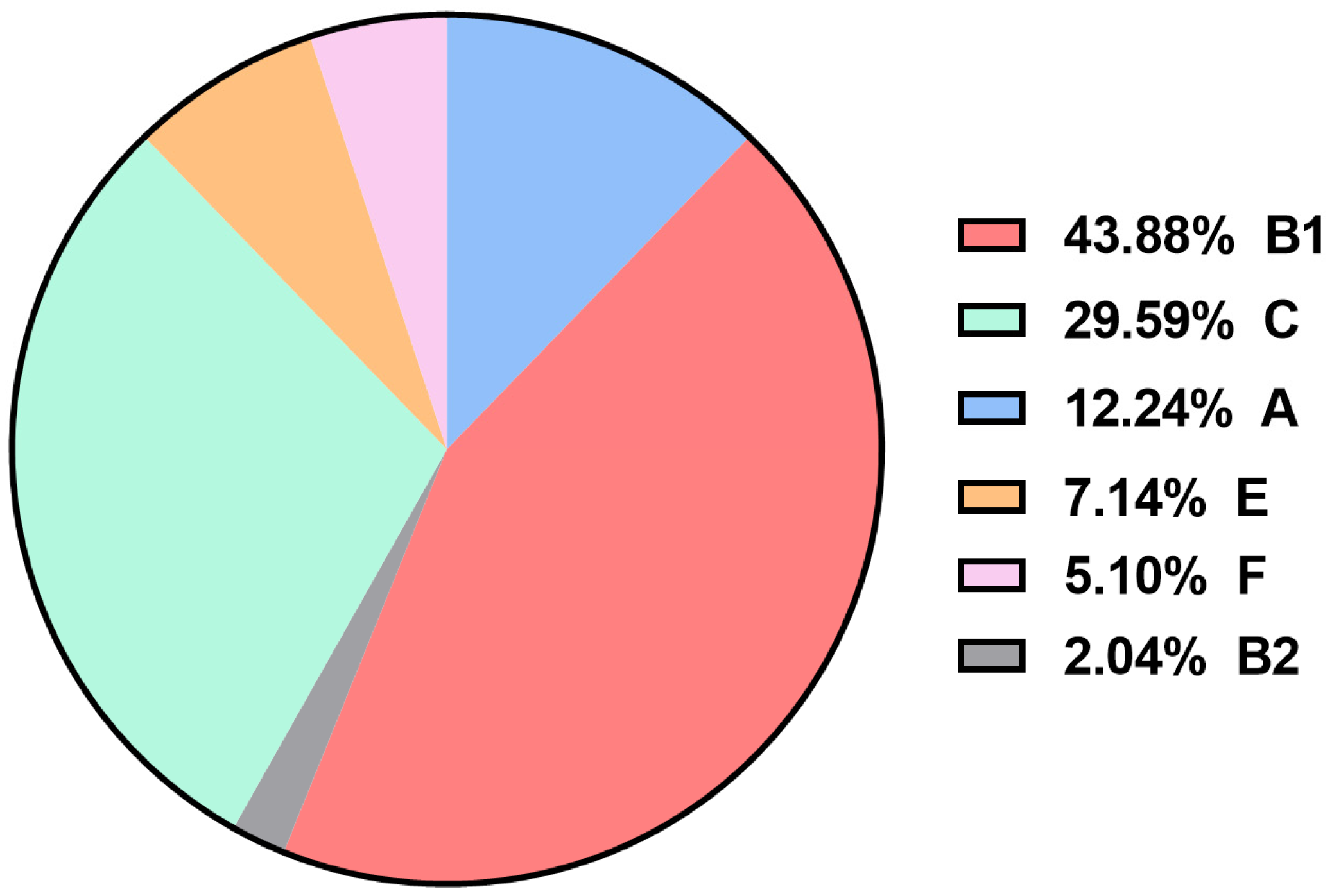
Phylogenetic analysis based on the novel Clermont’s phylogenetic typing scheme showed that most of the APEC isolates belonged to phylogroup B1 (43.88%, 43/98), C (29.59%, 29/98), and A (12.24%, 12/98). Only (7.14%, 7/98) of the APEC isolates belonged to phylogroup E, (5.10%, 5/98) to phylogroup F, and (2.04%, 2/98) to phylogroup B2, while none of the APEC isolates belonged to phylogroup D or clade I (Figure 2 and Table S3).

3.4. Virulence Gene Detection and Distribution

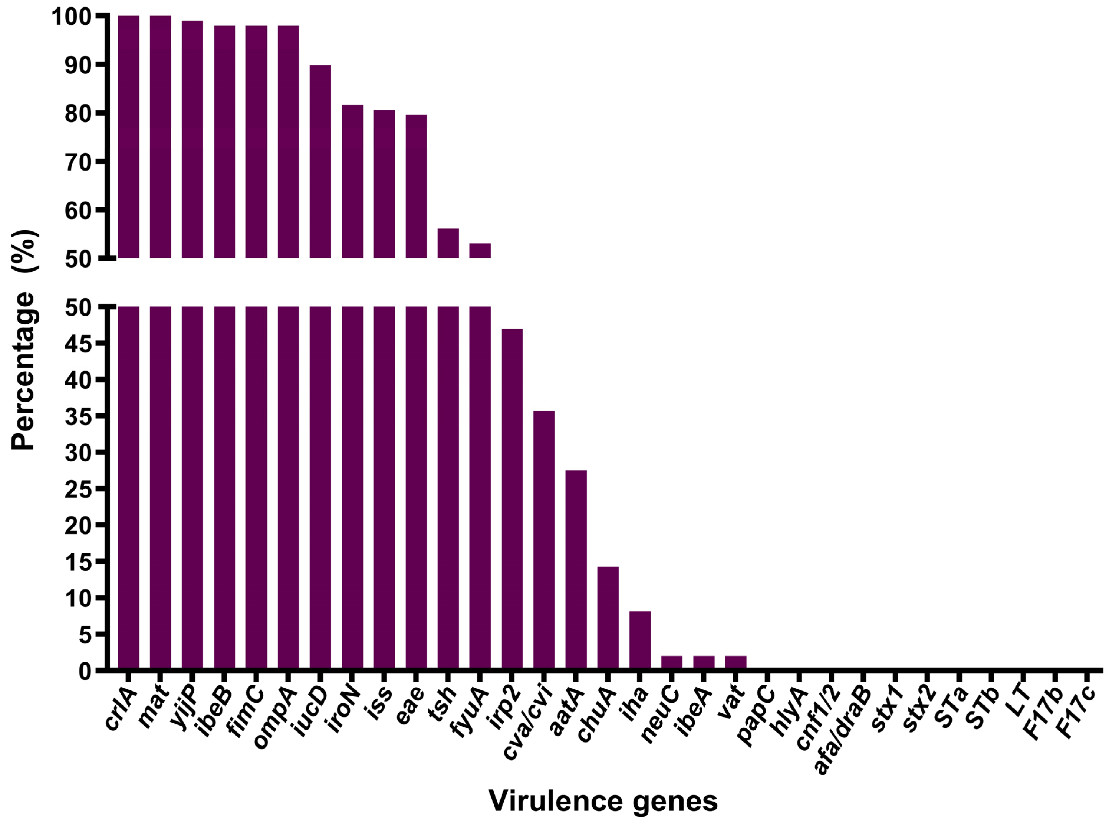
The virulence genotyping showed that mat and crlA genes were present in all 98 (100%) APEC isolates. The yijP gene was detected in 97 (98.98%) isolates. fimC, ibeB, and ompA were found in 96 (97.96%) isolates. The iucD, iroN, iss, and eae genes were present in 88 (89.80%), 80 (81.63%), 79 (80.61%), and 78 (79.59%) isolates, respectively. tsh, fyuA, irp2, cva/cvi, and aatA were identified in 55 (56.12%), 52 (53.06%), 46 (46.94%), 35 (35.71%), and 27 (27.55%) isolates, respectively. Additionally, 14 (14.29%) isolates tested positive for chuA and 8 (8.16%) for iha, while ibeA, vat, and neuC were only detected in 2 (2.04%) isolates. None of the APEC isolates carried papC, cnf1/2, stx1, stx2, F17b, F17c, hlyA, STa, STb, LT, or afa/draB genes (Figure 3 and Table S4). All APEC isolates in this study contained at least eight virulence genes.

3.5. Distribution of Virulence Genes Across Serotypes and Phylogenetic Groups

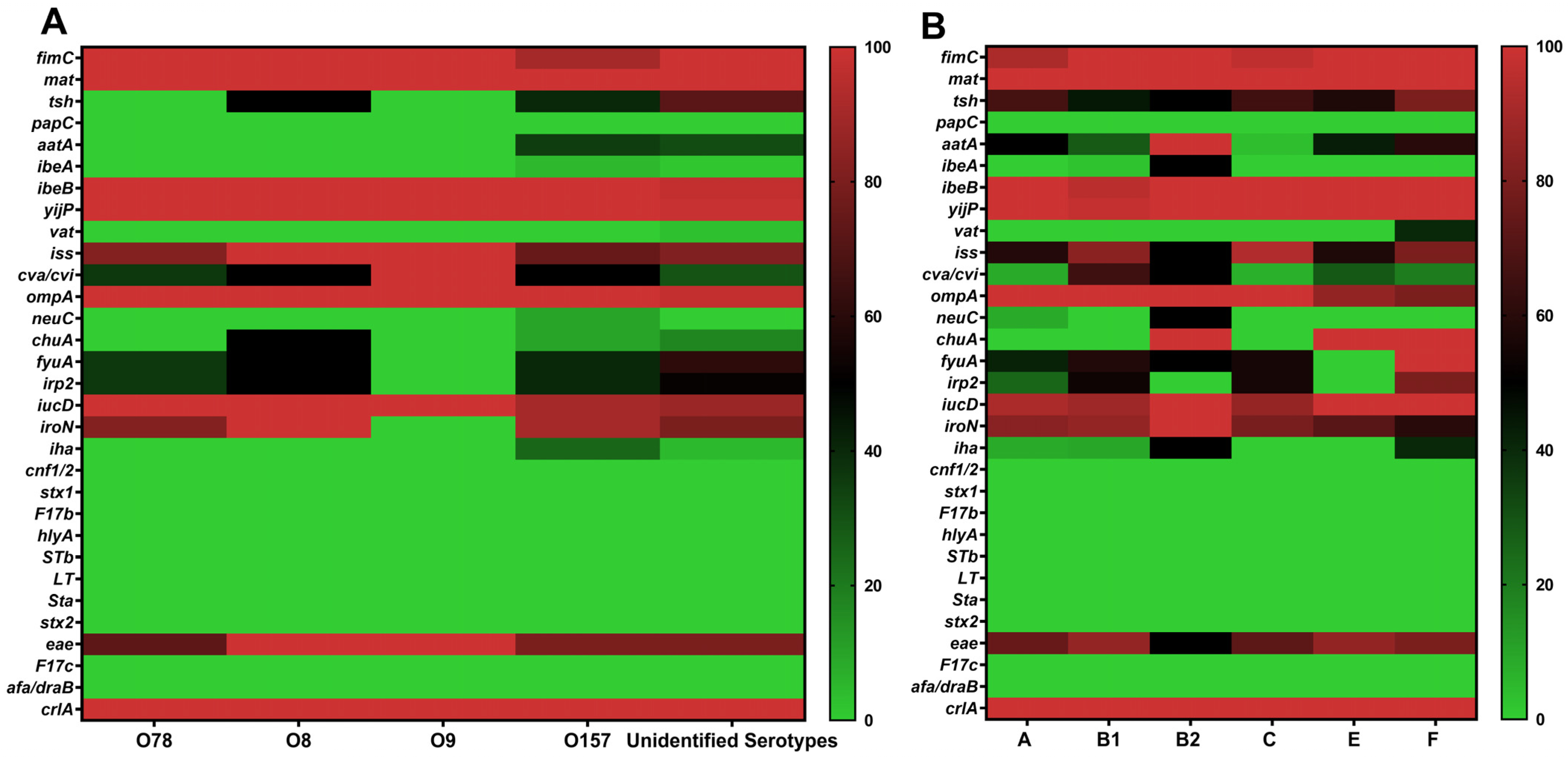
The relationship between virulence genes and serotypes was examined (Table 1 and Figure 4A). The fimC, ibeB, yijP, iss, ompA, iucD, eae, and crlA genes showed a similar distribution among serotypes O157, O78, O8, O9, and unidentified serotype isolates. The cva/cvi gene had relatively similar percentages in O157, O78, O8, and the unidentified serotypes was also found in the single O9 isolate. The mat, iroN, fyuA, and irp2 genes, although present at comparable frequencies in serotypes O157, O78, O8, and unidentified serotype isolates, were absent in O9. Among the studied genes, aatA had a comparable detection rate in O157 (35%) and unidentified serotype isolates (31.25%), but it was completely absent in O78, O8, and O9. The chuA gene was detected in 50% of O8 isolates; in contrast, it was found in 10% of O157 and 17.19% of unidentified serotype isolates and not in O78 and O9. The iha gene was present in 25% of O157 isolates; however, its prevalence in unidentified serotype isolates was much lower (4.69%), and it was not detected in O78, O8, or O9. The neuC gene was only found in the O157 serotype (10%) and not detected in O78, O8, O9, or unidentified serotype isolates. The ibeA gene was present in a single O157 isolate (5%) and in one unidentified serotype isolate (1.56%), but not in O78, O8, or O9. The vat gene was absent in all defined serotypes (O157, O78, O8, and O9), with detection only in 3.13% of unidentified serotype isolates. The tsh gene was found in 40% of O157 isolates and was more common in unidentified serotype isolates (over 70%). It was detected in 50% of O8 isolates but was absent in O78 and O9 (Table 1 and Figure 4A).
The association between virulence genes and the identified phylogenetic groups was investigated (Table 2 and Figure 4B). The crlA, fimC, mat, ibeB, yijP, and iucD genes were significantly distributed across all phylogroups. In contrast, the iha, iroN, ibeA, neuC, and aatA genes were predominantly detected in phylogroup B2, with frequencies of either 50% or 100%, compared to the other groups. The vat gene was exclusively identified in phylogroup F (40%). Among the iron acquisition-related genes, chuA was widely distributed in phylogroups B2, E, and F (100% each), while irp2 and fyuA were highly prevalent in phylogroup F (80% and 100%, respectively). The intimin-encoding gene eae was less frequently detected in phylogroup B2 than in the other groups. The ompA gene was universally present in phylogroups A, B1, B2, and C (100% each), and slightly less prevalent in groups E and F (85.71% and 80%, respectively). The iss gene was strongly associated with isolates from phylogroups C, B1, and F, with respective frequencies of 93.10%, 83.72%, and 80%, compared to lower frequencies observed in the remaining groups. The iucD gene was predominantly found in isolates belonging to phylogroups B2, E, and F, while cva/cvi were mainly associated with phylogroup B1 (Table 2 and Figure 4B).
Notably, the co-occurrence of some pivotal virulence genes was observed in phylogroups. In phylogroup B2, one isolate, out of a total of two identified in this group, simultaneously carried neuC and ibeA. Similarly, in phylogroup F, two isolates harbored a combination of fyuA, chuA, irp2, and vat genes (Table 2 and Table S7).
As shown in Table 2, phylogenetic group F had the highest mean virulence score (MVS) of 14.20, followed closely by phylogenetic group B2 with an MVS of 13.50. While phylogenetic groups A, B1, C, and E showed comparable MVS, ranging from 11.08 to 11.97. The MVS for each phylogenetic group was calculated by dividing the total number of virulence genes identified in isolates of the same group (excluding the diarrheagenic gene eae) by the number of isolates within that group, as described by El-Shaer et al. [35].

3.6. Antibiotic Susceptibility Testing of APEC

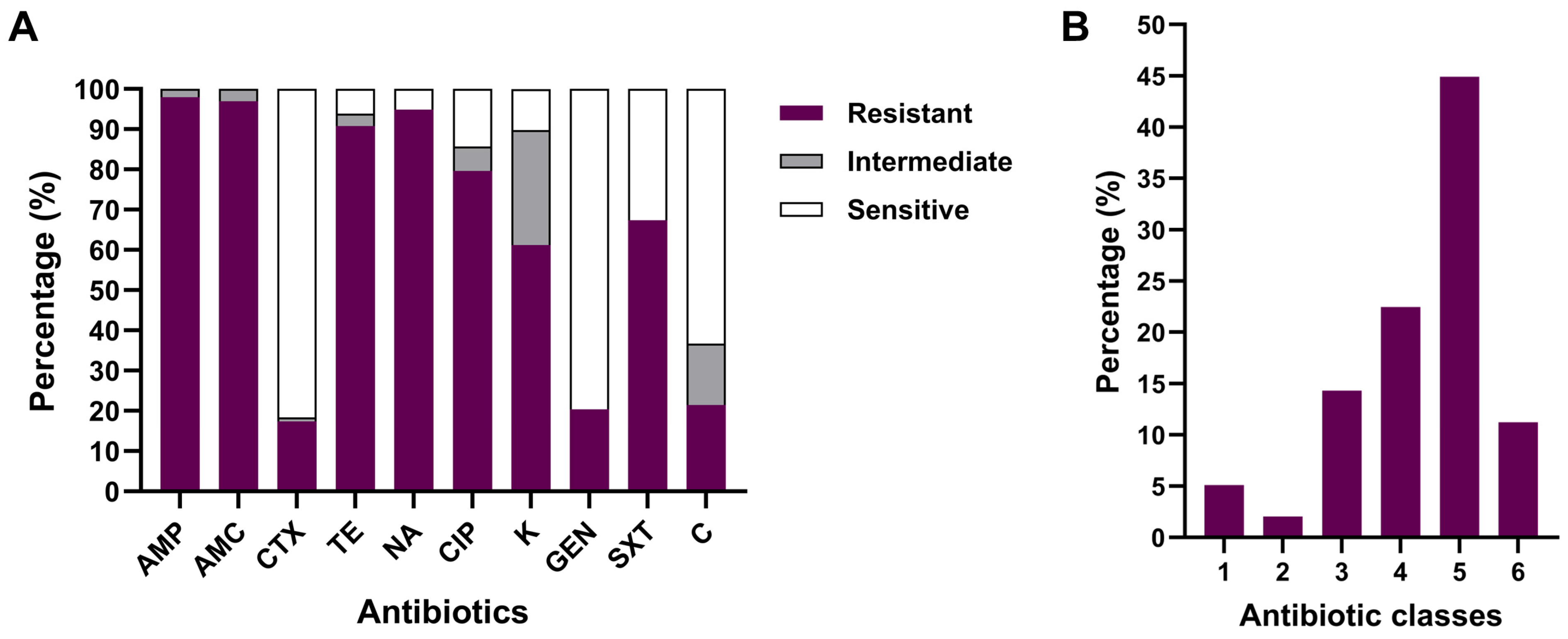
Our results indicated high levels of resistance toward ampicillin (97.96%), amoxicillin–clavulanic acid (96.94%), nalidixic acid (94.90%), tetracycline (90.82%), and ciprofloxacin (79.59%). Moderate resistance rates of 67.35% and 61.22% were recorded for trimethoprim–sulfamethoxazole and kanamycin, respectively, whereas low frequencies of resistance to chloramphenicol (21.40%), gentamicin (20.40%), and cefotaxime (17.30%) were observed (Figure 5A and Table S5). In addition, 91 isolates (92.86%) were resistant to at least three different families of antibiotics and consequently classified as multidrug-resistant (MDR) according to the criteria of Sweeney et al. [36]. The proportion of the APEC isolates that exhibited resistance to 5, 4, and 3 antibiotic categories was 44 (44.90%), 22 (22.45%), and 14 (14.29%), respectively, while 11 (11.22%), 5 (5.10%), and 2 (2.04%) of the APEC isolates were resistant to a panel of 6, 1, and 2 antibiotic classes (Figure 5B and Table S6), respectively.

4. Discussion

Our findings showed that the majority of the isolates were unidentified serotypes (64%). Previous studies conducted in Algeria and elsewhere have also reported a high frequency of unidentified serotypes of APEC isolates [37,38].
Unexpectedly, among the typeable isolates, O157 was the most frequently observed serotype, accounting for 20.41% of the tested APEC isolates. In Albania, E. coli O157 has been reported at a low prevalence among APEC isolates from birds affected by colibacillosis (7.67%) [39], whereas in Egypt, O157 has recently been identified as one of the predominant serotypes, with a prevalence similar to that observed in our study (20%) [40]. The pathogenicity of this serotype has been experimentally demonstrated in one-day-old chicks [41]. The O157 serotype is classically associated with Shiga-toxin-producing E. coli (STEC O157:H7), one of the most notorious foodborne pathogens causing severe human diseases such as hemorrhagic colitis and hemolytic uremic syndrome (HUS) [42]. The detection of serotype O157 in avian E. coli isolates suggests that poultry may serve as a potential zoonotic reservoir capable of transmitting these strains to humans [43].
None of the 98 APEC isolates were identified as belonging to serotypes O1 or O2, which is in accordance with previous reports from Egypt [44]. In contrast, a recent study conducted in northeast Algeria [20] demonstrated that serotypes O1 and O2 were detected in 31.30% and 33.20% of the isolates, respectively. The observed regional differences in Algeria suggest that APEC serotype distribution may be influenced by variations in poultry farming practices and local biosecurity measures. Furthermore, serotype O78 was observed at a prevalence of 11.22% in our study. This finding aligns with results obtained in Algeria and Poland, where approximately 14% of the isolates belonged to this serotype [20,45]. O78 has been widely reported as the most common serotype among APEC isolates in several countries [13,46,47]. Other serotypes identified in our study, such as O8 and O9, have also been documented in earlier research [45,48].
The analysis of the distribution of virulence genes among serotypes revealed that O157 possessed a multitude of such genes, several of which were also present in the classical APEC serotype O78. This suggests that the O157 serotype, most commonly associated with cattle, may represent an emerging serotype of APEC in Algeria, reflecting possible interspecies transmission dynamics within local poultry ecosystems. Similarly, O145, a serotype typically linked to Shiga-toxin-producing E. coli (STEC), has been identified as an APEC serotype of concern in China [49].
Our results, together with data from Egypt, suggest a changing trend within the population of disease-causing APEC serotypes in North African poultry production systems, likely driven by regional factors, and warrant special attention and continuous monitoring.
The species E. coli comprises eight distinct phylogenetic groups (A, B1, B2, C, D, E, F, and clade I) [10]. In the present study, phylogenetic analysis revealed a significant predominance of groups B1 and C, followed by A, E, F, and B2. These findings align with recent studies from South Korea and Brazil, which reported that most APEC isolates predominantly belong to phylogenetic group B1 [50,51]. A notable proportion (65%) of APEC isolates associated with this group were positive for the ColV plasmid, as confirmed by the presence of cva/cvi genes. These results indicate that, even in strains belonging to phylogenetic groups other than B2, the acquisition of virulent plasmids such as ColV can increase their ability to colonize extraintestinal sites and cause avian colibacillosis [5].
Previous investigations have shown that the distribution of phylogenetic groups among APEC populations varies in predominance between countries [11,51,52]. It appears that APEC differs from human ExPEC in terms of phylogenetic classification [11]. This difference may be explained by the fact that virulence genes essential for APEC pathogenesis are mainly plasmid-borne; consequently, Clermont’s phylogenetic grouping based on a limited number of chromosomal markers is often an inadequate method for accurately identifying the APEC pathotype [53].
APEC possesses diverse iron acquisition systems, including siderophores (yersiniabactin, aerobactin, salmochelin) as well as transporters to capture iron from the host’s body fluids [54]. These siderophores and transporters not only contribute to iron acquisition but also participate in APEC adhesion, invasion, the expression of additional virulence genes, protection from environmental stresses, colonization, and persistence in the host [1]. A significant proportion of the APEC isolates analyzed in this study harbored genes involved in iron scavenging mechanisms, such as iucD, iroN, fyuA, and irp2, with frequencies of 89.80%, 81.63%, 53.06%, and 46.94%, respectively. These findings are relatively comparable to reports from earlier studies conducted in China and Portugal [48,55]. In prior studies, the iucD, iroN, fyuA, and irp2 genes were reported to be more prevalent in APEC isolates than in AFEC isolates [8,55].
APEC isolates also exhibited a notable prevalence of adhesin genes, specifically mat (100%), crlA (100%), fimC (97.96%), and tsh (56.12%), whereas aatA was detected less frequently, with a prevalence of 27.55%. These findings align with previous research on ExPEC associated with Mink Hemorrhagic Pneumonia in China [56], which reported frequencies of 89.40%, 96.50%, and 15.3% for the mat, fimC, and aatA genes, respectively. In line with our data, Al-Kandari and Woodward [57] reported that the crlA gene, involved in regulating the curli fimbrial operon, was present in all APEC isolates (100%). Furthermore, Awawdeh et al. [58] observed a comparable prevalence of the tsh gene (55%) in Australia.
It is noteworthy that, in contrast to a previous study conducted in Algeria [15], which reported that all APEC isolates tested negative for the eae gene, our findings revealed a significantly higher prevalence (79.59%). The eae gene, located within a pathogenicity island known as the Locus of Enterocyte Effacement (LEE) in the genomes of Enteropathogenic E. coli (EPEC) and Enterohemorrhagic E. coli (EHEC), encodes intimin, which is an outer membrane adhesin protein involved in the development of attaching and effacing (A/E) lesions by mediating the intimate adherence of these pathogens to enterocytes [59]. Although none of the examined APEC isolates carried the stx1, stx2, STa, STb, or LT genes, the presence of the eae gene might nonetheless pose a serious risk to human beings. Consequently, further comparative molecular investigations between eae-positive APEC isolates and human diarrheagenic E. coli (EPEC, EHEC) are warranted to assess the zoonotic potential of APEC.
In the current study, consistent with results from previous reports [48,60], genes encoding invasins, yijP, and ibeB were present in 98.98% and 97.96% of the APEC isolates, respectively. The high prevalence of the yijP gene in APEC isolates suggests that it may play a pivotal role in APEC pathogenesis by facilitating the bacterial traversal of the blood–brain barrier, as this gene is known to be involved in the invasion of human brain microvascular endothelial cells (BMECs) by E. coli [61].
Regarding the genes encoding protectins, the ompA and iss were identified in 97.96% and 79.59% of the APEC isolates, respectively. These data are consistent with previous research conducted in China [48].
The colicin V (ColV) plasmid operon genes cva/cvi were detected in 35.71% of the APEC isolates. Similar findings have been reported in APEC isolates from broiler breeders in Mississippi, USA [4]. The low prevalence of these genes among the tested isolates does not align with the high frequency of other ColV plasmid-related genes, namely, iroN, iss, tsh, and iucD [62]. This disparity may be due to alternative genetic carriers other than ColV plasmids, such as ColBM-type plasmids, or it may result from rearrangements or deletions within parts of the ColV operon [63].
The IrgA homolog adhesin (Iha), encoded by the iha gene, has been shown to contribute to the urovirulence of UPEC strains, participating in both adhesion and iron uptake as a catecholate siderophore receptor [64,65]. As part of this study, the iha gene was found in 8.16% of the APEC isolates, and despite being rare, its presence suggests a potential role in APEC pathogenesis and may be indicative of overlapping pathogenic strategies between human and avian ExPEC strains. Further studies are needed to investigate the contribution of Iha to APEC virulence and its potential utility as a diagnostic or epidemiological marker for high-risk strains posing threats to both poultry and human health.
Interestingly, among all isolates examined, a single isolate carrying both the capsule K1 biosynthesis (neuC) and invasion-related (ibeA) genes was identified within phylogenetic group B2. Although rare, this combination is noteworthy for its potential role in enhancing immune evasion and invasion capabilities, which may increase the systemic virulence of phylogenetic group B2 strains [12,66], as this group has been shown to demonstrate higher pathogenicity in experimental chicken infection models [51]. The presence of this gene combination in APEC, which is characteristic of human NMEC strains, particularly within phylogenetic group B2, suggests a possible zoonotic threat [28,67].
An experimental study demonstrated that deleting the yersiniabactin system genes, fyuA and irp2, from the APEC genome significantly reduced the transcription of virulence genes, decreased adherence to DF-1 cells (immortalized chicken embryo fibroblasts), and attenuated pathogenicity in chicks [68]. Furthermore, the vacuolating auto transporter toxin, encoded by the vat gene, has been shown to induce vacuolating cytotoxic activity in chicken embryonic fibroblasts and contributes to the development of cellulitis as well as the respiratory/systemic form of avian colibacillosis [69]. In addition to their role in avian hosts, it has been demonstrated that E. coli strains carrying a gene combination of vat, fyuA, chuA, and yfcV are capable of efficiently colonizing the urinary tract [70]. In the present study, among phylogenetic group F isolates, two distinct virulence profiles were observed, each represented by a single isolate, and both characterized by the simultaneous presence of chuA, fyuA, irp2, and vat. These virulence profiles correlated with isolates exhibiting the highest MVS. These results support previous observations indicating that isolates originating from phylogroup F may be highly virulent to chickens and may act as the primary cause of colibacillosis [71]. Moreover, E. coli of avian origin from phylogroup F has recently been the subject of research, suggesting its potential zoonotic risk [72]
In vitro antimicrobial susceptibility tests of the 98 APEC isolates showed alarming levels of resistance to ampicillin (97.95%), amoxicillin–clavulanic acid (96.93%), nalidixic acid (94.89%), tetracycline (90.81%), and ciprofloxacin (79.59%). In contrast, the resistance rates for trimethoprim–sulfamethoxazole (67.34%) and kanamycin (61.22%) were relatively moderate. These findings align with a prior report from Eastern Algeria, demonstrating high resistance to ampicillin, nalidixic acid, tetracycline, and ciprofloxacin [73]. The observed levels of antibiotic resistance are not surprising given the excessive and inappropriate use of these antimicrobial agents in the Algerian poultry sector. Additionally, the notable resistance of APEC to antibiotics considered critically important for human health, such as beta-lactams, fluoroquinolones, and tetracyclines, could potentially reduce their effectiveness in treating human infections [74].
Resistance to cefotaxime (17.34%) is lower than previously reported in Algeria and Qatar [73,75]. Even though third-generation cephalosporins have not been used in Algerian poultry production systems, the occurrence of extended-spectrum cephalosporin-resistant E. coli presenting the extended-spectrum beta-lactamase (ESBL)-producing phenotype has been documented previously [20]. In addition, ESBL genes, including blaTEM-141 and blaCTX-M-55, carried on conjugative plasmids (IncF-type), can be horizontally transferred between E. coli and related species such as E. albertii [76]. From a One Health perspective, this horizontal gene transfer represents a potential risk to human and animal health, as these plasmids can disseminate resistance across bacterial populations in animals, humans, and the environment [77]. Continuous monitoring of plasmid-mediated resistance in livestock is therefore essential to anticipate zoonotic transmission and to guide antimicrobial stewardship strategies.
In this study, a low resistance level to gentamicin (20.41%) was observed. This finding aligns with results described in prior studies in Algeria and Turkey [21,78]. The noted resistance to gentamicin, despite being at low levels, may be attributed to its unofficial use, knowing that this antimicrobial has been banned for veterinary use in Algeria.
The resistance rate to chloramphenicol in our study was relatively low (21.43%). This is in agreement with findings from other researchers who have reported comparable levels of resistance [20,73]. Despite chloramphenicol being prohibited in Algeria since 2006 [79], some E. coli strains remain resistant, probably due to co-resistance, which occurs as a result of the frequent use of other antibiotic molecules. Reports have indicated that chloramphenicol co-resistance in E. coli, isolated from diseased cattle and swine, can arise from the use of other antibiotics such as ampicillin, dihydrostreptomycin, kanamycin, trimethoprim, sulfamethoxazole, and tetracycline [80,81].
In addition to resistance to each antibiotic, our findings indicated that 92.86% of the APEC isolates were multidrug-resistant (MDR), highlighting a significant concern. This is consistent with previous studies from Algeria (92%) [82], Tunisia (96%) [83], and China (100%) [48]. Indeed, the arsenal of effective antibiotics available for treating APEC infections has become increasingly limited, leading to a therapeutic impasse.

5. Conclusions

To the best of our knowledge, this work reports, for the first time, the potential emergence of O157 as one of the predominant serotypes of APEC in Algeria. This finding may indicate the possible existence of other serotypes associated with avian colibacillosis that have not yet been identified.
Additionally, the APEC isolates examined were predominantly assigned to phylogenetic groups B1 and C. These isolates presented a notable prevalence of various virulence genes commonly associated with the ExPEC pathotype, as well as high antibiotic resistance. Furthermore, our findings demonstrated that some APEC isolates from phylogenetic groups B2 and F share key virulence determinants with human ExPEC. These APEC may exhibit high virulence in avian hosts and could either act as human ExPEC or serve as a reservoir for their virulence genes. However, further investigations with a larger sample size are required to evaluate the extent of this genetic overlap, as the number of APEC isolates identified in phylogenetic groups B2 and F in this research was relatively low.
These findings underscore the importance of continuous surveillance of these pathogens in poultry flocks and the need for judicious antibiotic use to ensure effective management of APEC infections and mitigate the risk of zoonotic transmission.

Supplementary Materials

The following supporting information can be downloaded at https://www.mdpi.com/article/10.3390/ani15223324/s1, Table S1: PCR primers used to detect virulence and O-antigen genes in APEC; Table S2: Serotyping results of APEC isolates; Table S3: Distribution of phylogenetic groups among APEC isolates; Table S4: Frequencies of virulence genes in the 98 APEC isolates; Table S5: Prevalence of sensitive, intermediate, and resistant APEC isolates; Table S6: Prevalence of multidrug resistance (MDR) in APEC isolates; Table S7: Details of the APEC isolates.

Author Contributions

Conceptualization, S.W., I.B. and A.H.; methodology, S.W.; software, I.B.; validation, I.B., J.H., B.Z., X.W. and S.W.; formal analysis, I.B. and S.A.; investigation, I.B., J.H., B.Z., X.W., M.F. and S.W.; resources, I.B., J.H., B.Z., X.W., M.F. and S.W.; data curation, I.B., J.H., B.Z., X.W. and S.W.; writing—original draft preparation, I.B.; writing—review and editing, S.A. and S.W.; visualization, I.B.; supervision, A.H. and S.W.; project administration, S.W.; funding acquisition, I.B. and S.W. All authors have read and agreed to the published version of the manuscript.

Funding

This research was funded by the National Natural Science Foundation of China (32172856, 32302881), the Natural Science Foundation of Shanghai (23ZR1476600), Shanghai Magnolia Talent Program Pujiang Project (23PJ1416000), and the Agricultural Science and Technology Innovation Program of the Chinese Academy of Agricultural Sciences (CAAS-ASTIP-2021-SHVRI-07).

Institutional Review Board Statement

Ethical review and approval were not required for this study due to the source of the samples. Organ samples were obtained during post-mortem examinations of carcasses from birds that were already deceased. Poultry farmers voluntarily brought the birds to the Avian Pathology Service of the Institute of Veterinary Sciences for routine diagnostic purposes.

Informed Consent Statement

Not applicable.

Data Availability Statement

The data presented in this study are available within the manuscript.

Acknowledgments

We express our sincere gratitude for the valuable assistance and support provided by the staff of the Microbiology Laboratory and the Avian Pathology Service at the Institute of Veterinary Sciences of Tiaret.

Conflicts of Interest

The authors declare no conflicts of interest.

References

  1. Kathayat, D.; Lokesh, D.; Ranjit, S.; Rajashekara, G. Avian Pathogenic Escherichia coli (APEC): An Overview of Virulence and Pathogenesis Factors, Zoonotic Potential, and Control Strategies. Pathogens 2021, 10, 467. [Google Scholar] [CrossRef]
  2. Nolan, L.K.; Vaillancourt, J.-P.; Barbieri, N.L.; Logue, C.M. Colibacillosis. In Diseases of Poultry; John Wiley & Sons, Ltd.: Hoboken, NJ, USA, 2020; pp. 770–830. ISBN 978-1-119-37119-9. [Google Scholar] [CrossRef]
  3. Guabiraba, R.; Schouler, C. Avian Colibacillosis: Still Many Black Holes. FEMS Microbiol. Lett. 2015, 362, fnv118. [Google Scholar] [CrossRef]
  4. Joseph, J.; Jennings, M.; Barbieri, N.; Zhang, L.; Adhikari, P.; Ramachandran, R. Characterization of Avian Pathogenic Escherichia coli Isolated from Broiler Breeders with Colibacillosis in Mississippi. Poultry 2023, 2, 24–39. [Google Scholar] [CrossRef]
  5. Johnson, T.J.; Wannemuehler, Y.; Doetkott, C.; Johnson, S.J.; Rosenberger, S.C.; Nolan, L.K. Identification of Minimal Predictors of Avian Pathogenic Escherichia coli Virulence for Use as a Rapid Diagnostic Tool. J. Clin. Microbiol. 2008, 46, 3987–3996. [Google Scholar] [CrossRef]
  6. Kemmett, K.; Humphrey, T.; Rushton, S.; Close, A.; Wigley, P.; Williams, N.J. A Longitudinal Study Simultaneously Exploring the Carriage of APEC Virulence Associated Genes and the Molecular Epidemiology of Faecal and Systemic E. coli in Commercial Broiler Chickens. PLoS ONE 2013, 8, e67749. [Google Scholar] [CrossRef] [PubMed]
  7. Schouler, C.; Schaeffer, B.; Brée, A.; Mora, A.; Dahbi, G.; Biet, F.; Oswald, E.; Mainil, J.; Blanco, J.; Moulin-Schouleur, M. Diagnostic Strategy for Identifying Avian Pathogenic Escherichia coli Based on Four Patterns of Virulence Genes. J. Clin. Microbiol. 2012, 50, 1673–1678. [Google Scholar] [CrossRef] [PubMed]
  8. Rodriguez-Siek, K.E.; Giddings, C.W.; Doetkott, C.; Johnson, T.J.; Nolan, L.K. Characterizing the APEC Pathotype. Vet. Res. 2005, 36, 241–256. [Google Scholar] [CrossRef] [PubMed]
  9. Watts, A.; Wigley, P. Avian Pathogenic Escherichia coli: An Overview of Infection Biology, Antimicrobial Resistance and Vaccination. Antibiotics 2024, 13, 809. [Google Scholar] [CrossRef]
  10. Clermont, O.; Christenson, J.K.; Denamur, E.; Gordon, D.M. The Clermont Escherichia coli Phylo-Typing Method Revisited: Improvement of Specificity and Detection of New Phylo-Groups. Environ. Microbiol. Rep. 2013, 5, 58–65. [Google Scholar] [CrossRef]
  11. Logue, C.M.; Wannemuehler, Y.; Nicholson, B.A.; Doetkott, C.; Barbieri, N.L.; Nolan, L.K. Comparative Analysis of Phylogenetic Assignment of Human and Avian ExPEC and Fecal Commensal Escherichia coli Using the (Previous and Revised) Clermont Phylogenetic Typing Methods and Its Impact on Avian Pathogenic Escherichia coli (APEC) Classification. Front. Microbiol. 2017, 8, 283. [Google Scholar] [CrossRef]
  12. Hu, J.; Afayibo, D.J.A.; Zhang, B.; Zhu, H.; Yao, L.; Guo, W.; Wang, X.; Wang, Z.; Wang, D.; Peng, H. Characteristics, Pathogenic Mechanism, Zoonotic Potential, Drug Resistance, and Prevention of Avian Pathogenic Escherichia coli (APEC). Front. Microbiol. 2022, 13, 1049391. [Google Scholar] [CrossRef]
  13. Awad, A.; Arafat, N.; Elhadidy, M. Genetic Elements Associated with Antimicrobial Resistance among Avian Pathogenic Escherichia coli. Ann. Clin. Microbiol. Antimicrob. 2016, 15, 59. [Google Scholar] [CrossRef]
  14. Velhner, M.; Todorović, D.; Grego, E.; Kišek, T.C.; Ljubojević, D.; Špik, V.C.; Pajić, M.; Kozoderović, G. Characterisation of Multidrug Resistant Escherichia coli from Poultry Litter and Poultry Carrying Virulence Genes for Evaluation of Poultry Farm Management. Eur. Poult. Sci. 2020, 84, 1–15. [Google Scholar] [CrossRef]
  15. Ranabhat, G.; Subedi, D.; Karki, J.; Paudel, R.; Luitel, H.; Bhattarai, R.K. Molecular Detection of Avian Pathogenic Escherichia coli (APEC) in Broiler Meat from Retail Meat Shop. Heliyon 2024, 10, e35661. [Google Scholar] [CrossRef]
  16. Ahmed, Z.S.; Hashad, M.E.; Atef, Y.; Badr, H.; Elhariri, M.; Kadry, M. Public Health Threat of Antimicrobial Resistance and Virulence Genes in Escherichia coli from Human-Chicken Transmission in Egypt. Sci. Rep. 2025, 15, 12627. [Google Scholar] [CrossRef] [PubMed]
  17. Ali, F.; Hacène, I.; Mohamed, B. Structuring and Development of Poultry Sectors in Algeria: Limits of Modernization Policies: The Case of the” Turkey” Sector (2000–2020). Agric. Sci. Dig. 2024, 44, 1147–1153. [Google Scholar] [CrossRef]
  18. Alloui, N.; Ayachi, A. Biosecurity Practices in Algerian Poultry Farms. Online J. Anim. Feed Res. 2012, 2, 80–83. [Google Scholar]
  19. Kamel, K.; Merghad, A.; Barour, D.; Eddine Gherissi, D.; Khenenou, T. High Antimicrobial Resistance Rates and Multidrug Resistance in Enterobacteriaceae Isolates from Poultry in Souk Ahras Region, Algeria. Vet. World 2024, 17, 2709–2718. [Google Scholar] [CrossRef] [PubMed]
  20. Chenouf, N.S.; Messaï, C.R.; Carvalho, I.; Álvarez-Gómez, T.; Silva, V.; Zitouni, A.; Hakem, A.; Poeta, P.; Torres, C. Serogrouping and Molecular Characterization of ESBL-Producing Avian Pathogenic Escherichia coli from Broilers and Turkeys with Colibacillosis in Algeria. Antibiotics 2025, 14, 356. [Google Scholar] [CrossRef]
  21. Mohamed, L.; Ge, Z.; Yuehua, L.; Yubin, G.; Rachid, K.; Mustapha, O.; Junwei, W.; Karine, O. Virulence Traits of Avian Pathogenic (APEC) and Fecal (AFEC) E. coli Isolated from Broiler Chickens in Algeria. Trop. Anim. Health Prod. 2018, 50, 547–553. [Google Scholar] [CrossRef]
  22. Hu, Q.; Tu, J.; Han, X.; Zhu, Y.; Ding, C.; Yu, S. Development of Multiplex PCR Assay for Rapid Detection of Riemerella Anatipestifer, Escherichia coli, and Salmonella Enterica Simultaneously from Ducks. J. Microbiol. Methods 2011, 87, 64–69. [Google Scholar] [CrossRef]
  23. Iguchi, A.; Iyoda, S.; Seto, K.; Morita-Ishihara, T.; Scheutz, F.; Ohnishi, M. Escherichia coli O-Genotyping PCR: A Comprehensive and Practical Platform for Molecular O Serogrouping. J. Clin. Microbiol. 2015, 53, 2427–2432. [Google Scholar] [CrossRef] [PubMed]
  24. Wang, S.; Meng, Q.; Dai, J.; Han, X.; Han, Y.; Ding, C.; Liu, H.; Yu, S. Development of an Allele-Specific PCR Assay for Simultaneous Sero-Typing of Avian Pathogenic Escherichia coli Predominant O1, O2, O18 and O78 Strains. PLoS ONE 2014, 9, e96904. [Google Scholar] [CrossRef]
  25. Wei, J.; Zhu, H.; Zhang, B.; Zhang, Y.; Wang, Y.; Yao, L.; Afayibo, D.J.A.; Qi, J.; Tian, M.; Ding, C.; et al. Establishment and Application of Multiplex PCR Method for Escherichia coli O8/O9/O149 and O157 Serotypes. Chin. J. Vet. Sci. 2022, 52, 691–697. [Google Scholar] [CrossRef]
  26. Meng, Q.; Wang, S.; Han, X.; Han, Y.; Ding, C.; Dai, J.; Yu, S. Multiplex PCR Assay for Detection of Virulence Genes in Avian Pathogenic Escherichia coli. Wei Sheng Wu Xue Bao = Acta Microbiol. Sin. 2014, 54, 696–702. [Google Scholar] [CrossRef]
  27. Maurer, J.J.; Brown, T.P.; Steffens, W.L.; Thayer, S.G. The Occurrence of Ambient Temperature-Regulated Adhesins, Curli, and the Temperature-Sensitive Hemagglutinin Tsh among Avian Escherichia coli. Avian Dis. 1998, 42, 106–118. [Google Scholar] [CrossRef]
  28. Ewers, C.; Li, G.; Wilking, H.; Kieβling, S.; Alt, K.; Antáo, E.-M.; Laturnus, C.; Diehl, I.; Glodde, S.; Homeier, T. Avian Pathogenic, Uropathogenic, and Newborn Meningitis-Causing Escherichia coli: How Closely Related Are They? Int. J. Med. Microbiol. 2007, 297, 163–176. [Google Scholar] [CrossRef]
  29. Wang, S.; Xia, Y.; Dai, J.; Shi, Z.; Kou, Y.; Li, H.; Bao, Y.; Lu, C. Novel Roles for Autotransporter Adhesin AatA of Avian Pathogenic Escherichia coli: Colonization during Infection and Cell Aggregation. FEMS Immunol. Med. Microbiol. 2011, 63, 328–338. [Google Scholar] [CrossRef]
  30. Wang, S.; Zhang, S.; Liu, Z.; Liu, P.; Shi, Z.; Wei, J.; Shao, D.; Li, B.; Ma, Z. Molecular Characterization of Enterohemorrhagic E. coli O157 Isolated from Animal Fecal and Food Samples in Eastern China. Sci. World J. 2014, 2014, 946394. [Google Scholar] [CrossRef]
  31. Bihannic, M.; Ghanbarpour, R.; Auvray, F.; Cavalié, L.; Châtre, P.; Boury, M.; Brugère, H.; Madec, J.-Y.; Oswald, E. Identification and Detection of Three New F17 Fimbrial Variants in Escherichia coli Strains Isolated from Cattle. Vet. Res. 2014, 45, 76. [Google Scholar] [CrossRef]
  32. Nowaczek, A.; Dec, M.; Stępień-Pyśniak, D.; Urban-Chmiel, R.; Marek, A.; Różański, P. Antibiotic Resistance and Virulence Profiles of Escherichia coli Strains Isolated from Wild Birds in Poland. Pathogens 2021, 10, 1059. [Google Scholar] [CrossRef]
  33. Wang, S.; Dai, J.; Meng, Q.; Han, X.; Han, Y.; Zhao, Y.; Yang, D.; Ding, C.; Yu, S. DotU Expression Is Highly Induced during in Vivo Infection and Responsible for Virulence and Hcp1 Secretion in Avian Pathogenic Escherichia coli. Front. Microbiol. 2014, 5, 588. [Google Scholar] [CrossRef] [PubMed]
  34. Clinical and Laboratory Standards Institute. Performance Standards for Antimicrobial Susceptibility Testing, 29th ed.; CLSI Supplement M100; Clinical and Laboratory Standards Institute: Wayne, PA, USA, 2019. [Google Scholar]
  35. El-Shaer, S.; Abdel-Rhman, S.H.; Barwa, R.; Hassan, R. Virulence Characteristics, Serotyping and Phylogenetic Typing of Clinical and Environmental Escherichia coli Isolates. Jundishapur J. Microbiol. 2018, 11, 1. [Google Scholar] [CrossRef]
  36. Sweeney, M.T.; Lubbers, B.V.; Schwarz, S.; Watts, J.L. Applying Definitions for Multidrug Resistance, Extensive Drug Resistance and Pandrug Resistance to Clinically Significant Livestock and Companion Animal Bacterial Pathogens. J. Antimicrob. Chemother. 2018, 73, 1460–1463. [Google Scholar] [CrossRef] [PubMed]
  37. Halfaoui, Z.; Menoueri, N.M.; Bendali, L.M. Serogrouping and Antibiotic Resistance of Escherichia coli Isolated from Broiler Chicken with Colibacillosis in Center of Algeria. Vet. World 2017, 10, 830. [Google Scholar] [CrossRef]
  38. Koutsianos, D.; Athanasiou, L.V.; Mossialos, D.; Franzo, G.; Cecchinato, M.; Koutoulis, K.C. Investigation of Serotype Prevalence of Escherichia coli Strains Isolated from Layer Poultry in Greece and Interactions with Other Infectious Agents. Vet. Sci. 2022, 9, 152. [Google Scholar] [CrossRef]
  39. Shtylla, T.; Circella, E.; Madio, A.; Boci, J.; Çabeli, P.; Kumbe, I.; Camarda, A. Biological characteristics and pathogenicity of avian Escherichia coli strains from Albanian poultry flocks. Sci. Pap. Ser. D Anim. Sci. 2012, 55, 11–14. [Google Scholar]
  40. Enany, M.E.; Ramadan, B.A.; Eid, S.; Abo Hashem, M.E.S. Prevalence and Serotyping of E. coli Isolated from Broiler Chickens at Ismailia Governorate. Suez Canal Vet. Med. J. SCVMJ 2024, 29, 393–401. [Google Scholar] [CrossRef]
  41. Azza, A.; Dahshan, A.H.M.; El-Nahass, E.-S.; Abd El-Mawgoud, A.I. Pathogenicity of Escherichia coli O157 in Commercial Broiler Chickens. Beni-Suef Univ. J. Basic Appl. Sci. 2018, 7, 620–625. [Google Scholar] [CrossRef]
  42. Ameer, M.A.; Wasey, A.; Salen, P. Escherichia coli (E. coli 0157 H7). In StatPearls; StatPearls Publishing LLC: Treasure Island, FL, USA, 2025; Available online: https://europepmc.org/article/nbk/nbk507845 (accessed on 25 August 2025).
  43. Dipineto, L.; Santaniello, A.; Fontanella, M.; Lagos, K.; Fioretti, A.; Menna, L.F. Presence of Shiga Toxin—Producing Escherichia coli O157:H7 in Living Layer Hens. Lett. Appl. Microbiol. 2006, 43, 293–295. [Google Scholar] [CrossRef]
  44. Awad, A.M.; El-Shall, N.A.; Khalil, D.S.; El-Hack, M.E.A.; Swelum, A.A.; Mahmoud, A.H.; Ebaid, H.; Komany, A.; Sammour, R.H.; Sedeik, M.E. Incidence, Pathotyping, and Antibiotic Susceptibility of Avian Pathogenic Escherichia coli Among Diseased Broiler Chicks. Pathogens 2020, 9, 114. [Google Scholar] [CrossRef]
  45. Wilczyński, J.; Stępień-Pyśniak, D.; Wystalska, D.; Wernicki, A. Molecular and Serological Characteristics of Avian Pathogenic Escherichia coli Isolated from Various Clinical Cases of Poultry Colibacillosis in Poland. Animals 2022, 12, 1090. [Google Scholar] [CrossRef]
  46. Runcharoon, K.; Favro, M.E.; Logue, C.M. Longitudinal Analysis of Avian Pathogenic Escherichia coli (APEC) Serogroups and Pathotypes from Avian Colibacillosis in Georgia: A Continued Investigation–Year 2 Analysis. Poult. Sci. 2025, 104, 104722. [Google Scholar] [CrossRef]
  47. Ibrahim, R.A.; Cryer, T.L.; Lafi, S.Q.; Basha, E.-A.; Good, L.; Tarazi, Y.H. Identification of Escherichia coli from Broiler Chickens in Jordan, Their Antimicrobial Resistance, Gene Characterization and the Associated Risk Factors. BMC Vet Res 2019, 15, 159. [Google Scholar] [CrossRef] [PubMed]
  48. Afayibo, D.J.A.; Zhu, H.; Zhang, B.; Yao, L.; Abdelgawad, H.A.; Tian, M.; Qi, J.; Liu, Y.; Wang, S. Isolation, Molecular Characterization, and Antibiotic Resistance of Avian Pathogenic Escherichia coli in Eastern China. Vet. Sci. 2022, 9, 319. [Google Scholar] [CrossRef] [PubMed]
  49. Wang, Z.; Zheng, X.; Guo, G.; Hu, Z.; Miao, J.; Dong, Y.; Xu, Z.; Zhou, Q.; Wei, X.; Han, X.; et al. O145 May Be Emerging as a Predominant Serogroup of Avian Pathogenic Escherichia coli (APEC) in China. Vet. Microbiol. 2022, 266, 109358. [Google Scholar] [CrossRef]
  50. Jeong, J.; Lee, J.-Y.; Kang, M.-S.; Lee, H.-J.; Kang, S.-I.; Lee, O.-M.; Kwon, Y.-K.; Kim, J.-H. Comparative Characteristics and Zoonotic Potential of Avian Pathogenic Escherichia coli (APEC) Isolates from Chicken and Duck in South Korea. Microorganisms 2021, 9, 946. [Google Scholar] [CrossRef]
  51. Borges, K.A.; Furian, T.Q.; De Brito, B.G.; De Brito, K.C.T.; Da Rocha, D.T.; Salle, C.T.P.; Moraes, H.L.D.S.; Do Nascimento, V.P. Characterization of Avian Pathogenic Escherichia coli Isolates Based on Biofilm Formation, ESBL Production, Virulence-Associated Genes, and Phylogenetic Groups. Braz. J. Microbiol. 2023, 54, 2413–2425. [Google Scholar] [CrossRef]
  52. Tohmaz, M.; Askari Badouei, M.; Kalateh Rahmani, H.; Hashemi Tabar, G. Antimicrobial Resistance, Virulence Associated Genes and Phylogenetic Background Versus Plasmid Replicon Types: The Possible Associations in Avian Pathogenic Escherichia coli (APEC). BMC Vet. Res. 2022, 18, 421. [Google Scholar] [CrossRef]
  53. Ovi, F.; Zhang, L.; Nabors, H.; Jia, L.; Adhikari, P. A Compilation of Virulence-Associated Genes That Are Frequently Reported in Avian Pathogenic Escherichia coli (APEC) Compared to Other E. coli. J. Appl. Microbiol. 2023, 134, lxad014. [Google Scholar] [CrossRef] [PubMed]
  54. Nawaz, S.; Wang, Z.; Zhang, Y.; Jia, Y.; Jiang, W.; Chen, Z.; Yin, H.; Huang, C.; Han, X. Avian Pathogenic Escherichia coli (APEC): Current Insights and Future Challenges. Poult. Sci. 2024, 103, 104359. [Google Scholar] [CrossRef]
  55. Paixao, A.C.; Ferreira, A.C.; Fontes, M.; Themudo, P.; Albuquerque, T.; Soares, M.C.; Fevereiro, M.; Martins, L.; De Sá, M.C. Detection of Virulence-Associated Genes in Pathogenic and Commensal Avian Escherichia coli Isolates. Poult. Sci. 2016, 95, 1646–1652. [Google Scholar] [CrossRef]
  56. Yu, Y.; Hu, B.; Fan, H.; Zhang, H.; Lian, S.; Li, H.; Li, S.; Yan, X.; Wang, S.; Bai, X. Molecular Epidemiology of Extraintestinal Pathogenic Escherichia coli Causing Hemorrhagic Pneumonia in Mink in Northern China. Front. Cell. Infect. Microbiol. 2021, 11, 781068. [Google Scholar] [CrossRef] [PubMed]
  57. Al-Kandari, F.; Woodward, M.J. Genotypic and Phenotypic Diversity Differences of Presumptive Commensal and Avian Pathogenic E. coli. Br. Poult. Sci. 2019, 60, 79–86. [Google Scholar] [CrossRef]
  58. Awawdeh, L.; Forrest, R.; Turni, C.; Cobbold, R.; Henning, J.; Gibson, J. Virulence—Associated Genes in Faecal and Clinical Escherichia coli Isolates Cultured from Broiler Chickens in Australia. Aust. Vet. J. 2024, 102, 398–406. [Google Scholar] [CrossRef] [PubMed]
  59. Bhunia, A.K. Escherichia Coli. In Foodborne Microbial Pathogens: Mechanisms and Pathogenesis; Springer New York: New York, NY, 2018; pp. 249–269. ISBN 978-1-4939-7349-1. [Google Scholar] [CrossRef]
  60. Fu, Q.; Su, Z.; Cheng, Y.; Wang, Z.; Li, S.; Wang, H.; Sun, J.; Yan, Y. Clustered, Regularly Interspaced Short Palindromic Repeat (CRISPR) Diversity and Virulence Factor Distribution in Avian Escherichia coli. Res. Microbiol. 2017, 168, 147–156. [Google Scholar] [CrossRef] [PubMed]
  61. Wang, Y.; Huang, S.-H.; Wass, C.A.; Stins, M.F.; Kim, K.S. The Gene Locus yijP Contributes to Escherichia coli K1 Invasion of Brain Microvascular Endothelial Cells. Infect. Immun. 1999, 67, 4751–4756. [Google Scholar] [CrossRef]
  62. Johnson, T.J.; Siek, K.E.; Johnson, S.J.; Nolan, L.K. DNA Sequence of a ColV Plasmid and Prevalence of Selected Plasmid-Encoded Virulence Genes among Avian Escherichia coli Strains. J. Bacteriol. 2006, 188, 745–758. [Google Scholar] [CrossRef]
  63. Johnson, T.J.; Johnson, S.J.; Nolan, L.K. Complete DNA Sequence of a ColBM Plasmid from Avian Pathogenic Escherichia coli Suggests That It Evolved from Closely Related ColV Virulence Plasmids. J. Bacteriol. 2006, 188, 5975–5983. [Google Scholar] [CrossRef]
  64. Johnson, J.R.; Jelacic, S.; Schoening, L.M.; Clabots, C.; Shaikh, N.; Mobley, H.L.T.; Tarr, P.I. The IrgA Homologue Adhesin Iha Is an Escherichia Coli Virulence Factor in Murine Urinary Tract Infection. Infect. Immun. 2005, 73, 965–971. [Google Scholar] [CrossRef]
  65. Léveillé, S.; Caza, M.; Johnson, J.R.; Clabots, C.; Sabri, M.; Dozois, C.M. Iha from an Escherichia coli Urinary Tract Infection Outbreak Clonal Group A Strain Is Expressed In Vivo in the Mouse Urinary Tract and Functions as a Catecholate Siderophore Receptor. Infect. Immun. 2006, 74, 3427–3436. [Google Scholar] [CrossRef]
  66. Wang, S.; Niu, C.; Shi, Z.; Xia, Y.; Yaqoob, M.; Dai, J.; Lu, C. Effects of ibeA Deletion on Virulence and Biofilm Formation of Avian Pathogenic Escherichia coli. Infect. Immun. 2011, 79, 279–287. [Google Scholar] [CrossRef]
  67. Johnson, T.J.; Wannemuehler, Y.; Johnson, S.J.; Stell, A.L.; Doetkott, C.; Johnson, J.R.; Kim, K.S.; Spanjaard, L.; Nolan, L.K. Comparison of Extraintestinal Pathogenic Escherichia coli Strains from Human and Avian Sources Reveals a Mixed Subset Representing Potential Zoonotic Pathogens. Appl. Environ. Microbiol. 2008, 74, 7043–7050. [Google Scholar] [CrossRef]
  68. Tu, J.; Xue, T.; Qi, K.; Shao, Y.; Huang, B.; Wang, X.; Zhou, X. The irp2 and fyuA Genes in High Pathogenicity Islands Are Involved in the Pathogenesis of Infections Caused by Avian Pathogenic Escherichia coli (APEC). Pol. J. Vet. Sci. 2016, 19, 21–29. [Google Scholar] [CrossRef]
  69. Parreira, V.R.; Gyles, C.L. A Novel Pathogenicity Island Integrated Adjacent to the thrW tRNA Gene of Avian Pathogenic Escherichia coli Encodes a Vacuolating Autotransporter Toxin. Infect. Immun. 2003, 71, 5087–5096. [Google Scholar] [CrossRef] [PubMed]
  70. Spurbeck, R.R.; Dinh, P.C.; Walk, S.T.; Stapleton, A.E.; Hooton, T.M.; Nolan, L.K.; Kim, K.S.; Johnson, J.R.; Mobley, H.L.T. Escherichia coli Isolates That Carry vat, fyuA, chuA, and yfcV Efficiently Colonize the Urinary Tract. Infect. Immun. 2012, 80, 4115–4122. [Google Scholar] [CrossRef] [PubMed]
  71. Murase, T.; Ozaki, H. Relationship between Phylogenetic Groups of Escherichia coli and Pathogenicity among Isolates from Chickens with Colibacillosis and Healthy Chickens. Poult. Sci. 2022, 101, 102007. [Google Scholar] [CrossRef] [PubMed]
  72. Zhuge, X.; Zhou, Z.; Jiang, M.; Wang, Z.; Sun, Y.; Tang, F.; Xue, F.; Ren, J.; Dai, J. Chicken—Source Escherichia coli Within Phylogroup F Shares Virulence Genotypes and Is Closely Related to Extraintestinal Pathogenic E. coli Causing Human Infections. Transbounding Emerg. Dis. 2021, 68, 880–895. [Google Scholar] [CrossRef]
  73. Aberkane, C.; Messaï, A.; Messaï, C.R.; Boussaada, T. Antimicrobial Resistance Pattern of Avian Pathogenic Escherichia coli with Detection of Extended-Spectrum β-Lactamase-Producing Isolates in Broilers in East Algeria. Vet. World 2023, 16, 449. [Google Scholar] [CrossRef]
  74. Karpov, D.S.; Kazakova, E.M.; Kovalev, M.A.; Shumkov, M.S.; Kusainova, T.; Tarasova, I.A.; Osipova, P.J.; Poddubko, S.V.; Mitkevich, V.A.; Kuznetsova, M.V. Determinants of Antibiotic Resistance and Virulence Factors in the Genome of Escherichia coli APEC 36 Strain Isolated from a Broiler Chicken with Generalized Colibacillosis. Antibiotics 2024, 13, 945. [Google Scholar] [CrossRef]
  75. Johar, A.; Al-Thani, N.; Al-Hadidi, S.H.; Dlissi, E.; Mahmoud, M.H.; Eltai, N.O. Antibiotic Resistance and Virulence Gene Patterns Associated with Avian Pathogenic Escherichia coli (APEC) from Broiler Chickens in Qatar. Antibiotics 2021, 10, 564. [Google Scholar] [CrossRef]
  76. Guo, W.; Wang, D.; Wang, X.; Wang, Z.; Zhu, H.; Hu, J.; Zhang, B.; Qi, J.; Tian, M.; Bao, Y.; et al. Identification and Characterization of a Plasmid Co-Harboring blaCTX-M-55 and blaTEM-141 in Escherichia Albertii from Broiler in China. J. Integr. Agric. 2025, 24, 3212–3221. [Google Scholar] [CrossRef]
  77. Al-Khalaifah, H.; Rahman, M.H.; Al-Surrayai, T.; Al-Dhumair, A.; Al-Hasan, M. A One-Health Perspective of Antimicrobial Resistance (AMR): Human, Animals and Environmental Health. Life 2025, 15, 1598. [Google Scholar] [CrossRef]
  78. Ocak, F.; Çöven, F.; Ertunç, E.; Eğilmez, T.; Türkyilmaz, S. Determination of the Most Significant Serotypes and Antimicrobial Susceptibilities of Avian Pathogenic Escherichia coli Isolates in Turkey. Eur. Poult. Sci. 2018, 82, 1–12. [Google Scholar] [CrossRef]
  79. Barour, D.; Berghiche, A.; Boulebda, N. Antimicrobial Resistance of Escherichia coli Isolates from Cattle in Eastern Algeria. Vet. World 2019, 12, 1195. [Google Scholar] [CrossRef]
  80. Bischoff, K.M.; White, D.G.; Hume, M.E.; Poole, T.L.; Nisbet, D.J. The Chloramphenicol Resistance Gene cmlA Is Disseminated on Transferable Plasmids That Confer Multiple-Drug Resistance in Swine Escherichia coli. FEMS Microbiol. Lett. 2005, 243, 285–291. [Google Scholar] [CrossRef] [PubMed]
  81. Harada, K.; Asai, T.; Kojima, A.; Ishihara, K.; Takahashi, T. Role of Coresistance in the Development of Resistance to Chloramphenicol in Escherichia coli Isolated from Sick Cattle and Pigs. Am. J. Vet. Res. 2006, 67, 230–235. [Google Scholar] [CrossRef] [PubMed]
  82. Halfaoui, Z.; Rahab, H.; Achek, R.; Menoueri, M.N. First Report of Detection of Mcr-1 and Virulence Genes in Avian Pathogenic Escherichia coli in the Center of Algeria. Iran. J. Vet. Res. 2024, 25, 5. [Google Scholar] [CrossRef]
  83. Dhaouadi, S.; Soufi, L.; Hamza, A.; Fedida, D.; Zied, C.; Awadhi, E.; Mtibaa, M.; Hassen, B.; Cherif, A.; Torres, C. Co-Occurrence of Mcr-1 Mediated Colistin Resistance and β-Lactamase-Encoding Genes in Multidrug-Resistant Escherichia coli from Broiler Chickens with Colibacillosis in Tunisia. J. Glob. Antimicrob. Resist. 2020, 22, 538–545. [Google Scholar] [CrossRef]
Figure 1. Serotype classification of the 98 APEC isolates.
Animals 15 03324 g001
Figure 2. Phylogenetic group composition of APEC isolates.
Animals 15 03324 g002
Figure 3. Percentage distribution of virulence genes in APEC isolates.
Animals 15 03324 g003
Figure 4. Distribution of virulence genes according to serotypes and phylogenetic groups. (A) Virulence gene percentages across identified serotypes. (B) Distribution of virulence genes based on phylogenetic groups.
Animals 15 03324 g004
Figure 5. Results of antimicrobial sensitivity testing (A) Frequencies of APEC isolates according to their antibiotic susceptibility patterns. AMP: Ampicillin, AMC: Amoxicillin–clavulanic acid, CTX: Cefotaxime, TE: Tetracycline, NA: Nalidixic acid, CIP: Ciprofloxacin, K: Kanamycin, GEN: Gentamycin, SXT: Trimethoprim–Sulfamethoxazole, C: Chloramphenicol. (B) Proportion of Multidrug-Resistant (MDR) APEC isolates.
Animals 15 03324 g005
Table 1. Distribution of virulence genes based on APEC serotypes.
Virulence GenesO157O78O8O9Unidentified Serotypes
n (%)n (%)n (%)n (%)n (%)
crlA20 (100)11 (100)2 (100)1 (100)64 (100)
mat20 (100)11 (100)2 (100)0 (0)64 (100)
yijP20 (100)11 (100)2 (100)1 (100)63 (98.44)
ibeB20 (100)11 (100)2 (100)1 (100)62 (96.88)
fimC18 (90)11 (100)2 (100)1 (100)64 (100)
ompA20 (100)11 (100)2 (100)1 (100)62 (96.88)
iucD18 (90)11 (100)2 (100)1 (100)56 (87.50)
iroN18 (90)9 (81.82)2 (100)0 (0)51 (79.69)
iss15 (75)9 (81.82)2 (100)1 (100)52 (81.25)
zeae16 (80)8 (72.73)2 (100)1 (100)51 (79.69)
tsh8 (40)0 (0)1 (50)0 (0)46 (71.88)
fyuA8 (40)4 (36.36)1 (50)0 (0)39 (60.94)
irp28 (40)4 (36.36)1 (50)0 (0)33 (51.56)
cva/cvi10 (50)4 (36.36)1 (50)1 (100)19 (29.69)
aatA7 (35)0 (0)0 (0)0 (0)20 (31.25)
chuA2 (10)0 (0)1 (50)0 (0)11 (17.19)
iha5 (25)0 (0)0 (0)0 (0)3 (4.69)
neuC2 (10)0 (0)0 (0)0 (0)0 (0)
ibeA1 (5)0 (0)0 (0)0 (0)1 (1.56)
vat0 (0)0 (0)0 (0)0 (0)2 (3.13)
Table 2. Distribution of virulence genes within the identified APEC phylogenetic groups.
Virulence GenesNo. (%) of APEC Isolates by Phylogenetic Group
A (n = 12)B1 (n = 43)B2 (n = 2)C (n = 29)E (n = 7)F (n = 5)
crlA12 (100)43 (100)2 (100)29 (100)7 (100)5 (100)
mat12 (100)43 (100)2 (100)29 (100)7 (100)5 (100)
yijP12 (100)42(97.67)2 (100)29 (100)7 (100)5 (100)
ibeB12 (100)41(95.35)2 (100)29 (100)7 (100)5 (100)
fimC11 (91.67)43 (100)2 (100)28 (96.55)7 (100)5 (100)
ompA12 (100)43 (100)2 (100)29 (100)6 (85.71)4 (80)
iucD11 (91.67)38 (88.37)2 (100)25 (86.21)7 (100)5 (100)
iroN10 (83.33)37 (86.05)2 (100)23 (79.31)5 (71.43)3 (60)
iss7 (58.33)36 (83.72)1 (50)27 (93.10)4 (57.14)4 (80)
eae9 (75)37 (86.05)1 (50)21 (72.41)6 (85.71)4 (80)
tsh8 (66.67)19 (44.19)1 (50)19 (65.52)4 (57.14)4 (80)
fyuA5 (41.67)25 (58.14)1 (50)16 (55.17)0 (0)5 (100) b
irp23 (25)23 (53.49)0 (0)16 (55.17)0 (0)4 (80) b
cva/cvi1 (8.33)28 (65.12)1 (50)2(6.90)2 (28.57)1 (20)
aatA6 (50)12 (27.91)2 (100)1(3.45)3 (42.86)3 (60)
chuA0 (0)0 (0)2 (100)0 (0)7 (100)5 (100) b
iha1 (8.33)4 (9.30)1 (50)0(0)0 (0)2 (40)
neuC1 (8.33)0 (0)1 (50) a0 (0)0 (0)0 (0)
ibeA0 (0)1 (2.33)1 (50) a0 (0)0 (0)0 (0)
vat0 (0)0 (0)0 (0)0 (0)0 (0)2 (40) b
MVS c11.08 11.9713.5011.1311.2814.20
a The simultaneous presence of the neuC and ibeA genes. b The vat, fyuA, irp2, and chuA genes were present simultaneously in the same isolates. c MVS (the sum of all virulence genes found in isolates in a given group/number of isolates in that group).
Disclaimer/Publisher’s Note: The statements, opinions and data contained in all publications are solely those of the individual author(s) and contributor(s) and not of MDPI and/or the editor(s). MDPI and/or the editor(s) disclaim responsibility for any injury to people or property resulting from any ideas, methods, instructions or products referred to in the content.