Article

A Functional Regulatory Variant of FGF9 Gene Affected the Body Weight in Hu Sheep

1
College of Animal Science and Technology, Gansu Agricultural University, Lanzhou 730070, China
2
State Key Laboratory of Herbage Improvement and Grassland Agro-Ecosystems, Key Laboratory of Grassland Livestock Industry Innovation, Ministry of Agriculture and Rural Affairs, Engineering Research Center of Grassland Industry, Ministry of Education, College of Pastoral Agriculture Science and Technology, Lanzhou University, Lanzhou 730020, China
3
Institute of Animal Science, Xinjiang Academy of Animal Sciences, Urumqi 830011, China
*
Author to whom correspondence should be addressed.
Animals 2025, 15(16), 2375; https://doi.org/10.3390/ani15162375
Submission received: 8 July 2025 / Revised: 5 August 2025 / Accepted: 8 August 2025 / Published: 13 August 2025
(This article belongs to the Section Animal Genetics and Genomics)

Simple Summary

Body weight is a critical economic indicator. In this study, we collected the body weight data of a large homogeneous population of sheep, and the single nucleotide polymorphisms of FGF9 gene were verified and analyzed. Additionally, we identified regulatory elements in the hypophysis tissue by CUT&Tag and ATAC-seq methods and found that the functional regulatory variant can control body weight by integrating the results of single nucleotide polymorphisms and epigenomic data. This study generated a valuable epigenetic dataset (ATAC-seq, H3K27ac, and H3K4me3) in sheep hypophysis tissue and provided a novel functional variant for understanding of body weight variation in sheep.

Abstract

Body weight (BW) is a critical economic trait closely linked to livestock meat production performance and producer profitability. In the present study, we measured individual BW of 1070 male Hu sheep at six growth stages (80, 100, 120, 140, 160, and 180 days of age) and conducted descriptive statistical analyses. Results showed that the coefficient of variation (CV) for BW at each stage was higher than 13%. Additionally, we investigated the expression patterns of the FGF9 gene and its associations with single nucleotide polymorphisms (SNPs) and BW. Quantitative real-time PCR (qRT-PCR) revealed FGF9 is significantly higher expressed in the hypophysis tissues that in the other tested tissues. Association analyses indicated that the SNP FGF9:c.382-1264C>T was significantly associated with BW across different measurement periods. Finally, we performed assay for transposase-accessible chromatin using sequencing (ATAC-seq) and cleavage under targets and tagmentation (CUT&Tag) techniques to identify the OCRs (open chromatin regions) and regulatory regions in the hypophysis tissues of Hu sheep, and we obtained an average of 221243, 52692, and 20957.5 peaks in the ATAC-seq, H3K27ac, and H3K4me3 data per sample. Simultaneously, by integrating the SNPs of FGF9 and hypophysis tissue’s epigenomic data, we found that the SNP FGF9:c.382-1264C>T was located in the adjacent OCR- and H3K27ac-modified peaks. Therefore, we propose that the SNP FGF9:c.382-1264C>T plays an important role in regulating body weight in sheep. Overall, this study generated a valuable epigenetic dataset (ATAC-seq, H3K27ac, and H3K4me3) in sheep hypophysis tissue and identified a novel functional regulatory variant for improving body weight in sheep.

1. Introduction

Sheep, as small ruminants, are widely raised globally, providing a critical source of dairy products, meat, and wool for human consumption and playing an indispensable role in the agricultural economy [1,2]. In the sheep industry, body weight is a key economic trait, closely linked to mutton production efficiency, the economic benefits of husbandry enterprises, and breeders’ enthusiasm [2,3]. Increasing body weight is an effective strategy to enhance meat performance and profitability for producers. Improving animal meat performance is a primary goal in most breeding programs under China’s National Genetic Improvement Plan for Livestock and Poultry (2021–2035). Body weight is a complex trait regulated by several major genes and numerous minor genes [4]. Additionally, with the rapid advancement of molecular genetics and sequencing technologies, molecular breeding has been applied to the genetic improvement of livestock and poultry. Compared with traditional breeding methods, it not only accelerates genetic progress but also reduces costs. Simultaneously, the implementation of the Functional Annotation of Animal Genomes (FAANG) consortium and Farm Animal GTEx (FarmGTEx) has provided valuable data for identifying causative variants underlying important traits in livestock [5,6]. However, to date, only a limited number of major genes and molecular markers associated with body weight have been identified, and the underlying genetic mechanisms remain incompletely understood. Therefore, identifying key genes and molecular genetic markers for body weight traits in sheep is crucial, with the aim of advancing marker-assisted selection.
In the present study, we selected the FGF9 gene as a candidate gene based on our previous genome-wide selection signature analysis of indigenous and improved sheep breeds using whole-genome resequencing data [7]. FGF9 is a member of the fibroblast growth factor (FGF) family that mainly binds receptors FGFR2 and FGFR3 [8,9] and plays a crucial role in regulating cell growth, embryonic development, skeletal development [10,11], energy metabolism, and obesity [12,13]. For example, Sun et al. found that FGF9 acts as an inhibitory factor in the browning of white adipocytes and is associated with obesity in mice and humans [12]. In chickens, studies have indicated that FGF9 is involved in lipid formation and regulation [14] and plays a vital role in progesterone production in hierarchical granulosa cells [15]. In pigs, Wang et al. reported that the FGF9 gene may be involved in the regulation of embryo implantation [16]. In sheep, the existing literature has shown that the FGF9 gene plays an important role in early gonadal development and hair follicle growth and development [17,18]. Additionally, Li et al. identified an association between the FGF9 gene and weaning weight in sheep through genome-wide association study [19]. Hu sheep is a unique Chinese sheep breed known for its high rearing rate, along with traits such as high fecundity, early maturity, and adaptability to housed feeding. However, the breed exhibits lower growth rates, which limits its production efficiency in the livestock industry.
Hence, the aim of this study is to analyze the expression characteristics of the FGF9 gene in various tissues of 6-month-old Hu sheep and to identify the functional regulatory variants of the FGF9 gene that affect sheep body weight by integrating epigenomic data.

2. Materials and Methods

2.1. Collection of Phenotypic Data and Samples

In total, 1070 male Hu lambs with detailed birth information were randomly selected as the experimental population. These lambs were sourced from large-scale Hu sheep farms, namely Lanzhou Tianxin Sheep Industry Co. Ltd. (Lanzhou, China), Changxing Yongsheng Husbandry Co. Ltd. (Huzhou, China), Shandong Runlin Sheep Industry Co. Ltd. (Linqing, China), Hangzhou Pangda Agricultural Development Co. Ltd. (Hangzhou, China), Jinchang Zhongtian Sheep Industry Co. Ltd. (Jinchang, China), and Zhongsheng Huamei Sheep Industry Development Co. Ltd (Qiangyang, China). After weaning at 56 days old, they were transferred to Minqin Defu Agriculture Co. Ltd (Wuwei, China). and raised in the same feeding regime and management conditions until they were 180 days old. Throughout the experiment, all lambs had free access to water and food and were weighed and their weight recorded in the morning before feeding at 80, 100, 120, 140, 160, and 180 days using a calibrated electronic scale. Meanwhile, blood samples were collected from each sheep by puncturing the jugular at 180 days of age. We used tubes containing heparin sodium as an anticoagulant, and blood samples were stored at −20 °C until DNA extraction. After slaughter, 36 samples were collected for tissue expression profile analysis from nine tissues (hypophysis, liver, longissimus dorsi, lymph, rumen, spleen, kidney, heart, and tail fat) of four 6-month-old Hu sheep that were randomly selected and had similar body weights. In addition, we selected two half-sib male Hu lambs aged 6 months to collect the hypophysis tissue samples, which were cleaned with phosphate-buffered saline (PBS) and then transferred into 2 mL cryogenic vials for ATAC-seq and CUT&Tag.

2.2. DNA, RNA Extraction and Quality Control

Genomic DNA were isolated from 1070 blood samples using an EasyPure Blood Genomic DNA Kit (TransGen Biotech, Beijing, China) in accordance with the manufacturer’s instruction. Total RNA was extracted for 36 samples from nine tissues (hypophysis, liver, longissimus dorsi, lymph, rumen, spleen, kidney, heart, and tail fat) of four sheep with TRIzol reagent (TransGen, Beijing, China) according to the manufacturer’s instruction. After extraction, the DNA and RNA quality were checked by using NanoDrop 2000 spectrophotometer (Thermo Scientific, Waltham, MA, USA) and agarose gel electrophoresis. Finally, the DNA samples were diluted to 40 ng/μL and stored at −20 °C for PCR amplification and SNP genotyping. The total RNA was used for subsequent quantitative real-time PCR (qRT-PCR) analysis.

2.3. SNP Identification and Genotyping

To verify the polymorphisms of FGF9 gene in Hu sheep population, we firstly converted the coordinates of the three sites (NC_019467.2:g.35570188A>G, NC_019467.2:g.35570233T>C, and NC_019467.2:g.35571510C>T) from Oar_v4.0 to Oar_rambouillet_v1.0 (FGF9:c.438A>G, FGF9:c.393T>C, and FGF9:c.382-1264C>T) by sequence alignment (analysis performed in December 2022). Secondly, primer pairs containing these three SNPs and other exon regions were designed with Oligo 7.0 software using the Oar_rambouillet_v1.0 genomic sequences. Detailed information of the primers is shown in Supplementary Table S1. The PCR reaction volume and conditions were as described in our previous study [20], and the PCR products were qualified by agarose gel electrophoresis and then sequenced using the Sanger sequencing method in Tsingke (Xi’an, China) with the primer the same as the amplification primer. Finally, the SNPs were genotyped using the Sequenom MassARRAY® SNP technique (Beijing Compass Biotechnology Co., Ltd., Beijing, China) and competitive allele-specific FRET-based PCR (KASPar) assays (LGC Genomics). The PCR primers of genotyping were designed and are listed in Supplementary Table S2, and the detailed information about the system and procedures were described in a previously published method [21,22].

2.4. Quantitative Real-Time PCR Analysis

First-strand cDNA was generated from 200 ng of total RNA using an Evo M-MLV RT Kit with gDNA Clean for qPCR (Accurate Biotechnology Co., Ltd., Changsha, China) as recommended by the manufacturer and was stored at −20 °C. The relative expression of the FGF9 gene was determined in nine tissues of four sheep by qRT-PCR. Specific primers were designed using Oligo 7.0 software and were synthesized by Tsingke (Xi’an, China). The qRT-PCR was performed on CFX384 system (Bio-Rad, Hercules, CA, USA) and SYBR Green Premix Pro Taq HS qPCR Kit (Accurate Biotechnology Co., Ltd., Changsha, China); β-actin was used as the endogenous reference gene. The qRT-PCR procedure adopted the two-step method, which was as follows: 95 °C for 30 s, 40 cycles of 95 °C for 5 s, and 60 °C for 30 s. The relative expression levels of FGF9 gene in nine tissues were calculated using 2−ΔΔCt method. Primer sequences are listed in Table 1.

2.5. ATAC-Seq and CUT&Tag Library Construction and Sequencing

In this study, ATAC-seq and CUT&Tag experiments were completed in Wuhan Yingzi Gene Technology Co., Ltd. (Wuhan, China). The sequencing library was compiled following a previously described method with slight modifications [23,24]. Briefly, the frozen tissue sample was crushed in liquid nitrogen and homogenized into cell suspensions with 1 mL ice-cold PBS. The nuclei were isolated and incubated with the Tn5 transposase reaction mixture at 37 °C for 1 h. The library was then purified and amplified using DNA Purification and Concentration Kit (Genstone Biotech, TD413, Sydney, Australia) and NEBNext High-Fidelity 2X PCR Master Mix (NEB, M0541L) in accordance with the manufacturer’s instruction, followed by purification using KAPA Pure Beads (Kapa Biosystems, KS8002). Finally, the Illumina HiSeq X Ten paired-end 150 bp (PE150) platform was used for sequencing. For CUT&Tag, the nuclei were isolated and combined with concanavalin A-coated magnetic beads (BioMag Plus, BP531, Fishers, IN, USA) for 20 min at room temperature. Subsequently, the cell magnetic beads were combined with the corresponding antibodies (H3 lysine 4 trimethylation: H3K4me3: 04-745, Millipore; H3 lysine 27 acetylation: H3K27ac: ab4729, Abcam; and IgG: AC005, ABclonal, Woburn, MA, USA). After 1 h incubation at room temperature, the primary antibody was removed and incubated with a secondary antibody (goat anti-rabbit IgG: ab6702, Abcam, Cambridge, UK) for 1 h at room temperature, followed by three washes with Dig-Wash Buffer. Next, the protein G-Tn5 complex was added and incubated for 1 h at room temperature and was washed using 1× Dig-300 Buffer (Vazyme, TD901-TD902, Nanjing, China) and incubated in Mg2+ activation system (Invitrogen™, AM9530G, Waltham, MA, USA) in 37 °C for 1 h. Finally, the sodium dodecyl sulfate (SDS) buffer (Invitrogen™, 15553-027) was used to stop tagmentation reaction, the Tagment DNA Extract Beads (Novoprotein, N245) were used to extract DNA, and amplification was performed using NEBNext High-Fidelity 2X PCR Master Mix and adapting sequencing connector primers. Finally, the PCR product was purified with the KAPA Pure Beads and sequenced on an Illumina HiSeq X Ten PE150.

2.6. ATAC-Seq and CUT&Tag Data Analysis

For ATAC-seq and CUT&Tag analysis, two biological replicate samples were included for each assay, and each replicate was processed and analyzed independently. The raw reads were filtered using fastp (version 0.23.1) software with default parameters. After trimming, the quality of the reads was assessed using FastQC (version 0.11.9) software. The clean reads were mapped onto the sheep reference genome (Oar_rambouillet_v1.0) with Bowtie2 (version 2.2.4) software, and the mapping data were then filtered with Samtools (version 1.12) and Picard (version 3.1.1) software. Finally, the peak was called using MACS2 (version 2.2.6) with the parameter -g 2869914396 -p 0.01 --nomodel --shift -75 --extsize 150 -B --SPMR --keep-dup all --call-summits. The peak was annotated by using the ChIPseeker package [25]. The BEDTools intersect -wa -wb was used to obtain the intersection of SNP and peak. The results were visualized using IGV (version 2.14.1) browser.

2.7. Statistical Analysis

The descriptive statistics analysis for phenotypic data of body weight traits was conducted using the describe () function from the psych package in R software. For analyzing single nucleotide polymorphism (SNP)-related parameters, including allele frequencies, genotypic frequencies, and population genetic indices [polymorphic information content (PIC), expected heterozygosity (He), and Hardy–Weinberg equilibrium (HWE) tests], we utilized two approaches: an online tool (http://www.msrcall.com/Gdicall.aspx, accessed on 15 June 2022) and the SNPassoc package in R (version 4.1.1).
To explore the association between FGF9 genotypes and body weight traits, a general linear model (GLM) was employed. The model was structured as follows:
Yij = μ + Genotypei + Farmj + εij
In this model, Yij denotes the phenotypic observation value of body weight traits, μ represents the overall population mean; Genotypei is the fixed effect of the ith genotype; Farmj is the effect of the jth farm; εij indicates random error. Statistical significance was determined at a threshold of p < 0.05. For pairwise comparisons of body weight among different genotypes, the Tukey HSD test was conducted, and all phenotype values are presented as mean ± standard deviation (SD).

3. Results

3.1. Descriptive Statistics for Body Weight Traits

In the present study, the individual body weight (BW) of male Hu lambs (n = 1070) was measured and recorded at different growth stages. The abbreviated name of body weight traits as well as the descriptive statistics of phenotype data are provided in Table 2. The results showed that the mean values for body weight of six periods were 19.42 kg, 24.55 kg, 30.37 kg, 36.07 kg, 41.87 kg, and 46.80 kg, respectively. The coefficient of variation (CV) of body weight at each stage was higher than 13%, and the CV for the weight of BW80 was the highest to 20.73%. In addition, the phenotypic values for body weight of six periods are approximately normally distributed with small kurtosis (−0.061 to 0.242) and skewness (−0.009 to 0.245) values, as their absolute values were less than 1.

3.2. Expression Features Analysis of FGF9 Gene in Sheep

The quantitative RT-PCR was performed to investigate the expression features of FGF9 gene in different tissues (hypophysis, liver, longissimus dorsi, lymph, rumen, spleen, kidney, heart, and tail fat), and the results indicated that the FGF9 gene was widely expressed in tested tissues, with the highest expression level detected in the hypophysis; in contrast, the spleen, tail fat, and liver showed relatively lower expression levels compared to other tissues, as shown in Figure 1.

3.3. Detection and Genotyping of FGF9 Polymorphism in Hu Sheep

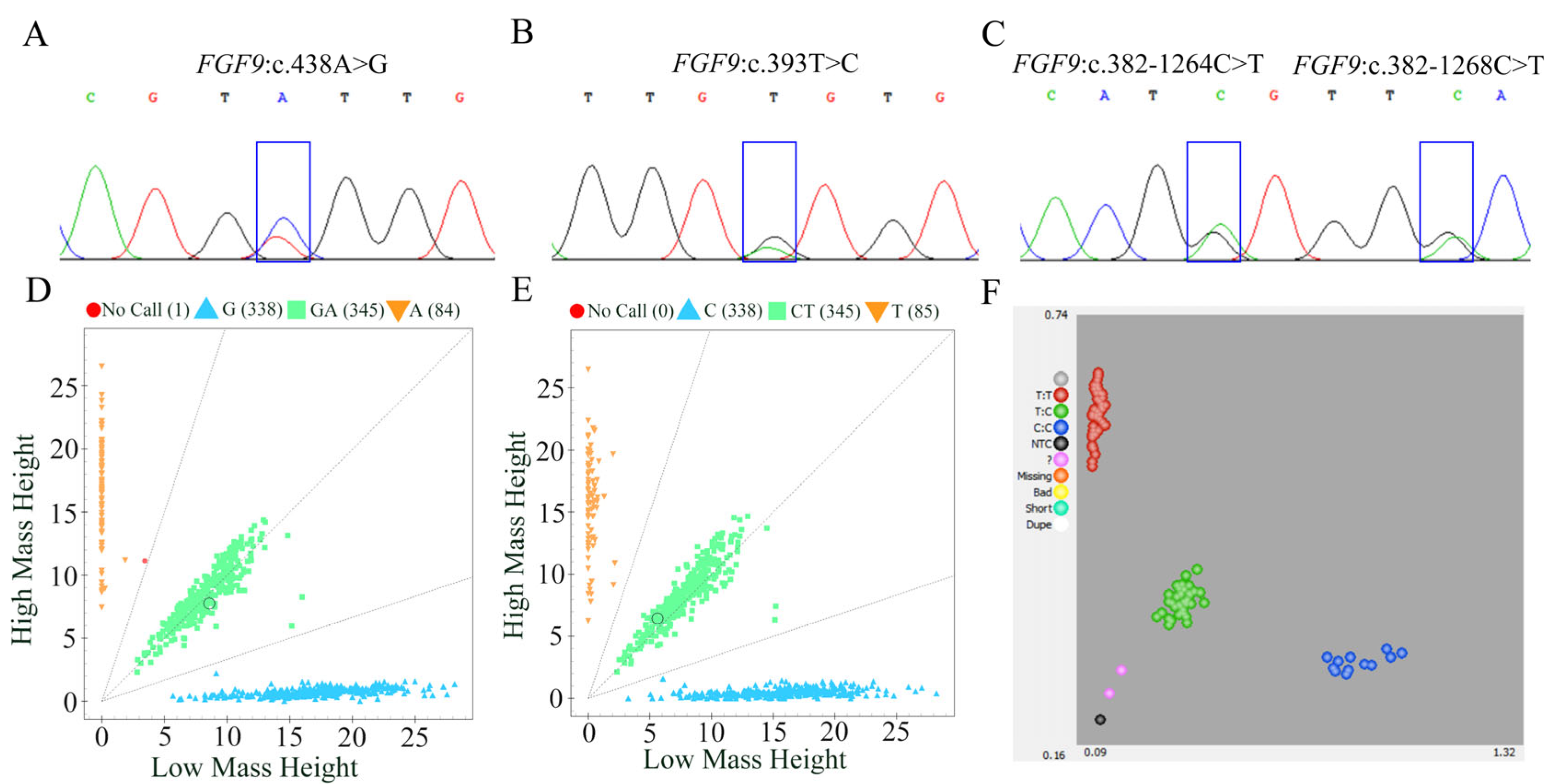
In our previous study, genome-wide selection tests comparing landraces and improved sheep breeds revealed strong selection signals in the genomic region of FGF9 [7]. In the present study, leveraging previously generated genomic data [7], we analyzed allele frequencies of these three variants across distinct populations. Results revealed that at the FGF9:c.438A>G locus, the A allele was detected at 0.73 in Chinese indigenous sheep, with the G allele at 0.27. Conversely, imported sheep exhibited a reciprocal pattern: the G allele predominated at 0.73, while the A allele was restricted to 0.27. For FGF9:c.393T>C, alleles T and C were skewed toward the T allele in Chinese indigenous sheep (0.73 and 0.27, respectively). In imported sheep, the C allele was significantly elevated to 0.72, with the T allele reduced to 0.28. For FGF9:c.382-1264C>T, the T allele occurred at 0.73 in Chinese indigenous sheep, with the C allele at 0.27. In imported sheep, the C allele predominated (0.71), while the T allele was restricted to 0.29 (Figure S1). To further investigate the polymorphism of the aforementioned SNPs and other exon regions of the FGF9 gene in the Hu sheep population with accurately recorded body weight data, we amplified sequences containing these SNPs and other exon regions of the FGF9 gene using specific PCR primer pairs (Supplementary Table S1), with DNA pools from 20 Hu sheep as templates. Sanger sequencing of the PCR products identified four SNPs in the FGF9 gene (Figure 2). Among these, two SNPs were only 3 bp apart, so a total of three SNPs were selected for genotyping in the experimental population (Figure 2).

3.4. Population Genetic Parameter Analysis of SNPs in FGF9 Gene

Population genetic parameters analyses of three SNPs in FGF9 gene were performed according to the genotyping data in the Hu sheep experimental population, and the results are shown in Figure S2. The results revealed that these three SNPs exhibited moderate polymorphism (polymorphism information content, PIC: 0.25 < PIC < 0.5) and were in Hardy–Weinberg equilibrium (HWE, p > 0.05). Furthermore, linkage disequilibrium analysis for the SNPs FGF9:c.438A>G and FGF9:c.393T>C, using the aforementioned genotyping data, showed that the two SNPs were in a state of strong linkage (Figure S3).

3.5. Association of Polymorphisms in FGF9 with Body Weight in Hu Sheep

The results of association analysis indicated that the SNP FGF9:c.438A>G was not significantly associated with body weight, while the SNP FGF9:c.382-1264C>T was significantly associated with body weight at six stages (Table 3). Moreover, except BW180, the body weight of Hu sheep at different periods with the TT genotype was the highest and was significantly higher than those in the animals with the CC genotype (p < 0.05).

3.6. Epigenetic Data (ATAC-seq, H3K4me3, and H3K427ac) Quality Control of Hypophysis Tissue in Sheep

To further explore the potential regulatory roles of the SNPs related to sheep body weight, we characterized open chromatin regions using assay for transposase-accessible chromatin using sequencing and also identified the genomic localization of histone H3K27ac and H3K4me3 by CUT&Tag methods in sheep hypophysis tissue. In total, 702,867,172; 160,076,532; and 201,741,220 raw reads were obtained in ATAC-Seq, H3K27ac, and H3K4me3, respectively. After filtration, 693,308,570; 149,604,394; and 181,906,606 clean reads were obtained. Comparison of these clean reads to the sheep reference genome (Oar_rambouillet_v1.0) revealed mapping rates ranging from 97.40% to 99.64%, and the average relative strand cross-correlation coefficient (RSC) and fraction of reads in peaks (FRiP) were 1.43 ± 0.25 and 0.55 ± 0.26, respectively (Table 4). These results showed our data could be used for subsequent analyses.

3.7. Comprehensive Profiling of Genetic Variants and Epigenomic Characteristics

We obtained an average of 221,243; 52692; and 20,957.5 peaks in the ATAC-seq, H3K27ac, and H3K4me3 data (Figure 3A), with average lengths of 801.46, 1783.64, and 2467.03 bp, respectively (Figure 3B). Most OCRs were annotated to non-coding regions, mainly including introns (41.58%) and distal intergenic regions (32.98%), followed by promoter (16.19%), exons (5.77%), while H3K27ac and H3K4me3 peaks were mainly annotated to the promoter region (H3K27ac: 33.37%; H3K4me3: 72.15%) of the genes (Figure 3D). In addition, three epigenetic marks (ATAC-seq, H3K4me3, and H3K27ac) signals were more enriched near the transcription start sites (TSS) of genes (Figure 3C). Finally, we integrated the epigenetic data with the SNPs of FGF9 gene, the results indicated higher OCR ~900 bp downstream of the SNP FGF9:c.382-1264C>T, and the SNP was also localized to the downstream peak (~700 bp) of H3K27ac modification, suggesting this polymorphism may influence FGF9 gene expression by altering chromatin accessibility or transcription factor binding, whereas other SNPs were not located in the adjacent OCR- and H3K27ac-modified peaks (Figure 4). Therefore, we suspect that the SNP FGF9:c.382-1264C>T plays an important role in regulating body weight in sheep.

4. Discussion

Improving meat production is a primary objective in mutton sheep breeding. While previous studies have mapped 329 quantitative trait loci (QTLs) associated with body weight traits based on records in the Sheep QTL Database (released 27 December 2023; https://www.animalgenome.org/cgi-bin/QTLdb/OA/index), most candidate variants reside in non-coding genomic regions [26,27,28]; only a limited number of causative SNPs have been identified for body weight traits in sheep. Therefore, identifying functional regulatory variations plays an important role in enhancing body weight in current sheep breeding programs.
In the present study, we measured and recorded the body weight of male Hu lambs (n = 1070) from 80 to 180 days of age under the same feeding regime and management conditions. We found that body weight traits exhibit great potential for selection. These phenotypic data provide a valuable resource for identifying key genetic variations underlying body weight. Additionally, based on our previous studies, we selected the FGF9 gene as a candidate gene for follow-up experiments. qRT-PCR results indicated that the relative expression of FGF9 was higher in the hypophysis than in other tested tissues; the hypophysis plays a central role in regulating growth and metabolism [29,30]. Previous studies have shown that FGF9 acts as a regulator of bone development and identified that a FGF9 loss-of-function mutation impairs early joint formation in a mouse model [31]. Additionally, Huang et al. found that FGF9 inhibits the myogenic differentiation of C2C12 and human muscle cells, which suggests that FGF9 may play an important role in modulating myogenesis [32]. In the present study, therefore, three SNPs of FGF9 gene were genotyped in a Hu population with accurate body weight data records. Genotyping results revealed that these SNPs exhibited moderate polymorphism (0.25 < PIC < 0.5) and conformed to Hardy–Weinberg equilibrium (p > 0.05) in the experimental population. This indicates that the studied Hu sheep population harbors sufficient genetic variation and maintains a stable genetic structure, thereby laying a robust foundation for subsequent analyses of associations between these SNPs and body weight traits. Furthermore, based on data from our previous studies data, we found that the allele frequencies of these loci differ significantly between Chinese indigenous sheep breeds and imported sheep breeds [7]. This divergence is likely associated with the differences in growth rate, body weight, and other traits between indigenous and imported sheep breeds.
To further explore whether these variant loci are associated with body weight traits, we performed association analyses in the Hu sheep population. The results revealed that the SNP FGF9:c.382-1264C>T was significantly associated with body weight, with all BW phenotype values of animals carrying the TT and TC genotypes being significantly higher than those of animals carrying the CC genotype. In addition, in the experimental population, the CC genotype was rare (~11.8%; 118 individuals), while TT (~48.1%; 481 individuals) and TC (~46.8%; 468 individuals) dominated. Notably, TT and TC showed minimal phenotypic divergence, implying that T-allele carriers (TT/TC) uniformly confer growth advantages. Selecting for T alleles could thus improve weight gain, which is consistent with the high frequency of T-allele-bearing genotypes in the population. Additionally, Li et al. identified the FGF9 gene as a key candidate gene influencing weaning weight in Hu sheep via a genome-wide association study [19], which supports our findings. Although this SNP was located in the intronic region, increasing evidence has indicated that non-coding SNPs can regulate enhancer activity, promoter activity, and the expression of distant target genes through spatial interactions—especially when they are localized in epigenetically active regions such as OCRs- or H3K27ac-modified peaks (a marker of active enhancers/promoters). For example, Pan et al. identified an SNP in these muscle-specific active strong enhancers, which may regulate the expression of ALPK2 and serve as a candidate causal variant contributing to average daily gain in pig [33]. Zhu et al. demonstrated that the locus most significantly associated with 8-week-old body weight in chickens (position 170,526,091 bp within CAB39L) is localized to the OCR of the duodenum. This locus may exert regulatory effects on 8-week-old body weight (BW8) by modulating the transcriptional activity of CAB39L [34]. Smemo et al. found that the top SNP in the intronic region of the FTO gene association with obesity by regulating the expression of the IRX3 and IRX5 genes nearby [35]. Miao et al. identified two SNPs near the BMP2 affecting the loin muscle depth in pig by integrating GWAS and 3D epigenomics [36]. Therefore, in this study, we identified the regulatory elements with ATAC-seq and CUT&Tag and performed integrated analysis with SNPs data; the result indicated the SNP FGF9:c.382-1264C>T was localized in the adjacent OCR- and H3K27ac-modified peaks. While OCRs are critical for enabling transcription factor binding and facilitating interactions between regulatory elements and gene promoters, H3K27ac is a well-characterized epigenetic marker of active enhancers—regions that drive transcriptional activation by looping to target gene promoters [37,38]. Together, these epigenetic signatures strongly suggest that this SNP resides within a functionally active genomic segment, where allelic variation could alter the accessibility of the chromatin landscape or disrupt the binding affinity of transcription factors or co-activators associated with H3K27ac-mediated enhancement. In addition, protein–protein interaction network analysis of FGF9 revealed its interactions with fibroblast growth factor receptors (FGFR1/2/3/4), transforming growth factor beta-1 (TGFB1), SRY-box transcription factor 9 (SOX9), and bone morphogenetic proteins (BMP2/4), implying that FGF9 may play multifaceted roles in developmental biology and the maintenance of tissue homeostasis. Therefore, these results suggest the functional regulatory variant may regulate FGF9 expression and is an important candidate mutation affecting body weight in sheep. Nonetheless, further in-depth investigations into the relationships between genotype, gene expression, and body weight are warranted to validate and expand these findings.

5. Conclusions

Our study demonstrates the functional regulatory variant FGF9:c.382-1264C>T as a promising candidate marker for improving weight traits in sheep. However, to fully translate these findings into practical breeding applications, further investigations are required: firstly to clarify the functional mechanisms by which FGF9 regulates weight traits and secondly to validate its utility across diverse sheep breeds. Such efforts will strengthen the practical applicability of these findings in breeding programs.

Supplementary Materials

The following supporting information can be downloaded at https://www.mdpi.com/article/10.3390/ani15162375/s1. Figure S1: Gene frequency and genetic parameters of SNPs for FGF9 gene in Hu sheep population. (A,B) Gene structure map and SNP location of FGF9 gene. (C) Genotype frequencies of three SNPs in FGF9 gene. (D). Genotypic frequency of three SNPs in FGF9 gene. (E) Genetic parameters of three SNPs in FGF9 gene. He, heterozygosity; PIC, polymorphism information content; HWE, Hardy–Weinberg equilibrium. Figure S2: Linkage disequilibrium analysis for the SNPs FGF9:c.438A>G and FGF9:c.393T>C. Table S1: Details of primer sequences used for qRT-PCR and PCR amplification. Table S2: Primer information of each SNP locus. Table S3: Annotation and distribution of epigenetic mark peaks.

Author Contributions

Conceptualization, X.Z., D.Z. and W.W. (Weimin Wang); methodology, X.Z.; software, X.Z., J.C., X.L., Y.Z. (Yukun Zhang) and Y.Z. (Yuan Zhao); validation, D.X., P.C. and D.Z.; formal analysis, H.T.; investigation, L.Z., H.T., F.L. and X.Z.; resources, F.L. and W.W. (Weiwei Wu); data curation, D.Z.; writing—original draft preparation, X.Z.; writing—review and editing, X.Z. and D.Z., visualization, X.Z.; supervision, W.W. (Weimin Wang); project administration, W.W. (Weimin Wang); funding acquisition, F.L. and W.W (Weimin Wang). All authors have read and agreed to the published version of the manuscript.

Funding

This research was supported by the National Key R&D Young Scientists Project (2022YFD1302000), the Tianchi Talent Introduction Program of China, the Fuxi Young Talents Program of Gansu Agricultural University (Gaufx-03Y11), the Key Science Technology Project of Wuwei City (WW23A03ZDQ001), and Science and Technology Plan Project of Gansu Province (24CXGH007).

Institutional Review Board Statement

All procedures involving blood and tissue samples collection and animal care were approved by the Ethics Committee of Gansu Agricultural University (Permit No. GSAU-Eth-AST-2022-022). All sample collection were performed in strict accordance with relevant guidelines and regulations.

Informed Consent Statement

Not applicable.

Data Availability Statement

The data that support the findings of this study are available from the corresponding author upon reasonable request.

Acknowledgments

We gratefully acknowledge the support of all members for their assistance in animal sampling, measurements, and data input. All authors have read and agreed to the published version of the manuscript.

Conflicts of Interest

The authors declare no conflicts of interest.

Abbreviations

The following abbreviations are used in this manuscript:
ATAC-seqAssay for transposase-accessible chromatin-sequencing
BWBody weight
CVCoefficient of variation
Cut&TagCleavage under targets and tagmentation
FGF9Fibroblast growth factor 9
H3K27acHistone H3 lysine 27 acetylation
H3K4me3Histone H3 lysine 4 trimethylation
OCROpen chromatin region
qRT-PCRQuantitative real-time PCR
SNPsSingle nucleotide polymorphisms

References

  1. Mohamadipoor Saadatabadi, L.; Mohammadabadi, M.; Amiri Ghanatsaman, Z.; Babenko, O.; Stavetska, R.; Kalashnik, O.; Kucher, D.; Kochuk-Yashchenko, O.; Asadollahpour Nanaei, H. Signature selection analysis reveals candidate genes associated with production traits in Iranian sheep breeds. BMC Vet. Res. 2021, 17, 369. [Google Scholar] [CrossRef]
  2. Tao, L.; He, X.; Pan, L.; Wang, J.; Gan, S.; Chu, M. Genome-wide association study of body weight and conformation traits in neonatal sheep. Anim. Genet. 2020, 51, 336–340. [Google Scholar] [CrossRef]
  3. Al-Mamun, H.A.; Kwan, P.; Clark, S.A.; Ferdosi, M.H.; Tellam, R.; Gondro, C. Genome-wide association study of body weight in Australian Merino sheep reveals an orthologous region on OAR6 to human and bovine genomic regions affecting height and weight. Genet. Sel. Evol. 2015, 47, 66. [Google Scholar] [CrossRef]
  4. Lu, Z.; Yue, Y.; Yuan, C.; Liu, J.; Yang, B. Genome-Wide Association Study of Body Weight Traits in Chinese Fine-Wool Sheep. Animals 2020, 10, 170. [Google Scholar] [CrossRef] [PubMed]
  5. FAANG Consortium; Andersson, L.; Archibald, A.L.; Bottema, C.D.; Brauning, R.; Burgess, S.C.; Burt, D.W.; Casas, E.; Cheng, H.H.; Clarke, L.; et al. Coordinated international action to accelerate genome-to-phenome with FAANG, the Functional Annotation of Animal Genomes project. Genome Biol. 2015, 16, 4–9. [Google Scholar]
  6. Liu, S.; Gao, Y.; Canela-Xandri, O.; Wang, S.; Yu, Y.; Cai, W.; Li, B.; Xiang, R.; Chamberlain, A.J.; Pairo-Castineira, E. A multi-tissue atlas of regulatory variants in cattle. Nat. Genet. 2022, 54, 1438–1447. [Google Scholar] [CrossRef]
  7. Zhang, D.; Zhang, X.; Li, F.; Yuan, L.; Li, X.; Zhang, Y.; Zhao, Y.; Zhao, L.; Wang, J.; Xu, D.; et al. Whole-genome resequencing reveals molecular imprints of anthropogenic and natural selection in wild and domesticated sheep. Zool. Res. 2022, 43, 695–705. [Google Scholar] [CrossRef]
  8. He, P.; Zhong, S.; Lin, S.; Xia, Z.; Wang, L.; Han, Y.; Xu, D.; Hu, S.; Li, X.; Li, P.; et al. FGF9 is required for Purkinje cell development and function in the cerebellum. iScience 2024, 27, 109039. [Google Scholar] [CrossRef]
  9. Huang, K.; Wang, Y.A.-O.X.; Zhu, J.; Xiong, Y.A.-O.; Lin, Y.A.-O. Regulation of fibroblast growth factor 9 on the differentiation of goat intramuscular adipocytes. Anim. Sci. J. 2021, 92, e13627. [Google Scholar] [CrossRef]
  10. Wang, S.; Li, Y.; Jiang, C.; Tian, H. Fibroblast growth factor 9 subfamily and the heart. Appl. Microbiol. Biotechnol. 2018, 102, 605–613. [Google Scholar] [CrossRef]
  11. Wang, L.; Roth, T.; Abbott, M.; Ho, L.; Wattanachanya, L.; Nissenson, R.A. Osteoblast-derived FGF9 regulates skeletal homeostasis. Bone 2017, 98, 18–25. [Google Scholar] [CrossRef]
  12. Sun, Y.; Wang, R.; Zhao, S.; Li, W.; Liu, W.; Tang, L.; Wang, Z.; Wang, W.; Liu, R.; Ning, G.; et al. FGF9 inhibits browning program of white adipocytes and associates with human obesity. J. Mol. Endocrinol. 2019, 62, 79–90. [Google Scholar] [CrossRef] [PubMed]
  13. Shamsi, F.; Xue, R.; Huang, T.L.; Lundh, M.; Liu, Y.; Leiria, L.O.; Lynes, M.D.; Kempf, E.; Wang, C.-H.; Sugimoto, S.; et al. FGF6 and FGF9 regulate UCP1 expression independent of brown adipogenesis. Nat. Commun. 2020, 11, 1421. [Google Scholar] [CrossRef] [PubMed]
  14. Zhang, R.; Yao, F.; Cheng, X.; Yang, M.; Ning, Z. Identification of candidate genomic regions for egg yolk moisture content based on a genome-wide association study. BMC Genom. 2023, 24, 110. [Google Scholar] [CrossRef] [PubMed]
  15. Guo, Y.; Zhang, Y.; Wang, Y.; Chen, Q.; Sun, Y.; Kang, L.; Jiang, Y. Phosphorylation of LSD1 at serine 54 regulates genes involved in follicle selection by enhancing demethylation activity in chicken ovarian granulosa cells. Poult. Sci. 2024, 103, 103850. [Google Scholar] [CrossRef]
  16. Wang, Y.; Hu, T.; Wu, L.; Liu, X.; Xue, S.; Lei, M. Identification of non-coding and coding RNAs in porcine endometrium. Genomics 2017, 109, 43–50. [Google Scholar] [CrossRef]
  17. Gao, X.; Yao, X.; Yang, H.; Deng, K.; Guo, Y.; Zhang, T.; Zhang, G.; Wang, F. Role of FGF9 in sheep testis steroidogenesis during sexual maturation. Anim. Reprod. Sci. 2018, 197, 177–184. [Google Scholar] [CrossRef]
  18. Jia, Q.; Zhang, S.; Wang, D.; Liu, J.; Luo, X.; Liu, Y.; Li, X.; Sun, F.; Xia, G.; Zhang, L. Regulatory Effects of FGF9 on Dermal Papilla Cell Proliferation in Small-Tailed Han Sheep. Genes 2023, 14, 1106. [Google Scholar] [CrossRef]
  19. Li, T.; Xing, F.; Zhang, N.; Chen, J.; Zhang, Y.; Yang, H.; Peng, S.; Ma, R.; Liu, Q.; Gan, S.; et al. Genome-Wide Association Analysis of Growth Traits in Hu Sheep. Genes 2024, 15, 1637. [Google Scholar] [CrossRef]
  20. Zhang, D.; Zhang, X.; Li, F.; Yuan, L.; Zhang, Y.; Li, X.; Zhao, Y.; Song, Q.; Li, G.; Wang, W. Polymorphisms in ovine ME1 and CA1 genes and their association with feed efficiency in Hu sheep. J. Anim. Breed. Genet. 2021, 138, 589–599. [Google Scholar] [CrossRef]
  21. Zhang, D.; Zhang, X.; Li, F.; Li, C.; La, Y.; Mo, F.; Li, G.; Zhang, Y.; Li, X.; Song, Q.; et al. Transcriptome Analysis Identifies Candidate Genes and Pathways Associated with Feed Efficiency in Hu Sheep. Front. Genet. 2019, 10, 1183. [Google Scholar] [CrossRef]
  22. Leslie, F.; Cécile, G.; Jean-François, A. Development of Nuclear Microsatellite Loci and Mitochondrial Single Nucleotide Polymorphisms for the Natterjack Toad, Bufo (Epidalea) calamita (Bufonidae), Using Next Generation Sequencing and Competitive Allele Specific PCR (KASPar). J. Hered. 2016, 107, 660–665. [Google Scholar] [CrossRef]
  23. Zhao, Y.; Hou, Y.; Xu, Y.; Luan, Y.; Zhou, H.; Qi, X.; Hu, M.; Wang, D.; Wang, Z.; Fu, Y.; et al. A compendium and comparative epigenomics analysis of cis-regulatory elements in the pig genome. Nat. Commun. 2021, 12, 2217. [Google Scholar] [CrossRef]
  24. Zhang, D.; Cheng, J.; Li, X.; Huang, K.; Yuan, L.; Zhao, Y.; Xu, D.; Zhang, Y.; Zhao, L.; Yang, X.; et al. Comprehensive multi-tissue epigenome atlas in sheep: A resource for complex traits, domestication, and breeding. Imeta 2024, 3, e254. [Google Scholar] [CrossRef] [PubMed]
  25. Yu, G.; Wang, L.G.; He, Q.Y. ChIPseeker: An R/Bioconductor package for ChIP peak annotation, comparison and visualization. Bioinformatics 2015, 31, 2382–2383. [Google Scholar] [CrossRef] [PubMed]
  26. Cao, Y.; Song, X.; Shan, H.; Jiang, J.; Xiong, P.; Wu, J.; Shi, F.; Jiang, Y. Genome-Wide Association Study of Body Weights in Hu Sheep and Population Verification of Related Single-Nucleotide Polymorphisms. Front. Genet. 2020, 11, 588. [Google Scholar] [CrossRef] [PubMed]
  27. Zuo, P.; Zhang, C.; Gao, Y.; Zhao, L.; Guo, J.; Yang, Y.; Yu, Q.; Li, Y.; Wang, Z.; Yang, H. Genome-wide unraveling SNP pairwise epistatic effects associated with sheep body weight. Anim. Biotechnol. 2023, 34, 3416–3427. [Google Scholar] [CrossRef]
  28. Pasandideh, M.A.-O.; Gholizadeh, M.; Rahimi-Mianji, G. A genome-wide association study revealed five SNPs affecting 8-month weight in sheep. Anim. Genet. 2020, 51, 973–976. [Google Scholar] [CrossRef]
  29. Ji, X.; Shen, Q.; Wu, P.; Chen, H.; Wang, S.; Chen, D.; Yu, Y.; Guo, Z.; Wang, J.; Tang, G. Pituitary-Gland-Based Genes Participates in Intrauterine Growth Restriction in Piglets. Genes 2022, 13, 2141. [Google Scholar] [CrossRef]
  30. Zhang, S.A.-O.; Cui, Y.; Ma, X.A.-O.; Yong, J.; Yan, L.A.-O.; Yang, M.; Ren, J.; Tang, F.A.-O.; Wen, L.A.-O.; Qiao, J.A.-O. Single-cell transcriptomics identifies divergent developmental lineage trajectories during human pituitary development. Nat. Commun. 2020, 11, 5275. [Google Scholar] [CrossRef]
  31. Tang, L.A.-O.; Wu, M.; Lu, S.; Zhang, H.; Shen, Y.; Shen, C.; Liang, H.; Ge, H.; Ding, X.; Wang, Z.A.-O. Fgf9 Negatively Regulates Bone Mass by Inhibiting Osteogenesis and Promoting Osteoclastogenesis Via MAPK and PI3K/AKT Signaling. J. Bone Miner. Res. 2020, 36, 779–791. [Google Scholar] [CrossRef]
  32. Huang, J.; Wang, K.; Shiflett, L.A.; Brotto, L.; Bonewald, L.F.; Wacker, M.J.; Dallas, S.A.-O.X.; Brotto, M. Fibroblast growth factor 9 (FGF9) inhibits myogenic differentiation of C2C12 and human muscle cells. Cell Cycle 2019, 18, 3562–3580. [Google Scholar] [CrossRef] [PubMed]
  33. Pan, Z.; Yao, Y.; Yin, H.; Cai, Z.; Wang, Y.; Bai, L.; Kern, C.; Halstead, M.; Chanthavixay, G.; Trakooljul, N.; et al. Pig genome functional annotation enhances the biological interpretation of complex traits and human disease. Nat. Commun. 2021, 12, 5848. [Google Scholar] [CrossRef] [PubMed]
  34. Zhu, X.; Wang, Y.; Li, C.; Wu, H.; Zhang, R.; Hu, X. Chicken chromatin accessibility atlas accelerates epigenetic annotation of birds and gene fine-mapping associated with growth traits. Zool. Res. 2023, 44, 53–62. [Google Scholar] [CrossRef] [PubMed]
  35. Smemo, S.; Tena, J.J.; Kim, K.H.; Gamazon, E.R.; Sakabe, N.J.; Gómez-Marín, C.; Aneas, I.; Credidio, F.L.; Sobreira, D.R.; Wasserman, N.F.; et al. Obesity-associated variants within FTO form long-range functional connections with IRX3. Nature 2014, 507, 371–375. [Google Scholar] [CrossRef]
  36. Miao, Y.; Zhao, Y.; Wan, S.; Mei, Q.; Wang, H.; Fu, C.; Li, X.; Zhao, S.; Xu, X.; Xiang, T. Integrated analysis of genome-wide association studies and 3D epigenomic characteristics reveal the BMP2 gene regulating loin muscle depth in Yorkshire pigs. PLoS Genet. 2023, 19, e1010820. [Google Scholar] [CrossRef]
  37. Raisner, R.; Kharbanda, S.; Jin, L.; Jeng, E.; Chan, E.; Merchant, M.; Haverty, P.M.; Bainer, R.; Cheung, T.; Arnott, D.; et al. Enhancer Activity Requires CBP/P300 Bromodomain-Dependent Histone H3K27 Acetylation. Cell Rep. 2018, 24, 1722–1729. [Google Scholar] [CrossRef]
  38. Consortium, E.P. An integrated encyclopedia of DNA elements in the human genome. Nature 2012, 489, 57–74. [Google Scholar] [CrossRef]
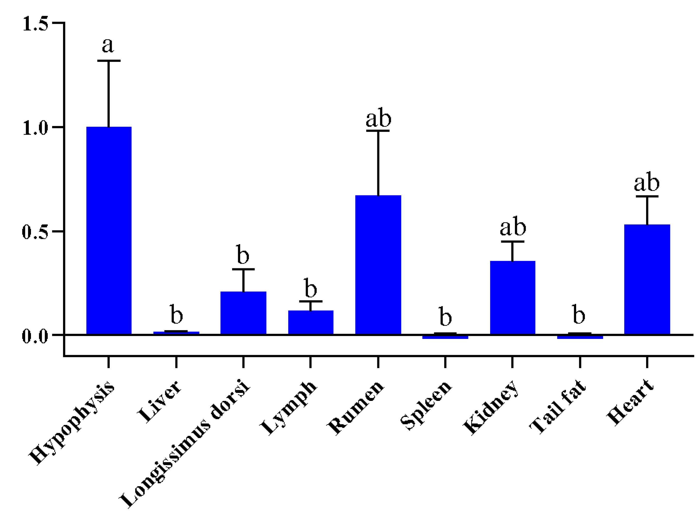
Figure 1. Relative expression of FGF9 mRNA in different tissues of the Hu sheep. Different lowercase letters indicate significant differences (p < 0.05).
Animals 15 02375 g001
Figure 2. Verification and genotyping of SNPs of FGF9 gene in sheep. (A,D) Sequence chromatogram (A) and Sequenom MassARRAY® genotyping (D) for the SNP FGF9:c.438A>G. (B,E) Sequence chromatogram (B) and Sequenom MassARRAY® genotyping (E) for the SNP FGF9:c.393T>C. (C,F) Sequence chromatogram (C) and KASP genotyping (F) for the SNP FGF9:c.382-1264C>T.
Animals 15 02375 g002
Figure 3. Characteristics of epigenetic data for hypophysis tissue in sheep. (A) Peak number of ATAC-seq, H3K27ac, and H3K4me3. (B) Peak length distribution of ATAC-seq, H3K27ac, and H3K4me3. (C) The distribution of epigenetic signals around TSSs of genes. TSS, transcription start site; TES, transcription end site. (D) Annotation and distribution of epigenetic mark peaks.
Animals 15 02375 g003
Figure 4. Epigenetic (ATAC, H3K27ac, and H3K4me3) signal on chromosome 10 containing FGF9 gene. Orange tracks represent ATAC-seq signals, green tracks represent H3K27ac signals, and cyan tracks represent H3K4me3 signal; the same color represents a biological repetition of the same assay.
Animals 15 02375 g004
Table 1. Details of primer sequences used for qRT-PCR.
Primer NamePrimer Sequence (5′-3′)Annealing Temperature (°C)Amplicon Size (bp)
FGF9-expression-FGAAGCTGCATTTAATCCCAAG60204 bp
FGF9-expression-RGATCACTTTTGGCTGTCTCC
β-actin-FTCCGTGACATCAAGGAGAAGC60256 bp
β-actin-RCCGTGTTGGCGTAGAGGT
Table 2. The summary statistics of body weight traits.
TraitsMeanSDMedianMinMaxSkewKurtosisCV (%)
BW8019.424.0219.309.5034.400.245−0.06120.73%
BW10024.554.8324.509.7842.400.0520.07319.68%
BW12030.375.3530.3013.8049.400.0130.13317.62%
BW14036.075.7436.1019.5556.300.0290.01615.91%
BW16041.876.1341.6025.1063.600.0700.12214.64%
BW18046.806.4146.8024.5570.20−0.0090.24213.70%
Table 3. Association analysis between variants of FGF9 gene and body weight in Hu sheep.
LocusGenotypeNo.BW80BW100BW120BW140BW160BW180
FGF9:c.438A>GGG47119.50 ± 0.1924.61 ± 0.2230.44 ± 0.2536.20 ± 0.2742.05 ± 0.2846.88 ± 0.30
GA47819.44 ± 0.1824.52 ± 0.2230.34 ± 0.2535.96 ± 0.2641.75 ± 0.2846.79 ± 0.29
AA11819.11 ± 0.3724.58 ± 0.4530.27 ± 0.4936.02 ± 0.5341.64 ± 0.5746.50 ± 0.59
FGF9:c.382-1264C>TTT48119.40 ± 0.18 a24.56 ± 0.22 a30.29 ± 0.24 a36.04 ± 0.26 a41.54 ± 0.27 a46.37 ± 0.29 ab
TC46819.33 ± 0.19 a24.53 ± 0.22 a30.40 ± 0.25 a36.15 ± 0.27 a41.69 ± 0.28 a46.65 ± 0.30 a
CC11818.36 ± 0.37 b23.49 ± 0.44 b29.20 ± 0.48 b34.92 ± 0.53 b40.44 ± 0.55 b45.41 ± 0.59 b
Note: BW, body weight. In the same column, numerical data marked with different lowercase letters indicate significant differences (p < 0.05).
Table 4. Summary of the epigenetic data (ATAC-seq, H3K4me3, and H3K427ac) for hypophysis tissue in sheep.
AssayRaw
Reads
Clean
Reads
Clean
Q20 (%)
Clean
Q30 (%)
Mapping Rate (%)RSCFRiP
ATAC-seq_1355,833,444351,268,39897.5293.3399.491.560.38
ATAC-seq_2347,033,728342,040,17297.3592.9599.641.870.36
H3K4me3_171,235,59466,164,15898.0693.5197.801.240.79
H3K4me3_2130,505,626115,742,44897.3391.5397.401.180.86
H3K27ac_168,907,38264,771,04898.4994.5598.301.420.23
H3K27ac_291,169,15084,833,34698.0193.1998.001.330.68
Disclaimer/Publisher’s Note: The statements, opinions and data contained in all publications are solely those of the individual author(s) and contributor(s) and not of MDPI and/or the editor(s). MDPI and/or the editor(s) disclaim responsibility for any injury to people or property resulting from any ideas, methods, instructions or products referred to in the content.