

M-Mode Echocardiography in Canine Veterinary Practice: A Comprehensive Review of Left Ventricular Measurements in 44 Different Dog Breeds

Simple Summary
Abstract
1. Introduction
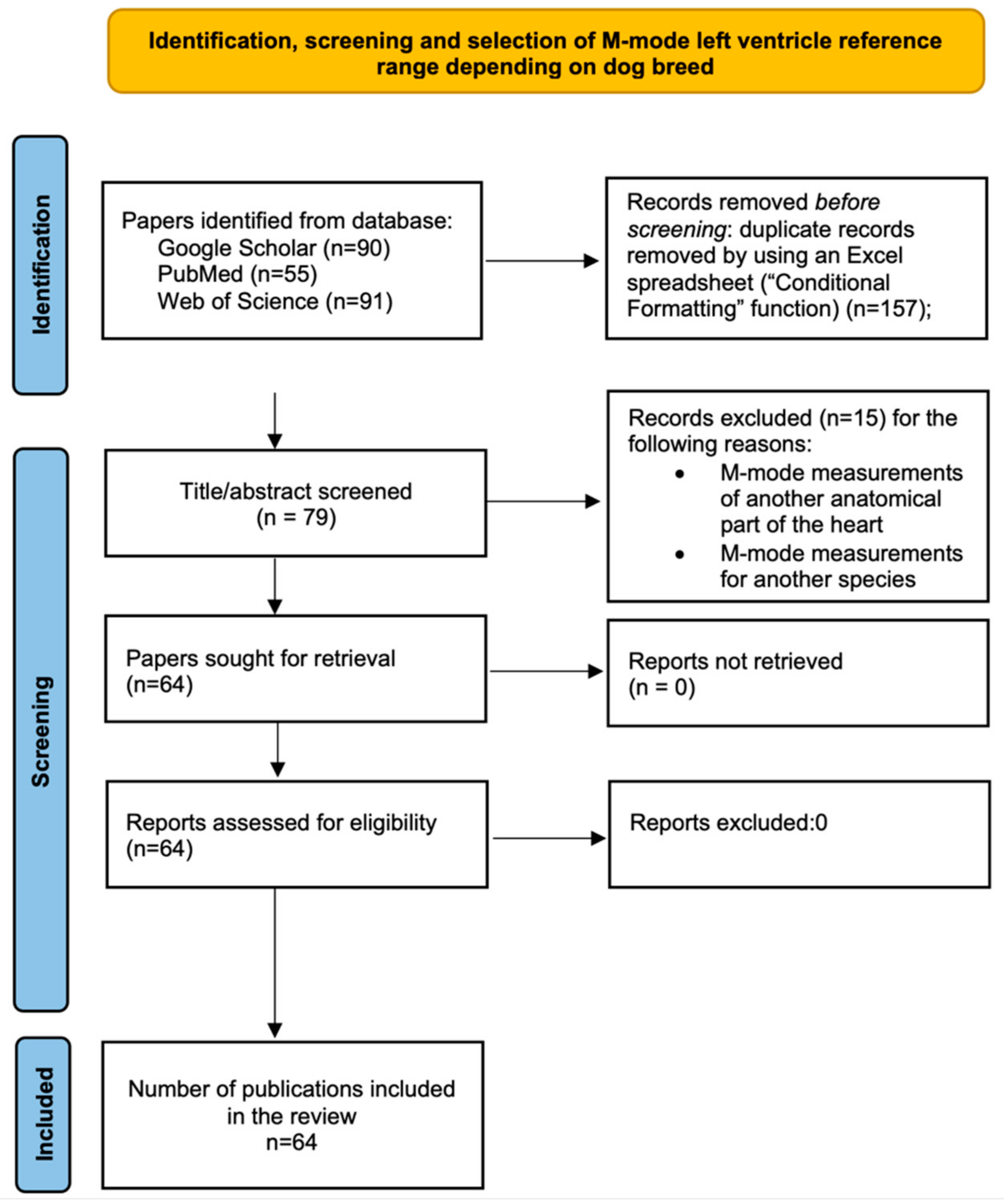
2. Materials and Methods
3. M-Mode Measurements of LV Based on Breed
4. Results and Discussions
5. Conclusions
Author Contributions
Funding
Institutional Review Board Statement
Informed Consent Statement
Data Availability Statement
Conflicts of Interest
References
- Boon, J.A. Veterinary Echocardiography; John Wiley & Sons: Hoboken, NJ, USA, 2011. [Google Scholar]
- De Lima, A.M.; Moreira, R.M.; Gomes, M.S.; Ramos, M.T.; dos Santos-Sousa, C.A.; Souza-Júnior, P.; Abidu-Figueiredo, M. Echocardiographic evaluation of working dogs of the Military Police of Rio de Janeiro: Effects of the breed and body weight. Braz. J. Vet. Med. 2022, 44, e001322. [Google Scholar] [CrossRef] [PubMed]
- Carerj, S.; Micari, A.; Trono, A.; Giordano, G.; Cerrito, M.; Zito, C.; Luzza, F.; Coglitore, S.; Arrigo, F.; Oreto, G. Anatomical M-Mode: An Old–New Technique. Echocardiography 2003, 20, 357–361. [Google Scholar] [CrossRef]
- Penninck, D.; d’Anjou, M.-A. Atlas of Small Animal Ultrasonography; John Wiley & Sons: Hoboken, NJ, USA, 1991. [Google Scholar]
- Noviana, D.; Paramitha, D.; Wulansari, R. Motion mode and two dimensional echocardiographic measurements of cardiac dimensions of Indonesian mongrel dogs. HAYATI J. Biosci. 2011, 18, 1–5. [Google Scholar] [CrossRef]
- Schober, K.E.; Baade, H. Comparability of left ventricular M-mode echocardiography in dogs performed in long-axis and short-axis. Vet. Radiol. Ultrasound 2000, 41, 543–549. [Google Scholar] [CrossRef] [PubMed]
- Smets, P.; Daminet, S.; Wess, G. Simpson’s method of discs for measurement of echocardiographic end-diastolic and end-systolic left ventricular volumes: Breed-specific reference ranges in Boxer dogs. J. Vet. Intern. Med. 2014, 28, 116–122. [Google Scholar] [CrossRef]
- Gonçalves, A.C.; Orton, E.C.; Boon, J.A.; Salman, M. Linear, logarithmic, and polynomial models of M-mode echocardiographic measurements in dogs. Am. J. Vet. Res. 2002, 63, 994–999. [Google Scholar] [CrossRef]
- Cornell, C.C.; Kittleson, M.D.; Torre, P.D.; Häggström, J.; Lombard, C.W.; Pedersen, H.D.; Vollmar, A.; Wey, A. Allometric scaling of M-mode cardiac measurements in normal adult dogs. J. Vet. Intern. Med. 2004, 18, 311–321. [Google Scholar] [CrossRef]
- Visser, L.C.; Ciccozzi, M.M.; Sintov, D.J.; Sharpe, A.N. Echocardiographic quantitation of left heart size and function in 122 healthy dogs: A prospective study proposing reference intervals and assessing repeatability. J. Vet. Intern. Med. 2019, 33, 1909–1920. [Google Scholar] [CrossRef]
- Keene, B.W.; Atkins, C.E.; Bonagura, J.D.; Fox, P.R.; Häggström, J.; Fuentes, V.L.; Oyama, M.A.; Rush, J.E.; Stepien, R.; Uechi, M. ACVIM consensus guidelines for the diagnosis and treatment of myxomatous mitral valve disease in dogs. J. Vet. Intern. Med. 2019, 33, 1127–1140. [Google Scholar] [CrossRef]
- Wess, G. Screening for dilated cardiomyopathy in dogs. J. Vet. Cardiol. 2022, 40, 51–68. [Google Scholar] [CrossRef]
- Dukes-McEwan, J.; Borgarelli, M.; Tidholm, A.; Vollmar, A.C.; Häggström, J.; Cardiomyopathy, E.T.f.C.D. Proposed guidelines for the diagnosis of canine idiopathic dilated cardiomyopathy. J. Vet. Cardiol. 2003, 5, 7–19. [Google Scholar] [CrossRef] [PubMed]
- Isayama, N.; Uchimura, Y.; Sasaki, K.; Maeda, E.; Takahashi, T.; Watanabe, M. Reference Values of M-mode Echocardiographic Parameter in Adult Toy Breed Dogs. Front. Vet. Sci. 2022, 9, 918457. [Google Scholar] [CrossRef] [PubMed]
- Misbach, C.; Lefebvre, H.P.; Concordet, D.; Gouni, V.; Trehiou-Sechi, E.; Petit, A.M.; Damoiseaux, C.; Leverrier, A.; Pouchelon, J.-L.; Chetboul, V. Echocardiography and conventional Doppler examination in clinically healthy adult Cavalier King Charles Spaniels: Effect of body weight, age, and gender, and establishment of reference intervals. J. Vet. Cardiol. 2014, 16, 91–100. [Google Scholar] [CrossRef]
- Wyatt, H.L.; Haendchen, R.V.; Meerbaum, S.; Corday, E. Assessment of quantitative methods for 2-dimensional echocardiography. Am. J. Cardiol. 1983, 52, 396–401. [Google Scholar] [CrossRef] [PubMed]
- Gugjoo, M.; Saxena, A.; Hoque, M.; Zama, M. M-mode echocardiographic study in dogs. Afr. J. Agric. Res. 2014, 9, 387–396. [Google Scholar]
- Crippa, L.; Ferro, E.; Melloni, E.; Brambilla, P.; Cavalletti, E. Echocardiographic parameters and indices in the normal Beagle dog. Lab. Anim. 1992, 26, 190–195. [Google Scholar] [CrossRef]
- Morrison, S.A.; Moise, N.S.; Scarlett, J.; Mohammed, H.; Yeager, A.E. Effect of breed and body weight on echocardiographic values in four breeds of dogs of differing somatotype. J. Vet. Intern. Med. 1992, 6, 220–224. [Google Scholar] [CrossRef]
- Kurosawa, T.; Sist, M.; Sanders, R. Echocardiographic variables in healthy North American Salukis. J. Vet. Cardiol. 2018, 20, 301–306. [Google Scholar] [CrossRef]
- Esser, L.C.; Borkovec, M.; Bauer, A.; Häggström, J.; Wess, G. Left ventricular M-mode prediction intervals in 7651 dogs: Population-wide and selected breed-specific values. J. Vet. Intern. Med. 2020, 34, 2242–2252. [Google Scholar] [CrossRef]
- Jacobs, G.; Mahjoob, K. Multiple regression analysis, using body size and cardiac cycle length, in predicting echocardiographic variables in dogs. Am. J. Vet. Res. 1988, 49, 1290–1294. [Google Scholar]
- De Madron, E. Update on normal TM (M-Mode) echocardiographic values in the dog. Prat. Medicale Chir. L Anim. Cie. 1995, 30, 647–658. [Google Scholar]
- Vollmar, A.C. Echocardiographic measurements in the Irish wolfhound: Reference values for the breed. J. Am. Anim. Hosp. Assoc. 1999, 35, 271–277. [Google Scholar] [CrossRef] [PubMed][Green Version]
- Kayar, A.; Gonul, R.; ERMAN OR, M.; Uysal, A. M-mode echocardiographic parameters and indices in the normal German shepherd dog. Vet. Radiol. Ultrasound 2006, 47, 482–486. [Google Scholar] [CrossRef] [PubMed]
- Muzzi, R.A.L.; Muzzi, L.A.L.; De Araujo, R.B.; Cherem, M. Echocardiographic indices in normal German shepherd dogs. J. Vet. Sci. 2006, 7, 193–198. [Google Scholar] [CrossRef]
- Bavegems, V.; Duchateau, L.; Sys, S.U.; De Rick, A. Echocardiographic reference values in whippets. Vet. Radiol. Ultrasound 2007, 48, 230–238. [Google Scholar] [CrossRef]
- Cunningham, S.; Rush, J.; Freeman, L.; Brown, D.; Smith, C. Echocardiographic ratio indices in overtly healthy Boxer dogs screened for heart disease. J. Vet. Intern. Med. 2008, 22, 924–930. [Google Scholar] [CrossRef] [PubMed]
- Jacobson, J.H.; Boon, J.A.; Bright, J.M. An echocardiographic study of healthy Border Collies with normal reference ranges for the breed. J. Vet. Cardiol. 2013, 15, 123–130. [Google Scholar] [CrossRef] [PubMed]
- Garncarz, M.; Parzeniecka-Jaworska, M.; Czopowicz, M.; Hulanicka, M.; Jank, M.; Szaluś-Jordanow, O. Reference intervals for transthoracic echocardiographic measurements in adult Dachshunds. Pol. J. Vet. Sci. 2018, 21, 779–788. [Google Scholar]
- Bayon, A.; del Palacio, M.F.; Montes, A.M.; Panizo, C.G. M-mode echocardiography study in growing Spanish mastiffs. J. Small Anim. Pract. 1994, 35, 473–479. [Google Scholar] [CrossRef]
- Koch, J.; Pedersen, H.; Jensen, A.; Flagstad, A. M-mode echocardiographic diagnosis of dilated cardiomyopathy in giant breed dogs. J. Vet. Med. Ser. A 1996, 43, 297–304. [Google Scholar] [CrossRef]
- Torre, P.D.; Kirby, A.; Church, D.; Malik, R. Echocardiographic measurements in Greyhounds, Whippets and Italian Greyhounds-dogs with a similar conformation but different size. Aust. Vet. J. 2000, 78, 49–55. [Google Scholar] [CrossRef]
- O’Leary, C.; Mackay, B.; Taplin, R.; Atwell, R. Echocardiographic parameters in 14 healthy English bull terriers. Aust. Vet. J. 2003, 81, 535–542. [Google Scholar] [CrossRef] [PubMed]
- Vurucu, M.; Ekinci, G.; Gunes, V. An echocardiographic study of breed-specific reference ranges in healthy French Bulldogs. Vet. Radiol. Ultrasound 2021, 62, 573–582. [Google Scholar] [CrossRef] [PubMed]
- Vatne, L.; Dickson, D.; Tidholm, A.; Caivano, D.; Rishniw, M. The effects of activity, body weight, sex and age on echocardiographic values in English setter dogs. J. Vet. Cardiol. 2021, 37, 26–41. [Google Scholar] [CrossRef] [PubMed]
- Vezzosi, T.; Ghinelli, R.; Ferrari, P.; Porciello, F. Reference intervals for transthoracic echocardiography in the American Staffordshire Terrier. J. Vet. Med. Sci. 2021, 83, 656–660. [Google Scholar] [CrossRef]
- Diez-Prieto, I.; García-Rodríguez, M.B.; Ríos-Granja, M.; Cano-Rábano, M.J.; Peña-Penabad, M.; Pérez-García, C.C. M-mode echocardiographic changes in growing beagles. J. Am. Assoc. Lab. Anim. Sci. 2010, 49, 31–35. [Google Scholar]
- Niimi, S.; Kobayashi, H.; Take, Y.; Ikoma, S.; Namikawa, S.; Fujii, Y. Reference intervals for echocardiographic measurements in healthy Chihuahua dogs. J. Vet. Med. Sci. 2022, 84, 754–759. [Google Scholar] [CrossRef]
- Lim, C.K.; Fosgate, G.T.; Green, H.W.; Kirberger, R.M. Two-dimensional left atrium–to–aorta ratios and left ventricular M-mode transthoracic echocardiographic measurements in clinically normal adult Dachshunds. Am. J. Vet. Res. 2016, 77, 374–382. [Google Scholar] [CrossRef]
- Locatelli, C.; Santini, A.; Bonometti, G.; Palermo, V.; Scarpa, P.; Sala, E.; Brambilla, P. Echocardiographic values in clinically healthy adult dogue de Bordeaux dogs. J. Small Anim. Pract. 2011, 52, 246–253. [Google Scholar] [CrossRef]
- Patata, V.; Vezzosi, T.; Marchesotti, F.; Domenech, O. Echocardiographic parameters in 50 healthy English bulldogs: Preliminary reference intervals. J. Vet. Cardiol. 2021, 36, 55–63. [Google Scholar] [CrossRef]
- Sisson, D.; Schaeffer, D. Changes in linear dimensions of the heart, relative to body weight, as measured by M-mode echocardiography in growing dogs. Am. J. Vet. Res. 1991, 52, 1591–1596. [Google Scholar] [PubMed]
- Hamabe, L.; Shimada, K.; Mandour, A.S.; Yoshida, T.; Hirose, M.; Hendawy, H.; El-Husseiny, H.M.; Tanaka, R. Evaluation of Left Ventricular Function in Healthy Retrievers Using Standard and 2D Speckle-Tracking Echocardiography. Vet. Sci. 2022, 9, 529. [Google Scholar] [CrossRef] [PubMed]
- Stephenson, H.; Fonfara, S.; Lopez-Alvarez, J.; Cripps, P.; Dukes-McEwan, J. Screening for dilated cardiomyopathy in Great Danes in the United Kingdom. J. Vet. Intern. Med. 2012, 26, 1140–1147. [Google Scholar] [CrossRef] [PubMed]
- Page, A.; Edmunds, G.; Atwell, R. Echocardiographic values in the Greyhound. Aust. Vet. J. 1993, 70, 361–364. [Google Scholar] [CrossRef]
- Snyder, P.S.; Sato, T.; Atkins, C.E. A comparison of echocardiographic indices of the nonracing, healthy greyhound to reference values from other breeds. Vet. Radiol. Ultrasound 1995, 36, 387–392. [Google Scholar] [CrossRef]
- Vörös, K.; Hetyey, C.; Reiczigel, J.; Czirok, G. M-mode and two-dimensional echocardiographic reference values for three Hungarian dog breeds: Hungarian Vizsla, Mudi and Hungarian Greyhound. Acta Vet. Hung. 2009, 57, 217–227. [Google Scholar] [CrossRef]
- Bodh, D.; Hoque, M.; Saxena, A.C. Echocardiographic study of healthy Indian Spitz dogs with normal reference ranges for the breed. Vet. World 2019, 12, 740. [Google Scholar] [CrossRef]
- Kayar, A.; Uysal, A. Determination of cardiac reference parameters using M-mode and 2-D echocardiographic techniques in adult Karabash dogs. Turk. J. Vet. Anim. Sci. 2004, 28, 39–46. [Google Scholar]
- Gugjoo, M.B. Determination of Radiographic, Electrocardiographic, Echocardiographic and Haemato-biochemical Cardiac Reference Values in Labrador Retriever. Ph.D. Thesis, Indian Veterinary Research Institute, Bareilly, India, 2011. [Google Scholar]
- Gugjoo, M.; Hoque, M.; Saxena, A.; Zama, M.S.; Dey, S. Reference values of M-mode echocardiographic parameters and indices in conscious Labrador Retriever dogs. Iran. J. Vet. Res. 2014, 15, 341. [Google Scholar]
- Saini, N.; Uppal, S.; Randhawa, C. Echocardiographic parameters and indices in healthy Labrador retriever dogs. Isr. J. Vet. Med. 2017, 72, 28–32. [Google Scholar]
- Tsai, C.H.; Huang, C.C.; Ho, C.C.; Claretti, M. Echocardiographic parameters and indices in 23 healthy Maltese dogs. J. Vet. Sci. 2021, 22, e60. [Google Scholar] [CrossRef] [PubMed]
- Ajibola, S.; Oyewale, J.; Oke, B.; Durotoye, L.; Adeliyi, T.; Rahman, S. Normal M-mode echocardiographic indices of Nigerian local dogs. J. Biol. Res.—Boll. Della Soc. Ital. Biol. Sper. 2017, 90. [Google Scholar] [CrossRef]
- Wiegel, P.S.; Nolte, I.; Mach, R.; Freise, F.; Bach, J.-P. Reference ranges for standard-echocardiography in pugs and impact of clinical severity of Brachycephalic Obstructive Airway Syndrome (BOAS) on echocardiographic parameters. BMC Vet. Res. 2022, 18, 1–13. [Google Scholar] [CrossRef] [PubMed]
- Seckerdieck, M.; Holler, P.; Smets, P.; Wess, G. Simpson’s method of discs in Salukis and Whippets: Echocardiographic reference intervals for end-diastolic and end-systolic left ventricular volumes. J. Vet. Cardiol. 2015, 17, 271–281. [Google Scholar] [CrossRef]
- Akasheva, D.U.; Plokhova, E.V.; Tkacheva, O.N.; Strazhesko, I.D.; Dudinskaya, E.N.; Kruglikova, A.S.; Pykhtina, V.S.; Brailova, N.V.; Pokshubina, I.A.; Sharashkina, N.V. Age-related left ventricular changes and their association with leukocyte telomere length in healthy people. PLoS ONE 2015, 10, e0135883. [Google Scholar] [CrossRef]
- Haskins, S.C. Comparative cardiovascular and pulmonary effects of sedatives and anesthetic agents and anesthetic drug selection for the trauma patient. J. Vet. Emerg. Crit. Care 2006, 16, 300–328. [Google Scholar] [CrossRef]
- Kellihan, H.; Stepien, R.; Hassen, K.; Smith, L. Sedative and echocardiographic effects of dexmedetomidine combined with butorphanol in healthy dogs. J. Vet. Cardiol. 2015, 17, 282–292. [Google Scholar] [CrossRef]
- Cruz-Benedetti, I.-C.; Bublot, I.; Ribas, T.; Fourel, I.; Vogl, C.; Dubois, C.; Milani, M.; Ida, K.K.; Portier, K. Pharmacokinetics of intramuscular alfaxalone and its echocardiographic, cardiopulmonary and sedative effects in healthy dogs. PLoS ONE 2018, 13, e0204553. [Google Scholar] [CrossRef]
- Boon, J.; Wingfield, W.E.; Miller, C.W. Ecocardiohraphic indices in the normal dog. Vet. Radiol. 1983, 24, 214–221. [Google Scholar] [CrossRef]
- Lombard, C.W. Normal values of the canine M-mode echocardiogram. Am. J. Vet. Res. 1984, 45, 2015–2018. [Google Scholar]
- Gooding, J.P.; Robinson, W.F.; Mews, G.C. Echocardiographic assessment of left ventricular dimensions in clinically normal English cocker spaniels. Am. J. Vet. Res. 1986, 47, 296–300. [Google Scholar] [PubMed]
- Dukes-McEwan, J.; French, A.T.; Corcoran, B.M. Doppler echocardiography in the dog: Measurement variability and reproducibility. Vet. Radiol. Ultrasound 2002, 43, 144–152. [Google Scholar] [CrossRef] [PubMed]


| Breed | No. of Cases | Age | Sex | Sedation | IVSd mm | LVIDd mm | LVPWd mm | IVSs mm | LVIDs mm | LVPWs mm | Study/ Reference |
|---|---|---|---|---|---|---|---|---|---|---|---|
| Afghan | 20 | 2–7 years | M and F | N-S | 8.0–12.0 | 33.0–52.0 | 7.0–11.0 | 8.0–18.0 | 20.0–37.0 | 9.0–18.0 | [19] |
| American Staffordshire Terrier | 57 | 1–10 years | M and F | N-S | 5.9–14.3 | 34.4–51.2 | 6.2–12.1 | 8.1–21.6 | 17.6–36.9 | 9.3–19.1 | [37] |
| Beagle | 25 | 28 weeks | M | N-S | 5–11 | 18–33 | 6–13 | 6–12 | 8–27 | 7–17 | [18] |
| 25 | F | 6–8 | 21–32 | 6–12 | 7–12 | 9–23 | 9–13 | ||||
| Beagle | 6 | 4 months | F | S | 7.0 ± 1.3 | 21.2 ± 1.6 | 6.4 ± 1.4 | 10.9 ± 1.9 | 12.2 ± 1.8 | 10.4 ± 1.5 | [38] |
| 6 | 7 months | 7.0 ± 1.1 | 25.5 ± 3.7 | 8.5 ± 1.7 | 11.0 ± 2.2 | 15.6 ± 2.0 | 11.5 ± 1.1 | ||||
| 6 | 10 months | 8.5 ± 1.7 | 25.4 ± 2.0 | 8.8 ± 1.6 | 11.6 ± 1.9 | 15.7 ± 1.8 | 12.8 ± 1.9 | ||||
| 6 | 13 months | 7.4 ± 1.9 | 27.6 ± 3.8 | 8.2 ± 1.6 | 12.2 ± 2.1 | 14.8 ± 2.4 | 13.3 ± 1.2 | ||||
| 6 | 17 months | 8.7 ± 0.6 | 26.9 ± 4.9 | 9.2 ± 1.6 | 13.5 ± 1.9 | 16.1 ± 1.6 | 13.4 ± 1.5 | ||||
| 6 | 21 months | 9.1 ± 1.4 | 27.6 ± 2.4 | 8.5 ± 0.7 | 13.5 ± 2.1 | 16.0 ± 3.1 | 12.5 ± 1.3 | ||||
| Border Collie | 20 | 2–12 years | M and F | N-S | 7.02–12.66 | 24.27–41.35 | 4.94–15.17 | 8.19–13.60 | 17.89–31.55 | 8.06–14.69 | [29] |
| Boxer | 81 | 2.1–11 years | M and F | N-S | 8.3–16.1 | 29.0–48.0 | 9.0–15.5 | 8.1–24.6 | 16.7–33.0 | 12.2–21.6 | [28] |
| Boxer | 37 | 1–14.5 years | M | N-S | - | 40.8 ± 3.0 | - | - | 28.1 ± 3.9 | - | [7] |
| 48 | 1–10.8 years | F | N-S | - | 38.0 ± 3.2 | - | - | 26.1 ± 3.0 | - | ||
| Cavalier King Charles Spaniel | 62 | ≥12 months | M | N-S | 4.8–8.8 | 23.3–41.1 | 5.2–8.5 | 6.9–14.1 | 13.1–24.6 | 8.2–14.0 | [15] |
| 72 | F | 4.8–8.2 | 21.4–34.7 | 4.9–8.4 | 7.0–14.0 | 10.9–21.0 | 7.8–14.8 | ||||
| Chihuahua | 25 | - | M and F | - | 5.2–6.5 | 15.83–18.53 | 5.1–6.1 | 6.95–8.22 | 9.01–11.52 | 6.6–8.12 | [14] |
| Chihuahua | 47 | 10 months–7 years | M and F | N-S | 2.9–10.00 | 13.6–23.9 | 3.0–6.6 | - | 6.5–13.4 | [39] | |
| Dachshund | 40 | 1–7 years | M and F | - | 4.6–7.8 | 21.6–34.5 | 5.2–8.6 | 6.7–10.8 | 11.1–21.1 | 7.2–12.0 | [40] |
| Dachshund | 9 | 9 months-16 years | M | N-S | 5.2–9.6 | 24.7–34.7 | 4.3–8.6 | 7.3–14.9 | 15.5–22.1 | 6.9–12.8 | [30] |
| 32 | F | N-S | 4.5–10.9 | 17.6–30.0 | 3.9–11.4 | 6.4–14.1 | 7.2–21.0 | 6.4–14.9 | |||
| Dobermann Pinscher | 8 | 3–5 years | - | - | 8.3 ± 1.2 | 41.6 ± 3.4 | 7.7 ± 1.2 | 11.8 ± 1.0 | 28.6 ± 2.5 | 12.2 ± 1.0 | [2] |
| Dogue de Bordeaux | 7 M and 24 F | 12–22 months | M | N-S | - | - | 12.1–12.3 | - | - | - | [41] |
| F | N-S | - | - | 10.81–11.05 | - | - | - | ||||
| 22–36 months | M | N-S | - | - | 12.56–12.7 | - | - | - | |||
| F | N-S | - | - | 11.1–11.38 | - | - | - | ||||
| 36–50 months | M | N-S | - | - | 12.89–13 | - | - | - | |||
| F | N-S | - | - | 11.62–11.7 | - | - | - | ||||
| 50–85 months | M | N-S | - | - | 13–13.2 | - | - | - | |||
| F | N-S | - | - | 11.82–12.54 | - | - | - | ||||
| 12–22 months | M and F | N-S | 10.14–12.49 | - | - | 12.41–16.38 | - | 14.32–17.61 | |||
| 22–36 months | 11.13–12.5 | - | - | 13.97–16.4 | - | 15.69–7.6 | |||||
| 36–50 months | 11.71–13.1 | - | - | 14.91–17.1 | - | 16.51–18.27 | |||||
| 50–85 months | 11.8–13.06 | - | - | 15.1–16.97 | - | 16.62–18.37 | |||||
| Dutch Shepherd | 5 | 3–5 years | - | - | 10.1 ± 0.9 | 38.9 ± 8.8 | 9.8 ± 1.3 | 13.8 ± 1.5 | 24.8 ± 5.7 | 13.0 ± 1.7 | [2] |
| English Bull Terrier | 14 | 9–30 months | M and F | N-S | 6–14 | 32–44 | 8–12 | 9–17 | kg (93 + 5)–kg (199 + 5) | 10–14 | [34] |
| English Bulldog | 50 | 1–14 years | M and F | N-S | 6.1–15 | 31.2–45.5 | 7–14.7 | 9–18.2 | 18.8–29 | 10.5–20 | [42] |
| English Pointer | 16 | 1 week | M and F | N-S | 2.0 ± 0.2 | 9.0 ± 0.7 | 2.1 ± 0.3 | 3.5 ± 0.4 | 5.1 ± 0.9 | 4.0 ± 0.5 | [43] |
| 16 | 2 weeks | 2.2 ± 0.5 | 11.9 ± 1.8 | 2.4 ± 0.4 | 4.1 ± 0.5 | 7.1 ± 1.1 | 4.5 ± 0.5 | ||||
| 16 | 4 weeks | 3.2 ± 0.6 | 14.5 ± 1.7 | 3.0 ± 0.3 | 4.8 ± 0.5 | 8.9 ± 1.5 | 5.0 ± 0.6 | ||||
| 16 | 8 weeks | 4.0 ± 0.6 | 20.3 ± 1.8 | 4.0 ± 0.6 | 6.4 ± 0.8 | 12.2 ± 1.6 | 6.9 ± 0.7 | ||||
| 16 | 3 months | 4.8 ± 0.7 | 26.1 ± 2.0 | 4.7 ± 0.8 | 7.7 ± 0.6 | 16.6 ± 1.6 | 8.3 ± 0.8 | ||||
| 16 | 6 months | 6.5 ± 0.8 | 35.8 ± 2.2 | 6.4 ± 0.7 | 9.9 ± 0.8 | 22.8 ± 2.2 | 10.6 ± 1.1 | ||||
| 16 | 9 months | 7.0 ± 1.0 | 38.1 ± 2.2 | 7.0 ± 0.6 | 10.7 ± 1.3 | 24.5 ± 1.8 | 11.5 ± 1.2 | ||||
| 16 | 12 months | 6.9 ± 1.1 | 39.2 ± 2.4 | 7.1 ± 0.7 | 10.6 ± 1.0 | 25.3 ± 2.4 | 11.5 ± 1.3 | ||||
| English Setter | 100 | 19–118 months | M and F | N-S | 6.6–12.8 | 32.7–55.6 | 6.9–12 | 7.1–16 | 21.6–40.5 | 8.8–15.6 | [36] |
| Flat-Coated Retriever | 16 | 1–11 years | M and F | N-S | 8.59 ± 0.93 | 40.70 ± 2.93 | 6.80–9.60 | 11.82 ± 1.37 | 28.42 ± 2.34 | 9.80–13.00 | [44] |
| French Bulldog | 42 | 1–10 years | M and F | N-S | 7.95 ± 0.99 | 33.50 ± 4.12 | 6.20–7.65 | 11.18 ± 1.69 | 21.23 ± 3.50 | 10.92 ± 1.41 | [35] |
| German Shepherd | 30 | - | M | N-S | 9.69 ± 1.528 | 50.70 ± 4.968 | 9.91 ± 1.293 | 13.90 ± 1.720 | 34.22 ± 3.560 | 13.94 ± 1.183 | [25] |
| 20 | - | F | N-S | 9.80 ± 1.355 | 49.26 ± 4.405 | 9.11 ± 1.145 | 14.42 ± 1.525 | 34.31 ± 3.157 | 13.22 ± 1.046 | ||
| - | 1–2 years | - | N-S | 9.93 ± 1.357 | 49.48 ± 4.416 | 9.73 ± 1.149 | 14.37 ± 1.528 | 33.76 ± 3.165 | 13.54 ± 1.051 | ||
| - | 3 years | - | N-S | 10.04 ± 1.321 | 50.01 ± 4.295 | 9.34 ± 1.119 | 14.30 ± 1.487 | 34.01 ± 3.079 | 13.40 ± 1.022 | ||
| - | 4 years | - | N-S | 9.27 ± 1.340 | 49.99 ± 4.361 | 9.70 ± 1.134 | 13.92 ± 1.509 | 33.79 ± 3.128 | 13.80 ± 1.038 | ||
| - | >5 years–8 years | - | N-S | 9.74 ± 1.442 | 50.43 ± 4.697 | 9.27 ± 1.222 | 14.04 ± 1.625 | 35.51 ± 3.364 | 13.58 ± 1.116 | ||
| German Shepherd | 60 | 1–5 years | M and F | - | 9.6 ± 0.9 | 41.7 ± 5.0 | 8.8 ± 1.1 | 14 ± 0.9 | 31 ± 5.1 | 13 ± 1.2 | [26] |
| German Shepherd | 10 | 3–5 years | - | - | 10.7 ± 1.7 | 41.1 ± 4.5 | 9.3 ± 1.0 | 14.4 ± 1.5 | 26.4 ± 4.8 | 12.7 ± 1.5 | [2] |
| Golden Retriever | 20 | 2–7 years | M and F | N-S | 8.0–13.0 | 37.0–51.0 | 8.0–12.0 | 10.0–17.0 | 18.0–35.0 | 10.0–19.0 | [19] |
| Golden Retriever | 16 | 1–11 years | M and F | N-S | 10.06 ± 1.17 | 39.89 ± 3.43 | 8.00–15.33 | 14.21 ± 1.45 | 26.67 ± 3.15 | 11.80–19.40 | [44] |
| Great Dane | 15 | 1–6 years | M and F | N-S | 12–16 | 44–59 | 10–16 | 14–19 | 34–45 | 11–19 | [32] |
| Great Dane | 14 | 48–143 months | M | N-S | - | 53.6 | - | - | 38.6 | - | [45] |
| 26 | F | N-S | - | 49.5 | - | - | 35.6 | - | |||
| Greyhound | 16 | 1–4 years | M and F | S | 9–14 | 36–49 | 10–15 | 11–16 | 27–37 | 13–18 | [46] |
| 16 | N-S | 8–14 | 40–49 | 9–14 | 10–17 | 29–38 | 12–18 | ||||
| Greyhound | 11 | 5.5 ± 2.5 years | M and F | N-S | 10–16 | 40–50 | 8–13 | 28–36 | [47] | ||
| Greyhound | 20 | 18 months–9 years | M and F | S | 11.9 | 42.7 | 12.9 | [33] | |||
| Hungarian Greyhound | 22 | 1–11 years | M and F | N-S | 8.6–15.5 | 37.6–50.4 | 9.8–15.6 | 10.1–19.5 | 21.7–33.3 | 12.9–19.3 | [48] |
| Hungarian Vizsla | 45 | 6 months–10 years | M and F | N-S | 7.1–14.0 | 30.2–52.3 | 8.4–15.5 | 8.8–16.6 | 18.4–36.4 | 10.4–20.2 | [48] |
| Indian Spitz | 12 | 3–5 years | M | N-S | 6–10 | 31–47 | 7–10 | 8–17 | 19–29 | 10–15 | [49] |
| 12 | F | N-S | 7–9 | 29–42 | 6–10 | 13–17 | 19–29 | 10–14 | |||
| Indonesian Mongrel | 4 | 2–5 years | M | - | 6.15–6.57 | 26.17–30.61 | 6.37–9.29 | 8.63–9.57 | 13.67–17.55 | 8.93–12.07 | [5] |
| 5 | F | - | 5.76–6.54 | 22.32–30.24 | 6.69–7.87 | 8.08–8.78 | 13.45–20.29 | 10.89–11.35 | |||
| Irish Wolfhound | 20 | 1–9 years | M and F | N-S | 9–14.5 | 46–59 | 9–13 | 11–17 | 33–45 | 11–17 | [32] |
| Irish Wolfhound | 262 | 12 months–8.5 years | M and F | N-S | 5.5–13.5 | 42.7–65.5 | 6.6–13.8 | 8.1–19 | 25.4–41.5 | 9.7–21.3 | [24] |
| Italian Greyhound | 20 | 18 months–7 years | M and F | S | 6.4 | 22.2 | 7.1 | - | - | - | [33] |
| Kangal | 25 | 2–6 years | M | 10.6 ± 1.28 | 55.8 ± 5.04 | 11.0 ± 1.52 | 15.3 ± 1.43 | 39.9 ± 4.29 | 16.2 ± 1.34 | [50] | |
| 25 | F | 10.1 ± 0.96 | 55.0 ± 4.17 | 9.4 ± 0.88 | 14.9 ± 1.19 | 38.9 ± 3.61 | 14.2 ± 1.39 | ||||
| Labrador Retriever | - | - | - | - | 5.6–13.5 | 27.00–45.30 | 6.20–11.30 | 9.10–16.60 | 14.50–36.80 | 8.10–20.9 | [51] |
| Labrador Retriever | 12 | 16 months–4 years | M | N-S | 5.6–12.5 | 29.4–45.3 | 6.8–11.3 | 8.1–20.8 | 14.5–36.8 | 9.4–14.7 | [52] |
| 12 | - | F | N-S | 6.0–13.5 | 30.4–42.6 | 6.2–10.3 | 10.2–15.3 | 21.0–30.8 | 9.1–14.6 | ||
| Labrador Retriever | 18 | - | M | N-S | 11.1 ± 0.5 | 39.3 ± 0.9 | 8.8 ± 0.3 | 13.5 ± 0.7 | 27.5 ± 0.8 | 11.7 ± 0.4 | [53] |
| 13 | - | F | N-S | 11.1 ± 0.6 | 36.5 ± 1.0 | 8.7 ± 0.4 | 13.2 ± 0.8 | 24.8 ± 1.0 | 11.8 ± 0.5 | ||
| 6 | 1–2 years | - | N-S | 10.8 ± 0.8 | 37.6 ± 1.5 | 8.1 ± 0.5 | 12.6 ± 1.2 | 27.6 ± 1.4 | 10.3 ± 0.7 | ||
| 10 | 2–3 years | - | N-S | 9.9 ± 0.6 | 37.9 ± 1.2 | 8.6 ± 0.4 | 12.6 ± 0.9 | 26.8 ± 1.1 | 11.0 ± 0.6 | ||
| 8 | 3–5 years | - | N-S | 12.9 ± 0.6 | 39.7 ± 1.1 | 8.8 ± 0.4 | 15.9 ± 0.9 | 25.6 ± 1.0 | 13.4 ± 0.5 | ||
| 7 | >5 years | - | N-S | 10.8 ± 0.7 | 36.2 ± 1.4 | 9.4 ± 0.5 | 12.3 ± 1.1 | 24.5 ± 1.3 | 12.4 ± 0.6 | ||
| Labrador Retriever | 14 | 1–12 years | M and F | N-S | 9.60 ± 1.95 | 40.61 ± 2.91 | 7.00–14.00 | 13.91 ± 2.52 | 27.93 ± 2.49 | 10.00–21.50 | [44] |
| Labrador Retriever | 13 | 3–5 years | - | - | 9.6 ± 1.2 | 39.7 ± 3.0 | 8.5 ± 1.1 | 13.6 ± 1.6 | 25.6 ± 3.4 | 13.1 ± 1.3 | [2] |
| Malinois Belgian Shepherd | 12 | 3–5 years | - | - | 10.1 ± 0.9 | 38.9 ± 8.8 | 9.8 ± 1.3 | 13.8 ± 1.5 | 24.8 ± 5.7 | 13.0 ± 1.7 | [2] |
| Maltese dog | 23 | 2–6 years | M and F | N-S | 3.70–6.50 | 15.80–29.40 | 3.70–6.20 | 5.50–10.10 | 8.30–19.00 | 5.60–9.40 | [54] |
| Miniature Poodle | 20 | 2–7 years | M and F | N-S | 4.0–6.0 | 16.0–28.0 | 4.0–6.0 | 6.0–10.0 | 8.0–16.0 | 6.0–10.0 | [19] |
| Mudi | 28 | 1–12 years | M and F | N-S | 6.6–10.4 | 28.4–41.0 | 7.0–11.3 | 7.2–12.8 | 17.7–27.4 | 8.2–15.6 | [48] |
| New Foundland | 27 | 1–11 years | M and F | N-S | 7–15 | 44–60 | 8–13 | 11–20 | 29–44 | 11–16 | [32] |
| Nigerian local dogs | 20 | >1 year | - | N-S | 9–18 | 15–27 | 5–13 | 14–23 | 6–14 | 9–21 | [55] |
| North American Saluki | 83 | >12 months | M and F | N-S | 7.00–14.00 | 36.67–55.35 | 7.47–11.51 | 10.00–18.00 | 25.00–42.58 | 10.00–16.77 | [20] |
| Papillon | 4 | - | M and F | - | 6.79–7.63 | 17.10–19.85 | 6.92–7.7 | 7.88–8.97 | 10.67–10.95 | 9.28–9.45 | [14] |
| Pug BOAS - | 21 | Over 24 months | M and F | N-S | 5.7–10.9 | 21.1–31.0 | 6.6–10.2 | 8.2–12.5 | 10.2–22.4 | 8.9–14.1 | [56] |
| Pug BOAS + | 21 | M and F | N-S | 5.5–12.8 | 19.4–31.9 | 6.6–9.4 | 8.2–14.8 | 14.6–24.3 | 4.4–13.4 | ||
| Saluki | 110 | ≥12 months | M and F | N-S | 7.7–13.8 | 33.0–47.3 | 8.4–12.9 | 8.9–15.7 | 23.2–39.8 | 10.1–17.1 | [57] |
| Spanish Mastiff | 13 | 1 month | - | N-S | 4.92 ± 0.15 | 16.60 ± 0.27 | 4.73 ± 0.16 | 7.81 ± 0.19 | 8.65 ± 0.29 | 7.61 ± 0.21 | [31] |
| 19 | 2 months | - | N-S | 5.97 ± 0.15 | 24.82 ± 0.71 | 5.76 ± 0.14 | 9.41 ± 0.24 | 15 ± 0.37 | 9.24 ± 0.20 | ||
| 20 | 3 months | - | N-S | 7.04 ± 0.22 | 28.29 ± 0.73 | 7.02 ± 0.25 | 10.88 ± 0.38 | 18.67 ± 0.58 | 10.69 ± 0.39 | ||
| 10 | 4 months | - | N-S | 7.20 ± 0.26 | 35.50 ± 0.60 | 6.60 ± 0.34 | 11.30 ± 0.21 | 22.70 ± 0.57 | 9.97 ± 0.37 | ||
| 11 | 5 months | - | N-S | 7.86 ± 0.41 | 36.77 ± 0.80 | 7.09 ± 0.36 | 12.09 ± 0.51 | 24.84 ± 0.38 | 11.23 ± 0.54 | ||
| 10 | 6 months | - | N-S | 8.15 ± 0.21 | 41.95 ± 1.20 | 7.60 ± 0.29 | 12.80 ± 0.33 | 27.55 ± 0.86 | 11.70 ± 0.45 | ||
| 10 | 7 months | - | N-S | 8.35 ± 0.25 | 42.75 ± 0.53 | 7.95 ± 0.30 | 13.05 ± 0.37 | 27.58 ± 0.55 | 12.25 ± 0.44 | ||
| 10 | 8 months | - | N-S | 8.80 ± 0.17 | 44.01 ± 1.25 | 8.25 ± 0.17 | 13.85 ± 0.33 | 28.26 ± 1.01 | 13 ± 0.22 | ||
| 10 | 9 months | - | N-S | 9.35 ± 0.36 | 44.61 ± 1.82 | 9.05 ± 0.38 | 14.35 ± 0.54 | 29.21 ± 0.88 | 13.80 ± 0.51 | ||
| 10 | 10 months | - | N-S | 10.63 ± 0.42 | 46.05 ± 2.11 | 10.10 ± 0.42 | 15.73 ± 0.54 | 27.51 ± 1.41 | 14.75 ± 0.50 | ||
| 11 | 11 months | - | N-S | 10.76 ± 0.32 | 47.20 ± 0.88 | 9.71 ± 0.28 | 16.25 ± 0.48 | 29.41 ± 0.83 | 14.71 ± 0.44 | ||
| 10 | 12 months | - | N-S | 11.83 ± 0.47 | 47.31 ± 1.09 | 10.80 ± 0.52 | 17.90 ± 0.61 | 29.70 ± 1.14 | 16.30 ± 0.72 | ||
| 12 | Over 13 months (2–4 years) | - | N-S | 9.76 ± 0.42 | 47.72 ± 1.35 | 9.71 ± 0.36 | 15.64 ± 0.50 | 29.01 ± 1.05 | 15.17 ± 0.43 | ||
| Toy Poodle | 40 | 3–9 years | M and F | - | 5.5–6.7 | 16.49–20.02 | 5.0–6.4 | 7.23–9.30 | 8.99–11.17 | 7.30–8.87 | [14] |
| Welsh Corgi | 20 | 2–7 years | M and F | N-S | 6.0–9.0 | 28.0–40.0 | 6.0–10.0 | 10.0–14.0 | 12.0–23.0 | 8.0–13.0 | [19] |
| Whippet | 20 | 18 months–7 years | M and F | S | 8,6 | 35.9 | 9 | [33] | |||
| Whippet | 105 | 10–169 months | M and F | N-S | 7.1–12.9 | 25.7–47.5 | 6.4–11.5 | 9.0–15.5 | 17.0–36.1 | 8.6–17.2 | [27] |
| Whippet | 119 | ≥12 months | M and F | N-S | 6.3–12.6 | 26.8–43.3 | 6.1–12.1 | 7.8–14.5 | 18.8–35.0 | 9.1–16.0 | [57] |
| Yorkshire | 7 | 3–9 years | M and F | - | 4.69–6.10 | 18.81–19.12 | 4.95–5.5 | 7.9–9.1 | 9.47–11.13 | 7.32–8.53 | [14] |
Disclaimer/Publisher’s Note: The statements, opinions and data contained in all publications are solely those of the individual author(s) and contributor(s) and not of MDPI and/or the editor(s). MDPI and/or the editor(s) disclaim responsibility for any injury to people or property resulting from any ideas, methods, instructions or products referred to in the content. |
© 2023 by the authors. Licensee MDPI, Basel, Switzerland. This article is an open access article distributed under the terms and conditions of the Creative Commons Attribution (CC BY) license (https://creativecommons.org/licenses/by/4.0/).
Share and Cite
Cerbu, M.; Cerbu, C.; Papuc, I. M-Mode Echocardiography in Canine Veterinary Practice: A Comprehensive Review of Left Ventricular Measurements in 44 Different Dog Breeds. Animals 2023, 13, 2986. https://doi.org/10.3390/ani13182986
Cerbu M, Cerbu C, Papuc I. M-Mode Echocardiography in Canine Veterinary Practice: A Comprehensive Review of Left Ventricular Measurements in 44 Different Dog Breeds. Animals. 2023; 13(18):2986. https://doi.org/10.3390/ani13182986
Chicago/Turabian StyleCerbu, Maria, Constantin Cerbu, and Ionel Papuc. 2023. "M-Mode Echocardiography in Canine Veterinary Practice: A Comprehensive Review of Left Ventricular Measurements in 44 Different Dog Breeds" Animals 13, no. 18: 2986. https://doi.org/10.3390/ani13182986
APA StyleCerbu, M., Cerbu, C., & Papuc, I. (2023). M-Mode Echocardiography in Canine Veterinary Practice: A Comprehensive Review of Left Ventricular Measurements in 44 Different Dog Breeds. Animals, 13(18), 2986. https://doi.org/10.3390/ani13182986