Article

Metastrongyloid Infection with Aelurostrongylus abstrusus, Troglostrongylus brevior, Oslerus rostratus and Angiostrongylus chabaudi in Feral Cats from the Canary Islands (Spain)

by
Katherine García-Livia
1,2,3,
Ricardo Reyes
1,4,
Virginia Amaro-Ramos
1,2,5,
Edgar Baz-González
1,2,5,
Natalia Martin-Carrillo
1,2,5,
Eligia Rodríguez-Ponce
6 and
Pilar Foronda
1,2,*
1
Instituto Universitario de Enfermedades Tropicales y Salud Pública de Canarias (IUETSPC), Universidad de La Laguna (ULL), Avda. Astrofísico F. Sánchez s/n, 38203 San Cristóbal de La Laguna, Spain
2
Department Obstetricia y Ginecología, Pediatría, Medicina Preventiva y Salud Pública, Toxicología, Medicina Legal y Forense y Parasitología, Universidad de La Laguna (ULL), Avda. Astrofísico F. Sánchez s/n, 38203 San Cristóbal de La Laguna, Spain
3
Programa de Doctorado en Biodiversidad y Conservación, Universidad de La Laguna (ULL), Avda. Astrofísico F. Sánchez s/n, 38203 San Cristóbal de La Laguna, Spain
4
Department Bioquímica, Microbiología, Biología Celular y Genética, Universidad de La Laguna (ULL), Avda. Astrofísico F. Sánchez s/n, 38203 San Cristóbal de La Laguna, Spain
5
Programa de Doctorado en Ciencias Médicas y Farmacéuticas, Desarrollo y Calidad de Vida, Universidad de La Laguna (ULL), Avda. Astrofísico F. Sánchez s/n, 38203 San Cristóbal de La Laguna, Spain
6
Department Patología Animal, Facultad de Veterinaria, Universidad de Las Palmas de Gran Canaria (ULPGC), Trasmontaña, Arucas, s/n, 35413 Las Palmas, Spain
*
Author to whom correspondence should be addressed.
Animals 2023, 13(13), 2168; https://doi.org/10.3390/ani13132168
Submission received: 27 April 2023 / Revised: 27 June 2023 / Accepted: 29 June 2023 / Published: 30 June 2023

Abstract

:

Simple Summary

Lung parasitosis in cats can be asymptomatic or produce a variety of symptoms ranging from mild to severe and potentially life-threatening. In Europe, lungworms have been studied mainly in domestic cats and European wildcats. However, studies on cats in Spain are scarce, especially in the Canary Islands, where Aelurostrongylus abstrusus has been the only lungworm species documented to date in feral cats. The present study was conducted in order to provide new epidemiological data on lungworm infections in feral cats in the Canary Archipelago. More than half of the cats analyzed in this study presented pulmonary nematodes, identifying a total of four metastrongylid species, namely A. abstrusus, Troglostrongylus brevior, Oslerus rostratus and Angiostrongylus chabaudi. Veterinary practitioners should consider these parasites as possible causes of respiratory pathologies in cats in the Canary Islands. In addition, control measures should be carried out to prevent the transmission of these respiratory parasites.

Abstract

Lungworms are a major cause of feline respiratory disease, frequently underdiagnosed due to its presentation of symptoms being similar to that of other feline respiratory pathologies. Epidemiological data about these nematodes are scarce in the Canary Islands (Spain). Given the veterinary importance of these parasites, the aim of the present study was to improve the current epidemiological knowledge of the lungworm species that could be affecting feral cats in this archipelago. A total of 29 feral cats from La Gomera were analyzed. The respiratory tract of each animal was inspected and the nematodes obtained were identified by morphological keys and molecular techniques. Metastrongylids were detected to be widely distributed throughout the island with a prevalence of 55.2% (16/29). The species Aelurostrongylus abstrusus, Troglostrongylus brevior, Oslerus rostratus and Angiostrongylus chabaudi were identified. Also, coinfections with A. chabaudi and O. rostratus were detected in two animals. The present study shows a high diversity of lungworms in feral cats in La Gomera, with the first report of A. chabaudi and T. brevior for the Canary Archipelago and the first citation of A. chabaudi in cats for Spain. The wide distribution and high prevalence found in this study indicate a high risk of exposure to pulmonary infections in cats.

1. Introduction

Lungworms are among the most important parasites affecting the respiratory system of cats worldwide [1,2]. In the last decade, these respiratory nematodes have gained the attention of the veterinary community because of the increasing number of infections reported in cats from many European countries, becoming a primary cause of respiratory disease in clinical practice and feline parasitology [2,3,4]. In fact, a study with more than 1900 cats from 12 European countries estimated that lungworms are the second-most frequently diagnosed group of parasites in cats after ascarid worms [3].
The transmission of respiratory nematodes in small felids can occur via the transmammary route [5] or by ingestion of gastropod intermediate hosts and, more frequently, by predation of paratenic hosts such as lizards, frogs, birds and rodents, among others, which are all part of the diet of cats [6]. Consequently, feral and outdoor cats are considered to be at higher risk of infection with these parasites than indoor cats, although indoor cats are also susceptible to infection, especially when the intermediate or paratenic hosts have easy access to the dwelling place [4,7,8,9,10]. Once an animal ingests the infective larvae, they migrate through the body of the animal and reach the respiratory tract, where they evolve into adulthood. Depending on the lungworm species, the adult stage may be located in the trachea, pulmonary arteries, alveoli, alveolar ducts, bronchi or bronchioles [11,12,13,14,15]. Later, adult females release eggs that, after hatching, produce L1 larvae and pass into the upper respiratory tract to be swallowed and excreted into the environment through feces [11,12,13,14,15].
The signs of lungworm infections range from mild to severe, with a variety of clinical signs and symptoms such as persistent cough, wheezing, sneezing, runny nose and labored breathing, among others [16]. All these symptoms are very similar to other feline respiratory pathologies, for example, feline asthma, allergic bronchitis or pneumonia; therefore, feline lungworms can be frequently underdiagnosed [2,16,17,18]. The anatomical location of adult nematodes, together with factors such as the age and immune status of the animal, the parasitic species and the degree of infection, may further exacerbate the severity of clinical symptoms, especially in cases of coinfection with multiple nematode species, thus compromising the life of the animal [1,19,20].
The most well-known and widespread lungworm reported in felids is the metastrongyloid Aelurostrongylus abstrusus, which is considered the most prevalent species in domestic cats and is endemic to many European countries [3,11,15,21,22]. In addition, other species such as the capillaroid Capillaria aerophila (syn. Eucoleus aerophilus) and the metastrongyloids Troglostrongylus spp., Oslerus rostratus and Angiostrongylus chabaudi, among others, have also been cited to affect cats [1,2,3,4,13,16,20,23].
Epidemiological surveys of lungworm species affecting cats in Spain recorded a general prevalence ranging from 0.3 to 24.1%, with the highest prevalence values found in stray cats studied by necropsies [10,24,25]. In addition, a recent European study on domestic cats showed that 6.5% of these animals in Spain were infected with lungworms [3]. The species A. abstrusus, T. brevior and O. rostratus are metastrongylid species frequently detected in cats in Spain and have been cited to affect feral, stray and owned cats from mainland Spain and the Balearic Islands [3,24,25,26,27,28].
In the Canary Islands (Spain), an archipelago composed of eight islands and islets located close to the northwest side of Africa, the overall prevalence of lungworms reported in cats in the available studies ranged from 8.9 to 28.3% [29,30,31,32]. In these studies, the species A. abstrusus and O. rostratus were cited as infecting feral cats from Gran Canaria and domestic cats from Tenerife, respectively [29,30,31,32]. In light of these reports and considering the respiratory pathology that these parasites can produce, the aim of this study was to improve the current epidemiological knowledge on the distribution of feline lungworms in the Canary Islands. For this purpose, the present study analyses the distribution, prevalence and identification of the metastrongylid species affecting feral cats from the Canary Island of La Gomera.

2. Materials and Methods

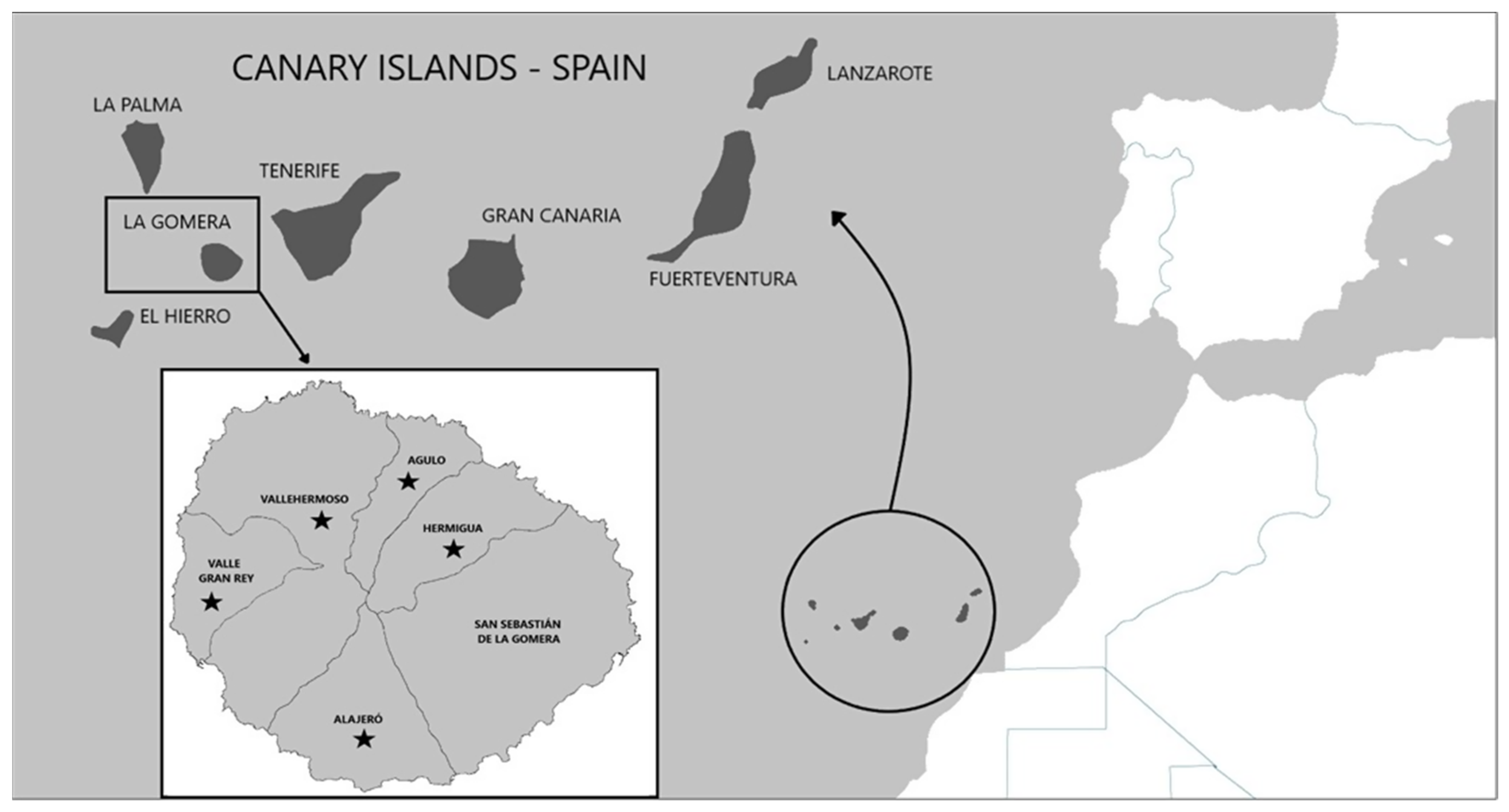
2.1. Sample Collection

Between June 2021 and July 2022, a total of 29 feral cats (Felis catus) from La Gomera, captured and provided already sacrificed by Gestión y Planeamiento Territorial y Medioambiental (GESPLAN) staff, with the authorization of Gobierno de Canarias (expedient number 2022/25073) and Excmo. Cabildo Insular de La Gomera (expedient numbers 1872/2021 and 1821/2022), were analyzed at the Instituto Universitario de Enfermedades Tropicales y Salud Pública de Canarias (Table S1).
The animals used in this study were obtained in the municipalities of Vallehermoso (n = 16), Valle Gran Rey (n = 6), Hermigua (n = 3), Agulo (n = 2), San Sebastián de La Gomera (n = 1) and Alajeró (n = 1) (Figure 1).
After dissection, the lungs and heart of each animal were examined for adult lungworms using a Leica M80 (Leica Mikrosysteme Vertrieb GmbH, Wetzlar, Germany) stereoscope. The nematodes obtained were washed in sterile phosphate-buffered saline (PBS) and preserved in 70% ethanol. The central part of a random selection of nematodes was reserved for DNA extraction and the anterior and posterior ends were clarified in lactophenol for morphological identification according to the available keys [7,23,33,34,35,36]. Digital images and measurements in µm were taken using an optical Leica DM2500 (Leica Microsystems, Heerbrugg, Switzerland) microscope and Leica LAS AF 4.12 software, respectively. In addition, a portion of positive lung tissue was fixed directly for histological analysis and the rest of the lung tissue remained frozen until molecular analysis.

2.2. Histological Analysis

To determine the existence of lesions, lungs positive for metastrongylids were taken randomly and processed for histological analysis following the standard protocols. Briefly, samples were fixed in 10% formalin solution and embedded in Paraplast®. Sections of a 5 μm thickness were obtained from each sample with a microtome (Shandon Finesse 325, ThermoFisher Scientific, MA, USA). The sections were stained with hematoxylin-erythrosine and Masson-Trichrome staining for tissue structure evaluation. Sections were analyzed using light microscopy (Leica DM 4000B, Leica, Barcelona, Spain) and images were captured with a camera (Leica DFC 300FX, Leica, Barcelona, Spain).

2.3. DNA Isolation

A portion of a lungworm-positive lung and the middle part of randomly selected nematodes were homogenized in 250 µL of a lysis solution containing 30 mM Tris-HCl (pH 8.0), 10 mM EDTA and 0.4% SDS. Then, 3 µL of proteinase K (20 ng/µL) was added to the samples and incubated at 56 °C overnight. After the inactivation of proteinase K, DNA was extracted according to the method described by Lopez et al. [37]. The quantity and quality of the extracted DNA were determined using a DeNovix DS-11+ spectrophotometer (DeNovix Inc., Wilmington, DE, USA). The DNA was stored at −20 °C until further processing.

2.4. PCR Amplification

A fragment of the internal transcribed spacer 2 region (ITS-2) was amplified using the primers NC1 (5′-ACGTCTGGTTCAGGGTTGTT-3′) and NC2 (5′-TTAGTTTCTTTTCCTCCGCT-3′) as previously described by Gasser et al. [38]. PCR reactions were performed in a total volume of 25 μL, including 10× buffer Mg2+ free (Bioline, London, UK), 2.5 μL of each dNTP (10 mM), 1 μL of each primer (12.5 ng/mL), 0.125 μL of Biotaq polymerase (5 U/mL) (Bioline, London), 0.75 mM MgCl2, 20–50 ng of genomic DNA and water. The ITS-2 fragment was amplified using the following cycling conditions: 2 min at 94 °C, followed by 35 cycles of denaturation at 94 °C for 1 min, annealing at 58 °C for 1 min and extension at 72 °C for 1 min, with a final extension step at 72 °C for 5 min. All PCR products were resolved on 1.5% agarose gel.

2.5. Sequencing and Phylogenetic Analysis

Positive amplicons were sequenced in both directions at Macrogen Inc. (Madrid, Spain). The obtained nucleotide sequences were edited with the MEGA X program [39] and subsequently aligned with other metastrongylid sequences using the Clustal-W program included in MEGA X. Minor corrections to increase the aligned sequence similarity and improve the inferences on any positional homology were then made by hand. A BLAST search was performed to elucidate any homologies or similarities with the sequences previously published in the GenBank database. Molecular identification was achieved using phylogenetic analysis through the neighbor-joining distance method with the p-distance model [40] and the maximum likelihood method with the Kimura 2-parameter model [41], both with at least 1000 bootstrap replications. Toxocara cati (Acc. Number: JN391472) was used as the outgroup.

3. Results

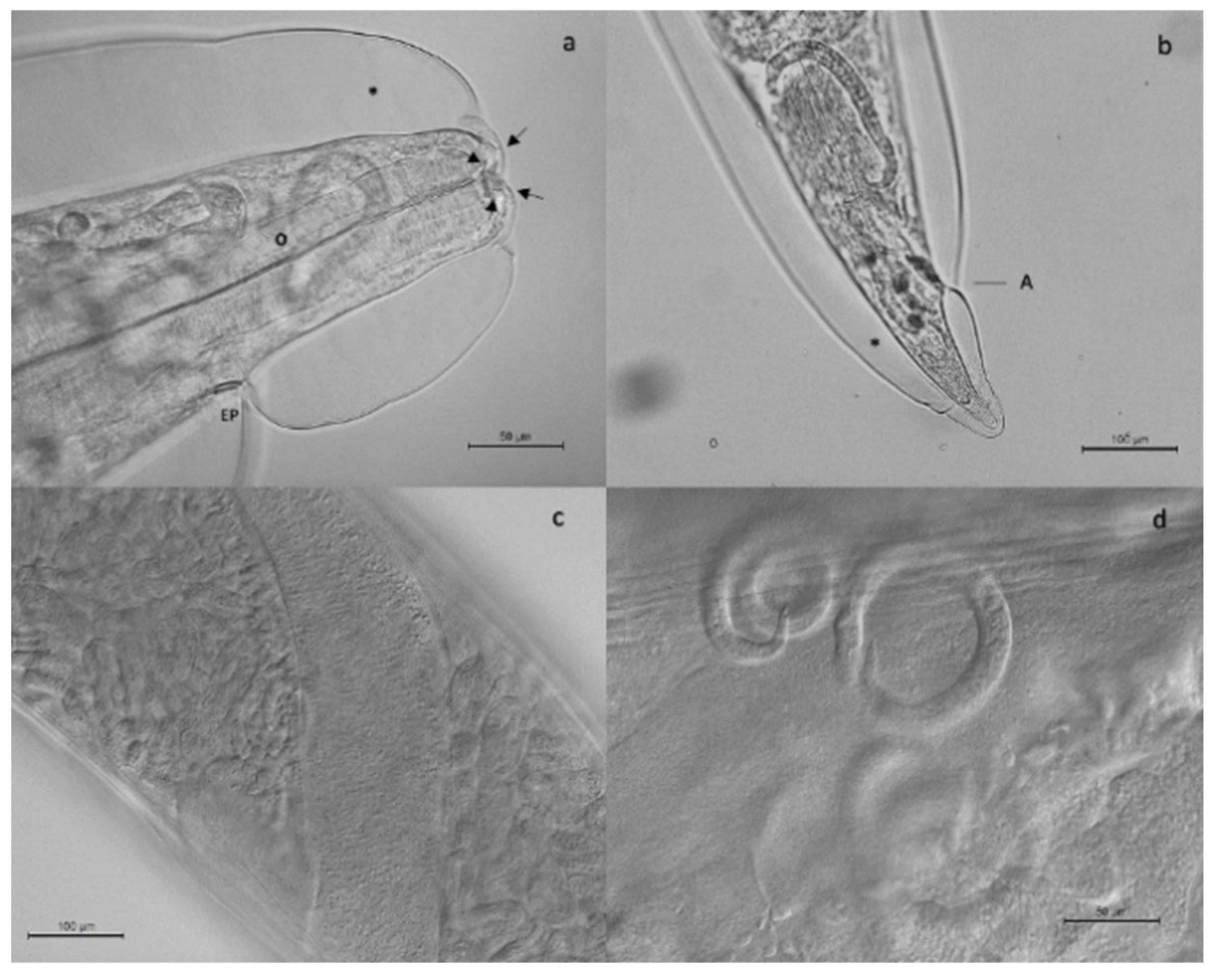
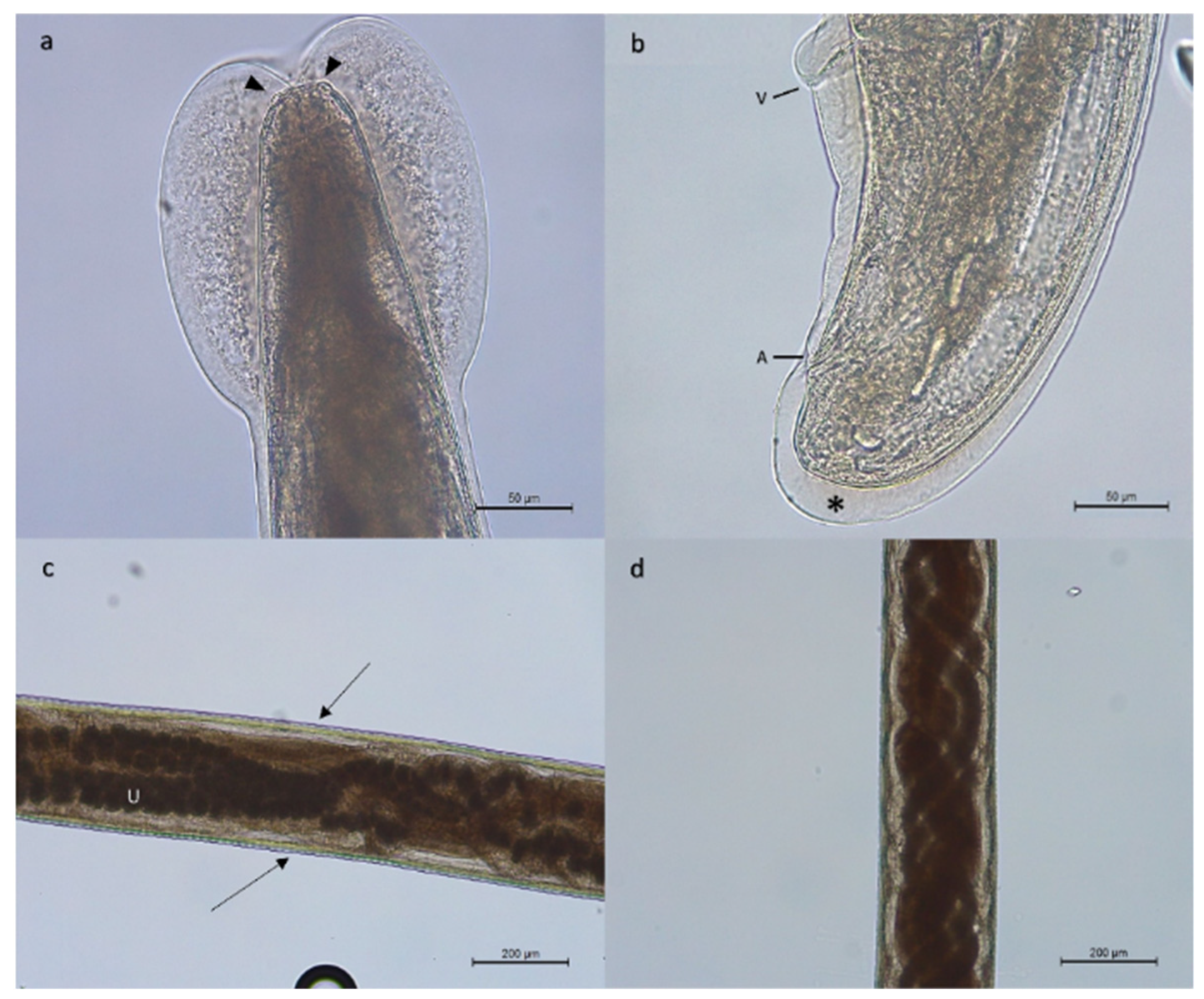
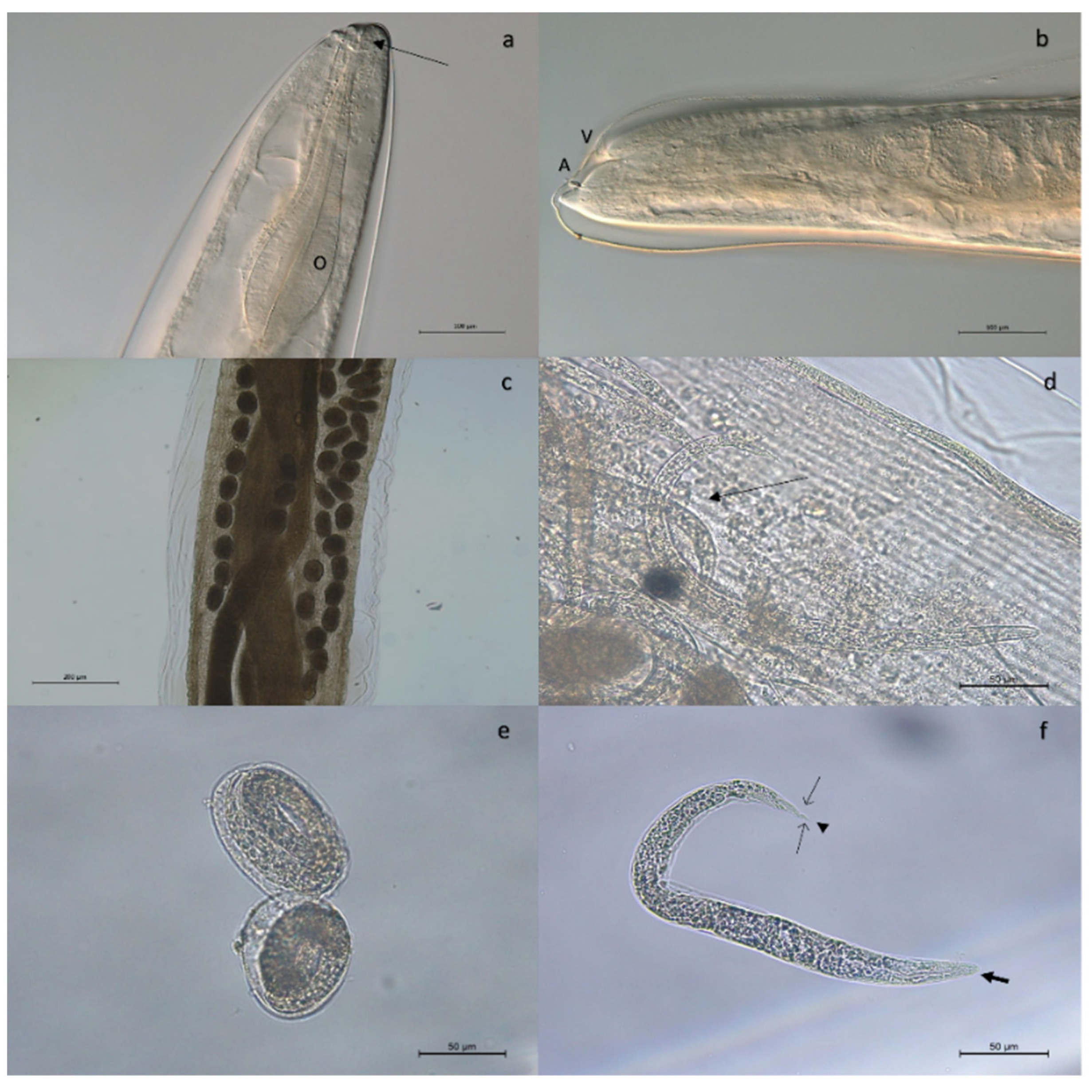
3.1. Morphological Analysis

Adult stages of pulmonary nematodes were detected in the lungs of 16 out of 29 feral cats analyzed (55.2%) from 5 out of 6 municipalities of La Gomera, concretely, Vallehermoso (43.7%; 7/16), Valle Gran Rey (50%; 3/6), Hermigua (66.6%; 2/3), Agulo (100%; 2/2) and Alajeró (100%; 1/1) (Figure 1). The mean intensity was 9.0 worms per animal, with an intensity range of 1 to 69. Gross tissue examination and morphological analysis allowed us to identify adult females of three metastrongylid species: T. brevior (Figure 2), A. chabaudi (Figure 3) and O. rostratus (Figure 4), while no males were detected. Additionally, it was not possible to morphologically identify two nematodes due to the damage to their ends suffered during isolation.
Adult females of T. brevior were 6–13 mm in length and 330–430 µm in width. Females of A. chabaudi were 21–22 mm in length and 260–270 µm in width. In the case of O. rostratus, worms were long and slender with a smooth integument. It was not possible to measure the total length of the females obtained, as they suffered damage during extraction due to being embedded in the peribronchial tissue surrounded by a fibrous tissue capsule.

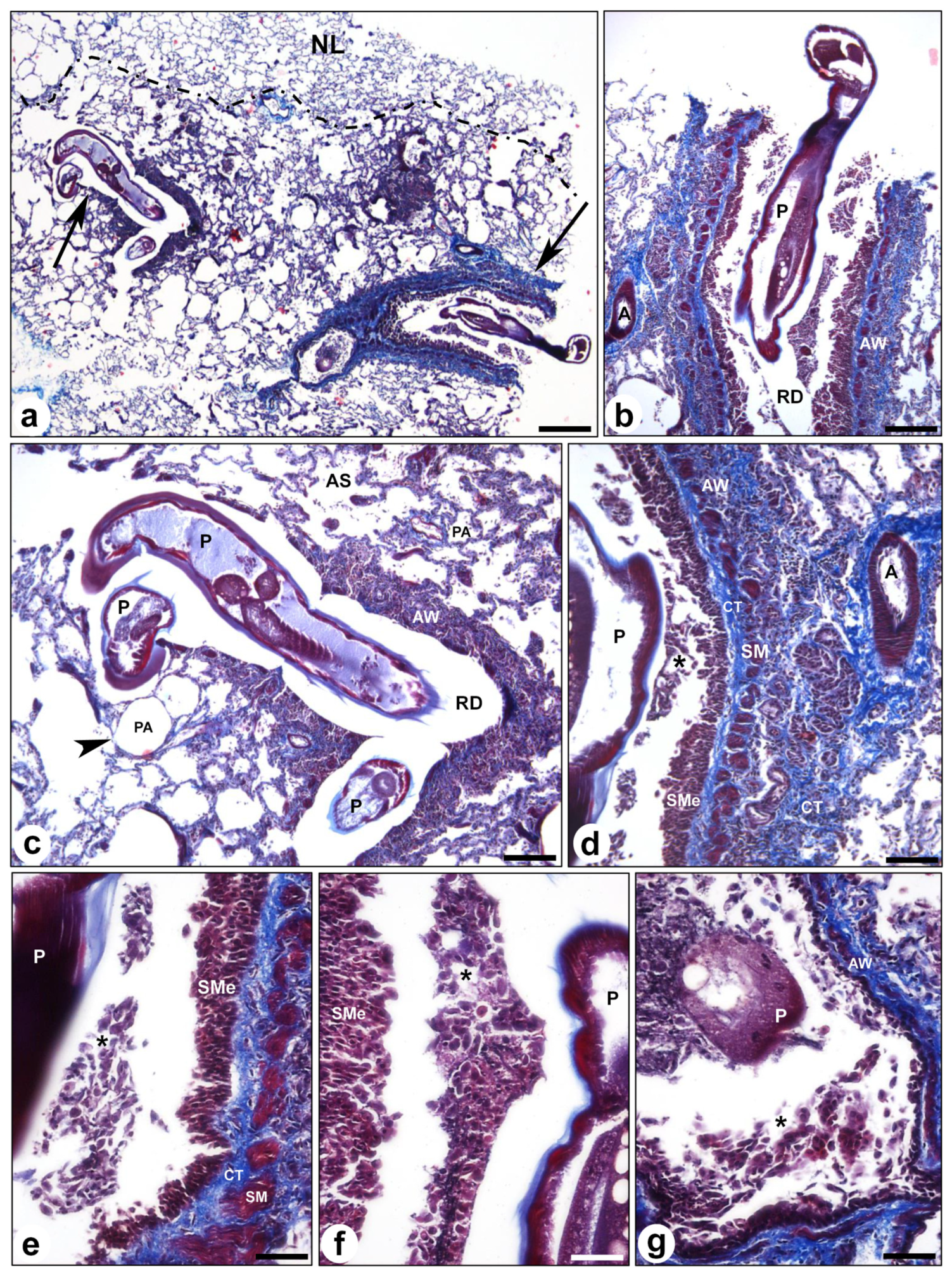
3.2. Histological Evaluation

The analyzed samples showed loss of normal lung tissue architecture with distortion of the alveolar structure and fibrosis in those areas where the parasite, O. rostratus, was located (Figure 5). Two granulomatous lesions were observed in the lung parenchyma containing the parasite, inside the respiratory ducts (Figure 5a–c). The lesions were characterized by airway wall hypertrophy with epithelial cell proliferation and squamous metaplasia instead of normal ciliated pseudostratified epithelium, an increase in connective tissue and smooth muscle hyperplasia (Figure 5d–g). Inflammatory infiltrate was also observed in the airway wall and around the parasite, with numerous macrophages, lymphocytes and eosinophils (Figure 5e–g).

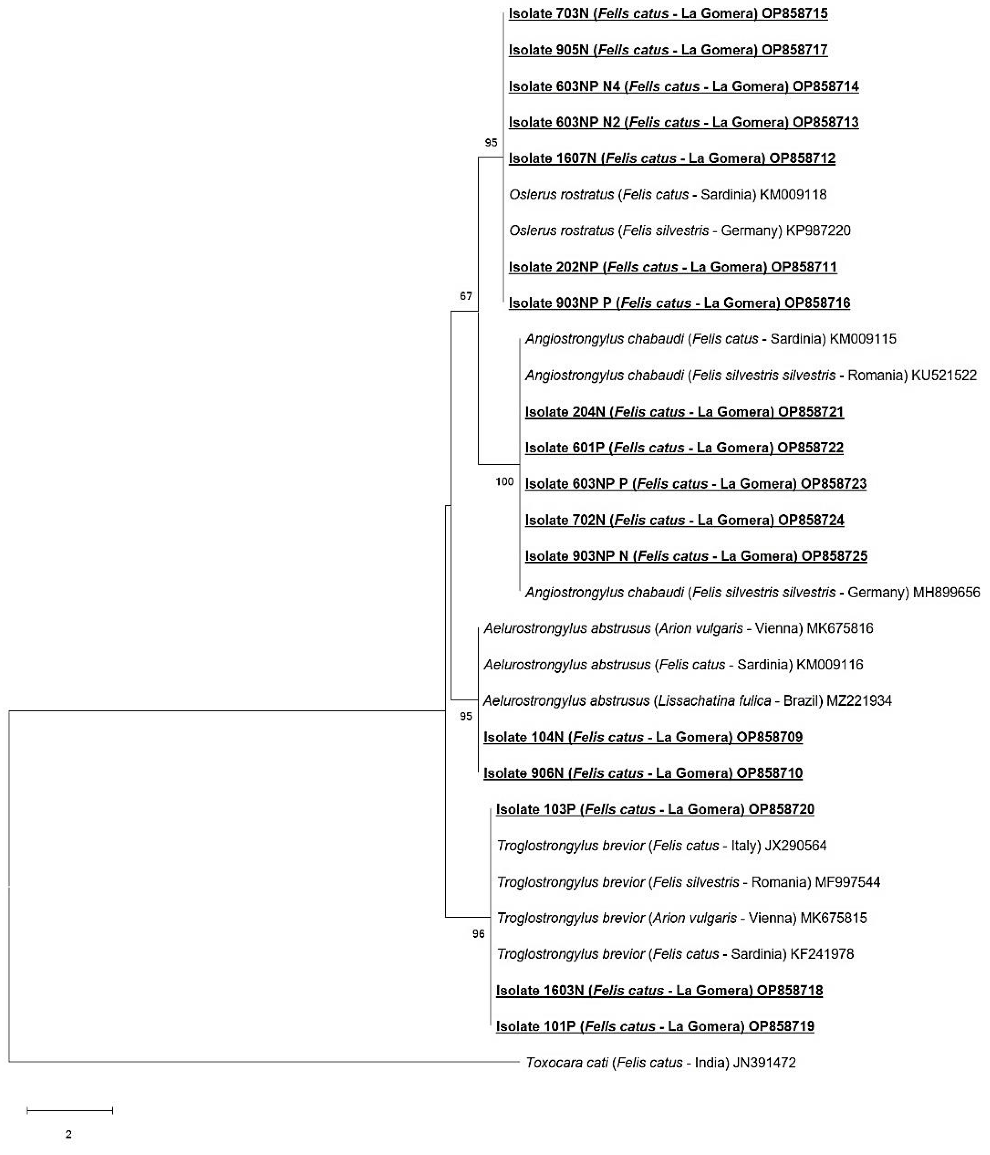
3.3. Molecular Examination

The results obtained in the molecular analysis confirmed the morphological results obtained, also identifying the species T. brevior, A. chabaudi and O. rostratus. Additionally, two nematodes that were not possible to identify morphologically due to damage to their ends during isolation were identified as A. abstrusus. The molecular analysis of the lung samples also confirmed the identity of previously obtained species and also detected coinfections with A. chabaudi and O. rostratus in two animals.
A total of seventeen nucleotide sequences were obtained. Troglostrongylus brevior was confirmed in three cats (isolates 101P, 103P and 1603N), displaying 99–100% homology with others T. brevior sequences (GenBank: MK675815, MH537789). Oslerus rostratus was identified in seven cats (isolates 202NP, 1607N, 603NPN2, 603NPN4, 703N, 905N and 903NPP), displaying 99% homology with an O. rostratus sequence (GenBank: KP987220). Angiostrongylus chabaudi was identified in five cats (isolates 204N, 702N, 903NPN, 601P and 603NPP), displaying 99–100% homology with other A. chabaudi sequences (GenBank: KU521522, MH899656). Aelurostrongylus abstrusus was identified in two cats (isolates 104N and 906), displaying 99–100% homology using BLAST with A. abstrusus sequences (GenBank: MK675816, MH779463). The obtained nucleotide sequences generated in this study are available in the GenBank database under the accession numbers OP858709-OP858725.
The results of the neighbor-joining (Figure 6) and maximum likelihood (Figure 7) analyses based on 358 bp of the alignment showed similar results, confirming the identity of the four metastrongylid species detected by BLAST. In both phylogenetic analyses, the sequences from feral cats obtained in this study were grouped into the A. abstrusus, T. brevior, O. rostratus and A. chabaudi clades with high bootstrap values (95–100%).
In this study, the cardio-pulmonary species O. rostratus and A. chabaudi were the most frequently detected species, with an occurrence of 27.6% (8/29) and 17.2% (5/29), respectively, followed by T. brevior (10.3%; 3/29) and A. abstrusus (6.9%; 2/29) (Table 1). Of the 16 feral cats positive for cardio-pulmonary parasites, 14 animals presented single infections with O. rostratus (n= 6), A. chabaudi (n = 3), T. brevior (n = 3) and A. abstrusus (n = 2). Coinfections with A. chabaudi and O. rostratus were detected in two animals.

4. Discussion

In recent years, the parasitosis of the respiratory system of felids has attracted the attention of the veterinary community due to their great geographical expansion and the epizootiological and biological characteristics that have been discovered in these nematodes [2,3,4].
In Europe, these pulmonary nematodes have been studied mainly in wildcats and domestic cats [15]. In the case of Spain, most available epidemiological investigations are based on wild animals such as wolves and foxes. However, studies related to cats from this region are scarce [42,43]. Therefore, the present study contributes to the knowledge of the epidemiology of the feline cardio-pulmonary nematodes affecting feral cats in Spain, providing new data on the prevalence and distribution of these parasites in the Canary Archipelago.
These nematodes have been previously cited worldwide, mostly the species A. abstrusus, which is frequently reported in cases of infections in domestic cats [1]. Other species, such as the metastrongyloids T. brevior, O. rostratus and A. chabaudi, have gained more veterinary relevance given the increase in the number of reports documented in wildcats and domestic cats, most of them geographically limited to Europe [2,4].
In this study, metastrongylid infections were detected in 16 feral cats from the island of La Gomera. A total of four species were successfully identified, concretely, A. abstrusus, T. brevior, O. rostratus and A. chabaudi. Up to now, only the species A. abstrusus and O. rostratus had been reported in feral and domestic cats of other Canary Islands, namely Gran Canaria and Tenerife, respectively [29,30,32].
The general prevalence obtained in our study was 55.2%, higher than the percentage reported in two other studies carried out in the Canary Islands, where 10.4% (5/48) and 28.3% (82/290) were reported in feral cats from Gran Canaria [30,31]. However, the prevalence of metastrongylid infections obtained in our study is similar to previous studies carried out in other insular areas, such as Sri Lanka, where 60% was reported [44].
There have been several studies carried out in island regions that have documented the presence of metastrongylid species in wildcats, stray, feral and domestic cats, such as Sri Lanka [44], Hawaii [45], Majorca [25], Ibiza [26], Sicily [23,35], Crete, Mykonos, Skopelos [46,47,48,49], Cyprus [50], Sardinia [51,52,53], Gran Canaria and Tenerife [29,30,31,32], among others. The most common lungworm species reported in these islands are A. abstrusus, T. brevior and O. rostratus. However, the cardio-pulmonary nematode A. chabaudi has only been previously reported in an adult cat on the island of Sardinia [51], with our study being the second report of A. chabaudi on islands.
Of all metastrongylid species detected in this study, O. rostratus was the most frequently detected species, with a 27.6% prevalence, followed by A. chabaudi (17.2%), T. brevior (10.3%) and A. abstrusus (6.9%). These results are in contrast to the previous data for Spain, where A. abstrusus was reported as the most prevalent metastrongylid species, with prevalence ranging between 1 and 10.4%, with the highest percentage being obtained for Gran Canaria [24,25,30].
Aelurostrongylus abstrusus is a parasite that affects feline lung tissues causing aelurostrongyliasis, a disease of the lower respiratory tract, whose serious infections can lead to verminous pneumonia which can be fatal [54,55,56,57]. This cosmopolitan nematode is the lungworm species most frequently diagnosed in felids, being reported in Europe, South America, Australia, the Middle East, Russia, the Far East, the USA, China and Africa [3,9,22,54,58,59,60,61,62,63,64,65,66,67]. The prevalence of A. abstrusus can vary significantly across Europe. It has been reported ranging from 0.4% in Croatia [68] to 43.1% in Albania [69], with Italy being the European country with the second-highest prevelance (26.5%) [70]. In addition, the prevalence rate of this lungworm species may vary within the same country (see [22]), for example, a 1% prevalence of A. abstrusus has been reported in feral cats from mainland Spain [24], while a 10.4% prevalence in feral cats from the island of Gran Canaria [30], with this percentage being similar to the prevalence obtained for A. abstrusus in our study.
Troglostrongylus brevior, after its first description in wild felids from Palestine [7], was reported mostly in wildcats from central Italy [4,13,71,72] until 2010, when it was first detected in domestic cats from Ibiza (Spain) [26]. In the last decade, T. brevior has been reported to infect domestic cats from the islands of Sicily and Sardinia in Italy, the islands of Crete, Mykonos and Skopelos in Greece, in the island state of Cyprus [35,46,47,50,73,74] and in continental areas such as Italy [74,75,76,77], Greece [47], Bulgaria [3], Spain [3], Romania [78,79] and Turkey [80]. Therefore, our study constitutes the first report of T. brevior in feral cats in the Canary Islands and the northwestern area close to the African continent.
The clinical manifestations of T. brevior are similar to those produced by A. abstrusus, being potentially fatal in kittens and cats less than one year old [1,35,48,79,81,82]. However, the lesions caused by both species are different: while T. brevior is longer in size and inhabits the bronchi and bronchioles of the definitive host, A. abstrusus is smaller and is localized in the alveolar ducts and the lung parenchyma of hosts [1,2,35,81]. Troglostrongylus brevior is usually reported with a lower prevalence in comparison to A. abstrusus. However, in certain regions, its prevalence is higher, as has been shown in ther region in our study and in other regions such as Greece [47], Cyprus [50], Italy [83] and Israel [84].
Oslerus rostratus, which can be found in the peri-bronchial tissues and in the bronchial walls, was first described as Anafilaroides rostratus in cats from Jerusalem [7]. Later, it was reported in cats from Sri Lanka [85], Hawaii [45], mainland Spain [86], Majorca and Ibiza [25,26], Sicily and Sardinia, [23,52] and Hungary [87]. Recently, O. rostratus has been cited in domestic cats from the Canary Island of Tenerife [32], with the present study being the first citation in feral cats. In mainland Spain, O. rostratus has been one of the least prevalent metastrongylid species detected in domestic cats, with a 1% prevalence reported [3]. However, in feral cats from the Spanish island of Majorca, O. rostratus has been reported with a 24.1% prevalence [25], being similar to that obtained in our study (27.6%).
Angiostrongylus chabaudi inhabits the small pulmonary arteries of wildcats and produces severe damage to the vascular system [11]. It was first described in wildcats from central Italy [88]. Later, it was reported in Sardinia [51] and in coinfection with A. abstrusus and T. brevior in a domestic cat in central Italy [76]. Subsequently, several studies have reported A. chabaudi in the European wildcat in Italy [89], Germany [90], Greece [36,91], Romania [92,93], Bulgaria [11] and, more recently, in Bosnia and Herzegovina [94]. Our study constitutes the first report on A. chabaudi in cats from Spain.
Although previous cases of coinfection with cardio-pulmonary nematodes in cats have been cited, such as A. abstrusus, T. brevior and A. chabaudi in a domestic cat in Italy [76], A. abstrusus and A. chabaudi in a wildcat in Bosnia and Herzegovina [94] and A. abstrusus, T. brevior, E. aerophilus and A. chabaudi in a wildcat in Greece [95], among others, our study constitutes the first report in cats of a mixed infection caused by A. chabaudi and O. rostratus.
Insular regions are particularly vulnerable to the effects of introduced species such as cats. These animals can interfere with the natural dynamics of populations and/or ecosystems and may originate or favor processes such as the introduction of parasites [96]. In the Canary Islands, cats have been introduced for more than 2300 years [97,98,99]. Considering the high diversity of metastrongylid species identified together with the high prevalence obtained in this study, lungworms could have been introduced to La Gomera with the cat at some point in the past.
On the other hand, the possibility should not be discarded that cats living in endemic areas could have traveled with their owners to the Canary Islands and lungworms have found a way to establish themselves in the archipelago, as was proposed in the finding of Gurltia paralysans, a parasite endemic to South America, that has been recently reported infecting a domestic cat in Tenerife [100]. In this sense, it is important to note that La Gomera has a high tourist influx of European travelers.
The results obtained in this study show that feral cats from La Gomera may play a role in the dissemination of lungworm species, increasing the risk of infection to both wild and domestic cats with outdoor access. Infection may occur with one or more species in coinfection, which would further aggravate the symptoms and worsen the health status of these animals. More studies are needed to better understand the distribution of these metastrongylid species in the Canary Archipelago. Furthermore, it is necessary to warn the veterinary community of the Canary Archipelago so that they can make a correct diagnosis in clinical cases with respiratory symptoms.

5. Conclusions

This study contributes to the knowledge of the epidemiology of lungworm parasites in cats, being the first report on the high diversity of metastrongylid species in feral cats in the Canaries, reporting A. chabaudi and T. brevior for the first time in this region and A. chabaudi in cats in Spain. The diversity of species and the high prevalence obtained indicate that wild and domestic feline populations in this region are exposed to lungworm infections with A. abstrusus, T. brevior, A. chabaudi and O. rostratus, which have been detected to be widely distributed on the island. More studies are needed to better understand the epidemiology of these parasites in the Canary Archipelago. Veterinary practitioners should consider these parasites as possible causes of respiratory pathologies in cats in the Canary Islands. Moreover, control measures should be carried out in order to prevent the transmission of these respiratory parasites.

Supplementary Materials

The following supporting information can be downloaded at: https://www.mdpi.com/article/10.3390/ani13132168/s1, Table S1: Detailed information related to the specimens of Felis catus captured in La Gomera (Canary Islands, Spain) analysed in this study and the results obtained.

Author Contributions

Conceptualization, P.F.; methodology, K.G.-L., R.R., V.A.-R., E.B.-G. and N.M.-C.; formal analysis, K.G.-L.; investigation, K.G.-L.; resources, P.F.; data curation, K.G.-L., R.R., V.A.-R., E.B.-G. and N.M.-C.; writing—original draft preparation, K.G.-L.; writing—review and editing, K.G.-L., R.R., V.A.-R., E.B.-G., N.M.-C., E.R.-P. and P.F.; supervision, P.F.; project administration, P.F.; funding acquisition, P.F. All authors have read and agreed to the published version of the manuscript.

Funding

This research was funded by ProID2021010013, funded by Consejería de Economía, Industria, Comercio y Conocimiento, Gobierno de Canarias and FEDER-FSE Canarias 2014–2020, “Programa de Apoyo a la Investigación María del Carmen Betencourt y Molina“ and by “Consejería de Transición Ecológica, Lucha contra el Cambio Climático y Planificación Territorial” (Gobierno de Canarias—Universidad de La Laguna agreement “Estudio de patógenos en aves migratorias y en especies exóticas en un escenario de cambio climático”). K.G.-L. was granted a predoctoral scholarship by the Spanish Ministry of Science, Innovation and Universities and Universidad de La Laguna (Becas M-ULL, 2019). E.B.-G. was granted a FPI predoctoral scholarship by “Agencia Canaria de Investigación, Innovación y Sociedad de la Información de la Consejería de Economía, Conocimiento y Empleo” and “Fondo Social Europeo (FSE) Programa Operativo integrado de Canarias 2014−2020, Eje 3 Tema Prioritario 74 (85%)” (TESIS2021010056).

Institutional Review Board Statement

The specimens of Felis catus used in this study were captured and provided already sacrificed by Gestión y Planeamiento Territorial y Medioambiental (GESPLAN) staff, with the authorization of Gobierno de Canarias (expedient number 2022/25073) and Excmo. Cabildo Insular de La Gomera (expedient numbers 1872/2021 and 1821/2022).

Informed Consent Statement

Not applicable.

Data Availability Statement

Not applicable.

Acknowledgments

We thank the staff of GESPLAN S.A. for supply the animals used in this study.

Conflicts of Interest

The authors declare no conflict of interest.

References

  1. Traversa, D.; Di Cesare, A. Feline lungworms: What a dilemma. Trends Parasitol. 2013, 29, 423–430. [Google Scholar] [CrossRef] [PubMed]
  2. Morelli, S.; Diakou, A.; Colombo, M.; Di Cesare, A.; Barlaam, A.; Dimzas, D.; Traversa, T. Cat respiratory nematodes: Current knowledge, novel data and warranted studies on clinical features, treatment and control. Pathogens 2021, 10, 454. [Google Scholar] [CrossRef] [PubMed]
  3. Giannelli, A.; Capelli, G.; Joachim, A.; Hinney, B.; Losson, B.; Kirkova, Z.; René-Martellet, M.; Papadopoulos, E.; Farkas, R.; Napoli, E.; et al. Lungworms and gastrointestinal parasites of domestic cats: A European perspective. Int. J. Parasitol. 2017, 47, 517–528. [Google Scholar] [CrossRef] [PubMed]
  4. Traversa, D.; Morelli, S.; Di Cesare, A.; Diakou, A. Felid Cardiopulmonary Nematodes: Dilemmas Solved and New Questions Posed. Pathogens 2021, 10, 30. [Google Scholar] [CrossRef]
  5. Bezerra-Santos, M.A.; Mendoza-Roldan, J.A.; Abramo, F.; Lia, R.P.; Tarallo, V.D.; Salant, H.; Brianti, E.; Baneth, G.; Otranto, D. Transmammary transmission of Troglostrongylus brevior feline lungworm: A lesson from our gardens. Vet. Parasitol. 2020, 285, 109215. [Google Scholar] [CrossRef]
  6. Lanszki, J.; Kletečki, E.; Trócsányi, B.; Mužinić, J.; Széles, G.L.; Purger, J.J. Feeding habits of house and feral cats (Felis catus) on small Adriatic islands (Croatia). North-West. J. Zool. 2016, 12, 336–348. [Google Scholar]
  7. Gerichter, C.B. Studies on the nematodes parasitic in the lungs of Felidae in Palestine. Parasitology 1949, 39, 251–262. [Google Scholar] [CrossRef]
  8. Beugnet, F.; Bourdeau, P.; Chalvet-Monfray, K.; Cozma, V.; Farkas, R.; Guillot, J.; Halos, L.; Joachim, A.; Losson, B.; Miro, G.; et al. Parasites of domestic owned cats in Europe: Co-infestations and risk factors. Parasites Vectors 2014, 7, 291. [Google Scholar] [CrossRef] [Green Version]
  9. Elsheikha, H.M.; Schnyder, M.; Traversa, D.; Di Cesare, A.; Wright, I.; Lacher, D.W. Updates on feline aelurostrongylosis and research priorities for de next decade. Parasites Vectors 2016, 9, 389. [Google Scholar] [CrossRef] [PubMed] [Green Version]
  10. Carretón Gómez, E.; Costa Rodríguez, N.; García Rodríguez, S.N.; Matos Rivero, J.I.; Falcon Cordón, Y.; Montoya Alonso, J.A.; García Morchon, R. Parásitos respiratorios felinos. Argos Inf. Vet. 2022, 235, 1–16. [Google Scholar]
  11. Giannelli, A.; Kirkova, Z.; Abramo, F.; Latrofa, M.S.; Campbell, B.; Zizzo, N.; Cantacessi, C.; Dantas-Torres, F.; Otranto, D. Angiostrongylus chabaudi in felids: New findings and a review of the literature. Vet. Parasitol. 2016, 228, 188–192. [Google Scholar] [CrossRef]
  12. Bowman, D.D. Respiratory system parasites of the dog and cat (Part II): Trachea and bronchi, and pulmonary vessels. In Companion and Exotic Animal Parasitology; Bowman, D.D., Ed.; International Veterinary Information Service: New York, NY, USA, 2000; pp. 1–15. [Google Scholar]
  13. Anderson, R.C. The superfamily Metastrongyloidea. In Nematode Parasites of Vertebrates. Their Development and Transmission, 2nd ed.; Anderson, R.C., Ed.; CABI Publishing: Wallingford, UK, 2000; pp. 129–229. [Google Scholar]
  14. Cowie, R.H. Pathways for transmission of angiostrongyliasis and the risk of disease associated with them. Hawai’i J. Med. Public Health 2013, 72, 70–74. [Google Scholar]
  15. Traversa, D.; Di Cesare, A. Diagnosis and management of lungworm infections in cats: Cornerstones, dilemmas and new avenues. J. Feline Med. Surg. 2016, 18, 7–20. [Google Scholar] [CrossRef] [PubMed] [Green Version]
  16. Traversa, D.; Guglielmini, C. Feline aelurostrongylosis and canine angiostrongylosis: A challenging diagnosis for two emerging verminous pneumonia infections. Vet. Parasitol. 2008, 157, 163–174. [Google Scholar] [CrossRef] [PubMed]
  17. Payo-Puente, P.; Botelho-Dinis, M.; Carvaja Uruena, A.M.; Payo-Puente, M.; Gonzalo-Orden, J.M.; Rojo-Vazquez, F.A. Prevalence study of the lungworm Aelurostrongylus abstrusus in stray cats of Portugal. J. Feline Med. Surg. 2008, 10, 242–246. [Google Scholar] [CrossRef] [PubMed]
  18. Foster, S.F.; Martin, P. Lower respiratory tract infections in cats: Reaching beyond empirical therapy. J. Feline Med. Surg. 2011, 13, 313–332. [Google Scholar] [CrossRef]
  19. Di Cesare, A.; Castagna, G.; Meloni, S.; Milillo, P.; Latrofa, S.; Otranto, D.; Traversa, D. Canine and feline infections by cardiopulmonary nematodes in Central and Southern Italy. Parasitol. Res. 2011, 109, 87–96. [Google Scholar] [CrossRef]
  20. Pennisi, M.G.; Hartmann, K.; Addie, D.D.; Boucraut-Baralon, C.; Egberink, H.; Frymus, T.; Gruffydd-Jones, T.; Horzinek, M.C.; Hosie, M.J.; Loret, A.; et al. Lungworm disease in cats: ABCD guidelines on prevention and management. J. Feline Med. Surg. 2015, 17, 626–636. [Google Scholar] [CrossRef]
  21. Traversa, D.; Di Cesare, A. Cardio-pulmonary parasitic nematodes affecting cats in Europe: Unraveling the past, depicting the present, and predicting the future. Front. Vet. Sci. 2014, 1, 11. [Google Scholar] [CrossRef] [Green Version]
  22. Elsheikha, H.M.; Wright, I.; Wang, B.; Schaper, R. Prevalence of feline lungworm Aelurostrongylus abstrusus in England. Vet. Parasitol. Reg. Stud. Rep. 2019, 16, 100271. [Google Scholar] [CrossRef] [Green Version]
  23. Brianti, E.; Gaglio, G.; Napoli, E.; Falsone, L.; Giannelli, A.; Annoscia, G.; Varcasia, A.; Giannetto, S.; Mazzullo, G.; Otranto, D. Feline lungworm Oslerus rostratus (Strongylida: Filaridae) in Italy: First case report and histopathological findings. Parasitol. Res. 2014, 13, 3853–3857. [Google Scholar] [CrossRef] [PubMed]
  24. Miro, G.; Montoya, A.; Jimenez, S.; Frisuelos, C.; Mateo, M.; Fuentes, I. Prevalence of antibodies to Toxoplasma gondii and intestinal parasites in stray, farm and household cats in Spain. Vet. Parasitol. 2004, 126, 249–255. [Google Scholar] [CrossRef] [PubMed]
  25. Millán, J.; Casanova, J.C. High prevalence of helminth parasites in feral cats in Majorca Island (Spain). Parasitol. Res. 2009, 106, 183–188. [Google Scholar] [CrossRef]
  26. Jefferies, R.; Vrhovec, M.G.; Wallner, N.; Catalan, D.R. Aelurostrongylus abstrusus and Troglostrongylus sp. (Nematoda: Metastrongyloidea) infections in cats inhabiting Ibiza, Spain. Vet. Parasitol. 2010, 173, 344–348. [Google Scholar] [CrossRef]
  27. Montoya, A.; García, M.; Gálvez, R.; Checa, R.; Marino, V.; Sarquis, J.; Barrera, J.P.; Rupérez, C.; Caballero, L.; Chicharro, C.; et al. Implications of zoonotic and vector-borne parasites to free-roaming cats in central Spain. Vet. Parasitol. 2018, 251, 125–130. [Google Scholar] [CrossRef] [PubMed]
  28. Remesar, S.; García-Dios, D.; Calabuig, N.; Díaz-Cao, J.M.; López-Lorenzo, G.; López, C.; Fernández, G.; Morrondo, P.; Panadero, R.; López, C.M.; et al. Cardiorespiratory nematodes and co-infections with gastrointestinal parasites in new arrivals at dog and cat shelters in north-western Spain. Transbound. Emerg. Dis. 2022, 69, e3141–e3153. [Google Scholar] [CrossRef]
  29. Valladares, B.; De Armas, F.; Del Castillo, A. A contribution to the knowledge of the pathology and immunopathology of the cat parasite Aelurostrongylus abstrusus (Railliet, 1898). In Proceedings of the European Multicolloquium of Parasitology, The Hague, The Netherlands, 7–11 September 1992. [Google Scholar]
  30. Rodríguez-Ponce, E.; González, J.F.; de Felipe, M.C.; Hernández, J.N.; Jaber, J.R. Epidemiological survey of zoonotic helminths in feral cats in Gran Canaria island (Macaronesian archipelago-Spain). Acta Parasitol. 2016, 61, 443–450. [Google Scholar] [CrossRef] [Green Version]
  31. Marbella, D.; Santana-Hernández, K.M.; Rodríguez-Ponce, E. Small islands as potential model ecosystems for parasitology: Climatic influence on parasites of feral cats. J. Helminthol. 2022, 96, e51. [Google Scholar] [CrossRef]
  32. García-Livia, K.; Valladares Salmerón, M.; Pacheco, S.; Valladares, B.; Foronda, P. Report of Oslerus rostratus (Strongylida: Filaroididae) in cats from the Canary Islands, Spain. Austral. J. Vet. Sci. 2022, 54, 145–150. [Google Scholar] [CrossRef]
  33. Seneviratna, P. Studies on Anafilaroides rostratus Gerichter, 1949 in cats: II. The life cycle. J. Helminthol. 1959, 33, 109–122. [Google Scholar] [CrossRef]
  34. Bowman, D.D.; Hendrix, C.M.; Lindsay, D.S.; Barr, S. Metastrongyloidea. In Feline Clinical Parasitology, 1st ed.; Bowman, D.D., Hendrix, C.M., Lindsay, D.S., Barr, S., Eds.; Iowa State University Press: Ames, IA, USA, 2002; pp. 271–272. [Google Scholar]
  35. Brianti, E.; Gaglio, G.; Giannetto, S.; Annoscia, G.; Latrofa, M.S.; Dantas-Torres, F.; Traversa, D.; Otranto, D. Troglostrongylus brevior and Troglostrongylus subcrenatus (Strongylida: Crenosomatidae) as agents of broncho-pulmonary infestation in domestic cats. Parasites Vectors 2012, 5, 178. [Google Scholar] [CrossRef] [Green Version]
  36. Diakou, A.; Psalla, D.; Migli, D.; Di Cesare, A.; Youlatos, D.; Marcer, F.; Traversa, D. First evidence of the European wildcat (Felis silvestris silvestris) as definitive host of Angiostrongylus chabaudi. Parasitol. Res. 2016, 115, 1235–1244. [Google Scholar] [CrossRef]
  37. López, C.; Clemente, S.; Almeida, C.; Brito, A.; Hernández, M. A genetic approach to the origin of Millepora sp. in the eastern Atlantic. Coral Reefs 2015, 34, 631–638. [Google Scholar] [CrossRef]
  38. Gasser, R.B.; Chilton, N.B.; Hoste, H.; Beveridge, I. Rapid sequencing of rDNA from single worms and eggs of parasitic helminths. Nucleic Acids Res. 1993, 21, 2525–2526. [Google Scholar] [CrossRef] [PubMed] [Green Version]
  39. Kumar, S.; Stecher, G.; Li, M.; Knyaz, C.; Tamura, K. MEGA X: Molecular evolutionary genetics analysis across computing platforms. Mol. Biol. Evol. 2018, 35, 1547–1549. [Google Scholar] [CrossRef] [PubMed]
  40. Saitou, N.; Nei, M. The neighbor-joining method: A new method for reconstructing phylogenetic trees. Mol. Biol. Evol. 1987, 4, 406–425. [Google Scholar]
  41. Kimura, M. A simple method for estimating evolutionary rate of base substitutions through comparative studies of nucleotide sequences. J. Mol. Evol. 1980, 16, 111–120. [Google Scholar] [CrossRef]
  42. Garrido-Castañé, I.; Ortuño, A.; Marco, I.; Castellà, J. Cardiopulmonary helminths in foxes from the Pyrenees. Acta Parasitol. 2015, 60, 712–715. [Google Scholar] [CrossRef] [PubMed]
  43. Martínez-Rondán, F.J.; Ruíz de Ybáñez, M.R.; López-Beceiro, A.M.; Fidalgo, L.E.; Berriatua, E.; Lahat, L.; Sacristán, I.; Oleaga, A.; Martínez-Carrasco, C. Cardiopulmonary nematode infections in wild canids: Does the key lie on host-prey-parasite evolution? Res. Vet. Sci. 2019, 126, 51–58. [Google Scholar] [CrossRef]
  44. Seneviratna, P. Observations on helminth infections in cats in Kandy district, Ceylon. Ceylon Vet. J. 1955, 3, 54–58. [Google Scholar]
  45. Ash, L.R. Helminth parasites of dogs and cats in Hawaii. J. Parasitol. 1962, 48, 63–65. [Google Scholar] [CrossRef] [PubMed]
  46. Diakou, A.; Di Cesare, A.; Aeriniotaki, T.; Traversa, D. First report of Troglostrongylus brevior in a kitten in Greece. Parasitol. Res. 2014, 113, 3895–3898. [Google Scholar] [CrossRef] [PubMed]
  47. Diakou, A.; Di Cesare, A.; Barros, L.A.; Morelli, S.; Halos, L.; Beugnet, F.; Traversa, D. Occurrence of Aelurostrongylus abstrusus and Troglostrongylus brevior in domestic cats in Greece. Parasites Vectors 2015, 8, 590. [Google Scholar] [CrossRef] [Green Version]
  48. Diakou, A.; Migli, D.; Dimzas, D.; Morelli, S.; Di Cesare, A.; Youlatos, D.; Lymberakis, P.; Traversa, D. Endoparasites of European Wildcats (Felis silvestris) in Greece. Pathogens 2021, 10, 594. [Google Scholar] [CrossRef]
  49. Morelli, S.; Diakou, A.; Di Cesare, A.; Schnyder, M.; Colombo, M.; Strube, C.; Dimzas, D.; Latino, R.; Traversa, D. Feline lungworms in Greece: Copromicroscopic, molecular and serological study. Parasitol. Res. 2020, 119, 2877–2889. [Google Scholar] [CrossRef]
  50. Diakou, A.; Sofroniou, D.; Di Cesare, A.; Kokkinos, P.; Traversa, D. Occurrence and zoonotic potential of endoparasites in cats of Cyprus and a new distribution area for Troglostrongylus brevior. Parasitol. Res. 2017, 116, 3429–3435. [Google Scholar] [CrossRef]
  51. Varcasia, A.; Tamponi, C.; Brianti, E.; Cabras, P.A.; Boi, R.; Pipia, A.P.; Giannelli, A.; Otranto, D.; Scala, A. Angiostrongylus chabaudi Biocca, 1957: A new parasite for domestic cats? Parasites Vectors 2014, 7, 588. [Google Scholar] [CrossRef] [PubMed] [Green Version]
  52. Varcasia, A.; Brianti, E.; Tamponi, C.; Pipia, A.P.; Cabra, P.; Mereu, M.; Dantas-Torres, F.; Scala, A.; Otranto, D. Simultaneous infection by four feline lungworm species and implications for the diagnosis. Parasitol. Res. 2015, 114, 317–321. [Google Scholar] [CrossRef]
  53. Tamponi, C.; Varcasia, A.; Pinna, S.; Melis, E.; Melosu, V.; Zidda, A.; Sanna, G.; Pipia, A.P.; Zedda, M.T.; Pau, S.; et al. Endoparasites detected in faecal samples from dogs and cats referred for routine clinical visit in Sardinia, Italy. Vet. Parasitol. Reg. Stud. Rep. 2017, 10, 13–17. [Google Scholar] [CrossRef]
  54. Yildiz, K.; Duru, S.Y.; Gokpinar, S. Alteration in blood gases in cats naturally infected with Aelurostrongylus abstrusus. J. Small Anim. Pract. 2011, 52, 376–379. [Google Scholar] [CrossRef]
  55. Mooney, E.T.; Rozanski, E.A.; King, R.G.P.; Sharp, C.R. Spontaneous pneumothorax in 35 cats (2001–2010). J. Feline Med. Surg. 2012, 14, 384–391. [Google Scholar] [CrossRef] [PubMed]
  56. Vezzosi, T.; Perrucci, S.; Parisi, F.; Morelli, S.; Maestrini, M.; Mennuni, G.; Traversa, D.; Poli, A. Fatal Pulmonary Hypertension and Right-Sided Congestive Heart Failure in a Kitten Infected with Aelurostrongylus abstrusus. Animals 2020, 10, 2263. [Google Scholar] [CrossRef] [PubMed]
  57. Scott, D.W. Current knowledge of aelurostrongylosis in the cat: Literature review and case reports. Cornell Vet. 1973, 63, 483–500. [Google Scholar] [PubMed]
  58. Foster, S.F.; Martin, P.; Allan, G.S.; Barrs, V.R.; Malik, R. Lower respiratory tract infections in cats: 21 cases (1995–2000). J. Feline Med. Surg. 2004, 6, 167–180. [Google Scholar] [CrossRef]
  59. Fiorello, C.V.; Robbins, R.G.; Maffei, L.; Wade, S.E. Parasites of free-ranging small canids and felids in the Bolivian Chaco. J. Zoo Wildl. Med. 2006, 37, 130–134. [Google Scholar] [CrossRef]
  60. Abu-Madi, M.A.; Al-Ahbabi, D.A.; Al-Mashhadani, M.M.; Al-Ibrahim, R.; Pal, P.; Lewis, J.W. Patterns of parasitic infections in faecal samples from stray cat populations in Qatar. J. Helminthol. 2007, 81, 281–286. [Google Scholar] [CrossRef]
  61. González, P.; Carbonell, E.; Urios, V.; Rozhnov, V.V. Coprology of Panthera tigris altaica and Felis bengalensis euptilurus from the Russian far east. J. Parasitol. 2007, 93, 948–950. [Google Scholar] [CrossRef]
  62. Gerdin, J.A.; Slater, M.R.; Makolinski, K.V.; Looney, A.L.; Appel, L.D.; Martin, N.M.; McDonough, S.P. Post-mortem findings in 54 cases of anesthetic associated death in cats from two spay–neuter programs in New York State. J. Feline Med. Surg. 2011, 13, 959–966. [Google Scholar] [CrossRef]
  63. Yang, Y.; Liang, H. Prevalence and risk factors of intestinal parasites in cats from China. BioMed Res. Int. 2015, 2015, 967238. [Google Scholar] [CrossRef] [Green Version]
  64. Di Cesare, A.; Laiacona, F.; Iorio, R.; Marangi, M.; Menegotto, A. Aelurostrongylus abstrusus in wild felids of South Africa. Parasitol. Res. 2016, 115, 3731–3735. [Google Scholar] [CrossRef] [PubMed]
  65. Penagos-Tabares, F.; Lange, M.K.; Chaparro-Gutiérrez, J.J.; Taubert, A.; Hermosilla, C. Angiostrongylus vasorum and Aelurostrongylus abstrusus: Neglected and underestimated parasites in South America. Parasites Vectors 2018, 11, 208. [Google Scholar] [CrossRef] [Green Version]
  66. Hoggard, K.R.; Jarriel, D.M.; Bevelock, T.J.; Verocai, G.G. Prevalence survey of gastrointestinal and respiratory parasites of shelter cats in northeastern Georgia, USA. Vet. Parasitol. Reg. Stud. Rep. 2019, 16, 100270. [Google Scholar] [CrossRef] [PubMed]
  67. Zottler, E.M.; Bieri, M.; Basso, W.; Schnyder, M. Intestinal parasites and lungworms in stray, shelter and privately owned cats of Switzerland. Parasitol. Int. 2019, 69, 75–81. [Google Scholar] [CrossRef] [PubMed]
  68. Grabarević, Ž.; Ćurić, S.; Tustonja, A.; Artuković, B.; Šimec, Z.; Ramadan, K.; Zivicnjak, T. Incidence and regional distribution of the lungworm Aelurostrongylus abstrusus in cats in Croatia. Vet. Arh. 1999, 69, 279–287. [Google Scholar]
  69. Knaus, M.; Kusi, I.; Rapti, D.; Xhaxhiu, D.; Winter, R.; Visser, M.; Rehbein, S. Endoparasites of cats from the Tirana area and the first report on Aelurostrongylus abstrusus (Railliet, 1898) in Albania. Wien. Klin. Wochenschr. 2011, 123, 31–35. [Google Scholar] [CrossRef]
  70. Genchi, M.; Ferrari, N.; Fonti, P.; de Francesco, I.; Piazza, C.; Viglietti, A. Relation between Aelurostrongylus abstrusus larvae excretion, respiratory and radiographic signs in naturally infected cats. Vet. Parasitol. 2014, 206, 182–187. [Google Scholar] [CrossRef]
  71. Paggi, L. Segnalazione, in Italia Centrale, di Troglostrongylus sp. parassita dei polmoni di felidi. Parassitologia 1959, 1, 80–81. [Google Scholar]
  72. Di Cesare, A.; Veronesi, F.; Traversa, D. Felid lungworms and heartworms in Italy: More questions than answers? Trends Parasitol. 2015, 31, 665–675. [Google Scholar] [CrossRef]
  73. Brianti, E.; Gaglio, G.; Napoli, E.; Falsone, L.; Giannetto, S.; Latrofa, M.S.; Giannelli, A.; Dantas-Torres, F.; Otranto, D. Evidence for direct transmission of the cat lungworm Troglostrongylus brevior (Strongylida: Crenosomatidae). Parasitology 2013, 140, 821–824. [Google Scholar] [CrossRef]
  74. Tamponi, C.; Varcasia, A.; Brianti, E.; Pipia, A.P.; Frau, V.; Pinna Parpaglia, M.L.; Sanna, G.; Garippa, G.; Otranto, D.; Scala, A. New insights on metastrongyloid lungworms infecting cats of Sardinia, Italy. Vet. Parasitol. 2014, 203, 222–226. [Google Scholar] [CrossRef]
  75. Traversa, D.; Romanucci, M.; Di Cesare, A.; Malatesta, D.; Cassini, R.; Lorio, R.; Seghetti, M.; Della Salda, L. Gross and histopathological changes associated with Aelurostrongylus abstrusus and Troglostrongylus brevior in a kitten. Vet. Parasitol. 2014, 201, 158–162. [Google Scholar] [CrossRef] [PubMed]
  76. Traversa, D.; Lepri, E.; Veronesi, F.; Paoletti, B.; Simonato, G.; Diaferia, M.; Di Cesare, A. Metastrongyloid infection by Aelurostrongylus abstrusus, Troglostrongylus brevior and Angiostrongylus chabaudi in a domestic cat. Int. J. Parasitol. 2015, 45, 685–690. [Google Scholar] [CrossRef]
  77. Di Cesare, A.; Di Francesco, G.; Frangipane di Regalbono, A.; Eleni, C.; De Liberato, C.; Marruchella, G.; Lorio, R.; Malatesta, D.; Romanucci, M.R.; Bongiovanni, L.; et al. Retrospective study on the occurrence of the feline lungworms Aelurostrongylus abstrusus and Troglostrongylus spp. in endemic areas of Italy. Vet. J. 2015, 203, 233–238. [Google Scholar] [CrossRef]
  78. Deak, G.; Ionică, A.M.; Mihalca, A.D.; Gherman, C.M. Troglostrongylus brevior: A new parasite for Romania. Parasites Vectors 2017, 10, 599. [Google Scholar] [CrossRef] [Green Version]
  79. Györke, A.; Dumitrache, M.O.; Kalmár, Z.; Paştiu, A.I.; Mircean, V. Molecular survey of metastrongyloid lungworms in domestic cats (Felis silvestris catus) from Romania: A retrospective study (2008–2011). Pathogens 2020, 9, 80. [Google Scholar] [CrossRef] [Green Version]
  80. Umur, Ş.; Barili, Ö.; Topçu, E.B.G.; Gürler, A.T. First report of a Troglostrongylus brevior case in a domestic cat in Turkey. Turk. J. Parasitol. 2020, 44, 176–178. [Google Scholar] [CrossRef]
  81. Giannelli, A.; Passantino, G.; Ramos, R.A.; Lo Presti, G.; Lia, R.P.; Brianti, E.; Dantas-Torres, F.; Papadopoulos, E.; Otranto, D. Pathological and histological findings associated with the feline lungworm Troglostrongylus brevior. Vet. Parasitol. 2014, 204, 416–419. [Google Scholar] [CrossRef]
  82. Traversa, D.; Della Salda, L.; Diakou, A.; Sforzato, C.; Romanucci, M.; Di Regalbono, A.F.; Lorio, R.; Colaberardino, V.; Di Cesare, A. Fatal Patent Troglostrongylosis in a litter of kittens. J. Parasitol. 2018, 104, 418–423. [Google Scholar] [CrossRef] [PubMed]
  83. Traversa, D.; Morelli, S.; Cassini, R.; Crisi, P.E.; Russi, I.; Grillotti, E.; Manzocchi, S.; Simonato, G.; Beraldo, P.; Viglietti, A.; et al. Occurrence of canine and feline extra-intestinal nematodes in key endemic regions of Italy. Acta Trop. 2019, 193, 227–235. [Google Scholar] [CrossRef] [PubMed]
  84. Salant, H.; Yasur-Landau, D.; Rojas, A.; Otranto, D.; Mazuz, M.L.; Baneth, G. Troglostrongylus brevior is the dominant lungworm infecting feral cats in Jerusalem. Parasitol. Res. 2020, 119, 3443–3450. [Google Scholar] [CrossRef]
  85. Seneviratna, P. Parasitic bronchitis in cats due to the nematode Anafilaroides rostratus Gerichter, 1949. J. Comp. Pathol. Ther. 1958, 68, 352–358. [Google Scholar] [CrossRef]
  86. Juste, R.A.; Garcia, A.L.; Mencia, L. Mixed infestation of a domestic cat by Aelurostrongylus abstrusus and Oslerus rostratus. Angew. Parasitol. 1992, 33, 56–90. [Google Scholar]
  87. Kiszely, S.; Gyurkovszky, M.; Solymosi, N.; Farkas, R. Survey of lungworm infection of domestic cats in Hungary. Acta Vet. Hung. 2019, 67, 407–417. [Google Scholar] [CrossRef] [PubMed] [Green Version]
  88. Biocca, E. Angiostrongylus chabaudi n. sp. parassita del cuore e dei vasi polmonari del gatto selvatico (Felis silvestris). Rend. Accad. Naz. Lincei 1957, 22, 526–532. [Google Scholar]
  89. Veronesi, F.; Traversa, D.; Lepri, E.; Morganti, G.; Vercillo, F.; Grelli, D.; Cassini, R.; Marangi, M.; Lorio, R.; Ragni, B.; et al. Occurrence of lungworms in European wildcats (Felis silvestris silvestris) of central Italy. J. Wildl. Dis. 2016, 52, 270–278. [Google Scholar] [CrossRef] [Green Version]
  90. Steeb, S.; Hirzmann, J.; Eskens, U.; Volmer, K.; Bauer, C. Lungenwurm-Infektionen bei der Europäischen Wildkatze. Kompakt Vet. 2014, 3, 9. [Google Scholar]
  91. Diakou, A.; Morelli, S.; Dimzas, D.; Di Cesare, A.; Capelli, G.; Parrinello, C.; Pollmeier, M.; Schaper, R.; Traversa, D. Efficacy of a moxidectin/imidacloprid spot-on formulation (Advocate®) for the treatment of Troglostrongylus brevior in naturally infected cats in a field study in Greece. Parasites Vectors 2019, 12, 519. [Google Scholar] [CrossRef]
  92. Gherman, C.M.; Ionică, A.M.; D’Amico, G.; Otranto, D.; Mihalca, A.D. Angiostrongylus chabaudi (Biocca, 1957) in wildcat (Felis silvestris silvestris, S) from Romania. Parasitol. Res. 2016, 115, 2511–2517. [Google Scholar] [CrossRef] [PubMed]
  93. Gherman, C.M.; Ionică, A.M.; Deak, G.; Chișamera, G.B.; Mihalca, A.D. Co-infection with Angiostrongylus chabaudi and Dirofilaria immitis in a wildcat, Felis silvestris from Romania—A case report. Acta Vet. Brno 2019, 88, 303–306. [Google Scholar] [CrossRef] [Green Version]
  94. Stevanović, O.; Diakou, A.; Morelli, S.; Paras, S.; Trbojević, I.; Nedić, D.; Sladojević, Z.; Kasagić, D.; Di Cesare, A. Severe verminous pneumonia caused by natural mixed infection with Aelurostrongylus abstrusus and Angiostrongylus chabaudi in a European wildcat from Western Balkan area. Acta Parasitol. 2019, 64, 411–417. [Google Scholar] [CrossRef]
  95. Diakou, A.; Dimzas, D.; Astaras, C.; Savvas, I.; Di Cesare, A.; Morelli, S.; Neofitos, K.; Migli, D.; Traversa, D. Clinical investigations and treatment outcome in a European wildcat (Felis silvestris silvestris) infected by cardio-pulmonary nematodes. Vet. Parasitol. Reg. Stud. Rep. 2020, 19, 100357. [Google Scholar] [CrossRef] [PubMed]
  96. Fernández-Palacios, M.J.; Morici, C. Ecología Insular/Island Ecology; Asociación Española de Ecología Terrestres (aeet)-Cabildo Insular de la Palma: Santa Cruz de La Palma, Spain, 2004; pp. 21–55. [Google Scholar]
  97. Hutterer, R. Remarks on a presumed record of Felis margarita from Tenerife. Vieraea 1990, 19, 169–174. [Google Scholar]
  98. País País, F.J. La Economía de Producción en la Prehistoria de la Isla de La Palma. La Ganadería; Dirección General del Patrimonio Histórico, Gobierno de Canarias: La Laguna, Spain, 1996. [Google Scholar]
  99. Fernández-Palacios, J.M. El impacto de los aborígenes sobre la naturaleza canarias. Makaronesia 2008, 10, 66–79. [Google Scholar]
  100. Udiz-Rodríguez, R.; García-Livia, K.; Valladares-Salmerón, M.; Dorta-Almenar, M.; Martín-Carrillo, N.; Martin-Alonso, A.; Izquierdo-Rodríguez, E.; Feliu, C.; Valladares, B.; Foronda, P. First ocular report of Gurltia paralysans (Wolffhügel, 1993) in cat. Vet. Parasitol. 2018, 255, 74–77. [Google Scholar] [CrossRef]
Figure 1. Geographical location of the Canary Islands and the municipalities of the island of La Gomera that were analyzed. Black stars represent the location of the feral cats that were positive for lungworms.
Animals 13 02168 g001
Figure 2. Adult female of Troglostrongylus brevior. The cephalic region (a) presented an oral opening surrounded by pairs of small papillae (arrows). The excretory pore (EP) was situated in the first third of the esophagus (O). The caudal region (b) was short and conical, note the anus (A). In both cephalic and caudal regions, the cuticle was inflated and folded (*). The gravid uterus (c,d) contained larvated eggs and developed larvae.
Animals 13 02168 g002
Figure 3. Adult female of Angiostrongylus chabaudi. Cephalic region (a) showing the cephalic vesicle, with a circular oral aperture surrounded by labial papillae (arrowheads). Caudal region (b) ventrally curved, showing the anal opening (A), the vulvar opening (V) and an inflated cuticle (*). Note the vulva is anterior to the anus. Middle of the body (c,d) showed transversal striation of the cuticle (arrows) and the intestine intertwined with the genital tube. Note the uterus (U) filled with eggs.
Animals 13 02168 g003
Figure 4. Adult female of Oslerus rostratus. The cephalic region (a) showed a retracted oral opening (arrow) and a club-shaped esophagus (O). Caudal region (b) surrounded by three protuberances, one of them longer than the other two giving the appearance of a tail. Note the vulva (V) located near the anus (A). The intestine (c) was dark brown and tubular, running through the body coiled around the uteri full of eggs (e) and developed larvae (d,f). The first-stage larvae (f) presented a rounded head with a central oral opening surrounded by a cuticular ring (bold arrow). The tail showed a deeper notch on the ventral side and a shallower one on the dorsal side (light arrows) with a minute cuticular spine at the proximal edge of the dorsal notch (arrowhead).
Animals 13 02168 g004
Figure 5. Histological evaluation. Representative images showing pulmonary lesions caused by Oslerus rostratus infection. (a) Panoramic image showing an area of the lung with preserved tissue architecture (above the dashed line) and an area affected by the infestation in which the presence of two granulomatous lesions (arrows) containing the parasite inside can be observed. (b,c) Magnification images of the lesions at different levels of the respiratory tract with alveolar dilatation (arrowhead). (dg) Magnification images showing hypertrophy of the airway wall in one of the lesions with the presence of intramural inflammatory infiltrate (*) as well as in the lumen of the duct surrounding the parasite. Note the extensive wall fibrosis (CT) with smooth muscle hyperplasia (SM) as well as the presence of squamous metaplasia (SM) of the respiratory epithelium. A: artery, AS: alveolar sac; AW: airway wall; CT: connective tissue; NL: normal lung; P: parasite; PA: pulmonary alveoli; RD: respiratory duct; SM: smooth muscle; SMe: squamous metaplasia; scale bars: (a) 1 mm, (b,c) 500 μm, (d) 250 μm, (eg) 100 μm. Masson-Trichrome staining.
Animals 13 02168 g005
Figure 6. Phylogenetic analysis, based on 358 bp fragment of the internal transcribed spacer 2 (ITS-2), using the neighbor-joining distance method and p-distance model with 1000 bootstrap replications. New sequences obtained in this study are typed in bold, underlined text. Toxocara cati was used as the outgroup.
Animals 13 02168 g006
Figure 7. Phylogenetic analysis, based on 358 bp fragment of the internal transcribed spacer 2 (ITS-2), using the maximum likelihood method and Kimura 2-parameter model with 1000 bootstrap replications. New sequences obtained in this study are typed in bold, underlined text. Toxocara cati was used as the outgroup.
Animals 13 02168 g007
Table 1. Metastrongylid species detected in feral cats and location in La Gomera, Canary Islands (Spain).
LocationFeral Cats +/n (P%)Aelurostrongylus abstrusus +/n (P%)Troglostrongylus brevior +/n (P%)Angiostrongylus chabaudi +/n (P%)Oslerus rostratus +/n (P%)
Vallehermoso7/16 (43.7%)-1/16 (6.2%)2/16 (12.5%)4/16 (25%)
Valle Gran Rey4/6 (66.6%)2/6 (33.3%)1/6 (16.6%)1/6 (16.6%)1/6 (16.6%)
Hermigua2/3 (66.6%)-1/3 (33.3%)-1/3 (33.3%)
Agulo2/2 (100%) 1/2 (50%)2/2 (100%)
Alajeró1/1 (100%)--1/1 (100%)-
San Sebastián de La Gomera0/1 (0%)----
TOTAL16/29 (55.2%)2/29
(6.9%)
3/29
(10.3%)
5/29
(17.2%)
8/29
(27.6%)
+/n: positive samples/sample size. (P%): prevalence.
Disclaimer/Publisher’s Note: The statements, opinions and data contained in all publications are solely those of the individual author(s) and contributor(s) and not of MDPI and/or the editor(s). MDPI and/or the editor(s) disclaim responsibility for any injury to people or property resulting from any ideas, methods, instructions or products referred to in the content.

