Review

Acid-Sensing Ion Channels in Zebrafish

Zebrafish Neuromorphology Lab, Department of Veterinary Sciences, University of Messina, 98168 Messina, Italy
*
Author to whom correspondence should be addressed.
These authors equally contributed to this work.
Animals 2021, 11(8), 2471; https://doi.org/10.3390/ani11082471
Submission received: 30 June 2021 / Revised: 20 August 2021 / Accepted: 21 August 2021 / Published: 23 August 2021
(This article belongs to the Special Issue Morpho-Physiological Evaluation of Fish Welfare)

Abstract

:

Simple Summary

The present review collects data regarding the presence of ASICs (acid-sensing ion channels) in zebrafish, which have become, over several years, an important experimental model for the study of various diseases. ASICs are a family of ion channels involved in the perception of different types of stimuli. They are excitatory receptors for extracellular H+ involved in synaptic transmission, the peripheral perception of pain and in chemical or mechanosensation.

Abstract

The ASICs, in mammals as in fish, control deviations from the physiological values of extracellular pH, and are involved in mechanoreception, nociception, or taste receptions. They are widely expressed in the central and peripheral nervous system. In this review, we summarized the data about the presence and localization of ASICs in different organs of zebrafish that represent one of the most used experimental models for the study of several diseases. In particular, we analyzed the data obtained by immunohistochemical and molecular biology techniques concerning the presence and expression of ASICs in the sensory organs, such as the olfactory rosette, lateral line, inner ear, taste buds, and in the gut and brain of zebrafish.

1. Introduction

During the last decade, different types of ion channels, including acid-sensing ionic channels (ASICs), calcium-activated potassium (SK1), voltage-gated potassium channels (KV2), and transient receptor potential (TRP) family members, have been identified and investigated in many vertebrate and invertebrate species. Encoded by several genes in all cells type, ion channels show a wide diversity in molecular structure, selectivity to ions, and mechanisms of operation. However, they share the general structural feature of a pore that provides a pathway for charged ions to cross the plasma membrane down their electrochemical gradient. The present chapter covers the intriguing acid-sensing ionic channels in different zebrafish organs, highlighting their structures and function.

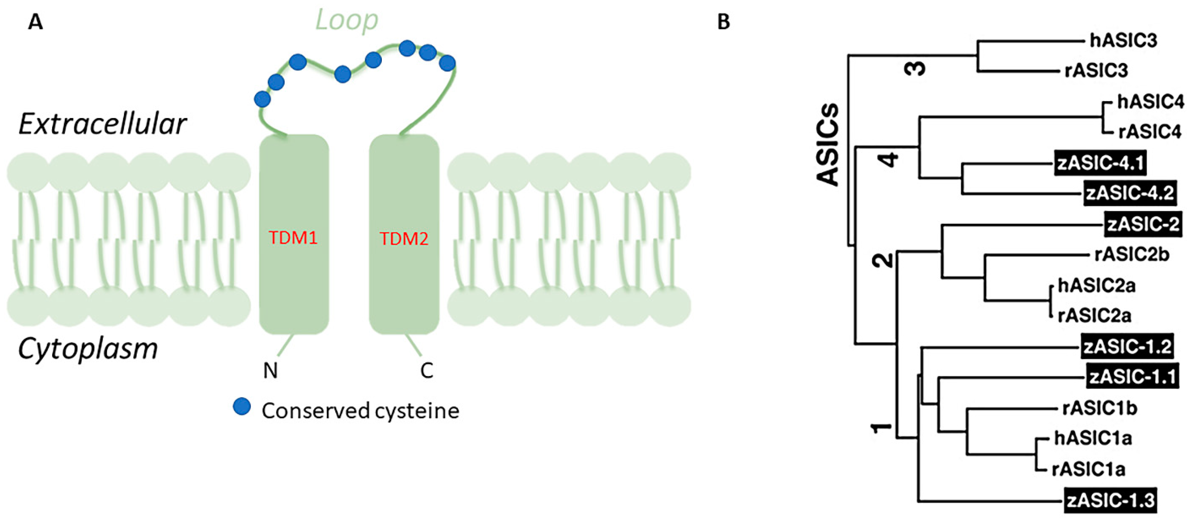
1.1. The Acid-Sensing Ion Channel Superfamily

Acid-sensing ion channels (ASICs) are Na+ channels gated by extracellular H+ and are widely expressed in the mammalian central and peripheral nerve systems [1]. ASICs are part of the degenerin/epithelial sodium (Na) channel (DEG/ENaC) superfamily whose feature is high permeability to Na that could be blocked by amiloride. Structurally, ASICs consist of two hydrophobic transmembrane domains (TMD) of 20 amino acids approximately, TMD1 and TMD2, a large domain of around 370 amino acids forming an extracellular loop of 14 conserved cysteines, and a kind of short cytoplasmic amino and carboxyl termini of 35–90 amino acids (Figure 1A) [2,3,4]. Six ASIC proteins, encoded by four genes, have been identified as ASIC1a, ASIC1b, ASIC2a, ASIC2b, ASIC3, and ASIC4 in mammals [5]. Functionally, ASICs monitor moderate deviations from the physiological values of extracellular pH and also participate in mechanoreception and nociception [2,6,7,8,9] or taste receptors [10,11]. ASICs are homo- or hetero-oligomeric assemblies of individual subunits [12,13,14]. The asic1a subunit is widely expressed in the central and peripheral nervous systems and contributes to synaptic transmission [6,15,16]. asic1b and asic3 are expressed in the peripheral nervous system where they would seem to be involved in pain perception [8,17,18,19]. The analgesic effects associated with the inhibition of those ASICs in animals suggest a role in pain. Particularly, in mice with targeted disruptions of the ASIC3 demonstrated the role of ASIC3 in modulating high-intensity stimuli perception, of great importance for the production of pain after tissue inflammation [8,17,19]. asic2a is extensively expressed in the brain and contributes to synaptic transmission [14,18]. asic4 transcripts are abundantly distributed in the adult wild-type mouse brain [20], but in humans, asic4 mRNA is expressed mainly in the pituitary gland while its expression in other parts of the brain is weak [21]. Moreover, ASICs are expressed in dorsal root ganglia (DRG), afferent gastrointestinal neurons [16] and in neurons of the myenteric and submucous plexuses [22].

1.2. ASICs in Zebrafish

Fish are, phylogenetically, the closest vertebrate group to amphibians and gave rise to mammals. Living in aquatic ecosystems, fish have different life histories and ecological pressures casting their evolution compared to mammals. An examination of the research findings from fish and mammals pointed up the extent of evolutionary conservation of ASICs across these animal taxa. Indeed, the proton-mediated activation of ASICs was highlighted in teleosts, while in a jawless fish and cartilaginous fish, ASICs were not activated by protons [23]. In addition, while in mammals the major routes for sodium chloride absorption are electroneutral absorption via the combined activity of electroneutral Na+/H+ exchangers and Cl−/HCO3− exchanger beside the electrogenic absorption via epithelial Na+ channels (ENaC) at the mucosal side of the intestinal epithelium, the Na+ uptake in the teleost was channelized by the ASIC family members [24]. ASIC, localized on the apical membranes of the ionocytes in FW trout gills, takes up Na+ from environmental FW in exchange for H+ through vacuolar-type H+-ATPase (VHA) [25]. In zebrafish, the involvement of the ASICs channels in Na+ uptake was proved [26].
The zebrafish is a model organism for studies in genetics and developmental biology that offers very advantageous features over other vertebrate models, including high fecundity, clear embryos, rapid development, straightforward analysis of gene expression, and detection of developmental abnormalities in mutant fish [27,28,29]. Similar to mammals, the zebrafish has six ASIC subunits denominated zASIC1.1, zASIC1.2, zASIC1.3, zASIC2, zASIC4.1, and zASIC4.2, encoded by six different genes. zASICs share the basic functional properties of mammalian ASICs: receptors of extracellular H+, Na+ selectivity, and the inhibiting effects of amiloride [30]. zasic1.1, zasic2, and zasic4.1 are orthologs of mammalian asic1, asic2, and asic4, respectively, while zasic1.2 and zasic1.3 are paralogs of zasic1.1 and, finally, zasic4.2, is a paralog of zasic4.1 (Figure 1B) [30]. The proteins coded by zasics have molecular masses of around 60 kDa and share 60–75% of amino acid identity with rat and human ASICs [31]. Single ASIC subunits assemble to create functional homomeric or heteromeric ASICs with different surface expression levels on the plasma membrane and consequently different properties. A small region post-TMD has been found to be important in the gating mechanisms of channels in the asic4 gene family [31]. ASICs have been demonstrated in zebrafish sensory organs, gut, gills, and brain using immunohistochemistry, molecular biology techniques, in situ hybridization, Western blot, and RT-PCR, where they ensure nerve transduction (Table 1). Additionally, as in rainbow trout, the ASICs involvement in sodium uptake has been demonstrated in adult zebrafish [31]. However, in larval zebrafish, the ASICs role in Na+ uptake has not been confirmed employing pharmacological and loss of function genetic approaches [32].

2. ASICs in Zebrafish Sensory Organs

Fish are able to transmit chemical or mechanical stimuli from the aquatic environment through a well-organized sensory system. The sensory organs in fish are represented by the olfactory rosette, lateral line, inner ear, taste buds, and chemosensory cells scattered throughout the epidermis and gills. ASICs have been observed in all the sensory organs of zebrafish, although with differences in the distribution between adult and larvae [33,34,35].

2.1. Lateral Line and Inner Ear

Neuromasts represent the sensory units of the lateral line system (LLS) of teleosts and are considered the morpho-functional unit of the LLS. In zebrafish, the neuromasts are grouped into superficial and deep neuromasts. They consist of centrally located sensory cells, supporting and mantle cells [36,37,38]. Lateral line ciliated sensory cells have their mechanosensitive stereocilia embedded in a gelatinous dome. The hair cells are involved in the perception of the water flow and movement [39]. They act as mechanoreceptors by converting mechanical stimuli into electrochemical signals [40]. The role of these cells is given by the presence on the sensory nerve endings and specialized sensory cells of mechanotransducer ion channels that generate a flow of ions in response to mechanical stimuli [7,41]. ASIC1 and ASIC3 immunoreactivity was found in the hair cells of neuromasts, while ASIC2 immunoreactivity was only observed in the nerves supplying the neuromasts. The nonsensory cells of the neuromasts, supporting and mantle cells, also displayed ASIC4 [42]. The inner ear consists of the labyrinth formed by three semicircular canals, the utricle, the vestibular organ, the sacculus, the organ of sound reception, and the lagena, the organ of orientation and hearing [43,44]. At the base of each semicircular canal, there is a dilated sac called a bony ampulla. Each ampulla has a sensory epithelium in the form of a round patch, the crista ampullaris [43,44]. The utricle, sacculus and lagena also have sensory cells termed macula, whose kinocilia and stereocilia are connected to the otoliths, dense calcareous structures [39,43,44]. ASIC1 and ASIC3 were observed in the inner ear neurosensory cells, while ASIC2 was demonstrated only in the nerves supplying them. The specific immunoreactivity for ASIC4 was not found [45].

2.2. Taste Buds

Taste buds are chemosensory organs devoted to evaluating food composition and detecting variations in the environmental chemical composition [46]. They are distributed in the skin surface of the head, lips, and barbels and the intraoral cavity and oropharyngeal apparatus. The taste buds are intraepithelial sensory organs localized on a minor dermal papilla and are onion shaped. They are composed of modified epithelial sensory, supporting and basal cells; the fusiform-like sensory cells are arranged vertically within the papilla [46]. According to their electron density, sensory cells are classified into two main populations, the dark cells, having an apex with many short microvilli, and the light cells characterized by one single large microvillus at the apical border [46]. A third type of sensory cells present in zebrafish taste buds was identified by low electron density and a brush-like apex with many tiny microvilli [46]. The basal poles of the sensory cells are reached by nerve endings coming from the facial, glossopharyngeal, or vagal nerves [47]. asic mRNA and protein were detected in the taste buds of adult zebrafish. The anti-ASIC1 antibody identified, by Western blot analysis, a protein band with an estimated molecular weight of about 60 kDa, the anti-ASIC2 antibody detected a single protein band with an estimated molecular weight of about 64 kDa, and also the antibody against ASIC4 detected a single band with an estimated molecular weight of 64kDa [48]. Western blot analysis did not detect any ASIC3 protein. The immunohistochemical results were in agreement with the results obtained by Western blot analysis. ASIC2 and ASIC4 were localized in taste buds or in the nerves supplying them. ASIC4 was found in the oral and cutaneous taste buds. ASIC1 and ASIC3 positive immunostaining was not detected in the taste buds of adult zebrafish. In most taste buds, ASIC4 was found in all cells, but, in some cases, ASIC4 was demonstrated only in a calretinin-expressing cell subtype [48,49].

2.3. Olfactory Epithelium

The olfactory organ consists of numerous lamellae that insert into a midline raphe, thus forming an oval-shaped rosette. In each lamella, there are distributed sensory and nonsensory regions located separately. Three types of receptor cells were identified in the olfactory epithelium: two with cilia or microvilli in the apical border, the third is a different type of sensory cell, the crypt cells with submerged microvilli, and cilia [50,51,52]. The ciliated and microvillous receptor cells are surrounded by supporting cells with small protrusions on their apical surfaces, while the crypt cells are surrounded by one or two specialized electron-lucent supporting cells with microvillous-like apices [51]. The nonsensory epithelium consists of goblet cells, ciliated nonsensory cells and epidermal cells with microridges [51,52,53]. In the olfactory rosette of adult zebrafish, asic2 mRNA and protein were observed. asic2 hybridization was observed in the luminal pole of the nonsensory epithelium, particularly, in the cilia basal bodies and ASIC2 immunoreactivity was found only in the cilia of the nonsensory cells. The immunoreaction for ASIC2 was never observed in the olfactory cells [54].

2.4. Retina

The zebrafish retina consists of the ganglion cell layer (GCL), inner plexiform layer (IPL), inner nuclear layer (INL), outer plexiform layer (OPL), and outer nuclear layer (ONL) [55]. Seven different cell types such as Müller cells and retinal ganglion cells (RGCs), amacrine cells (ACs), horizontal cells (HCs), bipolar cells (BCs), cones and rods are recognized in the zebrafish retina [55,56]. The structural similarities, the genome organization, the regulatory pathways controlling signal transduction and retinal development are highly conserved between zebrafish and humans [56,57]. For these reasons, the zebrafish visual system represents a powerful research tool and provides an excellent animal model to study retinal function and pathology [56,57].
In zebrafish, similarly to mammals, zasic1 mRNA was found in the whole eye of the adults [55]. The specific antibody against ASIC1 recognized, by Western blotting, a band with an estimated molecular weight of about 60 kDa, consistent with the estimated molecular mass of zASIC proteins in zebrafish. The expression of asic1 in zebrafish retina was analyzed using whole mount in situ hybridization on zebrafish larvae and immunostaining was detected on retina slices [55]. The zasic1 transcript was detected in 4–5 day post-hatch zebrafish larvae [55]. The immunostaining showed that the ASIC1 immunoreactivity was widely detected in the zebrafish retinal layers, the retinal ganglion cells layer, the inner plexiform layer, the inner nuclear layer, the outer plexiform layer, and the cone photoreceptor layer [55]. In adult zebrafish, mRNA encoding ASIC2 and ASIC4.2 but not zASIC4.1 was detected [33]. ASIC2 was expressed in the outer nuclear layer, the outer plexiform layer, the inner plexiform layer, the ganglion cell layer of the retina, and the optic nerve [33]. ASIC4 was found in the cone and rods layer and the retinal ganglion cell layer. It was also shown that the expression of asic2 and asic4.2 was affected by light and darkness [33].

3. ASICs in Zebrafish Gills

The gills of teleosts are composed of gill arches that provide support for the primary lamellae considered the functional unit of the gill, and are interspersed with five gill slits called chambers [58]. Their epithelium contains ion regulatory cells and, in addition, provides support for the secondary lamellae. The secondary lamellae represent the respiratory unit of the gill and extend from both sides and perpendicular to the longitudinal axis of the main filament [58]. The respiratory lamellae are formed by two epithelial layers separated by spaces due to the presence of pillar cells, through which blood circulates [58]. Freshwater fish, including teleosts, compensate for the loss of ions in a hypotonic environment through the uptake of Na+, Cl, and Ca2+ ions, which occurs through the activity of particular gill cells, specialized ionocytes defined as mitochondria-rich cells. Four types of ionocytes called mitochondria-rich cells (MRCs) involved in Na+ uptake have been demonstrated in zebrafish gills: VHA-rich cells (HR cells), NKA-rich cells (NaR cells), cells expressing Na+/Cl cotransporter (NCC cells), and K+-secreting cells (KS cells) [26]. Furthermore, a previous study has shown the involvement of skin keratinocytes in hypotonicity perception and the contribution of the aforenamed in the activation of innate immunity at an early developmental stage of zebrafish embryos through a transient potential receptor vanilloid 4 (TRPV4)/Ca2+/TGF-b–activated kinase 1 (TAK1)/NF-kB, by means of pharmacological and genetic inhibition experiments [59]. The expression pattern of different ASIC subunit mRNAs in zebrafish gill tissue was demonstrated by RT-PCR technique. All six ASIC mRNAs were observed in zebrafish gills. Expression of ASIC4.2, by immunoprecipitation, also identified a single band corresponding to ~65 kDa [26]. Furthermore, by immunohistochemistry, cells positive for anti-ASIC4.2 were found in the gills and interlamellar region of the gills. To clarify whether ASIC4.2 colocalizes with NaR cells or HR cells, the gills were double labelled with ASIC4.2 and VHA or NKA, a marker for HR and NaR-type MRC, respectively. Cells immunoreactive for anti-ASIC4.2 were also immunoreactive for anti-VHA. Finally, pharmacological studies in which specific ASIC inhibitors blocked Na+ uptake demonstrated the role of ASIC4.2 in regulating Na+ uptake in zebrafish exposed to low and ultra-low-sodium media at gill level [26].

4. ASICs in Zebrafish Gut

As in mammals, the enteric nervous system of adult zebrafish is organized into two plexuses, the myenteric and submucosal. Structurally, the myenteric plexus consists mainly of enteric neurons, while the submucous plexus has few neurons and numerous nerve fibers [60]. Specific ASIC2 immunoreactivity was found in the enteric nervous system of adult zebrafish as well as in scattered populations of enteroendocrine cells. Particularly, a subpopulation of neurons and nerve fibers were positive to ASIC2, mainly in the myenteric plexus and occasionally in the submucous one. Immunoreactivity for ASIC2 was also found in enteroendocrine epithelial cells in the gut wall. Most of these ASIC2 immunoreactive cells showed a central soma and two processes directed to the organ lumen and the submucous layer, where sometimes they were found close to ASIC2-positive nerve-fiber profiles [61].

5. ASICS in Zebrafish Brain

ASICs are widely expressed in the nervous system of zebrafish embryos and larvae. The expression of six zasic genes in zebrafish neurons was demonstrated through the in situ hybridization technique [30]. In general, the distribution patterns of zasic genes were different. Particularly, zasic1.1 was demonstrated within 30 h postfertilization (hpf) at anterior and posterior lateral line ganglia and optic sensory neurons and at 48 hpf, also in the trigeminal ganglia [30]. At 72 and 96 hpf, expression was observable throughout the central nervous system except for the eyes [30]. zasic1.2 was evident, although with weak expression at 48 hpf, in the ventral thalamus, ventral midbrain, ventral cerebellum and in the dorsal thalamus, hypothalamus and telencephalon along the anterior commissure. zasic1. 2 at 48 hpf was present in the dorsal midbrain (dMb) and olfactory bulb, whereas from 96 hpf it was also expressed in the tectum. zasic1.3 was expressed at 30 hpf in the ganglia of the lateral line while between 30 and 72 hpf in the telencephalon it was expressed along the anterior commissure tract, in the ventral thalamus, ventral midbrain, and ventral cerebellum [30]. By 48 hpf, expression was also evident in the dorsal thalamus and hypothalamus. At 96 hpf, expression was also strong in the habenula. zasic2 was observed at 30 hpf along the anterior commissure tract and at 48 hpf, also in the preoptic area, ventral thalamus, and ventral midbrain. At 72 and 96 hpf, it was present in the whole brain except for the dorsal forebrain and was also expressed in retinal ganglion cells. zasic4.1 at 48 hpf showed a similar pattern to that of zasic1.2. zasic4.1 was expressed in the dorsal midbrain and retinal ganglion cells. zasic4.2 as early as 24 and 30 hpf was expressed along the anterior commissure tract and in cells along the commissure tract. At 48 hpf, it was observable in the preoptic zone, posterior hypothalamus, ventral midbrain, cerebellum, and retinal ganglion cells [30].

6. Discussion

The presence of ASICs has been demonstrated in different organs of zebrafish, such as sensory organs, gills, gut, and brain. The expression of ASICs in the hair cells of LLS neuromasts and the neurosensory cells of the inner ear could support the hypothesis of an involvement in the transduction of mechanical stimuli from the aquatic environment similar to the role they play in mammalian inner ear cells. It is known that, functionally, ASICs control moderate deviations from the physiological values of extracellular pH. In the rat and mouse, ASIC1a and ASIC2a immunoreactivity was detected in small vestibular ganglion neurons and afferent fibers of the macula utricle and crista stroma, whereas ASIC2b, ASIC3, and ASIC4 were observed in vestibular ganglion neurons. It has also been demonstrated in these species that the acidification of the extracellular pH generates action potentials in vestibular neurons, confirming a function of ASICs in their excitability [62,63,64]. Regarding taste buds, conflicting data exist in mammals, i.e., ASIC2 has been proposed as a receptor for acidic taste although, in mice lacking the asic2 gene, behavioral responses to acidic taste stimuli remained unaffected [65,66,67]. There are significant differences in the expression and distribution of asics between mammals and zebrafish. Indeed, while asic4 was demonstrated in zebrafish, it was always absent in mouse and rat, asic1 and asic3 have always been absent in zebrafish while they were observed as RNAs in mice and rats. asic2 was observed in zebrafish and rats but not in mice [67,68]. These differences may be attributable to environmental and dietary differences between mammals and fish. However, the expression of asic2 and asic4 in taste buds would also suggest in zebrafish a role as a receptor for acid taste or an involvement of ASICs in taste cells, taste or more functions in zebrafish.
The localization of ASIC2 in the nonsensory epithelium of the olfactory rosette suggests that it is not involved in olfaction. However, the presence of immunoreactivity for ASIC2 in the basal bodies of the cilia of nonsensory cells, notoriously involved in the perception and transduction of mechanical and chemical stimuli, would suggest a role for this ion channel in other types of stimuli from the aquatic environment, pH changes, or movement of water. The presence of asic1a, asic2, and asic3 in the mammalian retina has been demonstrated [69,70]. Among the three aforementioned, asic1 is the most represented and is widely expressed in the retina and involved in retinal activity, particularly in cone function [71]. In mammals, the retina reacts to changes in local pH, presumably to control its acid/base environment in response to systemic acidosis with increased expression, also, of ASICs. The expression of ASICs in the developing and adult retina of zebrafish suggests that they participate, as in mammals, in the mechanism of vision [55,70]. As pH changes have been shown to be associated with pathological conditions, ASICs could also be involved in the pathogenesis of retinal disorders [72,73]. Moreover, it has also been shown that substances blocking the activity of ASIC may represent potential therapeutics in degenerative diseases, like optic nerve degeneration or ischemic retinal diseases [74,75]. Finally, in zebrafish as in mammals, asic1a, functionally expressed in retinal pigment epithelium (RPE) cells, might play an important role in the neuroprotection of RPE cells from oxidative stress [73]. The presence of asics in zebrafish gills, at mRNA and protein levels, was demonstrated, and their involvement in Na+ regulation of adult zebrafish acclimated to low-Na+ medium was validated using pharmacological blockade of Na+ uptake. Moreover, the presence of alternative transport mechanisms was also hypothesized. The differences found between zebrafish larvae and adults in response to specific inhibitors of ASICs, could be explained by a difference in the ionic compositions and pH used in the two different studies or could suggest the use of different Na+ uptake mechanisms in the two stages examined, as previously demonstrated in rainbow trout [25]. Numerous data have demonstrated ASICs in gastrointestinal DRG afferent neurons in mammalian myenteric and submucosal plexus neurons [76]. Indeed, these ion channels would appear to be involved in gastrointestinal physiology [13,16] in the regulation of acid secretion, motility, and mucosal protection in mammals [13,16,77,78]. Increased levels of asic3 have been observed in gastrointestinal disorders associated with inflammation, while disruption of the asic2 gene results in altered emptying from the gastrointestinal tract [22]. The role of ASIC2 in the zebrafish gut could be comparable to that observed in mammals, and thus they could be involved in the neuronal responses to acid challenge in the intestinal mucosa, or the tension of the intestinal walls [13,16]. On the other hand, the enteroendocrine cells that expressed positivity for ASIC2 might also be considered chemoreceptors and involved in the regulation of intestinal secretion, releasing intestinal hormones, and/or in maintaining intestinal wall tension. Finally, in the mammalian brain, ASICs are widely expressed in CNS neurons and microglia and play key roles in physiological activities such as synaptic plasticity, microglia Ca+ physiology, learning/memory, fear conditioning, and in pathological conditions such as brain ischemia, multiple sclerosis, epileptic seizures, depression-related behavior, anxiety disorders and malignant glioma [79]. In zebrafish, the different zasic genes were mainly expressed in the nervous system of embryos and larva, although the expression pattern of the six zasic genes, compared to mammals, was limited primarily to neurons and perhaps some glial cells. However, further studies are also needed in the adult zebrafish brain to demonstrate the exact role of ASICs in this experimental model. Therefore, such evidence on the involvement of these ion channels in both physiological and pathological processes in the central nervous system could suggest them as novel and effective therapeutic interventions for CNS diseases.

7. Conclusions

In zebrafish the pattern of the distribution of ASICs in different organs and tissues suggests their involvement in multiple physiological functions, including mechanosensation, hearing, temperature sensing, taste, vision, intestinal physiology and brain activity as previously demonstrated in mammals [80,81,82]. From the analysis of these data we can conclude that ASICs are essential for the proper functioning of various organs in zebrafish, and that the latter represents an important experimental model for the study of diseases related to alterations of these ion channels.

Author Contributions

Conceptualization, M.L., G.M. and A.G. conceived and designed the study as well as drafted the paper; M.L., A.G. and F.A. performed the literature review and drafted the paper; R.L., G.M., M.C.G., K.M., M.A. and A.G. critically revised the paper. All authors have read and agreed to the published version of the manuscript.

Funding

This research received no external funding.

Institutional Review Board Statement

Not applicable.

Data Availability Statement

Data sharing not applicable.

Conflicts of Interest

The authors declare no conflict of interest.

References

  1. Kress, M.; Waldmann, R. Chapter 8 Acid Sensing Ionic Channels. In Current Topics in Membranes; Academic Press: Cambridge, MA, USA, 2006; Volume 57, pp. 241–276. ISBN 978-0-12-815456-4. [Google Scholar]
  2. Sherwood, T.W.; Frey, E.N.; Askwith, C.C. Structure and Activity of the Acid-Sensing Ion Channels. Am. J. Physiol. Cell Physiol. 2012, 303, C699–C710. [Google Scholar] [CrossRef] [Green Version]
  3. Hanukoglu, I. ASIC and ENaC Type Sodium Channels: Conformational States and the Structures of the Ion Selectivity Filters. FEBS J. 2017, 284, 525–545. [Google Scholar] [CrossRef] [Green Version]
  4. Gründer, S.; Chen, X. Structure, Function, and Pharmacology of Acid-Sensing Ion Channels (ASICs): Focus on ASIC1a. Int. J. Physiol. Pathophysiol. Pharm. 2010, 2, 73–94. [Google Scholar]
  5. Krishtal, O. Receptor for Protons: First Observations on Acid Sensing Ion Channels. Neuropharmacology 2015, 94, 4–8. [Google Scholar] [CrossRef]
  6. Holzer, P. Acid-Sensitive Ion Channels and Receptors. In Sensory Nerves; Canning, B.J., Spina, D., Eds.; Springer: Berlin/Heidelberg, Germany, 2009; pp. 283–332. ISBN 978-3-540-79090-7. [Google Scholar]
  7. Delmas, P.; Coste, B. Mechano-Gated Ion Channels in Sensory Systems. Cell 2013, 155, 278–284. [Google Scholar] [CrossRef] [PubMed] [Green Version]
  8. Chen, C.-C.; Zimmer, A.; Sun, W.-H.; Hall, J.; Brownstein, M.J.; Zimmer, A. A Role for ASIC3 in the Modulation of High-Intensity Pain Stimuli. Proc. Natl. Acad. Sci. USA 2002, 99, 8992. [Google Scholar] [CrossRef] [PubMed] [Green Version]
  9. Zha, X. Acid-Sensing Ion Channels: Trafficking and Synaptic Function. Mol. Brain 2013, 6, 1. [Google Scholar] [CrossRef] [Green Version]
  10. Lin, W.; Ogura, T.; Kinnamon, S.C. Acid-Activated Cation Currents in Rat Vallate Taste Receptor Cells. J. Neurophysiol. 2002, 88, 133–141. [Google Scholar] [CrossRef] [Green Version]
  11. Ugawa, S. Identification of Sour-Taste Receptor Genes. Anat. Sci. Int. 2003, 78, 205–210. [Google Scholar] [CrossRef]
  12. Krishtal, O. The ASICs: Signaling Molecules? Modulators? Trends Neurosci. 2003, 26, 477–483. [Google Scholar] [CrossRef]
  13. Holzer, P. Acid-Sensitive Ion Channels in Gastrointestinal Function. Curr. Opin. Pharmacol. 2003, 3, 618–625. [Google Scholar] [CrossRef]
  14. Wemmie, J.A.; Price, M.P.; Welsh, M.J. Acid-Sensing Ion Channels: Advances, Questions and Therapeutic Opportunities. Trends Neurosci. 2006, 29, 578–586. [Google Scholar] [CrossRef] [PubMed]
  15. Lingueglia, E. Acid-Sensing Ion Channels in Sensory Perception. J. Biol. Chem. 2007, 282, 17325–17329. [Google Scholar] [CrossRef] [Green Version]
  16. Holzer, P. Acid Sensing by Visceral Afferent Neurones. Acta Physiol. 2011, 201, 63–75. [Google Scholar] [CrossRef]
  17. Lingueglia, E. Les canaux ioniques ASIC dans la douleur. Biol. Aujourd’hui 2014, 208, 13–20. [Google Scholar] [CrossRef]
  18. Hill, A.S.; Ben-Shahar, Y. The Synaptic Action of Degenerin/Epithelial Sodium Channels. Channels 2018, 12, 262–275. [Google Scholar] [CrossRef] [PubMed] [Green Version]
  19. Li, W.-G.; Xu, T.-L. ASIC3 Channels in Multimodal Sensory Perception. ACS Chem. Neurosci. 2011, 2, 26–37. [Google Scholar] [CrossRef] [PubMed] [Green Version]
  20. Hoshikawa, M.; Kato, A.; Hojo, H.; Shibata, Y.; Kumamoto, N.; Watanabe, M.; Ugawa, S. Distribution of ASIC4 Transcripts in the Adult Wild-Type Mouse Brain. Neurosci. Lett. 2017, 651, 57–64. [Google Scholar] [CrossRef]
  21. Gründer, S.; Geissler, H.S.; Bässler, E.L.; Ruppersberg, J.P. A New Member of Acid-Sensing Ion Channels from Pituitary Gland. Neuroreport 2000, 11, 1607–1611. [Google Scholar] [CrossRef] [PubMed]
  22. Yiangou, Y.; Facer, P.; Smith, J.; Sangameswaran, L.; Eglen, R.; Birch, R.; Knowles, C.; Williams, N.; Anand, P. Increased Acid-Sensing Ion Channel ASIC-3 in Inflamed Human Intestine. Eur. J. Gastroenterol. Hepatol. 2001, 13, 891–896. [Google Scholar] [CrossRef] [PubMed]
  23. Sneddon, L.U. Evolution of Nociception and Pain: Evidence from Fish Models. Philos. Trans. R. Soc. B Biol. Sci. 2019, 374, 20190290. [Google Scholar] [CrossRef]
  24. Takei, Y. The Digestive Tract as an Essential Organ for Water Acquisition in Marine Teleosts: Lessons from Euryhaline Eels. Zool. Lett. 2021, 7, 10. [Google Scholar] [CrossRef] [PubMed]
  25. Dymowska, A.K.; Schultz, A.G.; Blair, S.D.; Chamot, D.; Goss, G.G. Acid-Sensing Ion Channels Are Involved in Epithelial Na+ Uptake in the Rainbow Trout Oncorhynchus Mykiss. Am. J. Physiol. Cell Physiol. 2014, 307, C255–C265. [Google Scholar] [CrossRef] [PubMed] [Green Version]
  26. Dymowska, A.K.; Boyle, D.; Schultz, A.G.; Goss, G.G. The Role of Acid-Sensing Ion Channels in Epithelial Na+ Uptake in Adult Zebrafish (Danio Rerio). J. Exp. Biol. 2015, 218, 1244–1251. [Google Scholar] [CrossRef] [Green Version]
  27. He, Y.; Bao, B.; Li, H. Using Zebrafish as a Model to Study the Role of Epigenetics in Hearing Loss. Expert Opin. Drug Discov. 2017, 12, 967–975. [Google Scholar] [CrossRef]
  28. Nicolson, T. The Genetics of Hearing and Balance in Zebrafish. Annu. Rev. Genet. 2005, 39, 9–22. [Google Scholar] [CrossRef]
  29. Orlando, L. Odor Detection in Zebrafish. Trends Neurosci. 2001, 24, 257–258. [Google Scholar] [CrossRef]
  30. Paukert, M.; Sidi, S.; Russell, C.; Siba, M.; Wilson, S.W.; Nicolson, T.; Gründer, S. A Family of Acid-Sensing Ion Channels from the Zebrafish: Widespread Expression in the Central Nervous System Suggests a Conserved Role in Neuronal Communication. J. Biol. Chem. 2004, 279, 18783–18791. [Google Scholar] [CrossRef] [Green Version]
  31. Chen, X.; Polleichtner, G.; Kadurin, I.; Gründer, S. Zebrafish Acid-Sensing Ion Channel (ASIC) 4, Characterization of Homo- and Heteromeric Channels, and Identification of Regions Important for Activation by H+. J. Biol. Chem. 2007, 282, 30406–30413. [Google Scholar] [CrossRef] [Green Version]
  32. Zimmer, A.M.; Dymowska, A.K.; Kumai, Y.; Goss, G.G.; Perry, S.F.; Kwong, R.W.M. Assessing the Role of the Acid-Sensing Ion Channel ASIC4b in Sodium Uptake by Larval Zebrafish. Comp. Biochem. Physiol. Part A: Mol. Integr. Physiol. 2018, 226, 1–10. [Google Scholar] [CrossRef] [PubMed]
  33. Viña, E.; Parisi, V.; Sánchez-Ramos, C.; Cabo, R.; Guerrera, M.C.; Quirós, L.M.; Germanà, A.; Vega, J.A.; García-Suárez, O. Acid-Sensing Ion Channels (ASICs) 2 and 4.2 Are Expressed in the Retina of the Adult Zebrafish. Cell Tissue Res. 2015, 360, 223–231. [Google Scholar] [CrossRef]
  34. Detrich, H.W., 3rd; Westerfield, M.; Zon, L.I. The Zebrafish. Preface. Methods Cell Biol. 2011, 105, xxi–xxii. [Google Scholar] [CrossRef]
  35. Kotrschal, K.; Krautgartner, W.-D.; Hansen, A. Ontogeny of the Solitary Chemosensory Cells in the Zebrafish, Danio Rerio. Chem. Senses 1997, 22, 111–118. [Google Scholar] [CrossRef] [Green Version]
  36. Laurà, R.; Abbate, F.; Germanà, G.P.; Montalbano, G.; Germanà, A.; Levanti, M. Fine Structure of the Canal Neuromasts of the Lateral Line System in the Adult Zebrafish. Anat. Histol. Embryol. 2018, 47, 322–329. [Google Scholar] [CrossRef]
  37. Montalbano, G.; Capillo, G.; Laurà, R.; Abbate, F.; Levanti, M.; Guerrera, M.C.; Ciriaco, E.; Germanà, A. Neuromast Hair Cells Retain the Capacity of Regeneration during Heavy Metal Exposure. Ann. Anat.-Anat. Anz. 2018, 218, 183–189. [Google Scholar] [CrossRef]
  38. Germana, A.; Abbate, F.; González-Martı́nez, T.; del Valle, M.E.; de Carlos, F.; Germanà, G.; Vega, J.A. S100 Protein Is a Useful and Specific Marker for Hair Cells of the Lateral Line System in Postembryonic Zebrafish. Neurosci. Lett. 2004, 365, 186–189. [Google Scholar] [CrossRef] [PubMed]
  39. Bang, P.I.; Sewell, W.F.; Malicki, J.J. Morphology and Cell Type Heterogeneities of the Inner Ear Epithelia in Adult and Juvenile Zebrafish (Danio Rerio). J. Comp. Neurol. 2001, 438, 173–190. [Google Scholar] [CrossRef] [PubMed]
  40. Ghysen, A.; Dambly-Chaudière, C. The Lateral Line Microcosmos. Genes Dev. 2007, 21, 2118–2130. [Google Scholar] [CrossRef] [PubMed] [Green Version]
  41. Ranade, S.S.; Syeda, R.; Patapoutian, A. Mechanically Activated Ion Channels. Neuron 2015, 87, 1162–1179. [Google Scholar] [CrossRef] [Green Version]
  42. Abbate, F.; Madrigrano, M.; Scopitteri, T.; Levanti, M.; Cobo, J.L.; Germanà, A.; Vega, J.A.; Laurà, R. Acid-Sensing Ion Channel Immunoreactivities in the Cephalic Neuromasts of Adult Zebrafish. Ann. Anat.-Anat. Anz. 2016, 207, 27–31. [Google Scholar] [CrossRef] [PubMed]
  43. Popper, A.N. Organization of the Inner Ear and Auditory Processing. Fish. Neurobiol. 1983, 1, 125–177. [Google Scholar]
  44. Higgs, D.M.; Rollo, A.K.; Souza, M.J.; Popper, A.N. Development of Form and Function in Peripheral Auditory Structures of the Zebrafish (Danio Rerio). J. Acoust. Soc. Am. 2003, 113, 1145–1154. [Google Scholar] [CrossRef] [PubMed] [Green Version]
  45. Germanà, A.; Muriel, J.D.; Cobo, R.; García-Suárez, O.; Cobo, J.; Vega, J.A. Transient-Receptor Potential (TRP) and Acid-Sensing Ion Channels (ASICs) in the Sensory Organs of Adult Zebrafish. In Recent Advances in Zebrafish Researches; IntechOpen: London, UK, 2018. [Google Scholar]
  46. Hansen, A.; Reutter, K.; Zeiske, E. Taste Bud Development in the Zebrafish, Danio Rerio. Dev. Dyn. 2002, 223, 483–496. [Google Scholar] [CrossRef] [PubMed]
  47. Korsching, S.I. 3.23—Taste and Smell in Zebrafish. In The Senses: A Comprehensive Reference, 2nd ed.; Fritzsch, B., Ed.; Elsevier: Oxford, UK, 2020; pp. 466–492. ISBN 978-0-12-805409-3. [Google Scholar]
  48. Viña, E.; Parisi, V.; Cabo, R.; Laurà, R.; López-Velasco, S.; López-Muñiz, A.; García-Suárez, O.; Germanà, A.; Vega, J.A. Acid-Sensing Ion Channels (ASICs) in the Taste Buds of Adult Zebrafish. Neurosci. Lett. 2013, 536, 35–40. [Google Scholar] [CrossRef]
  49. Levanti, M.; Randazzo, B.; Viña, E.; Montalbano, G.; Garcia-Suarez, O.; Germanà, A.; Vega, J.A.; Abbate, F. Acid-Sensing Ion Channels and Transient-Receptor Potential Ion Channels in Zebrafish Taste Buds. Ann. Anat.-Anat. Anz. 2016, 207, 32–37. [Google Scholar] [CrossRef]
  50. Sepahi, A.; Kraus, A.; Casadei, E.; Johnston, C.A.; Galindo-Villegas, J.; Kelly, C.; García-Moreno, D.; Muñoz, P.; Mulero, V.; Huertas, M.; et al. Olfactory Sensory Neurons Mediate Ultrarapid Antiviral Immune Responses in a TrkA-Dependent Manner. Proc. Natl. Acad. Sci. USA 2019, 116, 12428–12436. [Google Scholar] [CrossRef] [Green Version]
  51. Hansen, A.; Zielinski, B.S. Diversity in the Olfactory Epithelium of Bony Fishes: Development, Lamellar Arrangement, Sensory Neuron Cell Types and Transduction Components. J. Neurocytol. 2005, 34, 183–208. [Google Scholar] [CrossRef] [PubMed]
  52. Hansen, A.; Eckart, Z. The Peripheral Olfactory Organ of the Zebrafish, Danio Rerio: An Ultrastructural Study. Chem. Senses 1998, 23, 39–48. [Google Scholar] [CrossRef]
  53. Parisi, V.; Guerrera, M.C.; Abbate, F.; Garcia-Suarez, O.; Viña, E.; Vega, J.A.; Germanà, A. Immunohistochemical Characterization of the Crypt Neurons in the Olfactory Epithelium of Adult Zebrafish. Ann. Anat.-Anat. Anz. 2014, 196, 178–182. [Google Scholar] [CrossRef] [PubMed]
  54. Viña, E.; Parisi, V.; Abbate, F.; Cabo, R.; Guerrera, M.C.; Laurà, R.; Quirós, L.M.; Pérez-Varela, J.C.; Cobo, T.; Germanà, A.; et al. Acid-Sensing Ion Channel 2 (ASIC2) Is Selectively Localized in the Cilia of the Non-Sensory Olfactory Epithelium of Adult Zebrafish. Histochem. Cell Biol. 2015, 143, 59–68. [Google Scholar] [CrossRef] [PubMed]
  55. Liu, S.; Wang, M.-X.; Mao, C.-J.; Cheng, X.-Y.; Wang, C.-T.; Huang, J.; Zhong, Z.-M.; Hu, W.-D.; Wang, F.; Hu, L.-F.; et al. Expression and Functions of ASIC1 in the Zebrafish Retina. Biochem. Biophys. Res. Commun. 2014, 455, 353–357. [Google Scholar] [CrossRef]
  56. Link, B.A.; Collery, R.F. Zebrafish Models of Retinal Disease. Annu. Rev. Vis. Sci. 2015, 1, 125–153. [Google Scholar] [CrossRef]
  57. Gestri, G.; Link, B.A.; Neuhauss, S.C.F. The Visual System of Zebrafish and Its Use to Model Human Ocular Diseases. Dev. Neurobiol. 2012, 72, 302–327. [Google Scholar] [CrossRef]
  58. Wilson, J.M.; Laurent, P. Fish Gill Morphology: Inside Out. J. Exp. Zool. 2002, 293, 192–213. [Google Scholar] [CrossRef]
  59. Galindo-Villegas, J.; Montalban-Arques, A.; Liarte, S.; de Oliveira, S.; Pardo-Pastor, C.; Rubio-Moscardo, F.; Meseguer, J.; Valverde, M.A.; Mulero, V. TRPV4-Mediated Detection of Hyposmotic Stress by Skin Keratinocytes Activates Developmental Immunity. J. Immunol. 2016, 196, 738–749. [Google Scholar] [CrossRef] [PubMed] [Green Version]
  60. Olsson, C. Autonomic Innervation of the Fish Gut. Acta Histochem. 2009, 111, 185–195. [Google Scholar] [CrossRef]
  61. Levanti, M.B.; Guerrera, M.C.; Calavia, M.G.; Ciriaco, E.; Montalbano, G.; Cobo, J.; Germanà, A.; Vega, J.A. Acid-Sensing Ion Channel 2 (ASIC2) in the Intestine of Adult Zebrafish. Neurosci. Lett. 2011, 494, 24–28. [Google Scholar] [CrossRef] [PubMed]
  62. Mercado, F.; López, I.A.; Acuna, D.; Vega, R.; Soto, E. Acid-Sensing Ionic Channels in the Rat Vestibular Endorgans and Ganglia. J. Neurophysiol. 2006, 96, 1615–1624. [Google Scholar] [CrossRef] [PubMed] [Green Version]
  63. Vega, R.; Rodríguez, U.; Soto, E. Acid-Sensing Ionic-Channel Functional Expression in the Vestibular Endorgans. Neurosci. Lett. 2009, 463, 199–202. [Google Scholar] [CrossRef]
  64. González-Garrido, A.; Vega, R.; Mercado, F.; López, I.A.; Soto, E. Acid-Sensing Ion Channels Expression, Identity and Role in the Excitability of the Cochlear Afferent Neurons. Front. Cell. Neurosci. 2015, 9, 483. [Google Scholar] [CrossRef] [Green Version]
  65. Lin, W.; Burks, C.A.; Hansen, D.R.; Kinnamon, S.C.; Gilbertson, T.A. Taste Receptor Cells Express PH-Sensitive Leak K+ Channels. J. Neurophysiol. 2004, 92, 2909–2919. [Google Scholar] [CrossRef] [Green Version]
  66. Liu, L.; Simon, S.A. Acidic Stimuli Activates Two Distinct Pathways in Taste Receptor Cells from Rat Fungiform Papillae. Brain Res. 2001, 923, 58–70. [Google Scholar] [CrossRef]
  67. Richter, T.A.; Dvoryanchikov, G.A.; Roper, S.D.; Chaudhari, N. Acid-Sensing Ion Channel-2 Is Not Necessary for Sour Taste in Mice. J. Neurosci. 2004, 24, 4088. [Google Scholar] [CrossRef] [PubMed] [Green Version]
  68. Shimada, S.; Ueda, T.; Ishida, Y.; Yamamoto, T.; Ugawa, S. Acid-Sensing Ion Channels in Taste Buds. Arch. Histol. Cytol. 2006, 69, 227–231. [Google Scholar] [CrossRef] [PubMed] [Green Version]
  69. Brockway, L.M.; Zhou, Z.-H.; Bubien, J.K.; Jovov, B.; Benos, D.J.; Keyser, K.T. Rabbit Retinal Neurons and Glia Express a Variety of ENaC/DEG Subunits. Am. J. Physiol. Cell Physiol. 2002, 283, C126–C134. [Google Scholar] [CrossRef] [PubMed] [Green Version]
  70. Ettaiche, M.; Guy, N.; Hofman, P.; Lazdunski, M.; Waldmann, R. Acid-Sensing Ion Channel 2 Is Important for Retinal Function and Protects against Light-Induced Retinal Degeneration. J. Neurosci. 2004, 24, 1005. [Google Scholar] [CrossRef] [PubMed]
  71. Ettaiche, M.; Deval, E.; Cougnon, M.; Lazdunski, M.; Voilley, N. Silencing Acid-Sensing Ion Channel 1a Alters Cone-Mediated Retinal Function. J. Neurosci. 2006, 26, 5800. [Google Scholar] [CrossRef]
  72. Tan, J.; Ye, X.; Xu, Y.; Wang, H.; Sheng, M.; Wang, F. Acid-Sensing Ion Channel 1a Is Involved in Retinal Ganglion Cell Death Induced by Hypoxia. Mol. Vis. 2011, 17, 3300–3308. [Google Scholar]
  73. Tan, J.; Xu, Y.; Liu, G.; Ye, X. Involvement of Acid-Sensing Ion Channel 1a in Functions of Cultured Human Retinal Pigment Epithelial Cells. J. Huazhong Univ. Sci. Technol. 2013, 33, 137–141. [Google Scholar] [CrossRef]
  74. Dibas, A.; Millar, C.; Al-Farra, A.; Yorio, T. Neuroprotective Effects of Psalmotoxin-1, an Acid-Sensing Ion Channel (ASIC) Inhibitor, in Ischemia Reperfusion in Mouse Eyes. Curr. Eye Res. 2018, 43, 921–933. [Google Scholar] [CrossRef]
  75. Miyake, T.; Nishiwaki, A.; Yasukawa, T.; Ugawa, S.; Shimada, S.; Ogura, Y. Possible Implications of Acid-Sensing Ion Channels in Ischemia-Induced Retinal Injury in Rats. Jpn. J. Ophthalmol. 2013, 57, 120–125. [Google Scholar] [CrossRef] [PubMed]
  76. Hughes, P.A.; Brierley, S.M.; Young, R.L.; Blackshaw, L.A. Localization and Comparative Analysis of Acid-Sensing Ion Channel (ASIC1, 2, and 3) MRNA Expression in Mouse Colonic Sensory Neurons within Thoracolumbar Dorsal Root Ganglia. J. Comp. Neurol. 2007, 500, 863–875. [Google Scholar] [CrossRef] [PubMed]
  77. Page, A.J.; Brierley, S.M.; Martin, C.M.; Price, M.P.; Symonds, E.; Butler, R.; Wemmie, J.A.; Blackshaw, L.A. Different Contributions of ASIC Channels 1a, 2, and 3 in Gastrointestinal Mechanosensory Function. Gut 2005, 54, 1408–1415. [Google Scholar] [CrossRef] [Green Version]
  78. Jones, R.C.W.; Xu, L.; Gebhart, G.F. The Mechanosensitivity of Mouse Colon Afferent Fibers and Their Sensitization by Inflammatory Mediators Require Transient Receptor Potential Vanilloid 1 and Acid-Sensing Ion Channel 3. J. Neurosci. 2005, 25, 10981. [Google Scholar] [CrossRef]
  79. Chu, X.-P.; Xiong, Z.-G. Physiological and Pathological Functions of Acid-Sensing Ion Channels in the Central Nervous System. Curr. Drug Targets 2012, 13, 263–271. [Google Scholar] [CrossRef] [PubMed] [Green Version]
  80. Baron, A.; Lingueglia, E. Pharmacology of Acid-Sensing Ion Channels—Physiological and Therapeutical Perspectives. Neuropharmacology 2015, 94, 19–35. [Google Scholar] [CrossRef]
  81. Omerbašić, D.; Schuhmacher, L.-N.; Bernal Sierra, Y.-A.; St Smith, E.J.; Lewin, G.R. ASICs and Mammalian Mechanoreceptor Function. Neuropharmacology 2015, 94, 80–86. [Google Scholar] [CrossRef] [PubMed] [Green Version]
  82. Whitfield, T.T. Zebrafish as a Model for Hearing and Deafness. J. Neurobiol. 2002, 53, 157–171. [Google Scholar] [CrossRef] [PubMed]
Figure 1. (A) Schematic view of one ASIC subunit: Each subunit has two hydrophobic transmembrane domains, a large cysteine-rich extracellular loop, and short intracellular N- and C- termini; (B) phylogenetic tree illustrating the relationship of zASICs and other ASICs from human (h) and rat (r) (Paukert et al., 2004 modified).
Animals 11 02471 g001
Table 1. Presence and localization of ASICs and ZASICs in adult and larvae zebrafish with techniques of molecular biology and immunohistochemistry.
ASIC1ASIC2ASIC3ASIC4ZASIC1ZASIC2ZASIC3ZASIC4
Neuromast++++----
Inner ear+++-----
Taste buds++-+----
Olfactory epithelium-+------
Retina++-++---
Gills---+----
Intestine-+------
Brain----++-+
Publisher’s Note: MDPI stays neutral with regard to jurisdictional claims in published maps and institutional affiliations.

