
Boswellia serrata Resin Extract in Diets of Nile Tilapia, Oreochromis niloticus: Effects on the Growth, Health, Immune Response, and Disease Resistance to Staphylococcus aureus

 , , and
Abstract
Simple Summary
Abstract
1. Introduction
2. Material and Methods
2.1. Boswellia Serrata Resin Extraction
2.2. Fish and Cultural Conditions
2.3. The Experimental Design and Diets Preparation
2.4. Growth Performance, Proximate Chemical Composition of the Whole-Fish Body, and Economic Efficiency of the Feed
2.5. Sampling
2.6. Blood Biochemical Parameters
2.7. Antioxidant Activity
2.8. Immunological Assessment
2.9. Histopathological Investigation
2.10. Bacterial Challenge
2.11. Statistical Analysis
3. Result
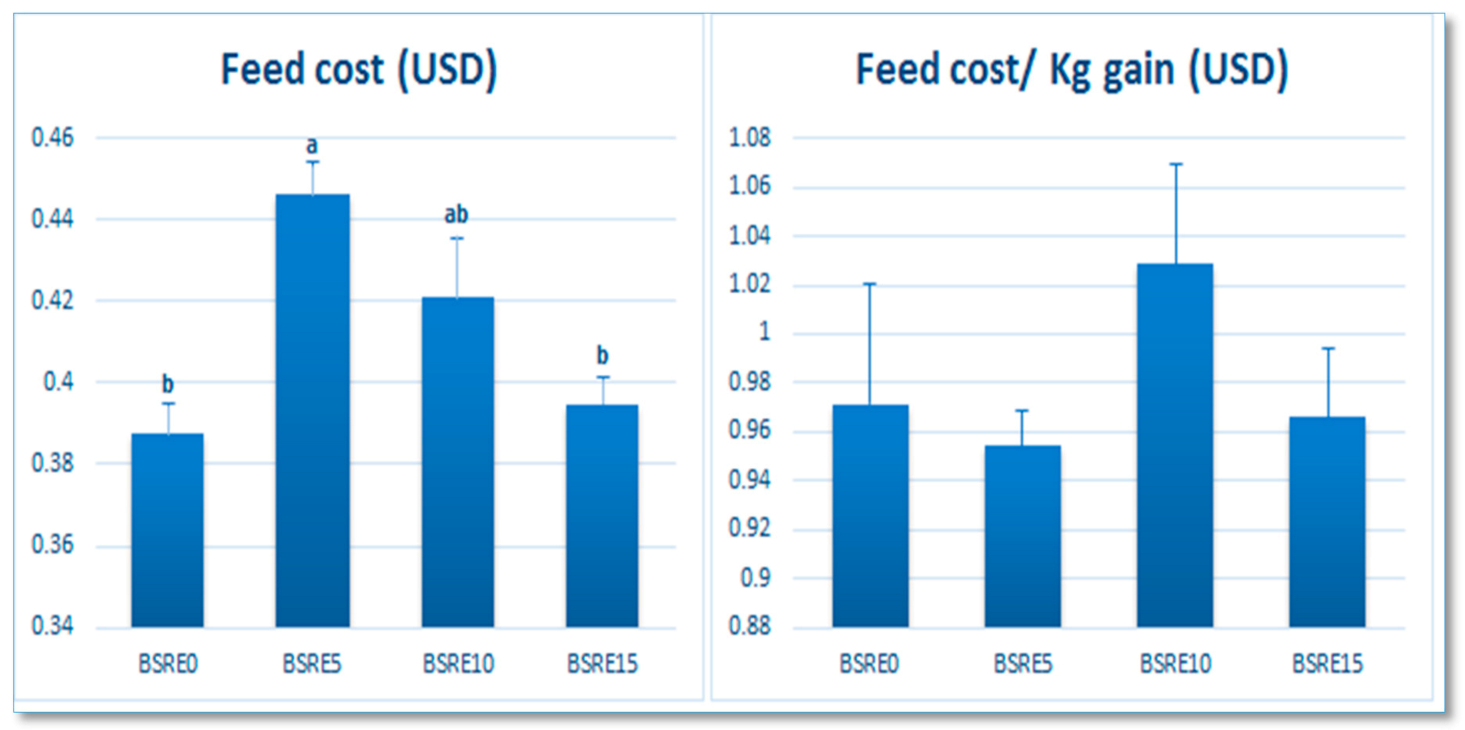
3.1. Growth Performance, Fish Whole-Body Composition, and Economic Value
3.2. Serum Biochemical Parameters
3.3. Antioxidant Activity and Immune Indices
3.4. Histological Finding
3.4.1. Gills
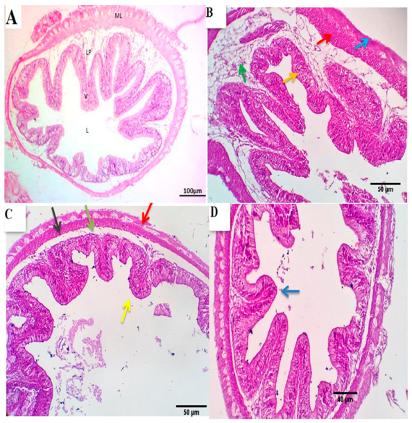
3.4.2. Intestine
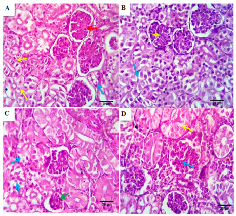
3.4.3. Kidney
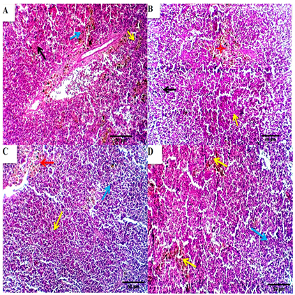
3.4.4. Spleen
3.5. Intestinal Morphometric Measurements
3.6. Challenge Test
4. Discussion
5. Conclusions
Author Contributions
Funding
Institutional Review Board Statement
Informed Consent Statement
Data Availability Statement
Acknowledgments
Conflicts of Interest
References
- Food and Agriculture Organization of the United Nations. The State of World Fisheries and Aquaculture 2018–Meeting the Sustainable Development Goals; FAO: Rome, Italy, 2018. [Google Scholar]
- Food and Agriculture Organization of the United Nations. Global Aquaculture Production 1950–2010; FAO: Rome, Italy, 2012. [Google Scholar]
- Allam, N.G.; Shabana, S.A.; Osman, Y.A.; Nouh, H.S. Prevalence of some virulence factors among Gram negative bacteria isolated from patients with lung infection and their antimicrobial susceptibility patterns. Egypt. J. Bot. 2019, 59, 633–643. [Google Scholar]
- Annabi, A.; Said, K.; Messaoudi, I. Cadmium: Bioaccumulation, histopathology and detoxifying mechanisms in fish. Am. J. Res. Commun. 2013, 1, 62. [Google Scholar]
- El-baz, R.; Rizk, D.E.; Barwa, R.; Hassan, R. Virulence characteristics and molecular relatedness of methicillin resistant Staphylococcus aureus harboring different staphylococcal cassette chromosome mec. Microb. Pathog. 2017, 113, 385–395. [Google Scholar] [CrossRef] [PubMed]
- Ahmed, S.H.; Tolba, S.; Al Zawahry, Y.A. Evaluation of the role of bla genes in beta lactam and methicillin resistant Staphylococcus aureus. Egypt. J. Bot. 2019, 59, 29–38. [Google Scholar] [CrossRef]
- Arfatahery, N.; Mirshafiey, A.; Abedimohtasab, T.; Zeinolabedinizamani, M. Study of the prevalence of Staphylococcus aureus in marine and farmed shrimps in Iran aiming the future development of a prophylactic vaccine. Procedia Vaccinol. 2015, 9, 44–49. [Google Scholar] [CrossRef]
- Amer, S.A.; Metwally, A.E.; Ahmed, S.A. The influence of dietary supplementation of cinnamaldehyde and thymol on the growth performance, immunity and antioxidant status of monosex Nile tilapia fingerlings (Oreochromis niloticus). Egypt. J. Aquat. Res. 2018, 44, 251–256. [Google Scholar] [CrossRef]
- Omar, A.E.; Al-Khalaifah, H.S.; Mohamed, W.A.; Gharib, H.S.; Osman, A.; Al-Gabri, N.A.; Amer, S.A. Effects of Phenolic-Rich Onion (Allium cepa L.) Extract on the Growth Performance, Behavior, Intestinal Histology, Amino Acid Digestibility, Antioxidant Activity, and the Immune Status of Broiler Chickens. Front. Vet. Sci. 2020, 7, 728. [Google Scholar] [CrossRef]
- Al-Khalaifah, H.; Khalil, A.A.; Amer, S.A.; Shalaby, S.I.; Badr, H.A.; Farag, M.F.; Altohamy, D.E.; Abdel Rahman, A.N. Effects of Dietary Doum Palm Fruit Powder on Growth, Antioxidant Capacity, Immune Response, and Disease Resistance of African Catfish, Clarias gariepinus (B.). Animals 2020, 10, 1407. [Google Scholar] [CrossRef] [PubMed]
- Thulin, M.; Warfa, A.M. The frankincense trees (Boswellia spp., Burseraceae) of northern Somalia and southern Arabia. Kew Bull. 1987, 42, 487–500. [Google Scholar] [CrossRef]
- Hosain, N.A.; Ghosh, R.; Bryant, D.L.; Arivett, B.A.; Farone, A.L.; Kline, P.C. Isolation, structure elucidation, and immunostimulatory activity of polysaccharide fractions from Boswellia carterii frankincense resin. Int. J. Biol. Macromol. 2019, 133, 76–85. [Google Scholar] [CrossRef]
- Hamm, S.; Bleton, J.; Connan, J.; Tchapla, A. A chemical investigation by headspace SPME and GC–MS of volatile and semi-volatile terpenes in various olibanum samples. Phytochemistry 2005, 66, 1499–1514. [Google Scholar] [CrossRef]
- Van Vuuren, S. Antimicrobial activity of South African medicinal plants. J. Ethnopharmacol. 2008, 119, 462–472. [Google Scholar] [CrossRef]
- Camarda, L.; Dayton, T.; Di Stefano, V.; Pitonzo, R.; Schillaci, D. Chemical composition and antimicrobial activity of some oleogum resin essential oils from Boswellia spp. (Burseraceae). Ann. Di Chim. J. Anal. Environ. Cult. Herit. Chem. 2007, 97, 837–844. [Google Scholar]
- Mannino, G.; Occhipinti, A.; Maffei, M.E. Quantitative Determination of 3-O-Acetyl-11-Keto-βBoswellic Acid (AKBA) and Other Boswellic Acids in Boswellia sacra Flueck (syn. B. carteri Birdw) and Boswellia serrata Roxb. Molecules 2016, 21, 1329. [Google Scholar] [CrossRef]
- Singh, B.; Kumar, R.; Bhandari, S.; Pathania, S.; Lal, B. Volatile constituents of natural Boswellia serrata oleo-gum-resin and commercial samples. Flavour Fragr. J. 2007, 22, 145–147. [Google Scholar] [CrossRef]
- Niebler, J.; Buettner, A. Identification of odorants in frankincense (Boswellia sacra Flueck.) by aroma extract dilution analysis and two-dimensional gas chromatography–mass spectrometry/olfactometry. Phytochemistry 2015, 109, 66–75. [Google Scholar] [CrossRef]
- Al-Harrasi, A.; Csuk, R.; Khan, A.; Hussain, J. Distribution of the anti-inflammatory and anti-depressant compounds: Incensole and incensole acetate in genus Boswellia. Phytochemistry 2019, 161, 28–40. [Google Scholar] [CrossRef] [PubMed]
- Herrmann, A.; Lechtenberg, M.; Hensel, A. Comparative isolation and structural investigations of polysaccharides from Boswellia serrata ROXB. and Boswellia carteri BIRDW. Planta Med. 2007, 73, YRW_003. [Google Scholar] [CrossRef]
- Unioni, E. European Union Register of Feed Additives pursuant to Regulation (EC) No 1831/2003. Annex I: List of additives. Europea n Union, Luxembourg: Verkossa. 2017. Available online: https://ec.europa.eu/food/sites/food/files/safety/docs/animal-feed-eureg-comm_register_feed_additives_1831-03.pdf (accessed on 11 December 2017).
- Schrott, E.; Laufer, S.; Lammerhofer, M.; Ammon, H. Extract from gum resin of Boswellia serrata decreases [IA. sub. 2]-antibody in a patient with ”Late onset Autoimmune Diabetes of the Adult”(LADA). Phytomed. Int. J. Phytother. Phytopharm. 2014, 21, 786–787. [Google Scholar]
- Siddiqui, M. Boswellia serrata, a potential anti-inflammatory agent: An overview. Indian J. Pharm. Sci. 2011, 73, 255. [Google Scholar]
- Umar, S.; Umar, K.; Sarwar, A.H.M.G.; Khan, A.; Ahmad, N.; Ahmad, S.; Katiyar, C.K.; Husain, S.A.; Khan, H.A. Boswellia serrata extract attenuates inflammatory mediators and oxidative stress in collagen induced arthritis. Phytomedicine 2014, 21, 847–856. [Google Scholar] [CrossRef]
- Zhao, W.; Entschladen, F.; Liu, H.; Niggemann, B.; Fang, Q.; Zaenker, K.S.; Han, R. Boswellic acid acetate induces differentiation and apoptosis in highly metastatic melanoma and fibrosarcoma cells. Cancer Detect. Prev. 2003, 27, 67–75. [Google Scholar] [CrossRef]
- CCAC. Canadian Council on Animal Care Guidelines on: The Care and Use of Fish in Research, Teaching and Testing; Canadian Council on Animal Care: Ottawa, ON, Canada, 2005. [Google Scholar]
- Water Environment Federation; American Public Health Association. Standard Methods for the Examination of Water and Wastewater; American Public Health Association: Washington, DC, USA, 1998. [Google Scholar]
- NRC. Nutrient Requirements of Fish and Shrimp; National Academies Press: Washington, DC, USA, 2011. [Google Scholar]
- AOAC. Official Methods of Analysis of AOAC International; AOAC: Rockville, MD, USA, 2000. [Google Scholar]
- El-Telbany, M.; Atallah, S. Some culture factors affecting the productive and economic efficiency of Mugil capito nursing in earthen pond system 9th Scientific Cingrees. Fac Vet. Med. Assiut Univ. 2000, 46, 19–20. [Google Scholar]
- Dunning, R.; Daniels, H. Hybrid Striped Bass Production in Ponds: Enterprise Budget; Southern Regional Aquaculture Center: Stoneville, MS, USA, 2001. [Google Scholar]
- Adeshina, I.; Jenyo-Oni, A.; Emikpe, B. Use of eugenia cayrophyllata oil as anaesthetic in farm raised african catfish clarias gariepinus juveniles. Egypt. J. Exp. Biol. (Zool.) 2016, 12, 71–76. [Google Scholar]
- Allain, C.C.; Poon, L.S.; Chan, C.S.; Richmond, W.; Fu, P.C. Enzymatic determination of total serum cholesterol. Clin. Chem. 1974, 20, 470–475. [Google Scholar] [CrossRef]
- McGowan, M.; Artiss, J.D.; Strandbergh, D.R.; Zak, B. A peroxidase-coupled method for the colorimetric determination of serum triglycerides. Clin. Chem. 1983, 29, 538–542. [Google Scholar] [CrossRef]
- Trinder, P. Determination of blood glucose using 4-amino phenazone as oxygen acceptor. J. Clin. Pathol. 1969, 22, 246. [Google Scholar] [CrossRef]
- Kaplan, A.; Savory, J. Evaluation of a cellulose-acetate electrophoresis system for serum protein fractionation. Clin. Chem. 1965, 11, 937–942. [Google Scholar] [CrossRef] [PubMed]
- Nishikimi, M.; Rao, N.A.; Yagi, K. The occurrence of superoxide anion in the reaction of reduced phenazine methosulfate and molecular oxygen. Biochem. Biophys. Res. Commun. 1972, 46, 849–854. [Google Scholar] [CrossRef]
- Aebi, H. Catalase in vitro. In Methods in Enzymology; Packer, L., Ed.; Academic Press: New York, NY, USA; Elsevier: Amsterdam, The Netherlands, 1984; Volume 105, pp. 121–126. [Google Scholar]
- Beutler, E. Improved method for the determination of blood glutathione. J. Lab. Clin. Med. 1963, 61, 882–888. [Google Scholar] [PubMed]
- Grinde, B. Lysozyme from rainbow trout, Salmo gairdneri Richardson, as an antibacterial agent against fish pathogens. J. Fish Dis. 1989, 12, 95–104. [Google Scholar] [CrossRef]
- Quade, M.J.; Roth, J.A. A rapid, direct assay to measure degranulation of bovine neutrophil primary granules. Vet. Immunol. Immunopathol. 1997, 58, 239–248. [Google Scholar] [CrossRef]
- Moshage, H. Simple and reliable measurement of nitric oxide metabolites in plasma. Clin. Chem. 2009, 55, 1881–1882. [Google Scholar] [CrossRef] [PubMed]
- Suvarna, S.; Layton, C.; Bancroft, J. The Hematoxylins and Eosin. Bancroft’s Theory and Practice of Histological Techniques, 7th ed.; Churchill Livingstone: London, UK, 2013; pp. 172–186. [Google Scholar]
- Pirarat, N.; Pinpimai, K.; Endo, M.; Katagiri, T.; Ponpornpisit, A.; Chansue, N.; Maita, M. Modulation of intestinal morphology and immunity in nile tilapia (Oreochromis niloticus) by Lactobacillus rhamnosus GG. Res. Vet. Sci. 2011, 91, e92–e97. [Google Scholar] [CrossRef]
- Zhou, F.; Song, W.; Shao, Q.; Peng, X.; Xiao, J.; Hua, Y.; Owari, B.N.; Zhang, T.; Ng, W.K. Partial replacement of fish meal by fermented soybean meal in diets for black sea bream, Acanthopagrus schlegelii, juveniles. J. World Aquac. Soc. 2011, 42, 184–197. [Google Scholar] [CrossRef]
- Scheidegger, E.; Fracalanzza, S.; Teixeira, L.; Cardarelli-Leite, P. RFLP analysis of a PCR-amplified fragment of the 16S rRNA gene as a tool to identify Enterococcus strains. Memórias Do Inst. Oswaldo Cruz 2009, 104, 1003–1008. [Google Scholar] [CrossRef][Green Version]
- Amend, D.F. Potency testing of fish vaccines. In Fish Biologics: Serodiagnostics and Vaccines; Karger: Berlin, Germany, 1981; pp. 447–454. [Google Scholar]
- Aman, M.; Ravishankar Rai, V.; Samaga, P.V. Antimicrobial and Phytochemical Screening of Boswellia serrata Roxb., Rhus mysorensis Heyne, Strychnos potatorum Linn. F. and Schefflera stellata Gaertn. Med. Aromat. Plant Sci. Biotechnol. 2010, 4, 69–72. [Google Scholar]
- Raja, A.F.; Ali, F.; Khan, I.A.; Shawl, A.S.; Arora, D.S. Acetyl-11-keto-β-boswellic acid (AKBA); targeting oral cavity pathogens. Bmc Res. Notes 2011, 4, 1–8. [Google Scholar] [CrossRef]
- Amer, S.A.; Osman, A.; Al-Gabri, N.A.; Elsayed, S.A.; El-Rahman, A.; Ghada, I.; Elabbasy, M.T.; Ahmed, S.A.; Ibrahim, R.E. The Effect of Dietary Replacement of Fish Meal with Whey Protein Concentrate on the Growth Performance, Fish Health, and Immune Status of Nile Tilapia Fingerlings, Oreochromis niloticus. Animals 2019, 9, 1003. [Google Scholar] [CrossRef] [PubMed]
- Pirarat, N.; Boonananthanasarn, S.; Krongpong, L.; Katagiri, T.; Maita, M. Effect of activated charcoal-supplemented diet on growth performance and intestinal morphology of Nile tilapia (Oreochromis niloticus). Thai J. Vet. Med. 2015, 45, 113–119. [Google Scholar]
- Lauriano, E.; Pergolizzi, S.; Capillo, G.; Kuciel, M.; Alesci, A.; Faggio, C. Immunohistochemical characterization of Toll-like receptor 2 in gut epithelial cells and macrophages of goldfish Carassius auratus fed with a high-cholesterol diet. Fish Shellfish Immunol. 2016, 59, 250–255. [Google Scholar] [CrossRef]
- Caspary, W.F. Physiology and pathophysiology of intestinal absorption. Am. J. Clin. Nutr. 1992, 55, 299S–308S. [Google Scholar] [CrossRef]
- Gabriel, N.N.; Qiang, J.; He, J.; Ma, X.Y.; Kpundeh, M.D.; Xu, P. Dietary Aloe vera supplementation on growth performance, some haemato-biochemical parameters and disease resistance against Streptococcus iniae in tilapia (GIFT). Fish Shellfish Immunol. 2015, 44, 504–514. [Google Scholar] [CrossRef]
- Mukherjee, D.; Ghosal, I.; Moniruzzaman, M.; De, M.; Chakraborty, S.B. Dietary Administration of Ethanol and Methanol Extracts of Withania somnifera Root Stimulates Innate Immunity, Physiological Parameters and Growth in Nile Tilapia Oreochromis Niloticus. Croat. J. Fish. 2019, 77, 107–118. [Google Scholar] [CrossRef]
- Wang, X.; Liu, R.; Zhang, W.; Zhang, X.; Liao, N.; Wang, Z.; Li, W.; Qin, X.; Hai, C. Oleanolic acid improves hepatic insulin resistance via antioxidant, hypolipidemic and anti-inflammatory effects. Mol. Cell. Endocrinol. 2013, 376, 70–80. [Google Scholar] [CrossRef] [PubMed]
- Qurishi, Y.; Hamid, A.; Zargar, M.; Singh, S.K.; Saxena, A.K. Potential role of natural molecules in health and disease: Importance of boswellic acid. J. Med. Plants Res. 2010, 4, 2778–2786. [Google Scholar]
- Jadhav, R.; Puchchakayala, G. Hypoglycemic and antidiabetic activity of flavonoids: Boswellic acid, ellagic acid, quercetin, rutin on streptozotocin-nicotinamide induced type 2 diabetic rats. Group 2012, 1, 100g. [Google Scholar]
- Yunoki, K.; Sasaki, G.; Tokuji, Y.; Kinoshita, M.; Naito, A.; Aida, K.; Ohnishi, M. Effect of dietary wine pomace extract and oleanolic acid on plasma lipids in rats fed high-fat diet and its DNA microarray analysis. J. Agric. Food Chem. 2008, 56, 12052–12058. [Google Scholar] [CrossRef] [PubMed]
- Fazio, F.; Saoca, C.; Casella, S.; Fortino, G.; Piccione, G. Relationship between blood parameters and biometric indices of Sparus aurata and Dicentrarcus labrax cultured in onshore tanks. Mar. Freshw. Behav. Physiol. 2015, 48, 289–296. [Google Scholar] [CrossRef]
- Fazio, F. Fish hematology analysis as an important tool of aquaculture: A review. Aquaculture 2019, 500, 237–242. [Google Scholar] [CrossRef]
- Polakof, S.; Panserat, S.; Soengas, J.L.; Moon, T.W. Glucose metabolism in fish: A review. J. Comp. Physiol. B 2012, 182, 1015–1045. [Google Scholar] [CrossRef]
- Sousa, A.A.; Lopes, D.L.; Emerenciano, M.G.; Nora, L.; Souza, C.F.; Baldissera, M.D.; Baldisserotto, B.; Alba, D.F.; Da Silva, A.S. Phosphatidylcholine in diets of juvenile Nile tilapia in a biofloc technology system: Effects on performance, energy metabolism and the antioxidant system. Aquaculture 2020, 515, 734574. [Google Scholar] [CrossRef]
- Castellano, J.M.; Guinda, A.; Delgado, T.; Rada, M.; Cayuela, J.A. Biochemical basis of the antidiabetic activity of oleanolic acid and related pentacyclic triterpenes. Diabetes 2013, 62, 1791–1799. [Google Scholar] [CrossRef]
- Liu, J. Pharmacology of oleanolic acid and ursolic acid. J. Ethnopharmacol. 1995, 49, 57–68. [Google Scholar] [CrossRef]
- Liu, J. Oleanolic acid and ursolic acid: Research perspectives. J. Ethnopharmacol. 2005, 100, 92–94. [Google Scholar] [CrossRef]
- Kew, M.C. Serum aminotransferase concentration as evidence of hepatocellular damage. Lancet 2000, 355, 591–592. [Google Scholar] [CrossRef]
- Cocchetto, D.M.; Tschanz, C.; Bjornsson, T.D. Decreased rate of creatinine production in patients with hepatic disease: Implications for estimation of creatinine clearance. Ther. Drug Monit. 1983, 5, 161–168. [Google Scholar] [CrossRef] [PubMed]
- Alexander, C.; Sahu, N.; Pal, A.; Akhtar, M. Haemato-immunological and stress responses of Labeo rohita (Hamilton) fingerlings: Effect of rearing temperature and dietary gelatinized carbohydrate. J. Anim. Physiol. Anim. Nutr. 2011, 95, 653–663. [Google Scholar] [CrossRef] [PubMed]
- Holderness, J.; Schepetkin, I.A.; Freedman, B.; Kirpotina, L.N.; Quinn, M.T.; Hedges, J.F.; Jutila, M.A. Polysaccharides isolated from Acai fruit induce innate immune responses. PLoS ONE 2011, 6, e17301. [Google Scholar] [CrossRef]
- Den Haan, J.M.; Kraal, G. Innate immune functions of macrophage subpopulations in the spleen. J. Innate Immun. 2012, 4, 437–445. [Google Scholar] [CrossRef]
- Ringø, E.; Myklebust, R.; Mayhew, T.M.; Olsen, R.E. Bacterial translocation and pathogenesis in the digestive tract of larvae and fry. Aquaculture 2007, 268, 251–264. [Google Scholar] [CrossRef]
- Ringø, E.; Løvmo, L.; Kristiansen, M.; Bakken, Y.; Salinas, I.; Myklebust, R.; Olsen, R.E.; Mayhew, T.M. Lactic acid bacteria vs. pathogens in the gastrointestinal tract of fish: A review. Aquac. Res. 2010, 41, 451–467. [Google Scholar]
- El-Asely, A.; Amin, R.; El-Habashi, N. Effect of dietary administration of Echinacea purpurea on immune responses, histopathological alteration and microbial safety in Nile tilapia (Oreochromis niloticus) infected with Aeromonas hydrophila. In Proceedings of the 5th Global Fisheries and Aquaculture Research Conference, Faculty of Agriculture, Cairo University, Giza, Egypt, 1–3 October 2012; pp. 100–114. [Google Scholar]
- Ledic-Neto, J.; Dotta, G.; Garcia, P.; Brum, A.; Gonçalves, E.L.T.; Martins, M.L. Haematology and melanoma crophage centers of Nile tilapia fed supplemented diet with propolis. Acta Sci. Biol. Sci. 2014, 36, 263–269. [Google Scholar] [CrossRef][Green Version]
- Brum, A.; Pereira, S.A.; Cardoso, L.; Chagas, E.C.; Chaves, F.C.M.; Mouriño, J.L.P.; Martins, M.L. Blood biochemical parameters and melanomacrophage centers in Nile tilapia fed essential oils of clove basil and ginger. Fish Shellfish Immunol. 2018, 74, 444–449. [Google Scholar] [CrossRef] [PubMed]
- Velasco, L.A.; Barros, J. Experimental larval culture of the Caribbean scallops Argopecten nucleus and Nodipecten Nodosus. Aquac. Res. 2008, 39, 603–618. [Google Scholar] [CrossRef]
- Engstad, R.E.; Robertsen, B.; Frivold, E. Yeast glucan induces increase in lysozyme and complement-mediated haemolytic activity in Atlantic salmon blood. Fish Shellfish Immunol. 1992, 2, 287–297. [Google Scholar] [CrossRef]
- Pratheepa, V.; Sukumaran, N. Effect of Euphorbia hirta plant leaf extract on immunostimulant response of Aeromonas hydrophila infected Cyprinus carpio. Peer J. 2014, 2, e671. [Google Scholar] [CrossRef]
- Das, A.; Nakhro, K.; Chowdhury, S.; Kamilya, D. Effects of potential probiotic Bacillus amyloliquifaciens FPTB16 on systemic and cutaneous mucosal immune responses and disease resistance of catla (Catla catla). Fish Shellfish Immunol. 2013, 35, 1547–1553. [Google Scholar] [CrossRef] [PubMed]
- Kurian, A.; Van Doan, H.; Tapingkae, W.; Elumalai, P. Modulation of mucosal parameters, innate immunity, growth and resistance against Streptococcus agalactiae by enrichment of Nile tilapia (Oreochromis niloticus) diet with Leucas aspera. Fish Shellfish Immunol. 2020, 97, 165–172. [Google Scholar] [CrossRef]
- Gaafar, A.; Soliman, M.; Ellakany, H.; Affr, N.; Elbialy, A.; Mona, S.Z.; Younes, A.; Abozahra, R. Comparative pathogenecity of methicillin-resistant Staphylococcus aureus (MRSA) in Nile tilapia (Oreochromis niloticus) and Tilapia zilli. Life Sci. J. 2015, 12, 186–194. [Google Scholar]
- Grema, H.A.; Geidam, Y.A.; Gadzama, G.B.; Ameh, J.A.; Suleiman, A. Methicillin resistant Staphylococcus aureus (MRSA): A review. Adv. Anim. Vet. Sci. 2015, 3, 79–98. [Google Scholar] [CrossRef]
- Atyah, M.; Zamri-Saad, M.; Siti-Zahrah, A. First report of methicillin-resistant Staphylococcus aureus from cage-cultured tilapia (Oreochromis niloticus). Vet. Microbiol. 2010, 144, 502–504. [Google Scholar] [CrossRef]
- Soliman, M.; Ellakany, H.; Gaafar, A.; Elbialy, A.; Zaki, M.; Younes, A. Epidemiology and antimicrobial activity of methicillin-resistant Staphylococcus aureus (MRSA) isolated from Nile tilapia (Oreochromis niloticus) during an outbreak in Egypt. Life Sci. J. 2014, 11, 1245–1252. [Google Scholar]
- Raphael, T.; Kuttan, G. Effect of naturally occurring triterpenoids glycyrrhizic acid, ursolic acid, oleanolic acid and nomilin on the immune system. Phytomedicine 2003, 10, 483–489. [Google Scholar] [CrossRef] [PubMed]
- Morikawa, T.; Abdel-Halim, O.B.; Matsuda, H.; Ando, S.; Muraoka, O.; Yoshikawa, M. Pseudoguaiane-type sesquiterpenes and inhibitors on nitric oxide production from Dichrocephala integrifolia. Tetrahedron 2006, 62, 6435–6442. [Google Scholar] [CrossRef]
- Matsuda, H.; Morikawa, T.; Sakamoto, Y. Antiinflammatory principles and three new labdane-type diterpenes, hedychilactones A, B, and C, from the rhizome of Hedychium coronarium Koeng. Heterocycles 2002, 56, 45–50. [Google Scholar] [CrossRef]
- Matsuda, H.; Morikawa, T.; Sakamoto, Y.; Toguchida, I.; Yoshikawa, M. Labdane-type diterpenes with inhibitory effects on increase in vascular permeability and nitric oxide production from Hedychium coronarium. Bioorganic Med. Chem. 2002, 10, 2527–2534. [Google Scholar] [CrossRef]
- Matsuda, H.; Morikawa, T.; Ando, S.; Oominami, H.; Murakami, T.; Kimura, I.; Yoshikawa, M. Absolute stereostructures of polypodane-and octanordammarane-type triterpenes with nitric oxide production inhibitory activity from guggul-gum resins. Bioorganic Med. Chem. 2004, 12, 3037–3046. [Google Scholar] [CrossRef]
- Yoshikawa, M.; Morikawa, T.; Oominami, H.; Matsuda, H. Absolute stereostructures of olibanumols A, B, C, H, I, and J from olibanum, gum-resin of Boswellia carterii, and inhibitors of nitric oxide production in lipopolysaccharide-activated mouse peritoneal macrophages. Chem. Pharm. Bull. 2009, 57, 957–964. [Google Scholar] [CrossRef]
- Ameur, W.B.; de Lapuente, J.; El Megdiche, Y.; Barhoumi, B.; Trabelsi, S.; Camps, L.; Serret, J.; Ramos-López, D.; Gonzalez-Linares, J.; Driss, M.R. Oxidative stress, genotoxicity and histopathology biomarker responses in mullet (Mugil cephalus) and sea bass (Dicentrarchus labrax) liver from Bizerte Lagoon (Tunisia). Mar. Pollut. Bull. 2012, 64, 241–251. [Google Scholar] [CrossRef]
- Ray, P.D.; Huang, B.-W.; Tsuji, Y. Reactive oxygen species (ROS) homeostasis and redox regulation in cellular signaling. Cell. Signal. 2012, 24, 981–990. [Google Scholar] [CrossRef]
- Radi, A.; Matkovics, B. Effects of metal ions on the antioxidant enzyme activities, protein contents and lipid peroxidation of carp tissues. Comp. Biochem. Physiol. Comp. Pharmacol. Toxicol. 1988, 90, 69–72. [Google Scholar] [CrossRef]
- Martínez-Álvarez, R.M.; Morales, A.E.; Sanz, A. Antioxidant defenses in fish: Biotic and abiotic factors. Rev. Fish Biol. Fish. 2005, 15, 75–88. [Google Scholar] [CrossRef]
- Ding, Y.; Chen, M.; Wang, M.; Li, Y.; Wen, A. Posttreatment with 11-keto-β-boswellic acid ameliorates cerebral ischemia–reperfusion injury: Nrf2/HO-1 pathway as a potential mechanism. Mol. Neurobiol. 2015, 52, 1430–1439. [Google Scholar] [CrossRef] [PubMed]
- Assimopoulou, A.; Zlatanos, S.; Papageorgiou, V. Antioxidant activity of natural resins and bioactive triterpenes in oil substrates. Food Chem. 2005, 92, 721–727. [Google Scholar] [CrossRef]
- Ebrahimpour, S.; Fazeli, M.; Mehri, S.; Taherianfard, M.; Hosseinzadeh, H. Boswellic acid improves cognitive function in a rat model through its antioxidant activity: Neuroprotective effect of boswellic acid. J. Pharmacopunct. 2017, 20, 10. [Google Scholar]
- Sharma, A.; Upadhyay, J.; Jain, A.; Kharya, M.; Namdeo, A.; Mahadik, K. Antioxidant activity of aqueous extract of Boswellia serrata. J. Chem. Bio. Phys. Sci. 2011, 1, 60–71. [Google Scholar]
- Afsar, V.; Reddy, Y.M.; Saritha, K. In vitro antioxidant activity and anti-inflammatory activity of methanolic leaf extract of Boswellia serrata. Int. J. Life Sci. Biotechnol. Pharm. Res. 2012, 4, 15–23. [Google Scholar]


| Ingredients | g kg−1 |
|---|---|
| Soybean meal 49% CP | 319.4 |
| Fish meal 70.7% CP | 150 |
| Yellow corn | 157.1 |
| Corn gluten 67% CP | 100 |
| Wheat flour | 100 |
| Wheat bran | 80 |
| Fish oil | 60 |
| Methionine | 3.5 |
| Premix 1 | 30 |
| Chemical composition (g kg−1) | |
| Crude protein | 374.76 |
| Crude fiber | 40.59 |
| Fat | 94.74 |
| NFE 2 | 427.53 |
| Ash | 62.35 |
| Lysine | 20.321 |
| Methionine | 10.88 |
| GE MJ/kg 3 | 20.70 |
| Parameters | BSRE0 | BSRE5 | BSRE10 | BSRE15 | SEM | Regression Analysis # | |
|---|---|---|---|---|---|---|---|
| Linear | Quadratic | ||||||
| IBW/fish (g) | 22.38 | 21.71 | 22.32 | 20.85 | 0.48 | 0.72 | 0.49 |
| FBW/fish (g) | 55.72 b | 60.71 a | 56.45 b | 54.96 b | 0.76 | 0.12 | 0.005 |
| TBWG/fish(g) | 33.34 b | 39.00 a | 34.13 b | 34.10 b | 0.78 | 0.54 | 0.01 |
| ADFI/ fish (g) | 0.74 b | 0.85 a | 0.80 ab | 0.75 b | 0.01 | 0.94 | 0.002 |
| Total FI/fish (g) | 41.68 b | 48.00 a | 45.28 ab | 42.47 b | 0.87 | 0.94 | 0.002 |
| FCR | 1.25 | 1.23 | 1.32 | 1.24 | 0.02 | 0.73 | 0.54 |
| PER | 2.13 | 2.16 | 2.01 | 2.14 | 0.03 | 0.69 | 0.52 |
| PPE | 0.98 b | 1.43 a | 1.28 a | 1.33 a | 0.05 | 0.001 | 0.001 |
| Parameters | Initial | BSRE0 | BSRE5 | BSRE10 | BSRE15 | SEM | Regression Analysis # | |
|---|---|---|---|---|---|---|---|---|
| Linear | Quadratic | |||||||
| DM% * | 20.55 | 24.13 | 27.45 | 25.05 | 27.17 | 0.57 | 0.06 | 0.37 |
| Crude protein % ** | 54.83 | 45.58 d | 65.92 a | 63.68 b | 61.86 c | 2.42 | 0.00 | 0.00 |
| Crude lipids % ** | 18.64 | 36.39 a | 18.50 b | 17.09 b | 13.38 c | 2.69 | 0.00 | 0.00 |
| Ash% ** | 22.36 | 15.35 bc | 13.89 c | 16.96 b | 21.03 a | 0.83 | 0.00 | 0.001 |
| Parameters | BSRE0 | BSRE5 | BSRE10 | BSRE15 | SEM | Regression Analysis # | |
|---|---|---|---|---|---|---|---|
| Linear | Quadratic | ||||||
| Triglycerides (mg dL−1) | 324.50 a | 255.00 b | 209.00 c | 160.50 d | 18.22 | 0.00 | 0.00 |
| Cholesterol (mg dL−1) | 220.50 a | 213.66 b | 206.50 c | 168.50 d | 6.07 | 0.00 | 0.00 |
| Glucose (mg dL−1) | 71.00 a | 68.30 b | 62.65 c | 61.65 c | 1.19 | 0.01 | 0.00 |
| Total protein (g dL−1) | 6.10 d | 7.70 c | 8.35 b | 8.90 a | 0.32 | 0.00 | 0.001 |
| Albumin (g dL−1) | 2.00 b | 2.35 ab | 2.40 ab | 2.55 a | 0.07 | 0.008 | 0.02 |
| Total globulin (g dL−1) | 4.90 c | 5.35 bc | 5.80 b | 6.50 a | 0.18 | 0.00 | 0.40 |
| α1 globulin (g dL−1) | 1.10 b | 1.27 a | 1.32 a | 1.35 a | 0.03 | 0.001 | 0.06 |
| α2 globulin (g dL−1) | 1.30 d | 1.52 c | 1.67 b | 1.78 a | 0.05 | 0.00 | 0.003 |
| ß globulin (g dL−1) | 1.10 c | 1.12 bc | 1.30 ab | 1.35 a | 0.03 | 0.002 | 0.78 |
| ɣ globulin (g dL−1) | 0.95 c | 1.40 b | 1.90 a | 2.10 a | 0.13 | 0.00 | 0.04 |
| ALT | 23.00 b | 18.70 c | 21.65 b | 31.45 a | 0.01 | 0.00 | 0.00 |
| Creatinine | 0.11 a | 0.10 a | 0.10 a | 0.07 b | 0.005 | 0.00 | 0.10 |
| Parameters | BSRE0 | BSRE5 | BSRE10 | BSRE15 | SEM | Regression Analysis # | |
|---|---|---|---|---|---|---|---|
| Linear | Quadratic | ||||||
| Antioxidant capacity | |||||||
| SOD (U L−1) | 3.77 d | 4.77 c | 6.36 b | 7.08 a | 0.39 | 0.00 | 0.39 |
| CAT (U L−1) | 124.00 d | 162.50 c | 190.00 b | 201.50 a | 9.01 | 0.00 | 0.00 |
| GSH (mmol L−1) | 2.67 b | 3.05 b | 3.75 a | 3.78 a | 0.14 | 0.00 | 0.11 |
| Immunological indices | |||||||
| Lysozyme (µg mL−1) | 4.61 c | 5.23 b | 6.42 a | 6.41 a | 0.23 | 0.00 | 0.00 |
| MPO (U L−1) | 18.50 d | 26.40 c | 30.10 b | 36.65 a | 1.99 | 0.00 | 0.00 |
| NO (µmol L−1) | 79.50 a | 40.50 b | 31.50 c | 29.00 d | 6.12 | 0.00 | 0.00 |
| Parameters | BSRE0 | BSRE5 | BSRE10 | BSRE15 | SEM | Regression Analysis # | |
|---|---|---|---|---|---|---|---|
| Linear | Quadratic | ||||||
| Villous height (µm) | 583.94 b | 709.31 a | 546.61 c | 418.88 d | 31.44 | 0.00 | 0.00 |
| Villous width (µm) | 208.25 d | 212.46 c | 232.32 b | 390.47 a | 22.71 | 0.00 | 0.00 |
| Mucosal thickness (µm) | 143.27 c | 266.63 a | 162.57 b | 140.09 d | 15.60 | 0.00 | 0.00 |
| Goblet cell count | 2.33 | 3.66 | 1.66 | 2.66 | 0.26 | 0.52 | 0.63 |
| IELI | 34.00 b | 17.00 c | 79.00 a | 37.33 b | 6.88 | 0.00 | 0.00 |
Publisher’s Note: MDPI stays neutral with regard to jurisdictional claims in published maps and institutional affiliations. |
© 2021 by the authors. Licensee MDPI, Basel, Switzerland. This article is an open access article distributed under the terms and conditions of the Creative Commons Attribution (CC BY) license (http://creativecommons.org/licenses/by/4.0/).
Share and Cite
Montaser, M.M.; El-sharnouby, M.E.; EL-Noubi, G.; El-Shaer, H.M.; Khalil, A.A.; Hassanin, M.; Amer, S.A.; El-Araby, D.A. Boswellia serrata Resin Extract in Diets of Nile Tilapia, Oreochromis niloticus: Effects on the Growth, Health, Immune Response, and Disease Resistance to Staphylococcus aureus. Animals 2021, 11, 446. https://doi.org/10.3390/ani11020446
Montaser MM, El-sharnouby ME, EL-Noubi G, El-Shaer HM, Khalil AA, Hassanin M, Amer SA, El-Araby DA. Boswellia serrata Resin Extract in Diets of Nile Tilapia, Oreochromis niloticus: Effects on the Growth, Health, Immune Response, and Disease Resistance to Staphylococcus aureus. Animals. 2021; 11(2):446. https://doi.org/10.3390/ani11020446
Chicago/Turabian StyleMontaser, Metwally M., Mohamed E. El-sharnouby, Gamal EL-Noubi, Heba M. El-Shaer, Alshimaa A. Khalil, Mohamed Hassanin, Shimaa A. Amer, and Doaa A. El-Araby. 2021. "Boswellia serrata Resin Extract in Diets of Nile Tilapia, Oreochromis niloticus: Effects on the Growth, Health, Immune Response, and Disease Resistance to Staphylococcus aureus" Animals 11, no. 2: 446. https://doi.org/10.3390/ani11020446
APA StyleMontaser, M. M., El-sharnouby, M. E., EL-Noubi, G., El-Shaer, H. M., Khalil, A. A., Hassanin, M., Amer, S. A., & El-Araby, D. A. (2021). Boswellia serrata Resin Extract in Diets of Nile Tilapia, Oreochromis niloticus: Effects on the Growth, Health, Immune Response, and Disease Resistance to Staphylococcus aureus. Animals, 11(2), 446. https://doi.org/10.3390/ani11020446