Article

Probiotic Properties and Immunomodulatory Activity of Lactobacillus Strains Isolated from Dairy Products

by
Luz María Rocha-Ramírez
1,*,
Ulises Hernández-Chiñas
2,
Silvia Selene Moreno-Guerrero
3,
Arturo Ramírez-Pacheco
3 and
Carlos A. Eslava
2
1
Unidad de Investigación en Enfermedades Infecciosas, Hospital Infantil de México Federico Gómez, Secretaria de Salud, Dr. Márquez No. 162, Col. Doctores, Delegación Cuauhtémoc, Ciudad de México 06720, Mexico
2
Unidad Periférica de Investigación Básica y Clínica en Enfermedades Infecciosas, Laboratorio de Patogenicidad Bacteriana, Departamento de Salud Pública/División de Investigación, Facultad de Medicina Universidad Nacional Autónoma de México, Unidad de Hemato-Oncología e Investigación, Hospital Infantil de México Federico Gómez/Facultad de Medicina, Universidad Nacional Autónoma de México, Dr. Márquez 162, Col. Doctores, Delegación Cuauhtémoc, Ciudad de México 06720, Mexico
3
Departamento de Hemato-Oncología, Hospital Infantil de México Federico Gómez. Dr. Márquez No. 162, Col. Doctores, Delegación Cuauhtémoc, Ciudad de México 06720, Mexico
*
Author to whom correspondence should be addressed.
Microorganisms 2021, 9(4), 825; https://doi.org/10.3390/microorganisms9040825
Submission received: 31 March 2021 / Revised: 8 April 2021 / Accepted: 9 April 2021 / Published: 13 April 2021
(This article belongs to the Special Issue The Role of Lactobacillus in Human Health)

Abstract

:
Lactobacilli species are an effective biotherapeutic alternative against bacterial infections and intestinal inflammatory disorders. However, it is important to evaluate their beneficial properties, before considering them as probiotics for medical use. In this study we evaluated some probiotic properties of Lactobacillus rhamnosus GG, Lactobacillus rhamnosus KLSD, Lactobacillus helveticus IMAU70129, and Lactobacillus casei IMAU60214 previously isolated from dairy products and as control Lactobacillus casei Shirota. Experimental evaluations revealed that all strains expressed hydrophobicity (25–40%), auto-aggregation (55–60%), NaCl tolerance (1–4%), adhesion to Caco-2 cells (25–33%), partial inhibition on adherence of Escherichia coli ATCC 35218, Salmonella Typhimurium ATCC 14028, and Staphylococcus aureus ATCC 23219. Cell-free supernatants (CFS) of Lactobacilli also inhibit growth of these pathogens. In immunomodulatory properties a reduction of interleukin-8 (IL-8) and nitric oxide (NO) release was observed in assays with Caco-2 cells stimulated with interleukin-1β (1 ng/mL), or lipopolysaccharide (0.1 µg/mL). On the other hand, the damage induced to Caco-2 cells with sodium dodecyl sulfate (SDS) was attenuated when the cultured cells were pretreated with L. rhamnosus KLDS, L. helveticus IMAU70129 and L. casei IMAU60214. These Lactobacilli possess probiotic properties determined by both an antagonistic activity on pathogenic bacteria and reduction in the inflammatory response of cells treated with SDS, a pro-inflammatory stimulant.

1. Introduction

The human gastrointestinal tract (GIT) is colonized by different microorganisms known as intestinal microbiota, which establish a symbiotic relationship with their host contributing to its homeostasis [1]. Intestinal microbiota contributes among other factors to intestinal mucosa maturation, functional digestion, metabolic homeostasis, protection against pathogens and immune regulation [2,3]. The microbiota of the GIT belongs to the Firmicutes, Bacteroides, Proteobacteria and Actinobacteria phyla principally and different studies have shown that gut microbiota composition changes are associated with inflammatory bowel diseases (IBD), type II diabetes, rheumatoid arthritis, autism, and Parkinson’s disease [4,5]. Clinical studies have shown positive effects on these alterations when probiotic microorganisms are consumed [5,6]. Probiotic microorganisms that belong to the genus Lactobacillus have been reclassified and renamed as genus Lacticaseibacillus, which includes Lacticaseibacillus casei (L.casei) Lacticaseibacillus rhamnosus (L. rhamnosus), and Lacticaseibacillus specie paracasei [7]. Species of Lactobacillaceae family as Lactobacillus acidophilus, Lactobacillus helveticus (L. helveticus) Lactiplantibacillus plantarum) among others as the well-characterized Lacticaseibacillus Shirota (formerly L. casei strain Shirota), exert a variety of beneficial effects on human health and some of them are considered potential probiotic bacteria [8,9]. Capabilities such as adherence to the epithelium, colonization interference with the colonization of pathogens by production of inhibitory components or competition of sites, and biofilm formation and immunomodulatory activities of intestinal epithelial cells (IEC, are important properties to consider a bacterium as a good probiotic [9,10]. In the IECs there are the sensors and cellular effector components to host defense against intestinal harm [11,12]. Studies with L. rhamnosus GG strain (LGG) show that it causesan increase in immunoglobulin A (IgA) producing cells in the intestinal mucosa, interferons are released and there is an improvement in the capture of antigens by lymphoid cells in Peyer’s patches [13]. In induced intestinal inflammatory conditions by sodium dextran sulfate administration, treatment with L. casei prevents the inflammation through down-regulation of neutrophil infiltration [14,15]. However, L. casei strain Shirota also activates macrophages that produced high interleukin-12 (IL-12) levels, while in chronic inflammatory diseases it exhibits a partial negative deregulation of the secretion of immunological markers [16,17,18]. The effects of L casei identified are diverse and can be beneficial or cause harm to the host [19,20]. That is why it is important to evaluate the properties of probiotics to define their potential use as beneficial microorganisms. In a previously study, L. rhamnosus GG, L. rhamnosus KLDS, L. helveticus IMAU70219 and L. casei IMAU70214 strains isolated from commercial fermented milk were evaluated showing immunomodulatory properties [21]. However, complete characterization is necessary to define their potential use as probiotic microorganisms. The aim of this study is to characterize L. rhamnosus GG, L. rhamnosus KLDS, L. helveticus IMAU70219 and L. casei IMAU70214 by analyzing their physicochemical properties and functional effects on the intestinal mucosa, with the purpose of understanding what cellular mechanisms are activated and their potential effect on health.

2. Materials and Methods

2.1. Bacterial Strains and Growth Conditions

Five strains of Lactobacilli (L. rhamnosus GG, L. rhamnosus KLDS, L. helveticus IMAU70129, L. casei IMAU60214 and L. casei Shirota) were analyzed. The Lactobacilli strains isolated from milk were kindly provided by Dr. Alma Cruz-Guerrero (Universidad Autónoma Metropolitana, Mexico) [22]. The strains were grown on MRS agar (Man, Ragosa and Sarp) and broth culture medium (Franklin Laker, NJ, USA) at 37 °C for 18–24 h. The reference L. casei strain Shirota was also grown in MRS broth under the same condition. Escherichia coli ATCC 35218, Salmonella Typhimurium ATCC 14028, and Staphylococcus aureus ATCC 23219 were used as control pathogens. These strains were grown overnight in a tryptic soy broth (TSB) medium at 37 °C. After incubation, the cultures were centrifuged (3000× g, 5 min, 4 °C) and washed twice in phosphate-buffered saline (PBS) pH = 7.4. Each bacterial pellet was suspended in sterile PBS at bacteria concentration adjusted to 1 × 10⁸ CFU/mL and used for the different assays. For plating of Lactobacillus strains and pathogen bacteria the count was performed with serial dilution (1:100, 1:1000) and 100 μL of the sample was seeded on MRS agar and TSB, respectively. Each test was performed in triplicate sample from three experiments.

2.2. Physicochemical Properties of Lactobacillus Strains

2.2.1. Cell Surface Hydrophobicity

The hydrophobicity of the cell surface was evaluated by affinity to organic solvent of the Lactobacillus strains in accordance with the method described by Kotzamanidis, et al. [23] with several modifications. Briefly, the bacterial cells were suspended in PBS solution at pH 7.4 to an optical density (OD600) of 0.72 corresponding to approximately 108 CFU/mL (A0). A bacterial suspension of 3 mL was mixed with 1 mL of hydrocarbon (xylene). The mixture was shaken in a vortex for 2 min and allowed to stand at 37 °C for 1 h for phase separation. The aqueous phase was gently collected, and the absorbance was measured at 600 nm. The surface hydrophobicity was calculated as:
Hydrophobicity (%) = (1 − A1/A0) × 100,
where A0 is initial absorbance and A1 is final absorbance.

2.2.2. Auto-Aggregation

The auto-aggregative ability of Lactobacillus strains was assessed as described previously by Archer, et al. [24] with some modifications. A 4 mL suspension of bacterial cells (108 CFU/mL) was prepared with PBS solution (pH = 7.4) shaken in a vortex for 10 s, followed by incubation without agitation at 37 ℃ during 1, 2, 3, 4, and 5 h. Each time the supernatant was collected, and the absorbance was determined at 600 nm using a quantum spectrophotometer (Amersham Biosciences, Little Chalfont, UK). The auto-aggregation was calculated as:
Auto-aggregation (%) = 1 − (A2/A0) × 100,
where A0 is the initial absorbance at 0 h (OD600) of 0.72 (A0) and A2 is the final absorbance at 1, 2, 3, 4, and 5 h of incubation.

2.3. NaCl Tolerance Assay

The salt tolerance of the Lactobacillus strains was evaluated as described by Archer, et al. [24] with slight modifications. MRS broth with different NaCl concentrations (1–6%) was inoculated with 10 µL of an overnight culture of the Lactobacillus strains and incubated at 37 ℃ for 18–24 h. The bacterial growth was then measured by examining the absorbance at 600 nm using a quantum spectrophotometer (Amersham Biosciences, Little Chalfont, UK).

2.4. Caco-2 Cells Assay

The Caco-2 epithelial cells of a human colon adenocarcinoma (ATCC, HTB-37) were cultured in Dulbecco’s modified Eagle’s minimal essential medium (DMEM; Gibco, Waltham, MA, USA) supplemented with 10% heat-inactivated fetal bovine serum (FBS; Gibco; Waltham, MA, USA) and penicillin (100 U/mL), and streptomycin (100 µg/mL) (Gibco, Waltham, MA, USA). The cells were incubated in a CO2 (5%) oven at 37 ℃. For the adhesion assay, the Caco-2 cells at 2 × 10⁵ cells/mL (final concentration), were deposited in 24-well tissue culture plates (Corning, NY, USA) containing coverslips (1 mm diameter). After the cells became fully differentiated they were maintained in a new DMEM medium 2 h prior to the adhesion assay, subsequently 100 µL with 108 CFU was incorporated in each well of tissue culture plates. The plates were incubated at 37 °C during 3 h under the same conditions specified earlier. To determine the Caco-2 cells adhesion of Lactobacillus strains, the cells were methanol (Merck, Darmstadt, Germany) and fixed during 10 min at room temperature. Then, the methanol was completely removed, and the cells were stained with Giemsa (Merck, Darmstadt, Germany) for 20 min at room temperature. To remove the excess stain the coverslips were washed with PBS (pH 7.4). The coverslips were air dried and mounted on glass slides using Entellan (Merck, Darmstadt, Germany). The fields of Caco-2 cells/Lactobacillus were observed under an oil immersion microscope at 400× magnification using a Nikon photomicroscope (Nikon Canada Inc., Richmond, BC, Canada).

2.5. Quantification of Adherence of Lactobacillus Strains

To determine the number of adhering bacteria, each Lactobacillus strain was added to a 1 mL 108 CFU/mL bacterial suspension to Caco-2 cells prepared as indicated in Section 2.4. These plates were incubated for 2 h at 37 ℃ under a 5% CO2 atmosphere. The incubation plates with cells were washed four times with a solution sterile of PBS (pH 7.4) to remove the non-adherent bacterial cells. Later the cells were detached by treating them with 100 µL 0.05% TritonX-100 for 5 min at room temperature. Each sample was diluted 10-fold and plated on MRS (Difco Agar, Franklin Lakes, NJ, USA) and incubated for 48 h. Each experimental assay was performed in triplicate. The percent of adhesion was determined by the following calculation:
Adhesion (%) = (V1 × 100)/V0,
where V0 is the initial (CFU/mL) viable count of the tested Lactobacillus strains and V1 is the viable bacteria count (CFU/mL) obtained from Caco-2 cells at the end of the experimental assay (i.e., at 2 h after treatment).

2.6. Adhesion Inhibition of Pathogen Bacteria by Lactobacillus Strains

This assay was performed using the method described by Jiang, et al. [25] with slight modifications. The Caco-2 cell adherence assay was performed as previously described, but the cells were pretreated with the Lactobacillus isolates at 2 h. Later the non-adherent Lactobacillus strains were removed with sterile PBS (pH 7.4) washing and then separately the pathogenic bacteria (E. coli ATCC 35218, S. Typhimurium ATCC 14028, and S. aureus ATCC 23219) were added (ratio 1:1) and incubated for another 1 h. In all cases, non-adherent bacteria were removed as mentioned, and the bacterial counts were determined as described previously in Section 2.1. The counts of the adhered pathogenic bacteria after each assay were calculated as:
% anti-adhesion = (1 − (the number of adherent CFU/mL of pathogenic bacteria pretreated with Lactobacillus strains/the number of adhered CFU/mL of pathogenic bacteria in Caco-2 cells non-pretreated with Lactobacillus strains)) × 100

2.7. Antimicrobial Activity of Cell-Free Supernatants (CFSs)

The antimicrobial activity of CFSs Lactobacillus strains were assessed against E. coli ATCC 35218 and S. Typhimurium ATCC 14028 Gram-negative bacteria and S. aureus ATCC 23219 Gram-positive bacteria using the modified agar well diffusion method described by Wang, et al. [26] with slight modifications. The bacterial inoculum was adjusted to 0.5 McFarland tube (1.5 × 10⁸ CFU/mL) and employed as inoculum in Mueller-Hinton agar plates (Difco Agar, Franklin Lakes, NJ, USA). The plates of 7 mm diameter wells were then filled with 100 µL of CFSs and incubated for 10 min at room temperature to allow diffusion into the agar, followed by incubation at 35 °C for 24 h. The antimicrobial activity of each Lactobacillus CFSs isolate was evaluated measuring the bacterial inhibition zones. The negative controls were MRS medium (alone) and MRS medium supplemented with tetracycline antibiotic as the positive control.

Preparation CFSs from Lactobacillus Strains

For the preparation of (CFSs), the 1% (v/v) inoculum of each Lactobacillus isolate was cultured in MRS broth medium and incubated at 37 ℃ for 48 h. The cells were separated by centrifugation at 5000 rpm for 15 min and the supernatant was collected and filtered through a sterilized 0.22 µm Millex-GV filter (Millipore, MA, USA). The CFS was adjusted to a pH of 6.5 with a sterile solution of 1 M sodium hydroxide (NaOH). Finally, the CFSs were stored in 500 µL aliquots at 4 °C.

2.8. Immunomodulatory Activitie of Lactobacillus Strains on the Secretion of IL-8 and NO-Induced by LPS on Caco-2 Cells

The protection effect of Lactobacillus strains against inflammation mediators (IL-8 and NO) of Caco-2 cells stimulated with 1 ng/mL IL-1β (Pharmingen, NJ, USA) or 0.1 µg/mL LPS of E. coli 0111:B4 for 24 h was evaluated measuring the IL-8 levels by ELISA assay kit (Pharmingen, NJ, USA) under basal conditions, with stimulated or without the Lactobacillus strains stimulus (ratio 1:1) after 24 h. The NO production levels were quantified by the Griess reaction in accordance with methods described by Archer, et al. [24]. The accumulation of nitrites, as a measure of the secretion of NO, was determined in the cell culture supernatant reading the absorbance at 570 nm using a microplate reader Fluoroskan Ascent FL (Thermo Fisher Scientific, Waltham, MA, USA). The culture medium alone was employed as the blank control.

2.9. Inflammatory Stress Protection Induced by Lactobacillus Strains

The protection effect of Lactobacillus strains on inflammatory stress was evaluated by Presti, et al. [27]. The inflammatory stress on Caco-2 cells was induced with SDS (Sigma, Darmstadt, Germany) at a concentration of 0.05% prepared in DMEM. Each one of the Lactobacillus strains was added to the wells at a concentration of 107 CFU/mL following incubation of 24 h. The cell viability was evaluated using the 3- (4,5-bromide dimethylthiazol-2-yl) -2,5-diphenyltetrazole (MTT) assay (Sigma, Darmstadt, Germany) at a concentration of 0.05% MTT in DMEM, followed by incubation for 4 h at 37 °C under a 5% CO2 atmosphere in the dark. After the medium was removed, 100 µL of dimethyl sulfoxide (DMSO; Sigma, Darmstadt, Germany) was added to each well and the reduction of MTT was measured at an absorbance of 570 nm using a microplate reader Fluoroskan Ascent FL (Thermo Fisher Scientific, Waltham, MA, USA) with DMSO alone serving as the blank control.

2.10. Statistical Analysis

The results are expressed as the mean ± standard deviation (SD) of three independent experiments. The statistical analysis was performed using the GraphPad Prism 5 (San Diego, CA, USA). The obtained data were subjected to one- or two-way analysis of variance (ANOVA) and p < 0.05 was considered statistically significant.

3. Results

3.1. Physicochemical Properties of Lactobacillus Strains

3.1.1. Cell Surface Hydrophobicity

The Lactobacillus strains displayed different values of hydrophobicity (Figure 1). L. rhamnosus KLDS, L. helveticus IMAU70129, and L. casei IMAU60214 showed a significant moderate hydrophobicity (31 ± 3.1%, 35.4 ± 2.4%, and 38.9 ± 3.4%), respectively after treatment with xylene. In contrast, lower values were observed for both L. rhamnosus GG (25 ± 2.0%) and Lacticaseibacillus strain Shirota (22 ± 2.5%), which was used as reference.

3.1.2. Auto-Aggregation

The auto-aggregation properties of the evaluated Lactobacillus strains showed variable scales which were time dependent (Figure 2). The analysis of L. rhamnosus KLDS, L. helveticus IMAU70129, and L. casei IMAU60214 showed high auto-aggregation levels to early time (3 h), while L. rhamnosus GG and L. casei Shirota showed lower activity in early incubation times which gradually increased after 5 h.

3.2. NaCL Tolerance

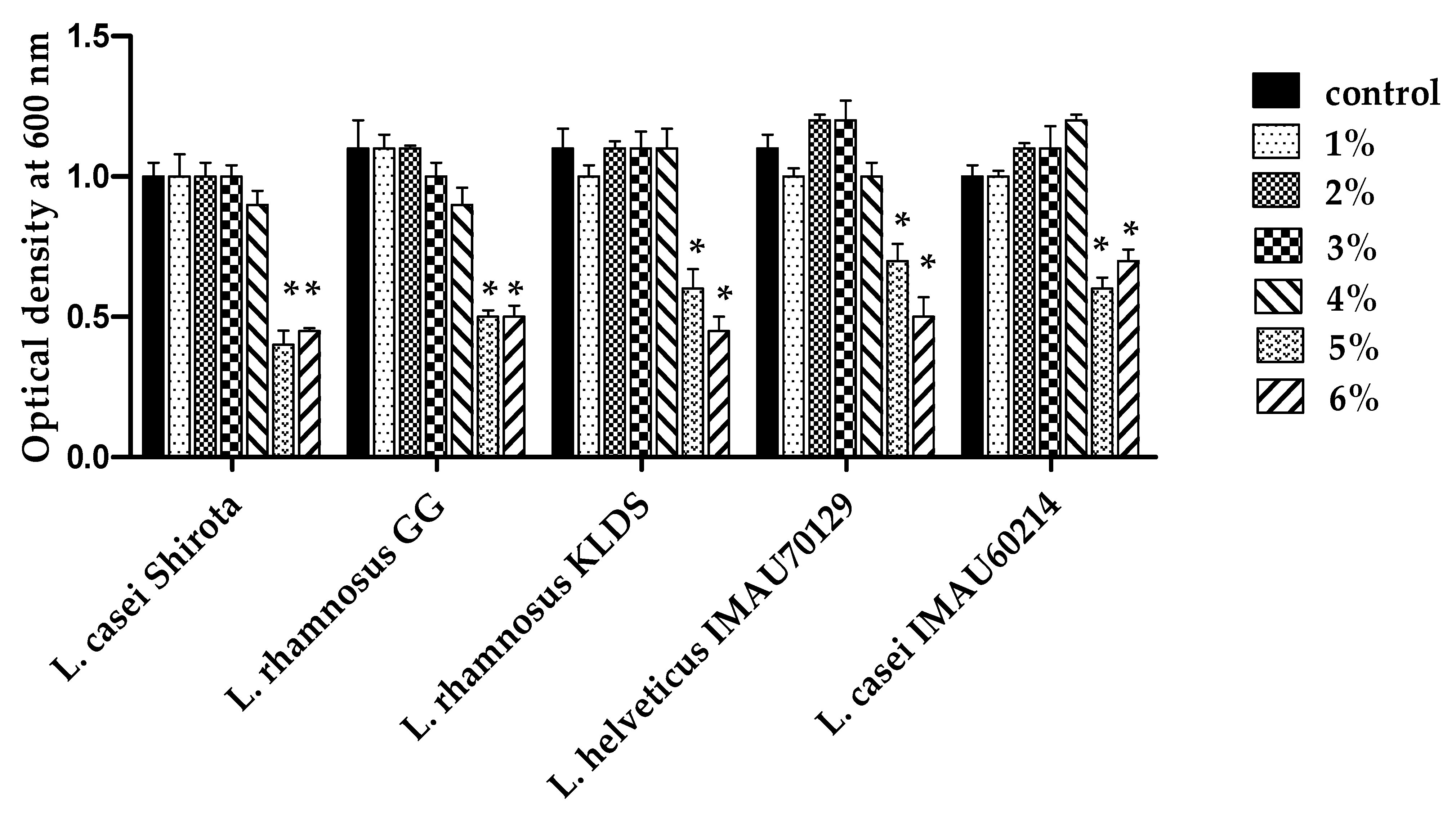
To investigate the effect of high concentrations of salt on growth, the Lactobacillus strains were cultured in MRS medium supplement with different NaCl concentration during 18 h. The observed results showed tolerance of Lactobacillus strains studied defined by the growth of bacteria to 1–4% NaCl concentrations (Figure 3).

3.3. Caco-2 Cells Adherence Assay

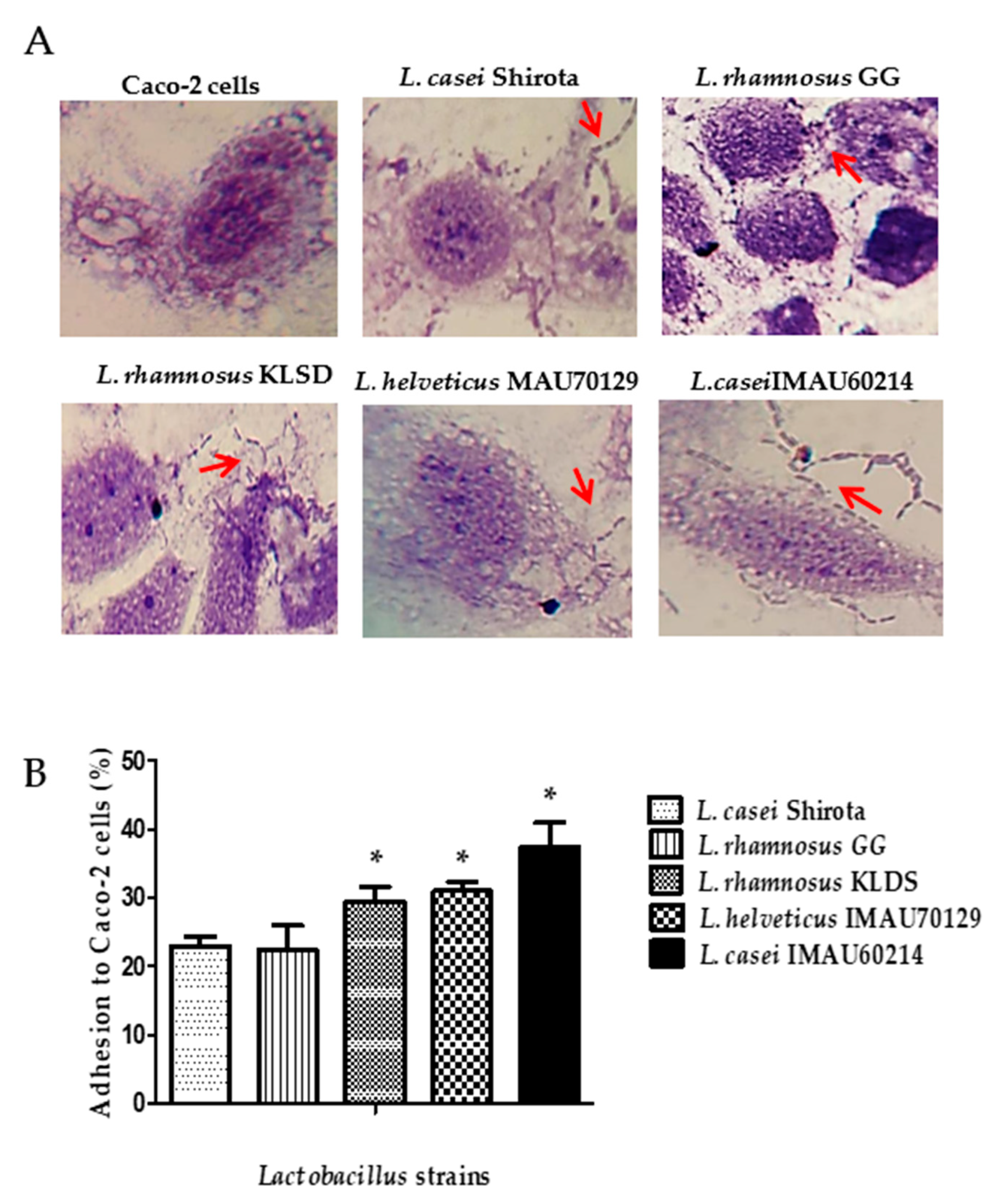
The qualitative test to determine the adherence capacity of L. rhamnosus GG, L. rhamnosus KLDS, L. helveticus IMAU70129, and L. casei IMAU60214 used the human epithelial Caco-2 cell line. This test showed the Lactobacillus strains adhere to cells after two hours of incubation (Figure 4A). The quantitative analysis report values varied from 25% to 35%. L. rhamnosus KLDS, L. helveticus IMAU70129, and L. casei IMAU60214 had higher adhesion, compared with L. casei strain Shirota (Figure 4B).

3.4. Adherence Inhibition of Pathogen Bacteria by Lactobacillus Strains

Lactobacillus strains were evaluated for their ability to interfere with the adhesion of human pathogens on epithelial Caco-2 cells. The results showed that some of the Lactobacillus strains evaluated induced a significant decrease in the adherence of pathogens to Caco-2 cells when compared with the reference probiotic L. casei strain Shirota (Figure 5). L. rhamnosus KLSD, L. helveticus IMAU70129, and L. casei IMAU60214 exhibited anti-adhesion effect against E. coli (30 ± 2.5%), S. Typhimurium (45 ± 3.5%) and S. aureus (50 ± 2.5%) respectively. In contrast, the anti-adhesion values observed with L. rhamnosus GG (25 ± 2.5%) and the reference probiotic L. casei strain Shirota (27 ± 3.1%) showed less activity.

3.5. Antimicrobial Activity of Cell-Free Supernatants (CFSs) from Lactobacillus Strains

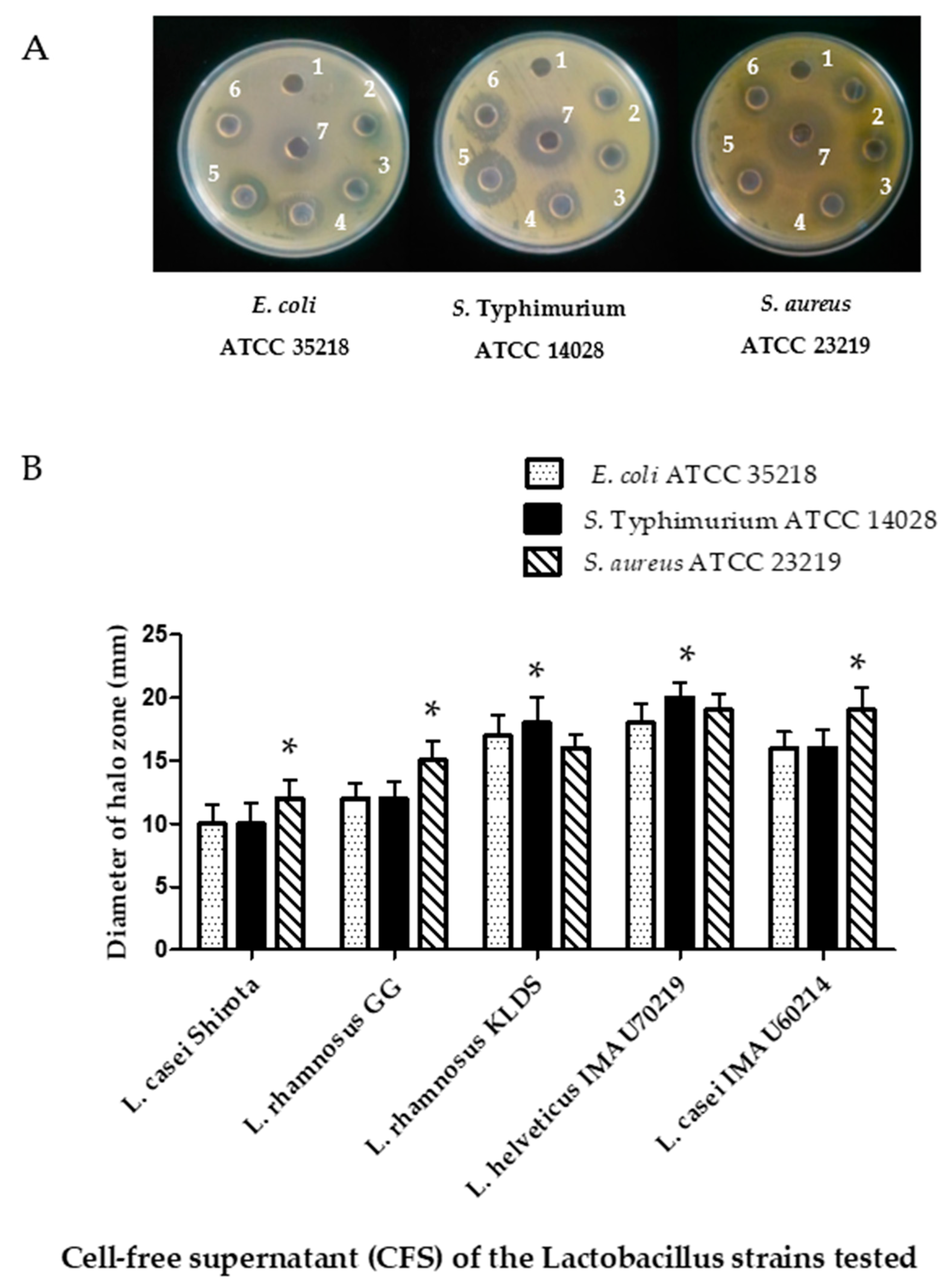
The obtained results in this assay shown that the cell-free supernatants tested inhibited the growing pathogenic bacteria evaluated (Figure 6A). The highest inhibition zone was recorded against S. aureus ATCC 29213 (16 mm), followed by S. Typhimurium ATCC 14028 (10 mm) and E. coli ATCC 35218 (12 mm) after 24 h of incubation (Figure 6B). L. helveticus IMAU70129, L. casei IMAU60214, and the reference probiotic L. casei strain Shirota showed higher inhibition zones than L. rhamnosus GG.

3.6. Immunomodulatory Activity of Lactobacillus Strains on the Secretion of IL-8 and NO-Induced by LPS on Caco-2 Cells

A feature to consider for a potential probiotic microorganism is its property of immunomodulation of the intestinal epithelium. An assay with Caco-2 cells incubated during 12 h with Lactobacillus strains induced a slight increase of IL-8 levels (250 ± 13 pg/mL), like those observed on unstimulated epithelial cells (150 ± 10 pg/mL). On the other hand, the incubation of Caco-2 cells with IL-1β (1 ng/mL) or LPS (0.1 µg/mL) significantly increased the IL-8 secretion levels (800 ± 15 pg/mL) (Figure 7). However, prior to stimulation of Caco-2 cells with IL-1β (1 ng/mL) or LPS (0.1 µg/mL), the cells were incubated with L. rhamnosus KLDS, L. helveticus IMAU70129, and L. casei IMAU60214 strains and a reduced secretion of IL-8 was observed (Figure 7A). was Another interesting observation was that stimulation with LPS of E. coli 011:B4 (0.1 µg/mL) of Caco-2 cells previously incubated for 12 h with L. rhamnosus KLDS, L. helveticus IMAU70129, and L. casei IMAU60214 inhibited NO production (Figure 7B).

3.7. Inflammatory Stress Protection Induced by Lactobacillus Strains

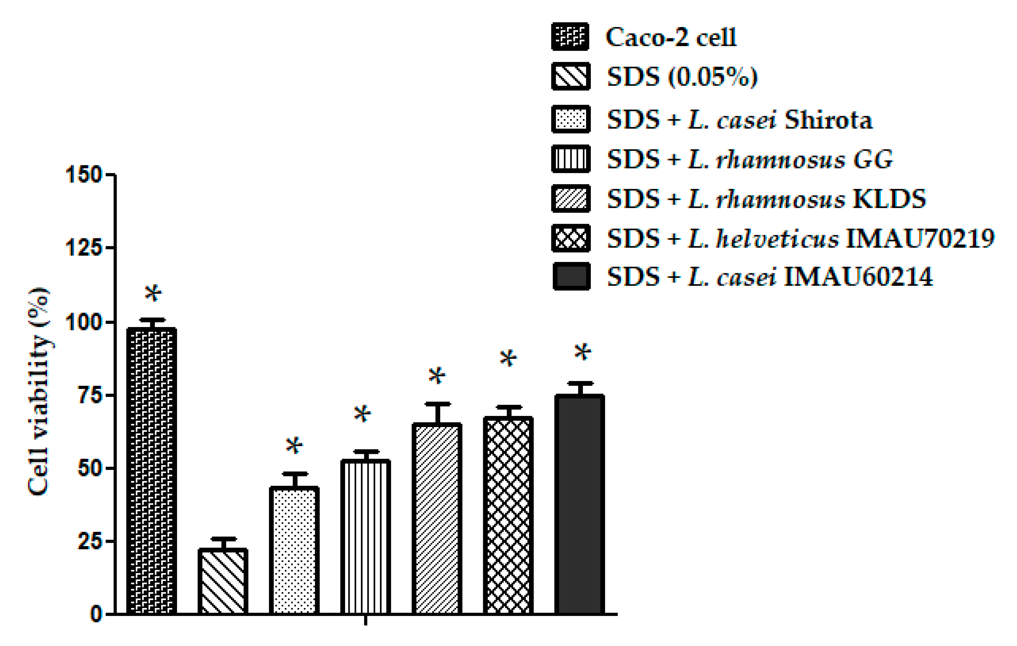
The in vitro inflammatory stress model of human epithelial Caco-2 cells exposed to SDS was used to evaluate the potential protective effect of Lactobacillus strains (L. rhamnosus GG, L. rhamnosus KLDS, L. helveticus IMAU70129, and L. casei IMAU60214). Co-incubation of Caco-2 cells with Lactobacillus strains for 24 h resulted in a significant reduction in the damage produced by SDS exposition (0.05%) compared with the untreated Caco-2 cells. In the assay L. rhamnosus KLDS, L. helveticus IMAU70129, and L. casei IMAU60214 exhibited a greater viability percent of the Caco-2 cells when compared to the cells only exposed to SDS (Figure 8).

4. Discussion

Human colonization by bacteria occurs at childbirth, as well as through breastfeeding and by environmental microorganism. Through breast milk the infant acquires different microorganisms, mainly Lactobacilli and bifidobacteria. The symbiotic relationship between human and microbiota contributes to establish a partnership that benefits both. The microorganisms acquire nutrients and a niche to reproduce and live while the host receives substrates that it is not capable of producing. In this way, it is protected against colonization by pathogenic microorganisms and the immune system matures [28]. Changes in the composition and structure of the intestinal microbiota have been linked to diseases such as diabetes, inflammatory syndromes such as ulcerative colitis, and neurological alterations like autism and Parkinson’s [29,30]. Changes in the intestinal microbiota have been related to diet and the inappropriate use of antimicrobials [2,8]. When microbiota alterations occur, the use of probiotic microorganisms is proposed. However, it is necessary to evaluate the specific characteristics and safety of the probiotic bacteria before using them to avoid causing damage to the host [31,32].
In this study, we evaluated different properties of the potential probiotic L rhamnosus KLDS, L. helveticus IMAU70129, and L. casei IMAU60214, which were isolated from commercial fermented products. Hydrophobicity, auto-aggregation, cell adhesion, antagonistic activity against enteric pathogens and immunomodulatory effects on Caco-2 cells were evaluated. The cell-surface hydrophobicity and cell-surface charge of bacteria are recognized as physicochemical variables for evaluating bacterial adhesion to surfaces [33]. In this study we observed that the hydrophobicity values of the Lactobacillus strains were similar to those observed in other probiotic strains reported elsewhere [34,35]. The hydrophobicity results (30–50%) support that the studied strains could be classified as having moderate hydrophobicity in accordance with the report by Jiang, et al. [25]. The hydrophobic characteristics of a strain have been implicated in bacteria adhesion to host tissues, conferring a competitive advantage on pathogen bacteria toward maintaining homeostasis in the host’s GIT [15,36,37].
Adherence between the same bacteria (auto-aggregation) helps bacteria adhere to the surface of the intestinal mucosa so that it persists in the intestine. Consequently, it competes with pathogenic bacteria and interferes with their establishment in the GIT to promote host health [38,39,40,41]. In the study the auto-aggregation results show that in L. casei IMAU60214 this property was very similar with what was observed in L. rhamnosus KLDS and L. helveticus IMAU70129.
Auto-aggregation and hydrophobicity are properties utilized as selective screening of potential probiotic strains. However, there is controversy when both properties correlate with epithelium adherence [42,43,44,45]. In this study, the cell adherence to Caco-2 cells, auto-aggregation, and hydrophobicity of the evaluated Lactobacillus strains was positive but with some differences between each other and with what we observed in the L. casei Shirota reference strain. This could be due to the difference in responses between strains which ultimately contributes to the bacteria interacting with the epithelium of their potential host. The Lactobacillus adherence initially could be associated with hydrophobic interactions and later specifically associated throughout bacterial structures as pilis which interact with cell receptors [46]. The identification and characterization of bacteria structures is of great importance in probiotic strain studies, since this would facilitate the study of the mechanisms associated with the beneficial properties of these microorganisms.
As previously mentioned, many different characteristics must be considered when selecting potential probiotic microorganisms. In addition to adherence, other properties need to be analyzed. Different studies have reported that several groups of probiotic strains can be protective, especially through interfering with the adherence and colonization of pathogenic bacteria [47,48]. In this study, anti-adhesion activity against pathogenic strains was analyzed (E. coli ATCC 35218; S. Typhimurium ATCC 12840 and S. aureus ATCC 23219). The results show interference of the pathogen bacteria (30–35% inhibition of adhered bacteria), similar to what was reported in studies performed with L. paracasei strains [49,50]. Although S. aureus does not act directly on the intestinal epithelium since it is through its enterotoxin, we evaluate the interference effect of Lactobacillus strains on this bacterium because dairy products can be enriched with probiotics and in this way they prevent staphylococci contamination.
The probiotic strains can also antagonize with pathogen bacteria both by antimicrobial activity mediated by metabolism secreted molecules or through immunomodulatory activities of the host’s immune system [9,12]. In this study, different growth inhibition of the selected pathogens induced by secreted products of Lactobacillus strains were observed. L. casei IMAU60214 and L. helveticus IMAU70129 show a more intense activity than the other studied strains, which was determined by the size of growth inhibition halos. Previous studies with L. reuteri isolated from humans show moderate inhibition against E. coli (inhibition of 7–10 mm in diameter) [50]. This is in contrast to what has been observed with the L. delbrueckii subspecies bulgaricus which exerted a substantial inhibition (21.1 mm in diameter) against E. coli [51]. It is also in contrast to what has been observed with L. plantarum B7 isolated from dyspeptic patients which induced moderate growth inhibition of H. pylori (11–13 mm) [52]. The differences in the bacterial growth inhibition effects observed in our study could be attributed to acidic products such as lactic and acetic acids, or compounds like hydrogen peroxide, ethanol, diacetyl, acetaldehyde, acetoin, reuterin, reutericyclin, and bacteriocins, among others. However, in the future it will be necessary to characterize the components responsible for these effects at the molecular level.
The immunomodulatory effect of probiotics has been considered as other one of the potential clinical effects of these microorganisms. This is why the term Bio-immunobiotics has been proposed to designate probiotic microorganisms with the ability to modulate the immune response [53]. In this study some immunomodulatory properties of the Lactobacillus strains analyzed were identified. One of these was the diminishing effect in the production of IL-8, similar to results observed with L. plantarum in previous studies [54,55]. The oxidative attenuation of NO that induces cytotoxic mechanisms under oxidative stress conditions was also evaluated. L. rhamnosus GG, L. helveticus IMAU70129, and L. casei IMAU60214 showed protective abilities against damage caused by SDS. This result is important because oxidative stress conditions can be extremely harmful to the epithelium, which suggests that these strains can contribute to reducing the inflammatory processes in the mucosa and may play a functional role in the prevention and control of several acute and chronic infectious diseases. Our research findings allow us to define these lactobacillus strains as probiotic candidates for the first time. However, the main limitation of the study is that the results were obtained by in vitro tests where the challenge and response conditions are controlled. Therefore, it is necessary to carry out studies in animal models and in humans to explore their safe use and explore the probable mechanisms of clinical application of these strains.

5. Conclusions

Studies of the gut microbiota on health and disease are of high importance, and the role of probiotics and prebiotics as modulators of the microbiota require studies to find the best candidates. The obtained results show that the Lactobacillus strains evaluated possess properties such as adherence to cultured cells, inhibition of the colonization and growth of pathogenic bacteria, and regulation of the inflammatory response. All of these are elements that make them good candidates for use as probiotics. However, it is important to evaluate their properties in animal models to ensure they are safe to use in clinical applications.

Author Contributions

Conceptualization, L.M.R.-R., U.H.-C. and C.A.E.; methodology, L.M.R.-R., S.S.M.-G. and A.R.-P.; investigation, L.M.R.-R., S.S.M.-G., A.R.-P. and C.A.E.; data curation, L.M.R.-R.; writing—original draft preparation, L.M.R.-R., S.S.M.-G. and A.R.-P.; project administration, L.M.R.-R.; funding acquisition, L.M.R.-R. All authors have read and agreed to the published version of the manuscript.

Funding

The work was supported by grants from the Fondos Federales México (HIM/2014/013 SSA 1120).

Institutional Review Board Statement

Not applicable.

Informed Consent Statement

Not applicable.

Data Availability Statement

The data presented in this study are available on request from the corresponding author.

Acknowledgments

We thank Mariano García Garibay and Alma Cruz Guerrero (from Departamento de Biotecnología, Universidad Autónoma Metropolitana-Iztapalapa, Mexico) for their donation of probiotic L. casei strain Shirota.

Conflicts of Interest

The authors declare no conflict of interest.

References

  1. Hooper, L.V.; Gordon, J.I. Commensal host-bacterial relationships in the gut. Science 2001, 292, 1115–1118. [Google Scholar] [CrossRef]
  2. Sekirov, I.; Russell, S.L.; Antunes, L.C.; Finlay, B.B. Gut microbiota in health and disease. Physiol. Rev. 2010, 90, 859–904. [Google Scholar] [CrossRef] [Green Version]
  3. Liévin, L.; Moal, V.; Servin, A.L. Anti-infective activities of lactobacillus strains in the human intestinal microbiota: From probiotics to gastrointestinal anti-infectious agents. Clin. Microbiol. Rev. 2014, 2, 167–199. [Google Scholar] [CrossRef] [PubMed] [Green Version]
  4. Weiss, G.A.; Hennet, T. Mechanisms and consequences of intestinal dysbiosis. Cell. Mol. Life Sci. 2017, 16, 2959–2977. [Google Scholar] [CrossRef] [PubMed] [Green Version]
  5. Plaza-Díaz, J.; Ruiz-Ojeda, F.J.; Vilchez-Padial, L.; Gil, A. Evidence of the Anti-Inflammatory Effects of Probiotics and Synbiotics in Intestinal Chronic Diseases. Nutrients 2017, 6, 555. [Google Scholar] [CrossRef] [PubMed] [Green Version]
  6. Walker, A.W.; Lawley, T.D. Therapeutic modulation of intestinal dysbiosis. Pharmacol. Res. 2013, 1, 75–86. [Google Scholar] [CrossRef] [PubMed]
  7. Zheng, J.; Wittouck, S.; Salvetti, E.; Franz, C.; Harris, H.M.B.; Mattarelli, P.; O’Toole, P.W.; Pot, B.; Vandamme, P.; Walter, J.; et al. A taxonomic note on the genus Lactobacillus: Description of 23 novel genera, emended description of thegenus Lactobacillus Beijerinck 1901, and union of Lactobacillaceae and Leuconostocaceae. Int. J. Syst. Evol. Microbiol. 2020, 70, 2782–2858. [Google Scholar] [CrossRef]
  8. Kanazawa, A.; Aida, M.; Yoshida, Y.; Kaga, H.; Katahira, T.; Suzuki, L.; Tamaki, S.; Sato, J.; Goto, H.; Azuma, K.; et al. Effects of Synbiotic Supplementation on Chronic Inflammation and the Gut Microbiota in Obese Patients with Type 2 Diabetes Mellitus: A Randomized Controlled Study. Nutrients 2021, 13, 558. [Google Scholar] [CrossRef]
  9. Duary, R.K.; Batish, V.K.; Grover, S. Immunomodulatory activity of two potential probiotic strains in LPS-stimulated HT-29 cells. Genes Nutr. 2014, 9, 398. [Google Scholar] [CrossRef] [Green Version]
  10. Reid, G. The growth potential for dairy probiotics. Int. Dairy J. 2015, 49, 16–22. [Google Scholar] [CrossRef] [Green Version]
  11. Goto, Y. Epithelial Cells as a Transmitter of Signals from Commensal Bacteria and Host Immune Cells. Front. Immunol. 2019, 10, 1–12. [Google Scholar] [CrossRef] [Green Version]
  12. Peterson, L.W.; Artis, D. Intestinal epithelial cells: Regulators of barrier function and immune homeostasis. Nat. Rev. Immunol. 2014, 14, 141–153. [Google Scholar] [CrossRef]
  13. Reid, G.; Jass, J.; Sebulsky, M.T.; McCormick, J.K. Potential uses of probiotics in clinical practice. Clin. Microbiol. Rev. 2003, 4, 658–672. [Google Scholar] [CrossRef] [Green Version]
  14. Chung, Y.W.; Choi, J.H.; Oh, T.Y.; Eun, C.S.; Han, D.S. Lactobacillus casei prevents the development of dextran sulphate sodium-induced colitis in Toll-like receptor 4 mutant mice. Clin. Exp. Immunol. 2008, 1, 182–189. [Google Scholar] [CrossRef]
  15. Tien, M.T.; Girardin, S.E.; Regnault, B.; Le Bourhis, L.; Dillies, M.A.; Coppée, J.Y.; Bourdet-Sicard, R.; Sansonetti, P.J.; Pédron, T. Anti-inflammatory effect of Lactobacillus casei on Shigella-infected human intestinal epithelial cells. J. Immunol. 2006, 2, 1228–1237. [Google Scholar] [CrossRef] [Green Version]
  16. Kaji, R.; Kiyoshima-Shibata, J.; Tsujibe, S.; Nanno, M.; Shida, K. Short communication: Probiotic induction of interleukin-10 and interleukin-12 production by macrophages is modulated by co-stimulation with microbial components. J. Dairy Sci. 2018, 4, 2838–2841. [Google Scholar] [CrossRef]
  17. Lai, H.H.; Chiu, C.H.; Kong, M.S.; Chang, C.J.; Chen, C.C. Probiotic Lactobacillus casei: Effective for Managing Childhood Diarrhea by Altering Gut Microbiota and Attenuating Fecal Inflammatory Markers. Nutrients 2019, 5, 1150. [Google Scholar] [CrossRef] [Green Version]
  18. Shokryazdan, P.; Sieo, C.C.; Kalavathy, R.; Liang, J.B.; Alitheen, N.B.; Faseleh, J.M.; Ho, Y.W. Probiotic potential of Lactobacillus strains with antimicrobial activity against some human pathogenic strains. BioMed Res. Int. 2014, 2014, 927268. [Google Scholar] [CrossRef] [PubMed] [Green Version]
  19. Ren, D.Y.; Li, C.; Qin, Y.Q.; Yin, R.L.; Du, S.W.; Ye, F.; Liu, H.F.; Wang, M.P.; Sun, Y.; Li, X.; et al. Lactobacilli reduce chemokine IL-8 production in response to TNF-α and Salmonella challenge of Caco-2 cells. BioMed Res. Int. 2013, 2013, 925219. [Google Scholar] [CrossRef] [PubMed] [Green Version]
  20. Meini, S.; Laureano, R.; Fani, L.; Tascini, C.; Galano, A.; Antonelli, A.; Rossolini, G.M. Breakthrough Lactobacillus rhamnosus GG bacteremia associated with probiotic use in an adult patient with severe active ulcerative colitis: Case report and review of the literature. Infection 2015, 6, 777–781. [Google Scholar] [CrossRef] [PubMed]
  21. Rocha-Ramírez, L.M.; Pérez-Solano, R.A.; Castañón-Alonso, S.L.; Moreno Guerrero, S.S.; Ramírez-Pacheco, A.; García Garibay, M.; Eslava, C. Probiotic Lactobacillus Strains Stimulate the Inflammatory Response and Activate Human Macrophages. J. Immunol. Res. 2017, 2017, 4607491. [Google Scholar] [CrossRef] [PubMed] [Green Version]
  22. Cruz-Guerrero, A.; Hernández-Sánchez, H.; Rodríguez-Serrano, G.; Gómez-Ruiz, L.; García-Garibay, M.; Figueroa-González, I. Commercial probiotic bacteria and prebiotic carbohydrates: A fundamental study on prebiotics uptake, antimicrobials production and inhibition of pathogens. J. Sci. Food. Agric. 2014, 94, 2246–2252. [Google Scholar] [CrossRef]
  23. Kotzamanidis, C.; Kourelis, A.; Litopoulou-Tzanetaki, E.; Tzanetakis, N.; Yiangou, M. Evaluation of adhesion capacity, cell surface traits and immunomodulatory activity of presumptive probiotic Lactobacillus strains. Int. J. Food. Microbiol. 2010, 140, 154–163. [Google Scholar] [CrossRef]
  24. Archer, A.C.; Halami, P.M. Probiotic attributes of Lactobacillus fermentum isolated from human feces and dairy products. Appl. Microbiol. Biotechnol. 2015, 99, 8113–8123. [Google Scholar] [CrossRef] [PubMed]
  25. Jiang, M.; Zhang, F.; Wan, C.; Xiong, Y.; Shah, N.P.; Wei, H.; Tao, X. Evaluation of probiotic properties of Lactobacillus plantarum WLPL04 isolated from human breast milk. J. Dairy Sci. 2016, 99, 1736–1746. [Google Scholar] [CrossRef] [Green Version]
  26. Wang, Y.; Xie, Q.; Zhang, Y.; Ma, W.; Ning, K.; Xiang, J.Y.; Cui, J.; Xiang, H. Combination of probiotics with different functions alleviate DSS-induced colitis by regulating intestinal microbiota, IL-10, and barrier function. Appl. Microbiol. Biotechnol. 2020, 104, 335–349. [Google Scholar] [CrossRef]
  27. Presti, I.; D’Orazio, G.; Labra, M.; La Ferla, B.; Mezzasalma, V.; Bizzaro, G.; Giardina, S.; Michelotti, A.; Tursi, F.; Vassallo, M.; et al. Evaluation of the probiotic properties of new Lactobacillus and Bifidobacterium strains and their in vitro effect. Appl. Microbiol. Biotechnol. 2015, 99, 5613–5626. [Google Scholar] [CrossRef] [PubMed]
  28. Hu, S.; Wang, L.; Jiang, Z. Dietary Additive Probiotics Modulation of the Intestinal Microbiota. Protein. Pept. Lett. 2017, 24, 382–387. [Google Scholar] [CrossRef] [PubMed]
  29. Kich, D.M.; Vincenzi, A.; Majolo, F.; Volken de Souza, C.F.; Goettert, M.I. Probiotic: Effectiveness nutrition in cancer treatment and prevention. Nutr. Hosp. 2016, 33, 1430–1437. [Google Scholar] [CrossRef] [Green Version]
  30. Yan, F.; Li, N.; Shi, J.; Li, H.; Yue, Y.; Jiao, W.; Wang, N.; Song, Y.; Huo, G.; Li, B. Lactobacillus acidophilus alleviates type 2 diabetes by regulating hepatic glucose, lipid metabolism and gut microbiota in mice. Food Funct. 2019, 10, 5804–5815. [Google Scholar] [CrossRef]
  31. Casellas, F.; Burgos, R.; Marcos, A.; Santos, J.; Ciriza-de-Los-Ríos, C.; García-Manzanares, Á.; Polanco, I.; Puy-Portillo, M.; Villarino, A.; Lema-Marqués, B.; et al. Consensus document on exclusion diets in irritable bowel syndrome (IBS). Nutr. Hosp. 2018, 35, 1450–1466. [Google Scholar] [CrossRef] [Green Version]
  32. Liu, J.; Hu, D.; Chen, Y.; Huang, H.; Zhang, H.; Zhao, J.; Gu, Z.; Chen, W. Strain-specific properties of Lactobacillus plantarum for prevention of Salmonella infection. Food Funct. 2018, 9, 3673–3682. [Google Scholar] [CrossRef]
  33. Archer, A.C.; Kurrey, N.K.; Halami, P.M. In vitro adhesion and anti-inflammatory properties of native Lactobacillus fermentum and Lactobacillus delbrueckii spp. J. Appl. Microbiol. 2018, 125, 243–256. [Google Scholar] [CrossRef]
  34. Castellanos, T.; Ascencio, F.; Bashan, Y. Cell-surface hydrophobicity and cell-surface charge of Azospirillum spp. FEMS Microbiol. Ecol. 1997, 24, 159–172. [Google Scholar] [CrossRef]
  35. Krasowska, A.; Sigler, K. How microorganisms use hydrophobicity and what does this mean for human needs? Front. Cell. Infect. Microbiol. 2014, 4, 112. [Google Scholar] [CrossRef] [PubMed] [Green Version]
  36. Xu, H.; Jeong, H.S.; Lee, H.Y.; Ahn, J. Assessment of cell surface properties and adhesion potential of selected probiotic strains. Lett. Appl. Microbiol. 2009, 49, 434–442. [Google Scholar] [CrossRef] [PubMed]
  37. Rokana, N.; Singh, B.P.; Thakur, N.; Sharma, C.; Gulhane, R.D.; Panwar, H. Screening of cell surface properties of potential probiotic lactobacilli isolated from human milk. J. Dairy Res. 2018, 85, 347–354. [Google Scholar] [CrossRef] [PubMed]
  38. Oh, N.S.; Joung, J.Y.; Lee, J.Y.; Kim, Y. Probiotic and anti-inflammatory potential of Lactobacillus rhamnosus 4B15 and Lactobacillus gasseri 4M13 isolated from infant feces. PLoS ONE 2018, 13, e0192021. [Google Scholar] [CrossRef] [PubMed]
  39. Panicker, A.S.; Ali, S.A.; Anand, S.; Panjagari, N.R.; Kumar, S.; Mohanty, A.K.; Behare, P.V. Evaluation of some in vitro probiotic properties of Lactobacillus fermentum Strains. J. Food Sci. Technol. 2018, 55, 2801–2807. [Google Scholar] [CrossRef]
  40. Dos Santos, A.S.; de Albuquerque, T.M.R.; de Brito Alves, J.L.; de Souza, E.L. Effects of Quercetin and Resveratrol on in vitro Properties Related to the Functionality of Potentially Probiotic Lactobacillus Strains. Front. Microbiol. 2019, 24, 2229. [Google Scholar] [CrossRef] [PubMed] [Green Version]
  41. Keller, M.K.; Hasslöf, P.; Stecksén-Blicks, C.; Twetman, S. Co-aggregation and growth inhibition of probiotic lactobacilli and clinical isolates of mutans streptococci: An in vitro study. Acta Odontol. Scand. 2011, 69, 263–268. [Google Scholar] [CrossRef]
  42. Matsuzaki, T. Immunomodulation by treatment with Lactobacillus casei strain Shirota. Int. J. Food Microbiol. 1998, 26, 133–140. [Google Scholar] [CrossRef]
  43. Kanmani, P.; Kim, H. Functional capabilities of probiotic strains on attenuation of intestinal epithelial cell inflammatory response induced by TLR4 stimuli. Biofactors 2019, 45, 223–235. [Google Scholar] [CrossRef]
  44. Tuo, Y.; Yu, H.; Ai, L.; Wu, Z.; Guo, B.; Chen, W. Aggregation and adhesion properties of 22 Lactobacillus strains. J. Dairy. Sci. 2013, 96, 4252–4257. [Google Scholar] [CrossRef] [PubMed] [Green Version]
  45. Pan, W.H.; Li, P.L.; Liu, Z. The correlation between surface hydrophobicity and adherence of Bifidobacterium strains from centenarians’ faeces. Anaerobe 2006, 3, 148–152. [Google Scholar] [CrossRef] [PubMed]
  46. Haddaji, N.; Mahdhi, A.K.; Krifi, B.; Ismail, M.B.; Bakhrouf, A. Change in cell surface properties of Lactobacillus casei under heat shock treatment. FEMS Microbiol. Lett. 2015, 9, fnv047. [Google Scholar] [CrossRef] [PubMed] [Green Version]
  47. Vlková, E.; Rada, V.; Smehilová, M.; Killer, J. Auto-aggregation and co-aggregation ability in bifidobacteria and clostridia. Folia Microbiol. 2008, 53, 263–269. [Google Scholar] [CrossRef] [PubMed]
  48. Malago, J.J.; Tooten, P.C.; Koninkx, J.F. Anti-inflammatory properties of probiotic bacteria on Salmonella-induced IL-8 synthesis in enterocyte-like Caco-2 cells. Benef. Microbes 2010, 1, 121–130. [Google Scholar] [CrossRef] [PubMed]
  49. Fonseca, H.C.; de Sousa, M.D.; Ramos, C.L.; Días, D.R.; Schwan, R.F. Probiotic Properties of Lactobacilli and Their Ability to Inhibit the Adhesion of Enteropathogenic Bacteria to Caco-2 and HT-29 Cells. Probiotics Antimicrob. Proteins 2021, 13, 102–112. [Google Scholar] [CrossRef]
  50. Alizadeh, B.B.; Noshad, M.; Falah, F. Inhibition of Escherichia coli adhesion to human intestinal Caco-2 cells by probiotic candidate Lactobacillus plantarum strain L15. Microb. Pathog. 2019, 136, 103677. [Google Scholar] [CrossRef]
  51. Sunanliganon, C.; Thong-Ngam, D.; Tumwasorn, S.; Klaikeaw, N. Lactobacillus plantarum B7 inhibits Helicobacter pylori growth and attenuates gastric inflammation. World J. Gastroenterol. 2012, 18, 2472–2480. [Google Scholar] [CrossRef]
  52. Jankowska, A.; Laubitz, D.; Antushevich, H.; Zabielski, R.; Grzesiuk, E. Competition of Lactobacillus paracasei with Salmonella enterica for adhesion to Caco-2 cells. J. Biomed. Biotechnol. 2008, 2008, 357964. [Google Scholar] [CrossRef] [PubMed] [Green Version]
  53. Tabashsum, Z.; Peng, M.; Alvarado-Martinez, Z.; Aditya, A.; Bhatti, J.; Romo, P.B.; Young, A.; Biswas, D. Competitive reduction of poultry-borne enteric bacterial pathogens in chicken gut with bioactive Lactobacillus casei. Sci. Rep. 2020, 10, 16259. [Google Scholar] [CrossRef] [PubMed]
  54. Boonma, P.; Spinler, J.K.; Venable, S.F.; Versalovic, J.; Tumwasorn, S. Lactobacillus rhamnosus L34 and Lactobacillus casei L39 suppress Clostridium difficile-induced IL-8 production by colonic epithelial cells. BMC Microbiol. 2014, 14, 177. [Google Scholar] [CrossRef] [PubMed] [Green Version]
  55. Perdigon, G.; Alvarez, S.; Rachid, M.; Agüero, G.; Gobbato, N. Immune system stimulation by probiotics. J. Dairy Sci. 1995, 78, 1597–1606. [Google Scholar] [CrossRef]
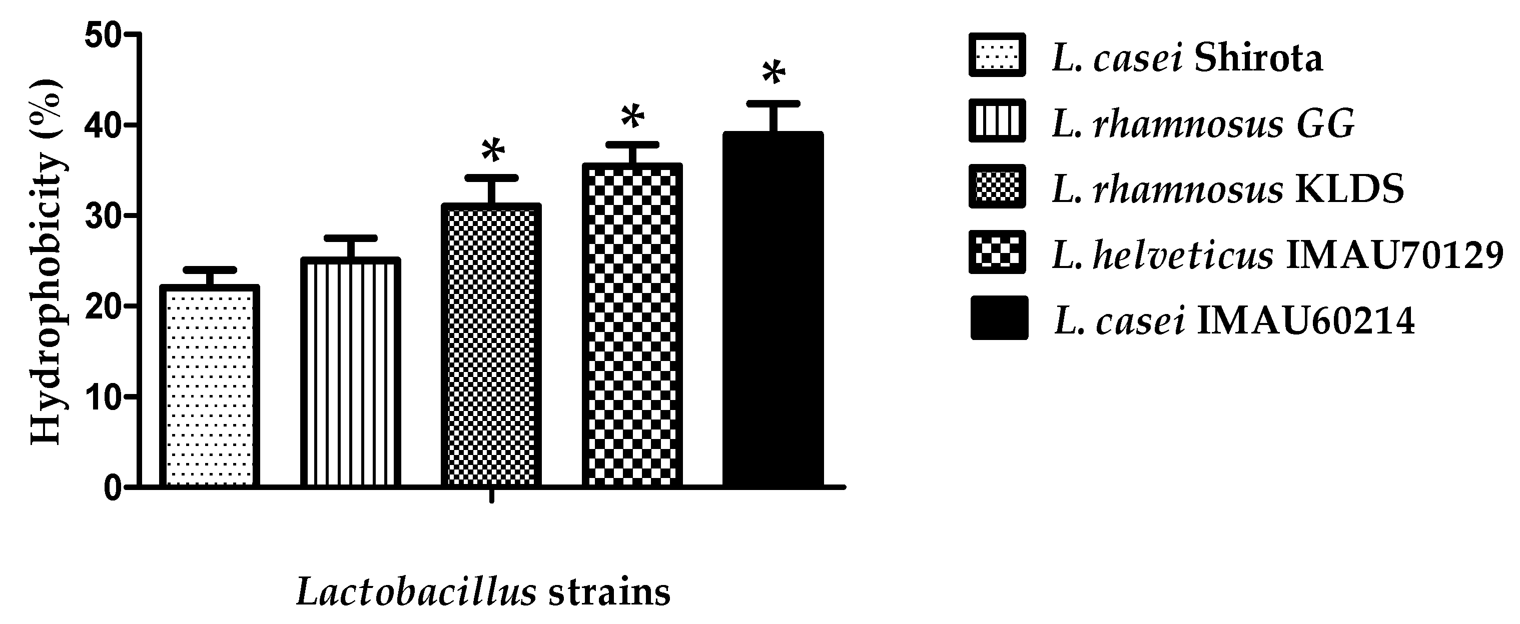
Figure 1. Lactobacillus strain hydrophobicity determined in xylene presence (1 h). Values are expressed as mean ± standard deviation (SD) in three different experiments. * significant difference among strains p < 0.05.
Microorganisms 09 00825 g001
Figure 2. Auto-aggregation of Lactobacillus strains as determined after various times of incubation durations at 37 °C. The values represent the mean value ± standard deviation (SD) of three different assays.
Microorganisms 09 00825 g002
Figure 3. NaCl tolerance of Lactobacillus strains. The tolerance to different salt concentrate ions was analyzed during 18 h at 37 °C. Values represent the mean ± standard deviation (SD) of three different assays. An MRS medium without NaCl was used as the control. * significant difference among strains at concentration of NaCl-MRS.
Microorganisms 09 00825 g003
Figure 4. Adhesion property of Lactobacillus strains to Caco-2 cells. (A) Representative microscopic visualization (400×). The red arrow indicates the evaluated Lactobacillus strains adhered to Caco-2 cells and the index score (number of Lactobacillus per Caco-2 cells). (B) Percentage of adhesion on Caco-2 cells. The values represent the mean ± SD of three separated experiments. * significant difference among strains (p < 0.05).
Microorganisms 09 00825 g004
Figure 5. Adherence inhibition effect of Lactobacillus strains against pathogenic bacteria as well as Escherichia coli ATCC 35218 and Salmonella Typhimurium ATCC 40128 and Staphylococcus aureus ATCC 23219. The values represent the mean ± SD of three different assays. * significant among Lactobacillus strains (p < 0.05).
Microorganisms 09 00825 g005
Figure 6. Antimicrobial activity of cell-free supernatant (CFS) of Lactobacillus strains against pathogenic bacteria. (A) Well 1, control MRS (alone) or negative (not-inhibitory); Well 2, L. rhamnosus GG; Well 3, L. rhamnosus KLDS; Well 4, L. helveticus IMAU70129; Well 5, L. casei IMAU60214; Well 6, L. casei strain Shirota; Well 7, MRS with tetracycline antibiotic (positive showed inhibitory activity). (B) Zone inhibition in diameter (mm). Values represent the mean ± SD of three different assays. * significant difference among Lactobacillus strains (p < 0.05).
Microorganisms 09 00825 g006
Figure 7. Immunomodulatory activity of Lactobacillus strains on the secretion of interleukin-8 (IL)-8 and NO on Caco-2 cells. (A) Baseline IL-8 response and production in an IL-1β (1 ng/mL) stimulated inflammatory environment. (B) Nitric oxide production at baseline and under an inflammatory environment stimulated with LPS (0.1 µg/mL). Values represent the mean ± SD (n = 3). * significant differences among Lactobacillus strains (p < 0.05).
Microorganisms 09 00825 g007
Figure 8. Protective effect on Caco-2 cells induced by Lactobacillus strains. The residual cell viability was measured by the mean MTT assay. The results are expressed in percentages. Values represent the mean ± SD of three separated assays. * significant differences among Lactobacillus strains versus SDS 0.05% (p < 0.05).
Microorganisms 09 00825 g008
Publisher’s Note: MDPI stays neutral with regard to jurisdictional claims in published maps and institutional affiliations.