Article

Antimicrobial Activity of Lactococcus lactis subsp. lactis Isolated from a Stranded Cuvier’s Beaked Whale (Ziphius cavirostris) against Gram-Positive and -Negative Bacteria

Laboratory of Aquatic Animal Physiology, Department of Marine Science and Resources, Graduate School of Bioresource Sciences, Nihon University, 1866 Kameino, Fujisawa 252-0880, Japan
*
Author to whom correspondence should be addressed.
Microorganisms 2021, 9(2), 243; https://doi.org/10.3390/microorganisms9020243
Submission received: 2 January 2021 / Revised: 20 January 2021 / Accepted: 22 January 2021 / Published: 25 January 2021
(This article belongs to the Special Issue Wildlife Microbiology 2.0)

Abstract

:
In the present study, we isolated and characterized Lactococcus lactis (L. lactis) subsp. lactis from a female Cuvier’s beaked whale (Ziphius cavirostris) stranded in Shizuoka, Japan. Only five isolates (CBW1-5), grown on Lactobacilli de Man Rogosa Sharpe (MRS) agar plates prepared using 50% artificial seawater, were positive in L. lactis species-specific primer PCR. Their 16S rRNA sequences were highly similar to those of L. lactis subsp. lactis JCM 5805T. The Gram reaction, motility, gas production from glucose, catalase production, and growth conditions were consistent with those of the type strain. Additionally, carbohydrate utilization of the strains was consistent with previously reported marine organism-derived strains. The pH-neutralized cell-free culture supernatant of strain CBW2 inhibited the growth of Bacillus subtilis subsp. subtilis ATCC 6051 and Vibrio alginolyticus ATCC 17749, whereas protease treatment eliminated or diminished its inhibitory activity. The strain possesses a precursor of the nisin structural gene (nisA), which showed 100% homology with nisin Z, and nisin biosynthesis-related genes (nisB, nisC, nisT, nisP, nisF, nisI, and nisRK), suggesting that the strain produces a nisin-like substance. This study provides fundamental information on whale-derived L. lactis subsp. lactis which may be useful for reducing the carriage of B. subtilis subsp. subtilis and V. alginolyticus.

1. Introduction

Some strains of lactic acid bacteria (LAB), such as Enterococcus, Lactobacillus, Lactococcus, and Pediococcus, have received attention as beneficial organisms [1]. LAB create a low-pH environment, by fermenting several nutrients, and produce antimicrobial factors, which can prevent contamination by pathogenic bacteria [1,2]. In addition, the interaction between LAB and the intestinal mucosa of the host is associated with immunostimulating and immunomodulatory effects [3]. Therefore, many studies have reported the potential benefits of using various LAB strains in the food industry [4,5] and LAB-associated health improvement in animals and humans [6,7,8].
Bacteriocins are gene-encoded, ribosomally-synthesized antimicrobial peptides produced by both Gram-positive and -negative microorganisms [9]. Bacteriocins derived from LAB have a wide variety of potential applications, such as preventing contamination by pathogenic bacteria in food and as alternatives to antibiotics for medical and veterinary use [10]. Nisin, produced by some strains of L. lactis, is a widely studied bacteriocin. This heat-stable, non-toxic, cationic lantibiotic belongs to class I bacteriocins, according to the classification criteria of Klaenhammer [9]. It consists of 34 amino acids, including several unusual amino acids such as dehydroalanine, dehydrobutyrine, aminobutyric acid, lanthionine, and β-methyllanthionine [9,11]. Nisin can kill closely related bacteria but also a wide range of Gram-positive bacteria, including Staphylococcus aureus (S. aureus), Bacillus spp., and Clostridium spp., by forming pores in the cell membrane via binding to lipid II, a bacterial cell wall precursor, and causing the subsequent efflux of small cytoplasmic contents such as amino acids, nucleotides, and ions [11,12]. However, it cannot kill Gram-negative bacteria because of the structure of the outer membrane barrier, outward cytoplasmic membrane, and peptidoglycan layer, which is composed of lipopolysaccharide molecules in the outer leaflet and glycerophospholipids in the inner leaflet [11,13]. Because of its potency against various Gram-positive bacteria, nisin is widely used as a food preservative in processed cheeses, dairy products, and canned foods [9,14].
Few LAB strains have been isolated from cetaceans. Lactobacillus salivarius (Lact. salivarius) was isolated from the rectal contents of a captive common bottlenose dolphin (Tursiops truncatus). Its strains inhibit the growth of pathogens in marine mammals and humans and stimulate the production of tumor necrosis factor in mammalian cells [15]. Lactobacillus ceti (Lact. ceti) 142-2T was isolated from the liver and lungs of a stranded Cuvier’s beaked whale (Ziphius cavirostris) [16], and Weissella ceti (W. ceti) 1119-1A-09T was isolated from several organs of beaked whales (Mesoplodon bidens) [17]. Lactococcus garvieae (L. garvieae), which is suspected to be responsible for diseases in fish, was isolated from the muscle and kidney of a stranded common bottlenose dolphin [18,19]. L. lactis is not normally considered as part of the commensal microbiota [10] but has been isolated not only from terrestrial mammals [20] but also from marine organisms [21,22,23,24]. In addition, various studies showed that several L. lactis strains can survive transit through the mammalian gastrointestinal tract [10,25]. Therefore, L. lactis strains may also exist in cetacean gastrointestines as indigenous microbial members. In one study, L. lactis strains were isolated from cetaceans, specifically only from stranded harbor porpoises (Phocoena phocoena), and the clinical relevance between the isolated L. lactis strains and pneumonia as a secondary infection was stated, however, the fundamental characteristics of the strains were not investigated [26]. Thus, our knowledge regarding L. lactis strains in cetacean organs is limited. In this study, we aimed to determine the phenotypic characteristics and antimicrobial activities of L. lactis subsp. lactis strains isolated from a fecal sample of a stranded Cuvier’s beaked whale during an investigation of the gastrointestinal microbiota of the whale.

2. Materials and Methods

2.1. Sample Collection and Stranded Whale Species Identification

A female stranded whale with a body length of 5.61 m was found on the Miho coast, Shimizu, Shizuoka, Japan (35°00′38.6″ N, 138°31′53.2″ E, Figure S1) on 18 September 2019. After wiping the skin around the vent using 70% ethanol, a fecal sample was collected using a sterile spoon and stored in Kenki-Porter II (Terumo, Tokyo, Japan), an anaerobic transport vial tube, at 4 °C until further experimentation in the laboratory.
The species of the stranded whale was identified by morphological examination and genetic investigation using a primer set (Table 1) for amplification of a 550-bp fragment of the mitochondrial DNA D-loop region. The reaction mixture consisted of 0.5 μL of each primer (10 μM), 2.5 μL of 10× PCR buffer for Blend Taq (Toyobo, Osaka, Japan), 2.5 μL dNTPs (2 mM), 0.25 μL of Blend Taq Plus polymerase (Toyobo), 17.75 μL of distilled water, and 1 μL of template crude DNA. The PCR conditions were as follows: initial denaturation at 94 °C for 2 min, 30 cycles of denaturation at 94 °C for 30 s, annealing at 55 °C for 30 s, and extension at 72 °C for 30 s.

2.2. LAB Isolation from the Fecal Sample

A loopful of fecal sample was inoculated on Lactobacilli MRS agar plates (BD Difco, Franklin Lakes, NJ, USA), prepared with 50% artificial seawater and containing 1% CaCO3, and incubated at 37 °C until colonies developed under anaerobic conditions, established using Anaeropack (Mitsubishi Gas Chemical Company, Tokyo, Japan) and an anaerobic jar (Mitsubishi Gas Chemical Company). The screening of bacterial acid production was completed by checking for the formation of a clear zone around the colony on agar plates. The developed colonies showing acid production were stored as 20% glycerol stocks at −80 °C for subsequent phylogenic and biochemical analyses.

2.3. Screening for L. lactis Using Species-Specific Primers and 16S rRNA Gene Sequencing

Bacterial DNA was extracted from the bacterial strains by bead disruption in sterile 10% Triton X-100 (MP Biomedicals, Irvine, CA, USA) and then used as a template for PCR amplification. As an initial screening for L. lactis, PCR amplification was conducted as previously described [22] using a universal forward primer and an L. lactis-specific reverse primer (Table 1). The PCR amplicons were subjected to electrophoresis on a 2% agarose gel, and the presence of expected bands was examined using a Gel Doc EZ Imager (Bio-Rad, Hercules, CA, USA) under UV light.
Approximately 1500 bp of the bacterial 16S rRNA gene region were amplified by the universal primers (Table 1). The PCR conditions were as follows: initial denaturation at 94 °C for 2 min, 25 cycles of denaturation at 94 °C for 30 s, annealing at 55 °C for 30 s, and extension at 72 °C for 90 s. The composition of the PCR mixture was the same as that described above. The PCR amplicons were purified using a Wizard SV Gel and a PCR clean-up system (Promega, Madison, WI, USA) and directly sequenced using an ABI 3130xl Genetic Analyzer (Applied Biosystems, Foster City, CA, USA) with a BigDye™ Terminator v3.1 Cycle Sequencing Kit (Applied Biosystems). The obtained sequences were subjected to a similarity-based search against quality-controlled databases of 16S rRNA sequences in EzBioCloud [32]. Multiple sequence alignments with the related species in the genus Lactococcus obtained from the NCBI GenBank database (https://www.ncbi.nlm.nih.gov/) were performed using the CLUSTAL W program [33]. A phylogenetic tree was reconstructed using the neighbor-joining method [34] and Kimura’s two-parameter model [35] in MEGA X ver. 10.1.8 [36], and the tree topology was evaluated using 1000 bootstrap replicates.

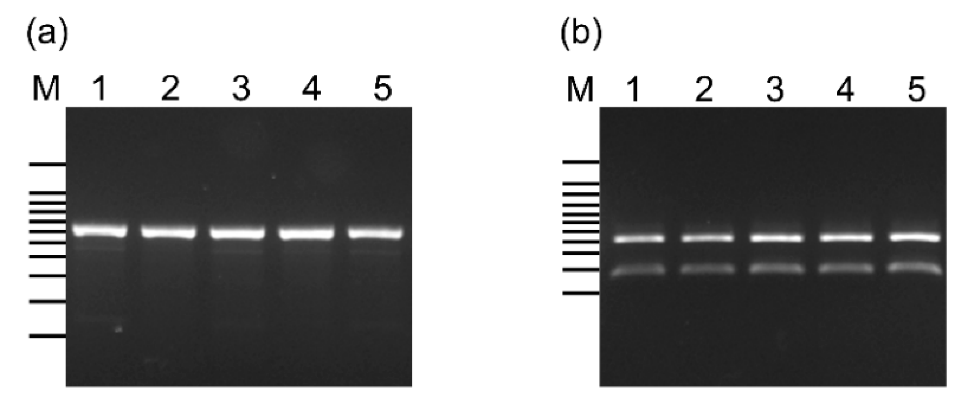
2.4. Classification of L. lactis Subspecies Using PCR-Restriction Fragment Length Polymorphism Analysis

Five isolates showing positive results by PCR amplification with L. lactis species-specific primers and the highest similarity to the 16S rRNA gene sequences of L. lactis subspecies strains were investigated using a primer set (Table 1) to amplify the gadB gene encoding glutamate decarboxylase, which was observed in L. lactis subsp. lactis but not in L. lactis subsp. cremoris [37]. The PCR conditions were as follows: initial denaturation at 94 °C for 2 min, 35 cycles of denaturation at 94 °C for 30 s, annealing at 50 °C for 30 s, extension at 72 °C for 1 min, and final extension at 72 °C for 5 min. Subsequently, the amplified fragments were digested using AseI endonuclease (Nippon Gene, Tokyo, Japan) following the manufacturer’s instructions. The expected PCR amplicon sizes were approximately 600 and 560 bp for L. lactis subsp. lactis and subsp. cremoris, respectively. The amplicon of the L. lactis subsp. lactis strain was cut into two fragments (approximately 190 and 410 bp in length) by the enzyme, whereas that of the L. lactis subsp. cremoris strain was not [31]. The cleaved amplicons were visualized on a 2% agarose gel using a Gel Doc EZ Imager (Bio-Rad).

2.5. Random Amplified Polymorphic DNA (RAPD)-PCR Analysis

Genomic DNA was extracted from the bacterial strains using a NucleoSpin Microbial DNA kit (Macherey-Nagel, Düren, Germany) according to the manufacturer’s instructions. To analyze the genetic diversity of the five isolates, random amplified polymorphic DNA (RAPD)-PCR analysis using M13 primer (5′-GAGGGTGGCGGTTCT-3′, [38]) was performed. The reaction mixture consisted of 2 μL of each primer (50 μM), 5 μL of 10× PCR buffer for Blend Taq (Toyobo), 5 μL of dNTPs (2 mM), 0.5 μL of Blend Taq Plus polymerase (Toyobo), 36.5 μL of distilled water, and 1 μL of template DNA (30 ng μL−1). The PCR conditions were as follows: initial denaturation at 94 °C for 2 min, 35 cycles of denaturation at 94 °C for 30 s, annealing at 50 °C for 1 min, and extension at 72 °C for 2 min. The PCR amplicons were visualized on a 1.5% agarose gel using a Gel Doc EZ Imager (Bio-Rad).

2.6. Biochemical Characterization

All strains were subjected to Gram reaction and motility tests, and the production of gas from glucose and catalase was assessed. The Gram reaction test were performed using 3% potassium hydroxide [39]. For the motility test, a single colony of the strains was inoculated into lysine indole motility semisolid agar (Eiken Chemical, Tokyo, Japan), and the presence or absence of the diffusion of bacteria growth was checked after incubation at 37 °C for 24 h. Gas production from glucose was tested using agar containing 1% glucose, 1.5% peptone, 0.5% sodium chloride, and 0.002% phenol red in Durham tubes [22]. Catalase production was examined in the presence of gas bubbles within 30 s after adding the purified strains to 1 mL of hydrogen peroxide (Fujifilm Wako Pure Chemical Corporation, Osaka, Japan). The final pH of the Lactobacilli MRS broth after two days of incubation at 37 °C was measured using an F-50 pH meter (Horiba, Kyoto, Japan). The carbohydrate fermentation profile was tested using the API 50 CH system (bioMérieux, Marcy-l’Étoile, France) with API 50 CHL medium (bioMérieux) following the manufacturer’s instructions.
To classify the L. lactis subspecies, the strains were incubated for two days in Lactobacilli MRS broth at pH 4.0, 9.2, and 10.0 at 37 °C, and on Lactobacilli MRS agar plates at 10 °C, 40 °C, and 45 °C. Additionally, the sodium chloride tolerance of the strains was investigated by incubation on Lactobacilli MRS agar plates containing 1%, 2%, 3%, 4%, 5%, 6%, 7%, 8%, 9%, 10%, and 15% sodium chloride at 37 °C for two days.

2.7. Antimicrobial Activity Assay

The agar well diffusion assay was performed as described previously [6,40], with some modifications. The suspension of the Gram-positive and -negative indicator bacterial strains (Table 2) was adjusted to McFarland Standard No. 0.5 and inoculated onto Muller Hinton agar plates (BD Difco). Well holes (5 mm in diameter) were hollowed out using a sterile alloy pipe and filled with 100 µL of cell-free supernatant of Lactobacilli MRS broth of each L. lactis subsp. lactis strain (adjusted to pH 7.0 by 2 N NaOH) obtained by repeated centrifugation at 5000× g for 10 min and 0.22-μm filter sterilization. The Muller Hinton agar plates with each indicator strain were incubated under the conditions shown in Table S1. The pH-neutralized, cell-free supernatants from the strains were treated with 3 mg mL−1 (final concentration) proteinase K (Fujifilm Wako Pure Chemical Corporation), trypsin (Fujifilm Wako Pure Chemical Corporation), lysozyme (MP Biomedicals), catalase (Tokyo Chemical Industry, Tokyo, Japan), pepsin (Nacalai Tesque, Kyoto, Japan), and lipase (Nacalai esque). The agar well diffusion assay of the enzyme-treated cell-free supernatants was performed against the indicator bacterial strains, whose growth was inhibited by the pH-neutralized, cell-free supernatants. Antimicrobial activity was determined by measuring the diameter (mm) of the inhibition zone around the well. Strains with inhibition zones less than 6 mm in diameter, between 6 and 10 mm, and more than 10 mm were classified as non-inhibitors, intermediate inhibitors, and strong inhibitors, respectively. The assay was performed in triplicate, and the inhibition zone of enzyme-treated, cell-free supernatants was compared with that of the pH-neutralized, cell-free supernatant treated as a control, by one-way analysis of variance followed by Dunnett’s test.

2.8. Genetical Analysis for Bacteriocin Production Potential

All L. lactis subsp. lactis strains were screened for the presence of the precursor of the nisin structural gene (nisA), which encodes the prepeptides of nisin, lacticin 481 and 3147, and lactococcin A, B, M, and 972, using the primer sets shown in Table 3. PCR amplification was performed under the following conditions: initial denaturation at 94 °C for 2 min, 35 cycles of denaturation at 94 °C for 30 s, annealing at 45 °C (for nisin) or 50 °C (for lacticin 481 and 3147 and lactococcin A, B, M, and 972) for 30 s, extension at 72 °C for 30 s, and final extension at 72 °C for 5 min.
Subsequently, all strains were subjected to PCR amplification targeting nisin biosynthesis-related genes. The primer sequences and targeted genes for each assay are shown in Table 3. The PCR conditions were as follows: initial denaturation at 94 °C for 2 min, 35 cycles of denaturation at 94 °C for 30 s, annealing at 42 °C (for nisC), 45 °C (for nisP and nisRK), and 55 °C (for nisB, nisT, nisI, and nisF) for 30 s, extension at 72 °C for 1 min, and final extension at 72 °C for 5 min.
The resulting amplicons for nisA were purified and sequenced as described above. The obtained DNA sequences were translated into amino acid sequences using the ExPASy translate tool [41], and then the blastp-based protein sequence similarity search tool in RiPPMiner, a ribosomally synthesized and post-translationally modified peptide (RiPP) database [42], was used to find the closest neighbors of RiPP peptides and predict the RiPP class of the gene product.

3. Results

3.1. Isolation of L. lactis Strains from a Stranded Cuvier’s Beaked Whale

The morphology of the stranded whale was identical to that of Cuvier’s beaked whale, and the mitochondrial DNA D-loop region sequence of the whale was homologous to that of the species (GenBank accession number: AB610404) at 100% identity.
We isolated 46 potential LAB isolates based on acid production. Of these, five strains, namely CBW1–5, showed L. lactis-specific bands (Figure S2). According to 16S rRNA gene sequencing, five strains had sequences highly homologous to that of L. lactis subsp. lactis JCM 5805T (99.04–99.93% similarity) (Table 4). These strains were clustered in the same group as L. lactis subsp. lactis using the neighbor-joining method and Kimura’s two-parameter model, with 100% bootstrap values based on 1000 replicates (Figure 1). In addition, the band patterns of the five strains amplified by RAPD-PCR analysis using the M13 primer differed from each other (Figure S3).

3.2. Classification of L. lactis Subspecies in the CBW1–5 Strains

The PCR amplicons of the CBW1–5 strains were approximately 600 bp in length (Figure 2a) with homologies to L. lactis subsp. lactis ATCC 19435 (GenBank accession number: AB067750) ranging from 97.6% to 98.8%. Their PCR products were digested by AseI endonuclease into two fragments of approximately 190 and 410 bp (Figure 2b).

3.3. Biochemical Characterization

The phenotypic characteristics of the strains are summarized in Table 5. The CBW1–5 isolates were identified as Gram-positive, non-motile, and catalase-negative bacterial strains that did not produce gas from glucose. The pH of Lactobacilli MRS broth decreased from 6.52 to 4.25–4.34 after two days of incubation. All five strains were grown on Lactobacilli MRS agar plates containing 1–7% sodium chloride at 37 °C for two days, but not on those containing more than 8% sodium chloride. Furthermore, all strains grew steadily in the broth at pH 4.0, 9.2, and 10.0, on the agar plate at 10 °C and at 40 °C, but not at 45 °C. All five strains utilized l-arabinose, ribose, d-xylose, galactose, d-glucose, d-fructose, d-mannose, mannitol, N-acetylglucosamine, amygdalin, arbutin, esculin, salicin, cellobiose, maltose, lactose, saccharose, trehalose, starch, β-gentiobiose, and gluconate (Table S2).

3.4. Antimicrobial Activity Assay

The antimicrobial activity of the cell-free supernatant of Lactobacilli MRS broth (pH 7.0) obtained from five L. lactis subsp. lactis strains against the indicator bacterial species is shown in Table 2. Only the supernatant of strain CBW2 inhibited the growth of V. alginolyticus ATCC 17749 and B. subtilis subsp. subtilis ATCC 6051. The antimicrobial activity of strain CBW2 against V. alginolyticus and B. subtilis subsp. subtilis was completely eliminated by proteinase K treatment (p = 0.000001 and 0.000001, respectively), whereas it was only moderately diminished by treatment with trypsin (p = 0.000001 and 0.023102, respectively) and pepsin (p = 0.000001 and 0.024799, respectively) (Table 6). In contrast, lysozyme, catalase, and lipase treatments had no effect on antimicrobial activity (Table 6).

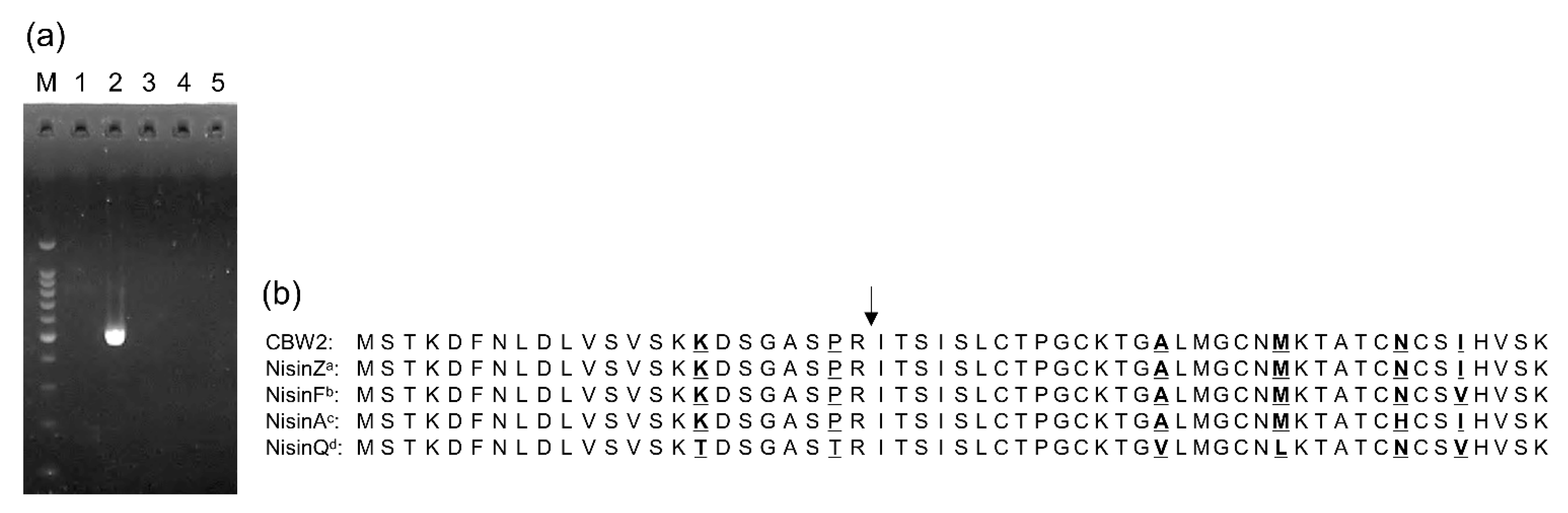
3.5. Genetic Evidence for Nisin Synthesis

We obtained an amplicon only from strain CBW2, the size of which was similar to that found in a previous study [43], using the precursor nisin structural gene (nisA) primer set (Figure 3a). Fifty-seven amino acid residues (MSTKDFNLDLVSVSKKDSGASPRITSISLCTPGCKTGALMGCNMKTATCNCSIHVSK) in the DNA amplicon were deduced, and the predicted RiPP class was lanthipeptideA with a leader peptide (MSTKDFNLDLVSVSKKDSGASPR) and core peptide (ITSISLCTPGCKTGALMGCNMKTATCNCSIHVSK). The amino acid sequences of the nisin prepeptides highlighted that the deduced amino acid sequence from strain CBW2 was 100% coincident with that of nisin Z (Figure 3b). We also obtained amplicons only from strain CBW2 using each primer set targeting nisB, nisC, nisT, nisP, nisF, nisI, and nisRK, and their amplicons were the expected sizes based on a previous study [52] (Table 7, Figure S4).

4. Discussion

We successfully isolated L. lactis subsp. lactis from a fecal sample of a Cuvier’s beaked whale. This is the first study to reveal the characteristics of these whale-derived strains. We obtained L. lactis-specific PCR products from five strains (named CBW1–5), indicating that they are related to L. lactis [22,29]. Bacterial 16S rRNA gene analysis and gadB amplification with subsequent endonuclease digestion genetically identified strains CBW1–5 as L. lactis subsp. lactis. Their phenotypic properties, including the Gram reaction, motility, catalase activity, gas production from glucose, and carbohydrate fermentation profile, were consistent with those of marine-derived L. lactis subsp. lactis [22,53]. Similar to the characteristics of L. lactis subsp. lactis and unlike those of L. lactis subsp. cremoris, the strains could grow at pH 9.2, 40 °C, and in 4% sodium chloride [54]. Hence, the CBW1–5 isolates were identified as L. lactis subsp. lactis. In addition, the results of RAPD-PCR using the M13 primer indicated that the isolated strains were genotypically diverse and not clonal, despite their high 16S rRNA gene sequence similarities against the same bacteria species, i.e., L. lactis subsp. lactis JCM 5805T.
The transmission route of the identified L. lactis subsp. lactis strains to Cuvier’s beaked whale is intriguing. L. lactis strains have been isolated from various specimens, including plants [55], milk, and wastewater tanks [56]. In marine environments, a high-density colony of L. lactis was observed in the clone library of the intestine of grass puffer (Takifugu niphobles) [57], and halotolerant (6% sodium chloride) L. lactis subsp. lactis strains were isolated from coastal fish and clams (Meretrix lamarckii) [22,53]. These marine-derived strains may have diverged from terrestrial-derived strains through food chains and the water cycle [58]. The five L. lactis subsp. lactis strains isolated in this study showed higher halotolerance (up to 7% sodium chloride) than that of strains derived from plants, freshwater fish, and cheese [53,58], indicating their adaptation to the marine environment. In addition, the growth of all five strains at pH 4.0 and 10.0, as well as their carbohydrate fermentation profiles, was consistent with those of marine-derived strains [22,53]. However, they were different from those of freshwater fish-derived strains [21,58] in terms of their ability to ferment trehalose and inulin, and from the cheese-starter culture in their ability to ferment l-arabinose, mannitol, amygdalin, saccharose, and gluconate [22]. L. lactis subsp. lactis has been isolated from marine organisms, such as various fish species and Bivalvia [22,53], whereas Cuvier’s beaked whales found at Suruga Bay, Pacific coast in central Japan, mainly consume squids [59]. Thus, isolation of L. lactis subsp. lactis strains from squids and comparison of their genetic and phenotypic characteristics with those of Cuvier’s beaked whale-derived strains are required to reveal the transmission route of the bacteria.
Only the pH-neutralized, cell-free supernatant of Lactobacilli MRS broth (pH 7.0) obtained from the L. lactis subsp. lactis strain CBW2 inhibited the growth of V. alginolyticus ATCC 17749 and B. subtilis subsp. subtilis ATCC 6051, whereas its antimicrobial activity was abolished or diminished by treatment with proteinase K, trypsin, and pepsin, suggesting that its antimicrobial activity is not pH-related and may be associated with its peptidic nature. In contrast, no loss of activity was observed after treatment with lysozyme, lipase, and catalase, indicating that the inhibitory activity of strain CBW2 was not due to lipid, polysaccharide moieties, or hydrogen peroxide. In addition, strain CBW2 possesses the structural gene for the nisin precursor (nisA), and deduction of its sequence revealed 100% coincidence with that of nisin Z. These results suggest that strain CBW2 produces the precursor of nisin Z-like substances.
Nisin biosynthetic genes, which are located on a conjugative transposon, consist of 11 genes, including nisA, and are typically organized into four operons: nisABTCIPRK, nisI, nisRK, and nisFEG [60]. The genes are involved in nisin maturation (nisBC) [61], export from bacterial cells (nisT) [62], cleaving leader peptide (nisP) [63], self-induction of the nisA promotor by the mature nisin (nisRK) [64], anti-nisin protection (nisI) [65], and anti-nisin immunity function (nisFEG) [66]. Therefore, we examined each gene using previously reported primer sets. We found that strain CBW2 possessed nisin biosynthesis-related gene, nisBTCIPRK, and a nisin immunity-related gene, nisF, which is a part of the downstream nisFEG operon. Taken together, these data indicate that strain CBW2 produces the precursors of nisin Z-like substances, processes the precursors, then releases the mature nisins through exocytosis. In addition, the strain may protect itself from nisin and promote nisin production using the mature nisin product.
In general, nisin shows strong antibacterial effects against Gram-positive bacteria, but not against Gram-negative bacteria unless used in combination with physical treatment or substances, such as ethylenediaminetetraacetic acid, sodium citrate, and trisodium phosphate, to destabilize their outer membrane [9,12]. In contrast, L. lactis subsp. lactis A164 produces a nisin-like bacteriocin that inhibits Salmonella typhimurium (S. typhimurium) [67]. L. lactis WFLU12, which encodes the nisin Z gene cluster and colicin V [68], isolated from the olive flounder (Paralichthys olivaceus), showed antagonistic activity against Edwardsiella tarda (E. tarda) [24]. Kuwano et al. [13] reported that purified nisin Z could permeabilize both Gram-positive and Gram-negative bacterial cytoplasmic membranes, and that nisin’s antibacterial mechanism against Gram-negative bacteria may differ from that against Gram-positive bacteria. However, the detailed mechanism underlying nisin’s inhibition of Gram-negative bacteria has not been fully elucidated. In this study, the Cuvier’s beaked whale-derived L. lactis subsp. lactis strain CBW2 phenotypically and genetically showed the potential to synthesize nisin-like substances that may contribute to inhibiting the growth of V. alginolyticus and B. subtilis subsp. subtilis. The mechanism of V. alginolyticus growth inhibition, however, remains unclear. Purification and characterization studies of the nisin-like substance produced by strain CBW2 will provide useful information on the mechanism underlying nisin’s action against Gram-negative bacteria.

5. Conclusions

This is the first study to isolate and characterize L. lactis subsp. lactis strains from a fecal sample of a whale. The strains’ tolerance to NaCl and carbohydrate metabolism profiles suggest their adaptation to the marine environment and the whale’s conceivable acquirement of the strains through the food chain. The supernatant of strain CBW2 inhibited the growth of Gram-positive and -negative bacteria, but its inhibitory activity was significantly diminished by protease treatment. Furthermore, strain CBW2 possessed the structural gene for the nisin precursor (nisA), which was 100% identical to that for nisin Z, and nisin biosynthesis-related genes (nisBTCIPRK and nisF). These results suggest that strain CBW2 produces nisin-like substances; however, further studies are needed to purify and molecularly characterize the nisin-like substance from strain CBW2 to reveal the mechanism underlying the inhibition of harmful bacteria, particularly Gram-negative bacteria.

Supplementary Materials

The following are available online at https://www.mdpi.com/2076-2607/9/2/243/s1. Table S1: Bacterial strains and the culture temperature and conditions used for the agar well diffusion assay. Table S2: Carbohydrate utilization profiles of the five strains detected in this study and those of reference strains determined using API 50 CH. Figure S1: The sampled stranded female Cuvier’s beaked whale (a) and the location where it was found in Shimizu, Shizuoka, Japan (b). Figure S2: PCR amplification using a universal primer (8F, [28]) and a Lactococcus lactis-specific primer (LacreR, [29]). Lane M: 100-bp ladder marker from 100 to 1000 bp and 1500 bp; lanes 1–5: strains CBW1–5. Figure S3: RAPD-PCR bands patterns of five Lactococcus lactis subsp. lactis strains. Lane M: molecular weight ladder marker; lanes 1–5: strains CBW1–5. Figure S4: PCR results for nisin biosynthesis-related genes in five Lactococcus lactis subsp. lactis strains isolated from the fecal sample of a stranded female Cuvier’s beaked whale. Lane M: 100-bp ladder marker from 100 to 1000 bp and 1500 bp; lanes 1–5: strains CBW1–5.

Author Contributions

Conceptualization, A.S. and M.S.; formal analysis, A.S.; funding acquisition, A.S. and M.S.; investigation, A.S.; resources, A.S.; writing—original draft preparation, A.S.; supervision, M.S.; writing—review and editing, M.S. Both authors have read and agreed to the published version of the manuscript.

Funding

This work was funded by the Grant-in-Aid from the Japan Society for the Promotion of Science (JSPS), a research fellowship for Young Scientists from the Ministry of Education, Culture, Sports, Science, and Technology of Japan (No. 20J10377) awarded to A.S., and the International Joint Research and Training of Young Researchers for Zoonosis Control in the Globalized World grant from the Ministry of Education, Culture, Sports, Science, and Technology of Japan.

Institutional Review Board Statement

Fecal sample collection was conducted under the permission and surveillance of the Fisheries Agency, Tokyo, Japan, and the Public Works Office, Transportation Infrastructure Department of Shizuoka Prefecture, Shizuoka, Japan. All experiments were carried out in compliance with Biosecurity guidelines and approved by the College of Bioresource Sciences, Nihon University (No. BHS15016; approved on 25 July 2020).

Informed Consent Statement

Not applicable.

Data Availability Statement

The 16S rRNA sequences of the five strains obtained in this study are registered in the DDBJ/GenBank/EMBL databases under the accession numbers LC570855–LC570859.

Acknowledgments

We are thankful to Akira Shiozaki of the National Museum of Nature and Science, Ibaraki, Japan, for his assistance with sample collection.

Conflicts of Interest

The authors declare no conflict of interest.

References

  1. Heczko, P.; Strus, M.; Kochan, P. Critical evaluation of probiotic activity. J. Physiol. Pharmacol. 2006, 57, 5–12. [Google Scholar] [PubMed]
  2. Guarner, F.; Khan, A.G.; Garisch, J.; Eliakim, R.; Gangl, A.; Thomson, A.; Krabshuis, J.; Lemair, T.; Kaufmann, P.; de Paula, J.A.; et al. World gastroenterology organisation global guidelines: Probiotics and prebiotics October 2011. J. Clin. Gastroenterol. 2011, 46, 468–481. [Google Scholar] [CrossRef] [PubMed]
  3. Fink, L.N.; Zeuthen, L.H.; Christensen, H.R.; Morandi, B.; Frøkiær, H.; Ferlazzo, G. Distinct gut-derived lactic acid bacteria elicit divergent dendritic cell-mediated NK cell responses. Int. Immunol. 2007, 19, 1319–1327. [Google Scholar] [CrossRef] [PubMed] [Green Version]
  4. Dantas, A.B.; Jesus, V.F.; Silva, R.; Almada, C.N.; Esmerino, E.A.; Cappato, L.P.; Silva, M.C.; Raices, R.S.L.; Cavalcanti, R.N.; Carvalho, C.C.; et al. Manufacture of probiotic minas frescal cheese with Lactobacillus casei Zhang. J. Dairy Sci. 2016, 99, 18–30. [Google Scholar] [CrossRef] [PubMed] [Green Version]
  5. Dal Bello, B.; Rantsiou, K.; Bellio, A.; Zeppa, G.; Ambrosoli, R.; Civera, T.; Cocolin, L. Microbial ecology of artisanal products from North West of Italy and antimicrobial activity of the autochthonous populations. LWT-Food Sci. Technol. 2010, 43, 1151–1159. [Google Scholar] [CrossRef]
  6. Tsuchida, S.; Kakooza, S.; Mbehang Nguema, P.P.; Wampande, E.M.; Ushida, K. Characteristics of gorilla-specific Lactobacillus isolated from captive and wild gorillas. Microorganisms 2018, 6, 86. [Google Scholar] [CrossRef] [PubMed] [Green Version]
  7. Kanamori, Y.; Hashizume, K.; Sugiyama, M.; Morotomi, M.; Yuki, N. Combination therapy with Bifidobacterium breve, Lactobacillus casei, and galactooligosaccharides dramatically improved the intestinal function in a girl with short bowel syndrome: A novel synbiotics therapy for intestinal failure. Dig. Dis. Sci. 2001, 46, 2010–2016. [Google Scholar] [CrossRef]
  8. Liu, Q.; Ni, X.; Wang, Q.; Peng, Z.; Niu, L.; Xie, M.; Lin, Y.; Zhou, Y.; Sun, H.; Pan, K.; et al. Investigation of lactic acid bacteria isolated from Giant Panda Feces for potential probiotics in vitro. Probiotics. Antimicrob. Proteins. 2019, 11, 85–91. [Google Scholar] [CrossRef] [PubMed]
  9. Klaenhammer, T.R. Genetics of bacteriocins produced by lactic acid bacteria. FEMS Microbiol. Rev. 1993, 12, 39–85. [Google Scholar] [CrossRef]
  10. Dobson, A.; Crispie, F.; Rea, M.C.; O’Sullivan, O.; Casey, P.G.; Lawlor, P.G.; Cotter, P.D.; Ross, P.; Gardiner, G.E.; Hill, C. Fate and efficacy of lacticin 3147-producing Lactococcus lactis in the mammalian gastrointestinal tract. FEMS Microbiol. Ecol. 2011, 76, 602–614. [Google Scholar] [CrossRef] [Green Version]
  11. Punyauppa-path, S.; Phumkhachorn, P.; Rattanachaikunsopon, P. Nisin: Production and mechanism of antimicrobial action. Int. J. Curr. Res. Rev. 2015, 7, 47. [Google Scholar]
  12. Schuller, F.; Benz, R.; Sahl, H.G. The peptide antibiotic subtilin acts by formation of voltage-dependent multi-state pores in bacterial and artificial membranes. Eur. J. Biochem. 1989, 182, 181–186. [Google Scholar] [CrossRef] [PubMed]
  13. Kuwano, K.; Tanaka, N.; Shimizu, T.; Nagatoshi, K.; Nou, S.; Sonomoto, K. Dual antibacterial mechanisms of nisin Z against Gram-positive and Gram-negative bacteria. Int. J. Antimicrob. Agents 2005, 26, 396–402. [Google Scholar] [CrossRef] [PubMed]
  14. Zhou, H.; Fang, J.; Tian, Y.; Lu, X.Y. Mechanisms of nisin resistance in Gram-positive bacteria. Ann. Microbiol. 2014, 64, 413–420. [Google Scholar] [CrossRef]
  15. Diaz, M.A.; Bik, E.M.; Carlin, K.P.; Venn-Watson, S.K.; Jensen, E.D.; Jones, S.E.; Gaston, E.P.; Relman, D.A.; Versalovic, J. Identification of Lactobacillus strains with probiotic features from the bottlenose dolphin (Tursiops truncatus). J. Appl. Microbiol. 2013, 115, 1037–1051. [Google Scholar] [CrossRef] [Green Version]
  16. Vela, A.I.; Fernandez, A.; los Monteros, A.E.; Goyache, J.; Herraez, P.; Tames, B.; Cruz, F.; Domínguez, L.; Fernandez-Garayzabal, J.F. Lactobacillus ceti sp. nov., isolated from beaked whales (Ziphius cavirostris). Int. J. Syst. Evol. Microbiol. 2008, 58, 891–894. [Google Scholar] [CrossRef]
  17. Vela, A.I.; Fernández, A.; De Quirós, Y.B.; Herráez, P.; Domínguez, L.; Fernández-Garayzábal, J.F. Weissella ceti sp. nov., isolated from beaked whales (Mesoplodon bidens). Int. J. Syst. Evol. Microbiol. 2011, 61, 2758–2762. [Google Scholar] [CrossRef] [Green Version]
  18. Evans, J.J.; Pasnik, D.J.; Klesius, P.H.; Al-Ablani, S. First report of Streptococcus agalactiae and Lactococcus garvieae from a wild bottlenose dolphin (Tursiops truncatus). J. Wildl. Dis. 2006, 42, 561–569. [Google Scholar] [CrossRef]
  19. Kawanishi, M.; Kojima, A.; Ishihara, K.; Esaki, H.; Kijima, M.; Takahashi, T.; Suzuki, S.; Tamura, Y. Drug resistance and pulsed-field gel electrophoresis patterns of Lactococcus garvieae isolates from cultured Seriola (yellowtail, amberjack and kingfish) in Japan. Lett. Appl. Microbiol. 2005, 40, 322–328. [Google Scholar] [CrossRef]
  20. Pot, B.; Devriese, L.A.; Ursi, D.; Vandamme, P.; Haesebrouck, F.; Kersters, K. Phenotypic identification and differentiation of Lactococcus strains isolated from animals. Syst. Appl. Microbiol. 1996, 19, 213–222. [Google Scholar] [CrossRef]
  21. Sugita, H.; Ohta, K.; Kuruma, A.; Sagesaka, T. An antibacterial effect of Lactococcus lactis isolated from the intestinal tract of the Amur catfish, Silurus asotus Linnaeus. Aquac. Res. 2007, 38, 1002–1004. [Google Scholar] [CrossRef]
  22. Itoi, S.; Abe, T.; Washio, S.; Ikuno, E.; Kanomata, Y.; Sugita, H. Isolation of halotolerant Lactococcus lactis subsp. lactis from intestinal tract of coastal fish. Int. J. Food Microbiol. 2008, 121, 116–121. [Google Scholar] [CrossRef] [PubMed]
  23. Sahnouni, F.; Boutibamaatallah, A.; Bouhadi, D.; Boutiba, Z. Characterization of bacteriocin produced by Lactococcus lactis ssp. lactis strains isolated from marine fish caught in the Algerian west coast. Türk Tarım Doğa Bilimleri Dergisi 2014, 1, 1838–1843. [Google Scholar]
  24. Nguyen, T.L.; Park, C.I.; Kim, D.H. Improved growth rate and disease resistance in olive flounder, Paralichthys olivaceus, by probiotic Lactococcus lactis WFLU12 isolated from wild marine fish. Aquaculture 2017, 471, 113–120. [Google Scholar] [CrossRef]
  25. Klijn, N.; Weerkamp, A.H.; De Vos, W.M. Genetic marking of Lactococcus lactis shows its survival in the human gastrointestinal tract. Appl. Environ. Microbiol. 1995, 61, 2771–2774. [Google Scholar] [CrossRef] [Green Version]
  26. Jepson, P.D.; Baker, J.R.; Kuiken, T.; Simpson, V.R.; Kennedy, S.; Bennett, P.M. Pulmonary pathology of harbour porpoises (Phocoena phocoena) stranded in England. Vet. Rec. 2000, 146, 721–728. [Google Scholar] [CrossRef]
  27. Dalebout, M.L.; Van Helden, A.; Van Waerebeek, K.; Baker, C.S. Molecular genetic identification of southern hemisphere beaked whales (Cetacea: Ziphiidae). Mol. Ecol. 1998, 7, 687–694. [Google Scholar] [CrossRef] [Green Version]
  28. Turner, S.; Pryer, K.M.; Miao, V.P.; Palmer, J.D. Investigating deep phylogenetic relationships among cyanobacteria and plastids by small subunit rRNA sequence analysis 1. J. Eukaryot. Microbiol. 1999, 46, 327–338. [Google Scholar] [CrossRef]
  29. Pu, Z.Y.; Dobos, M.; Limsowtin, G.K.Y.; Powell, I.B. Integrated polymerase chain reaction-based procedures for the detection and identification of species and subspecies of the Gram-positive bacterial genus Lactococcus. J. Appl. Microbiol. 2002, 93, 353–361. [Google Scholar] [CrossRef] [Green Version]
  30. Lane, D.J. 16S/23S rRNA sequencing. In Nucleic Acid Techniques in Bacterial Systematics; Stackebrandt, E., Goodfellow, M., Eds.; John Wiley & Sons: New York, NY, USA, 1991; pp. 115–175. [Google Scholar]
  31. Nomura, M.; Kobayashi, M.; Okamoto, T. Rapid PCR-based method which can determine both phenotype and genotype of Lactococcus lactis subspecies. Appl. Environ. Microbiol. 2002, 68, 2209–2213. [Google Scholar] [CrossRef] [Green Version]
  32. Yoon, S.H.; Ha, S.M.; Kwon, S.; Lim, J.; Kim, Y.; Seo, H.; Chun, J. Introducing EzBioCloud: A taxonomically united database of 16S rRNA gene sequences and whole-genome assemblies. Int. J. Syst. Evol. Microbiol. 2017, 67, 1613–1617. [Google Scholar] [CrossRef] [PubMed]
  33. Thompson, J.D.; Higgins, D.G.; Gibson, T.J. CLUSTAL W: Improving the sensitivity of progressive multiple sequence alignment through sequence weighting, position-specific gap penalties and weight matrix choice. Nucleic Acids Res. 1994, 22, 4673–4680. [Google Scholar] [CrossRef] [PubMed] [Green Version]
  34. Saitou, N.; Nei, M. The neighbor-joining method: A new method for reconstructing phylogenetic trees. Mol. Biol. Evol. 1987, 4, 406–425. [Google Scholar] [CrossRef] [PubMed]
  35. Kimura, M. A simple method for estimating evolutionary rates of base substitutions through comparative studies of nucleotide sequences. J. Mol. Evol. 1980, 16, 111–120. [Google Scholar] [CrossRef] [PubMed]
  36. Kumar, S.; Stecher, G.; Li, M.; Knyaz, C.; Tamura, K. MEGA X: Molecular evolutionary genetics analysis across computing platforms. Mol. Biol. Evol. 2018, 35, 1547–1549. [Google Scholar] [CrossRef] [PubMed]
  37. Nomura, M.; Kimoto, H.; Someya, Y.; Suzuki, I. Novel characteristic for distinguishing Lactococcus lactis subsp. lactis from subsp. cremoris. Int. J. Syst. Evol. Microbiol. 1999, 49, 163–166. [Google Scholar] [CrossRef] [Green Version]
  38. Huey, B.I.N.G.; Hall, J.E.F.F. Hypervariable DNA fingerprinting in Escherichia coli: Minisatellite probe from bacteriophage M13. J. Bacteriol. 1989, 171, 2528–2532. [Google Scholar] [CrossRef] [Green Version]
  39. Ryu, E. A simple method of differentiation between Gram-positive and Gram-negative organisms without staining. Kitasato Arch. Exp. Med. 1940, 17, 58–63. [Google Scholar]
  40. Neveling, D.P.; Ahire, J.J.; Laubscher, W.; Rautenbach, M.; Dicks, L.M. Genetic and phenotypic characteristics of a multi-strain probiotic for broilers. Curr. Microbiol. 2020, 77, 369–387. [Google Scholar] [CrossRef]
  41. Artimo, P.; Jonnalagedda, M.; Arnold, K.; Baratin, D.; Csardi, G.; De Castro, E.; Duvaud, S.; Flegel, V.; Fortier, A.; Gasteiger, E.; et al. ExPASy: SIB bioinformatics resource portal. Nucleic Acids Res. 2012, 40, 597–603. [Google Scholar] [CrossRef]
  42. Agrawal, P.; Khater, S.; Gupta, M.; Sain, N.; Mohanty, D. RiPPMiner: A bioinformatics resource for deciphering chemical structures of RiPPs based on prediction of cleavage and cross-links. Nucleic Acids Res. 2017, 45, 80–88. [Google Scholar] [CrossRef] [PubMed] [Green Version]
  43. Alegría, Á.; Delgado, S.; Roces, C.; López, B.; Mayo, B. Bacteriocins produced by wild Lactococcus lactis strains isolated from traditional, starter-free cheeses made of raw milk. Int. J. Food Microbiol. 2010, 143, 61–66. [Google Scholar] [CrossRef] [PubMed] [Green Version]
  44. Martinez, B.; Suarez, J.E.; Rodriguez, A. Antimicrobials produced by wild lactococcal strains isolated from homemade cheeses. J. Food Prot. 1995, 58, 1118–1123. [Google Scholar] [CrossRef] [PubMed]
  45. Martínez-Cuesta, M.C.; Kok, J.; Herranz, E.; Peláez, C.; Requena, T.; Buist, G. Requirement of autolytic activity for bacteriocin-induced lysis. Appl. Environ. Microbiol. 2000, 66, 3174–3179. [Google Scholar] [CrossRef] [PubMed] [Green Version]
  46. Martinez, B.; Fernandez, M.; Suarez, J.E.; Rodriguez, A. Synthesis of lactococcin 972, a bacteriocin produced by Lactococcus lactis IPLA 972, depends on the expression of a plasmid-encoded bicistronic operon. Microbiology 1999, 145, 3155–3162. [Google Scholar] [CrossRef] [Green Version]
  47. Van Belkum, M.J.; Hayema, B.J.; Jeeninga, R.E.; Kok, J.; Venema, G. Organization and nucleotide sequences of two lactococcal bacteriocin operons. Appl. Environ. Microbiol. 1991, 57, 492–498. [Google Scholar] [CrossRef] [PubMed] [Green Version]
  48. Olasupo, N.A.; Schillinger, U.; Narbad, A.; Dodd, H.; Holzapfel, W.H. Occurrence of nisin Z production in Lactococcus lactis BFE 1500 isolated from wara, a traditional Nigerian cheese product. Int. J. Food Microbiol. 1999, 53, 141–152. [Google Scholar] [CrossRef]
  49. Li, H.; O’Sullivan, D.J. Heterologous expression of the Lactococcus lactis bacteriocin, nisin, in a dairy Enterococcus strain. Appl. Environ. Microbiol. 2002, 68, 3392–3400. [Google Scholar] [CrossRef] [Green Version]
  50. Ghrairi, T.; Manai, M.; Berjeaud, J.M.; Frere, J. Antilisterial activity of lactic acid bacteria isolated from rigouta, a traditional Tunisian cheese. J. Appl. Microbiol. 2004, 97, 621–628. [Google Scholar] [CrossRef]
  51. Veljovic, K.; Terzic-Vidojevic, A.; Vukasinovic, M.; Strahinic, I.; Begovic, J.; Lozo, J.; Ostojic, M.; Topisirovic, L. Preliminary characterization of lactic acid bacteria isolated from Zlatar cheese. J. Appl. Microbiol. 2007, 103, 2142–2152. [Google Scholar] [CrossRef]
  52. Fusieger, A.; Perin, L.M.; Teixeira, C.G.; de Carvalho, A.F.; Nero, L.A. The ability of Lactococcus lactis subsp. lactis bv. diacetylactis strains in producing nisin. Antonie van Leeuwenhoek 2019, 113, 651–662. [Google Scholar] [CrossRef] [PubMed]
  53. Itoi, S.; Uchida, J.; Takanashi, S.; Narita, T.; Abe, K.; Naya, S.; Sugita, H. The clam Meretrix lamarckii (Bivalvia: Veneridae) is a rich repository of marine lactic acid bacterial strains. Ann. Microbiol. 2014, 64, 1267–1274. [Google Scholar] [CrossRef]
  54. Teuber, M.; Geis, A. The genus lactococcus. In The Prokaryotes, 3rd ed.; Dworkin, M., Falkow, S., Rosenberg, E., Schleifer, K.-H., Stackebrandt, E., Eds.; Springer: New York, NY, USA, 2006; Volume 4, pp. 205–228. [Google Scholar]
  55. Kimoto, H.; Nomura, M.; Kobayashi, M.; Okamoto, T.; Ohmomo, S. Identification and probiotic characteristics of Lactococcus strains from plant materials. Jpn. Agric. Res. Q. 2004, 38, 111–117. [Google Scholar] [CrossRef] [Green Version]
  56. Klijn, N.; Weerkamp, A.H.; De Vos, W.M. Detection and characterization of lactose-utilizing Lactococcus spp. in natural ecosystems. Appl. Environ. Microbiol. 1995, 61, 788–792. [Google Scholar] [CrossRef] [PubMed] [Green Version]
  57. Shiina, A.; Itoi, S.; Washio, S.; Sugita, H. Molecular identification of intestinal microflora in Takifugu niphobles. Comp. Biochem. Physiol. Part D Genom. Proteom. 2006, 1, 128–132. [Google Scholar] [CrossRef]
  58. Itoi, S.; Yuasa, K.; Washio, S.; Abe, T.; Ikuno, E.; Sugita, H. Phenotypic variation in Lactococcus lactis subsp. lactis isolates derived from intestinal tracts of marine and freshwater fish. J. Appl. Microbiol. 2009, 107, 867–874. [Google Scholar] [CrossRef]
  59. Ohizumi, H.; Kishiro, T. Stomach contents of a Cuviers beaked whale (Ziphius cavirostris) stranded on the central Pacific coast of Japan. Aquat. Mamm. 2003, 29, 99–103. [Google Scholar] [CrossRef]
  60. Lubelski, J.; Rink, R.; Khusainov, R.; Moll, G.N.; Kuipers, O.P. Biosynthesis, immunity, regulation, mode of action and engineering of the model lantibiotic nisin. Cell. Mol. Life Sci. 2008, 65, 455–476. [Google Scholar] [CrossRef] [Green Version]
  61. Khusainov, R.; Heils, R.; Lubelski, J.; Moll, G.N.; Kuipers, O.P. Determining sites of interaction between prenisin and its modification enzymes NisB and NisC. Mol. Microbiol. 2011, 82, 706–718. [Google Scholar] [CrossRef]
  62. Qiao, M.; Saris, P.E. Evidence for a role of NisT in transport of the lantibiotic nisin produced by Lactococcus lactis N8. FEMS Microbiol. Lett. 1996, 144, 89–93. [Google Scholar] [CrossRef]
  63. Lagedroste, M.; Smits, S.H.; Schmitt, L. Substrate specificity of the secreted nisin leader peptidase NisP. Biochemistry 2017, 56, 4005–4014. [Google Scholar] [CrossRef] [PubMed]
  64. Cheigh, C.I.; Pyun, Y.R. Nisin biosynthesis and its properties. Biotechnol. Lett. 2005, 27, 1641–1648. [Google Scholar] [CrossRef]
  65. Siegers, K.; Entian, K.D. Genes involved in immunity to the lantibiotic nisin produced by Lactococcus lactis 6F3. Appl. Environ. Microbiol. 1995, 61, 1082–1089. [Google Scholar] [CrossRef] [PubMed] [Green Version]
  66. AlKhatib, Z.; Lagedroste, M.; Zaschke, J.; Wagner, M.; Abts, A.; Fey, I.; Kleinschrodt, D.; Smits, S.H. The C-terminus of nisin is important for the ABC transporter Nis FEG to confer immunity in Lactococcus lactis. Microbiologyopen 2014, 3, 752–763. [Google Scholar] [CrossRef] [PubMed]
  67. Choi, H.J.; Cheigh, C.I.; Kim, S.B.; Pyun, Y.R. Production of a nisin-like bacteriocin by Lactococcus lactis subsp. lactis A164 isolated from Kimchi. J. Appl. Microbiol. 2000, 88, 563–571. [Google Scholar] [CrossRef] [PubMed] [Green Version]
  68. Nguyen, T.L.; Kim, D.H. Genome-wide comparison reveals a probiotic strain Lactococcus lactis WFLU12 isolated from the gastrointestinal tract of olive flounder (Paralichthys olivaceus) harboring genes supporting probiotic action. Mar. Drugs 2018, 16, 140. [Google Scholar] [CrossRef] [PubMed] [Green Version]
Figure 1. Phylogenetic tree generated using the neighbor-joining method and Kimura’s two-parameter model based on the 16S rRNA gene sequences (1326 bp) representing the relationships between the isolated strains CBW1–5 and related species in the genus Lactococcus. The accession numbers are shown inside the parentheses. Only >50% bootstrap values based on 1000 replicates are shown. The scale bar represents the evolutionary distance of the nucleotide substitutions per site. Some species in the genus Leuconostoc are used as an outgroup.
Microorganisms 09 00243 g001
Figure 2. PCR amplification (a) and digestion with AseI (b) of the Lactococcus lactis subsp. lactis gadB gene. Lane M shows the 100-bp ladder marker from 100 to 1000 bp and 1500 bp and lanes 1–5 represent the CBW1, CBW2, CBW3, CBW4, and CBW5 strains, respectively.
Microorganisms 09 00243 g002
Figure 3. Specific PCR amplification of the structural gene for the nisin precursor (nisA) (a) and alignment of the proposed prepeptide sequences of the nisin variant (b). Lane M shows the 100-bp ladder marker from 100 to 1000 bp and 1500 bp and lanes 1–5 represent the CBW1, CBW2, CBW3, CBW4, and CBW5 strains, respectively. The National Center for Biotechnology Information (NCBI) GenBank accession numbers are: a CAA79467, b ABU45463, c CAA48380, and d BAG71479. Vertical arrow indicates a processing site. Substituted residues are marked in bold and underlined.
Microorganisms 09 00243 g003
Table 1. PCR primers used in this study.
UsePrimer NameGeneSequence (5′ to 3′)Reference
Whale species identificationt-Pro whalemitochondrial DNA D-loop regionTCACCCAAAGCTGRARTTCTA[27]
Dlp5CCATCGWGATGTCTTATTTAAGRGGAA
Lactococcus lactis-specific PCR8F16S rRNAAGAGTTTGATCCTGGCTCAG[28]
LacreRGGGATCATCTTTGAGTGAT[29]
Bacterial identification and phylogenetic analysis27F16S rRNAAGAGTTTGATCMTGGCTCAG[30]
1492RTACGGYTACCTTGTTACGACTT
Classification of Lactococcus lactis subspeciesgadB21gadBCGTTATGGATTTGATGGATATAAAGC[31]
GAD7ACTCTTCTTAAGAACAAGTTTAACAGC
Table 2. Bacterial strains used for agar well diffusion assay and their sensitivity to the cell-free supernatant (pH 7.0) of five Lactococcus lactis subsp. lactis strains isolated from the fecal sample of a stranded female Cuvier’s beaked whale a.
Antimicrobial Activity of the Cell-Free Supernatant (pH 7.0)
Indicator Bacteria SpeciesStrainCBW1CBW2CBW3CBW4CBW5
Vibrio alginolyticusATCC 17749++
Vibrio parahaemolyticusATCC 17802
Escherichia coliDSM 30083
Photobacterium damselae subsp. damselaeDSM 7482
Lactococcus lactis subsp. lactisATCC 19435
Lactococcus lactis subsp. cremorisATCC 19257
Lactococcus garvieaeATCC 43921
Lactococcus plantarumATCC 43199
Lactococcus raffinolactisATCC 43920
Enterococcus faecalisDSM 20478
Enterococcus hiraeATCC 8043
Enterococcus faeciumATCC 19434
Enterococcus canisDSM 17029
Staphylococcus xylosusATCC 29971
Staphylococcus epidermidisATCC 14990
Bacillus subtilis subsp. subtilisATCC 6051+
Streptococcus salivariusDSM 20560
a −, no inhibition; +, inhibition zone diameter between 6 and 10 mm; ++, inhibition zone diameter exceeding 10 mm.
Table 3. PCR primers used to detect common bacteriocins produced by L. lactis strains and nisin biosynthesis-related genes.
Primer NameGeneSequence (5′ to 3′)Reference
Nis-FnisACGGCTCTGATTAAATTCTGAAG[43]
Nis-RGGATTAGCTAGTAGTAACTGTTC
Lact481-FLacticin 481TCTGCACTCACTTCATTAGTTA[44]
Lact482-RAAGGTAATTACACCTCTTTTAT
Lact3147-FLacticin 3147TACTGGGGAAATAACGG[45]
Lact3148-RTGGACAAGTATTGGTAC
Lcn972-FLactococcin 972TTGTAGCTCCTGCAGAAGGAACATGG[46]
Lcn973-RGCCTTAGCTTTGAATTCTTACCAAAAG
LactABM-FLactococcin A, B, and MGAAGAGGCAATCAGTAGAG[43,47]
LactA-RLactococcin A geneGTGTTCTATTTATAGCTAATG[43]
LactB-RLactococcin B geneCCAGGATTTTCTTTGATTTACTTC[43]
LactM-RLactococcin M geneGTGTACTGGTCTAGCATAAG[47]
p4nisBAGAGAAGTTATTTACGATCAAC[48]
P5ATCTGACAACAAATCTTTTTGT
p6nisCTTCAGAGCAATATGAGG[48]
p7TATTAAGGCCACAATAAG
P06-FnisTGAAGAATACATGAAATGAGG[49]
P06-RTAACTTTCCAGCTGTCCC
P08-FnisIATTGTGGCCTTAATAGGG[49]
P08-RTAGCGACTTGTCAGAAGC
P11-FnisFCAGGTGCTACAAGATATCAG[49]
P11-RACAACTCCGCAATACCATCAG
Prim-NisP5nisPGGATTTGGTATCTGTTTCGAAG[50]
Prim-NisP3TCTTTCCCATTAACTTGTACTGTG
Nis3nisRKCAGTGCCATGGGTAAAAAATATTCAATGCG[51]
Nis4CTTAGAGAATTCTCTAATGAG
Table 4. Nearest phylogenetic neighbor of the five bacterial strains isolated from the fecal sample of a stranded Cuvier’s beaked whale according to the EzBioCloud 16S rRNA sequences database.
StrainSequence Length (bp)Nearest Phylogenetic NeighborSimilarity (%)Completeness (%)
CBW11420Lactococcus lactis subsp. lactis JCM 5805T99.9396.3
CBW2141499.6595.9
CBW3141299.8695.8
CBW4136899.7892.2
CBW5137699.0491.8
Table 5. Phenotypic characteristics of the five Lactococcus lactis subsp. lactis strains isolated from the fecal sample of a stranded female Cuvier’s beaked whale a.
CharacteristicsCBW1CBW2CBW3CBW4CBW5
Gram reaction+++++
Motility
Catalase
Gas from glucose
Final pH b4.254.314.324.294.34
Growth
NaCl (%)
1+++++
2+++++
3+++++
4+++++
5+++++
6+++++
7+++++
8
9
10
15
Temperature (°C)
10+++++
40+++++
45
pH
4.0+++++
9.2+++++
10.0+++++
a +, positive; −, negative. b Final pH of Lactobacilli MRS broth was measured after two days of incubation.
Table 6. Enzyme treatment effects on antimicrobial activity of the pH-neutralized, cell-free supernatant of strain CBW2 against Vibrio alginolyticus ATCC 17749 and Bacillus subtilis subsp. subtilis ATCC 6051.
Diameter of the Inhibition Zone ± SD (mm)
EnzymeVibrio alginolyticusBacillus subtilis subsp. subtilis
Control15.13 ± 0.158.38 ± 0.34
Proteinase K0.00 ± 0.00 **0.00 ± 0.00 **
Trypsin8.80 ± 0.20 **7.86 ± 0.10 *
Pepsin10.13 ± 0.12 **7.87 ± 0.25 *
Lysozyme15.13 ± 0.31 N.S.8.23 ± 0.16 N.S.
Catalase14.97 ± 0.25 N.S.8.30 ± 0.20 N.S.
Lipase14.83 ± 0.15 N.S.8.26 ± 0.25 N.S.
* and ** represent statistically significant differences compared with the control at p < 0.05 and < 0.01, respectively. N.S. indicates no significant differences.
Table 7. PCR results for nisin biosynthesis-related genes in five Lactococcus lactis subsp. lactis strains isolated from the fecal sample of a stranded female Cuvier’s beaked whale a.
GeneCBW1CBW2CBW3CBW4CBW5
nisA+
nisB+
nisC+
nisT+
nisI+
nisF+
nisP+
nisRK+
a +, positive; −, negative.
Publisher’s Note: MDPI stays neutral with regard to jurisdictional claims in published maps and institutional affiliations.

