Article

Sulfur and Methane-Oxidizing Microbial Community in a Terrestrial Mud Volcano Revealed by Metagenomics

by
Andrey V. Mardanov
,
Vitaly V. Kadnikov
,
Alexey V. Beletsky
and
Nikolai V. Ravin
*
Research Center of Biotechnology of the Russian Academy of Sciences, Institute of Bioengineering, 119071 Moscow, Russia
*
Author to whom correspondence should be addressed.
Microorganisms 2020, 8(9), 1333; https://doi.org/10.3390/microorganisms8091333
Submission received: 30 July 2020 / Revised: 27 August 2020 / Accepted: 28 August 2020 / Published: 31 August 2020
(This article belongs to the Special Issue Microbial Diversity in Extreme Environments)

Abstract

:
Mud volcanoes are prominent geological structures where fluids and gases from the deep subsurface are discharged along a fracture network in tectonically active regions. Microbial communities responsible for sulfur and methane cycling and organic transformation in terrestrial mud volcanoes remain poorly characterized. Using a metagenomics approach, we analyzed the microbial community of bubbling fluids retrieved from an active mud volcano in eastern Crimea. The microbial community was dominated by chemolithoautotrophic Campylobacterota and Gammaproteobacteria, which are capable of sulfur oxidation coupled to aerobic and anaerobic respiration. Methane oxidation could be enabled by aerobic Methylococcales bacteria and anaerobic methanotrophic archaea (ANME), while methanogens were nearly absent. The ANME community was dominated by a novel species of Ca. Methanoperedenaceae that lacked nitrate reductase and probably couple methane oxidation to the reduction of metal oxides. Analysis of two Ca. Bathyarchaeota genomes revealed the lack of mcr genes and predicted that they could grow on fatty acids, sugars, and proteinaceous substrates performing fermentation. Thermophilic sulfate reducers indigenous to the deep subsurface, Thermodesulfovibrionales (Nitrospirae) and Ca. Desulforudis (Firmicutes), were found in minor amounts. Overall, the results obtained suggest that reduced compounds delivered from the deep subsurface support the development of autotrophic microorganisms using various electron acceptors for respiration.

1. Introduction

Mud volcanoes are surface geological features representing the expression of a fracture network that often extends to several kilometers in depth [1]. They are usually found along fracture or fault zones associated with compressional tectonic regions [2,3,4]. Mud volcanoes can be diverse in form but are typically cone-shaped circular structures or pools, from which gaseous mud slurries are discharged, in some cases containing hydrocarbons [2,3]. Methane and CO2 appear to constitute the gaseous phase emitted from most mud volcanoes [5]. The contribution of mud volcanoes and seepages to the global greenhouse gas emissions is significant and, according to recent estimates, account for approximately 30% of the natural methane emission [6].
Mud volcanoes attracted the attention of microbiologists because they provide easy access to the deep subsurface biosphere since relatively rapid fluid transport in a fracture network enables limited alteration of fluid and gas until it is discharged to the surface [2]. Microbial communities associated with mud volcanoes located at the seafloor have been extensively studied [7,8,9,10,11,12]. More limited number of studies has focused on microbial communities in terrestrial mud volcanoes [13,14,15,16,17,18,19]. These studies revealed that archaeal communities are mostly composed of methanogens and anaerobic methanotrophs (ANME), while bacterial communities are more diverse and consist of members of various phyla. Among them, sulfate-reducing bacteria, presumably originating from deep subsurface fluids, were found at various terrestrial mud volcano sites [14,16,17,18,20]. Sulfate-reducers can form syntrophic consortia with anaerobic methanotrophic archaea (ANME) performing anaerobic oxidation of methane (AOM) [8,21]. AOM can also be coupled with the reduction of iron, manganese, nitrate, and nitrite [22,23,24]. Consistently, the composition of both archaeal and bacterial communities strongly depends on the concentrations of these potential electron acceptors [17,18].
The mud volcanoes selected for this study are located in the Kerch Peninsula, where the south-eastern end of Crimea extends between the Sea of Azov and the Black Sea. This region, known as the Kerch-Taman mud volcanic province, is one of the most manifested mud volcanism areas located in the Caucasus segment of the Alpine Himalayan belt [3]. This province includes more than 100 active mud volcanoes. Among them, there are episodically active violent mud volcanoes (up to 60 m high), continuously erupting small cone-shaped circular structures, and bubbling mud pools [25]. Until now, microbial communities of the mud volcanoes of the Kerch Peninsula have not been characterized.
The aim of this study was to uncover the composition and functional potential of a mud volcano microbial community. We report data on high-throughput sequencing of 16S rRNA gene amplicons and assembled metagenome of mud fluid. We successfully recovered high-quality metagenome-assembled genomes (MAGs) of the majority of the community members, which enabled accurate metabolic reconstruction of the microbial community, and revealed the primary microbial drivers of methane and sulfur cycling.

2. Materials and Methods

2.1. Sampling, Field Measurements, Chemical Analyses, and DNA Isolation

The mud volcano sampled in this study is located on the Bulganak mud volcano field, in the Bondarenkovo Village area, near the city of Kerch (45.4261° N 36.4780° E). Mud samples were collected on 18 May 2019, from a ‘bubbling pool’ in the central crater-like structure of the volcano. Samples were collected at a depth of 10–20 cm below the surface of the liquid into sterile 50 mL tubes avoiding gas bubbles. The temperature, pH, and Eh of the recovered fluids were determined on-site using pH-meter HI 8314 (Hanna Instruments, Germany). For chemical analysis, the mud samples were centrifuged at 12,000× g for 10 min. The supernatant was filtered through a 0.22-µm sterile filter (Merck Millipore, Darmstadt, Germany) and analyzed by inductively coupled plasma mass spectrometry (ICP-MS) and ion chromatography. Total DNA was extracted from 15 mL of the mud slurry using the DNeasy PowerMax Soil Kit (Qiagen, Hilden, Germany). About 30 ng of DNA was obtained.

2.2. 16S rRNA Gene Sequencing and Analysis

PCR amplification of 16S ribosomal RNA gene fragments spanning the V3–V6 variable regions was carried out using the universal primers 341F (5′-CCTAYGGGDBGCWSCAG-3′) and 806R (5′-GGACTACNVGGGTHTCTAAT-3′) [26]. PCR fragments were barcoded using the Nextera XT Index Kit v.2 (Illumina, San Diego, CA, USA). The PCR fragments were purified using Agencourt AMPure beads (Beckman Coulter, Brea, CA, USA) and quantitated using the Qubit dsDNA HS Assay Kit (Invitrogen, Carlsbad, CA, USA). Then, all of the amplicons were pooled together in equimolar amounts and sequenced on the Illumina MiSeq (2 × 300 nt paired-end reads). Paired-end overlapping reads were merged using FLASH v.1.2.11 [27]. The sequences were clustered into operational taxonomic units (OTUs) at 97% identity using the USEARCH program [28]; low quality reads, chimeric sequences, and singletons were removed during clustering by the USEARCH algorithm. To calculate OTU abundances, all reads (including singleton and low-quality reads) were mapped to OTU sequences at 97% global identity threshold by Usearch. The taxonomic identification of OTUs was performed by searches against the SILVA v.132 rRNA sequence database using the VSEARCH sintax algorithm [29]. The OTU sequences are provided in Supplementary Table S1.

2.3. Sequencing of Metagenomic DNA, Assembly, and Annotation of MAGs

Metagenomic DNA was sequenced using the Illumina HiSeq2500 platform according to the manufacturer’s instructions (Illumina). The sequencing of a paired-end (2 × 150 bp) TruSeq DNA library generated 59,783,865 read pairs. Adapter removal and trimming of low-quality sequences (Q < 33) were performed using Cutadapt v.1.8.3 [30] and Sickle v.1.33 [31], respectively. Trimmed reads were merged using FLASH v.1.2.11 [27]. The resulting merged and unmerged reads (about 12.8 Gbp in total) were de novo assembled into contigs using metaSPAdes genome assembler v.3.13.0 [32].
Contigs longer than 1500 bp were binned into clusters representing MAGs using CONCOCT v.0.4.1 [33] and MetaBAT v.2.12.1 [34]. The completeness of the MAGs and their possible contamination (redundancy) were estimated using CheckM v.1.05 [35] with lineage-specific marker genes. The assembled MAGs were taxonomically classified using the Genome Taxonomy Database Toolkit (GTDB-Tk) v.0.3.2 and Genome Taxonomy database (GTDB) [36]. The contig to MAG binning scheme was selected manually by comparing the results from MetaBAT and CONCOCT.
Gene search and annotation of MAGs were performed using the National Center for Biotechnology Information (NCBI) Prokaryotic Genome Annotation Pipeline [37] or RAST server 2.0 [38], followed by manual correction of the annotation by comparing the predicted protein sequences with the NCBI databases.

2.4. Genome -to-Genome Distance Estimation and Phylogenetic Analysis

The average amino acid identity (AAI) and average nucleotide identity (ANI) between the selected genomes was calculated using scripts from the Enveomics Collection [39].
GTDB-Tk v.0.3.2 was used to find single-copy marker genes in the MAGs and to construct a multiple sequence alignment of concatenated single-copy gene sequences, comprising of those from a given MAG and all species from the GTDB. A portion of the multiple sequence alignment generated in GTDB-Tk was used to construct a phylogenetic tree with PhyML v.3.3 [40] using default parameters. The level of support for internal branches was assessed using the Bayesian test in PhyML.

2.5. Nucleotide Sequence Accession Numbers

The raw data generated from 16S rRNA gene sequencing and metagenome sequencing have been deposited in the NCBI Sequence Read Archive (SRA) under the accession numbers SRR11917417 and SRR11917416, respectively (BioProject PRJNA636628). The annotated sequences of MAGs have been deposited in the GenBank database and are accessible via the BioProject PRJNA636628.

3. Results and Discussion

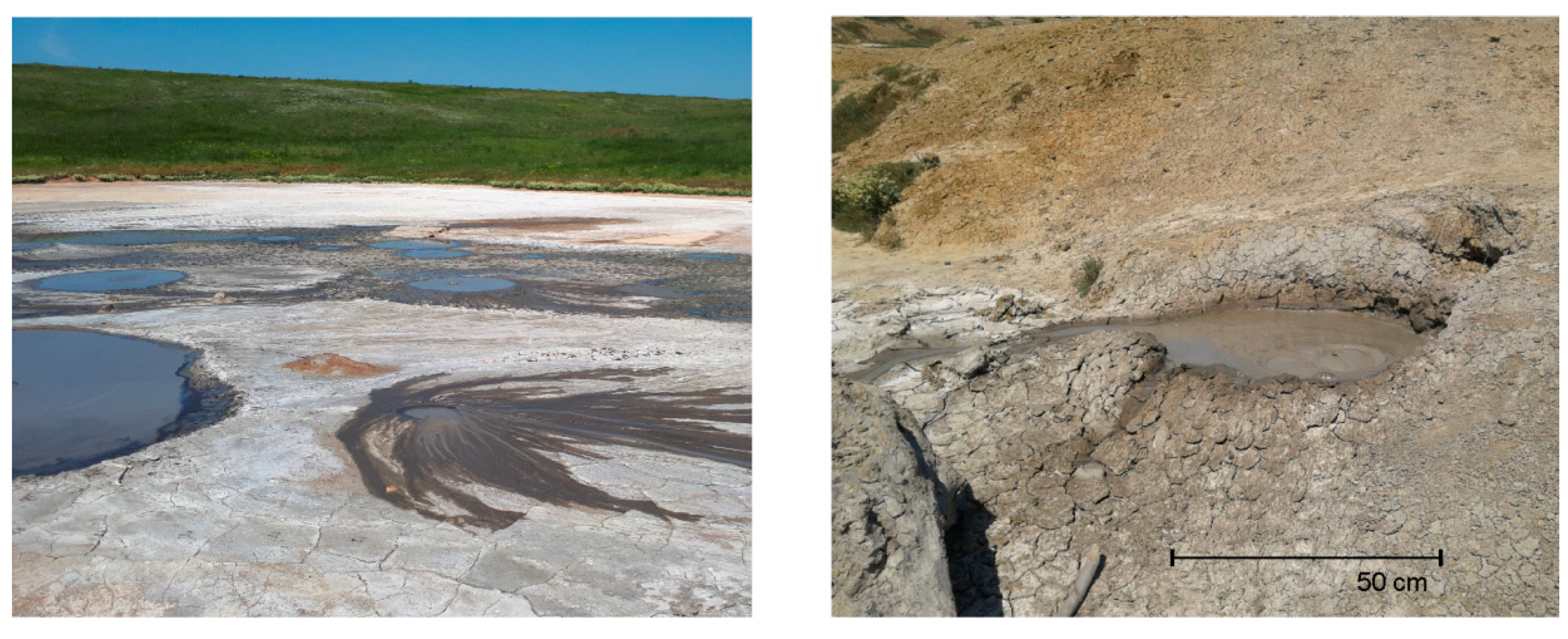
3.1. Study Site and Fluid Chemistry

The study site was a mud volcano located on the Bulganak mud volcano field near the city of Kerch. Here, there are more than ten mud volcanoes of two types: relatively large flat mud pools up to 15 m in diameter with liquid mud, and small conical volcanoes with more dense mud. The mud volcano in this study was a cone-shaped structure with a diameter of about 2 m and a height of about 50 cm. The top of the volcano had a central crater-like depression (50–70 cm in diameter), in which bubbling and fluid discharge out of the cone was observed (Figure 1).
The mud temperature was about 16 °C, a value consistent with the origin of the fluid from the subsurface. The fluid had a near neutral pH (about 7.43) and was moderately reduced (Eh = −123 mV). The chemical characteristics of the water extracted from the mud sample are shown in Table 1. The major cation was sodium, followed by boron, calcium, and potassium. Chloride and carbonate were dominant anions, while the concentration of sulfate was rather low (6.1 mg L−1). The mass concentration of nitrate, another potential electron acceptor for anaerobic respiration, was nearly twice as high as sulfate.

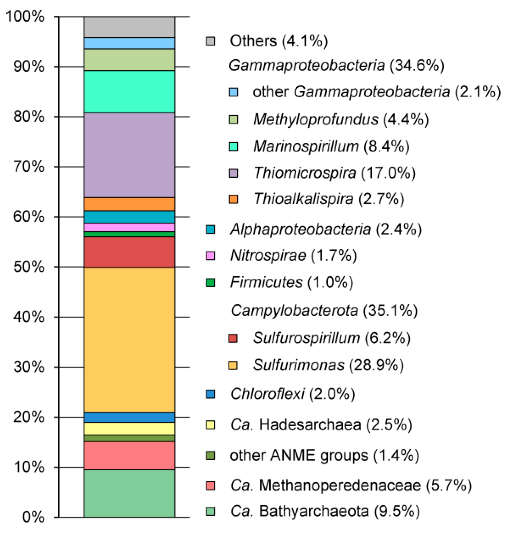
3.2. Microbial Community Structure Revealed by 16S rRNA Profiling

A total of 82,023 high-quality 16S rRNA reads were used to analyze microbial community composition. The pool of reads retrieved from the mud sample was dominated by 16S rRNA gene sequences of bacterial origin (79.7% of the total reads), and archaeal 16S rRNA gene reads accounted for 19.1% of the total reads. About 1.2% of read were assigned to chloroplasts probably appeared as a result of the ingress of allochthonous biomaterial from the surrounding area. The results of the taxonomic classification of the OTUs are shown in Figure 2 and the Supplemental Table S1.
The archaeal population was represented by members of Candidatus (Ca.) Bathyarchaeota (9.5% of the total reads), Euryarchaeota (Methanomicrobia at 7.0% and Halobacteria at 0.05%), and Ca. Hadesarchaea (2.5%). Nearly all Euryarchaeota belonged to lineages known to be involved in anaerobic methane oxidation, namely, Ca. Methanoperedenaceae (5.7%), ANME-3 (0.6%), ANME-2a-2b (0.5%), and ANME-1 (0.3%). Most of Ca. Bathyarchaeota was represented by two OTUs. A GenBank search revealed 16S rRNA sequences closely related (>98% identity) to both of these OTUs in terrestrial and marine mud volcanoes, groundwater, and marine sediments.
The phylum Ca. Hadesarchaea was represented by two OTUs. 16S rRNA gene sequences that were similar to both of them, as revealed by GenBank search, were detected in mud volcanoes worldwide.
The most abundant bacterial lineage, Campylobacterota (formerly Epsilonproteobacteria), accounted for 35.1% of the community and was represented by the genera Sulfurimonas (28.9%) and Sulfurospirillum (6.2%). Sulfurimonas species are chemolithoautotrophs that are typically found in sulfidic environments such as hydrothermal vents, marine sediments, sulfidic springs, and groundwater [41,42]. They can oxidize various reduced sulfur compounds (some species also hydrogen) using oxygen, nitrate, or nitrite as electron acceptors. Metabolically versatile Sulfurospirillum species can use various electron acceptors, including oxygen, nitrate, arsenate, sulfur, and halogenated compounds [43].
The second most frequent lineage was Gammaproteobacteria at an abundance of 34.6%, represented by the genera Thiomicrospira (17.0%), Marinospirillum (8.4%), Methyloprofundus (4.4%), Thioalkalispira (2.7%), Marinobacter (1.0%), Halomonas (0.6%), and Methylomicrobium (0.2%). Cultured members of the genera Thiomicrospira and Thioalkalispira are sulfur-oxidizing chemolithoautotrophs [44,45]. The genus Methyloprofundus currently consists of a single species, Methyloprofundus sedimenti, an obligate methanotroph from oceanic sediments [46]. The heterotrophic part of the community was represented by Marinospirillum, Marinobacter, and Halomonas, which are chemoorganotrophic aerobic halophilic bacteria that are typically found in saline environments, such as soda lakes, marine sediments, and saline soil [47,48,49,50].
About 2.4% of the 16S rRNA reads were assigned to Alphaproteobacteria of the family Rhodobacteraceae that is mostly comprised of aerobic heterotrophs. Chloroflexi, assigned to uncultured lineages of the classes Anaerolineae and Dehalococcoidia, was present at 2.0%. The phylum Nitrospirae (1.7%) was represented by a single OTU that was phylogenetically distant from cultured sulfate-reducing species of Thermodesulfovibrio. Among other recognized bacterial lineages, members of Aminicenantes, Actinobacteria, Aerophobetes, Atribacteria, Bacteroidetes, Betaproteobacteria, Deltaproteobacteria, Firmicutes, and Spirochaetes were found in minor amounts (<1% of reads).

3.3. Metagenome Sequencing and Assembly of MAGs

To assemble the composite genomes of the most abundant members of the microbial community, we sequenced the metagenome of the mud sample using the Illumina HiSeq2500 platform. A total of 12.8 Gbp of metagenomic sequences were assembled into contigs. These contigs were binned into 35 MAGs with more than 90% completeness and less than 10% contamination (Kmv03 to 05, Kmv11 to 37, and Kmv39 to 43), as estimated based on the presence of a set of single-copy conserved genes by CheckM (Supplementary Table S2).
Taxonomic classification of the MAGs based on the searches against GTDB [36] revealed the same major prokaryotic lineages that were detected by 16S rRNA profiling. To get insights into the metabolic capabilities of the microbial lineages that were most abundant in the microbiome or involved in important biotransformations, we analyzed several of the MAGs in detail.

3.4. Sulfur-Oxidizing Bacteria

Six MAGs were assigned to lineages of Campylobacterota and Gammaproteobacteria, which potentially oxidize reduced sulfur compounds. MAG Kmv11 was classified as a member of Sulfurimonas, and the less abundant MAG Kmv12, according in the GTDB taxonomy, represented genus-level division GCA-2733885; both are in the Sulfurimonadaceae family. Analysis of the Kmv11 genome revealed a set of genes described in other Sulfurimonas genomes that enable oxidation of sulfur compounds [51]. There are two sulfide quinone oxidoreductase (sqr) genes, flavocytochrome c sulfide dehydrogenase gene, and two gene clusters encoding sulfur oxidation enzymes, soxXYZAB and soxCDYZH. The presence of soxXYZAB, missing in some Sulfurimonas species [52], indicates the ability of Kmv11 bacterium to oxidize thiosulfate. The presence of periplasmic group 1a H2-uptake [NiFe] hydrogenase suggests that hydrogen also could be used as an electron donor. The Kmv11 genome encodes NADH-ubiquinone oxidoreductase, succinate dehydrogenase, cytochrome bc complex, and terminal reductases for aerobic and anaerobic respiration. The presence of Nap-type periplasmic nitrate reductase, cytochrome cd1-dependent nitrite reductase, nitrous oxide reductase, and nitric-oxide reductase indicates the ability of Sulfurimonas Kmv11 bacterium to perform complete denitrification. The presence of cytochrome c oxidase of the cbb3-type known to have a high affinity for oxygen [53], suggest that this bacterium could be capable of aerobic respiration under microaerophilic conditions.
Two MAGs were assigned to the family Sulfurospirillaceae proposed in the GTDB taxonomy. One of them, Kmv13, belonged to the genus Sulfurospirillum, while a less abundant MAG Kmv14 was assigned to the genus-level division UBA1877. Analysis of the Kmv13 genome revealed that contrary to Kmv11 Sulfurimonas, this bacterium lacked a complete sulfur-oxidation pathway since only a sulphide quinone oxidoreductase and a soxCDYZH cluster are present. Genome analysis indicated the possibility that various electron acceptors are used for respiration. The Kmv13 genome contained napAGHBFLD genes of periplasmic nitrate reductase, a nitrous oxide reductase gene cluster similar to the nos cluster in Sulfurospirillum arsenophilum [54], an ammonia-forming cytochrome c nitrite reductase nrfAH, a respiratory arsenate reductase, and a Psr/Psh family molybdopterin reductase that could be responsible for reduction of sulfur compounds, such as thiosulfate or polysulfide. In addition, the Sulfurospirillum Kmv13 genome contains a five gene cluster encoding three multiheme c-type cytochrome (MHC), an iron-sulfur electron-transfer PsrB-like protein, and a PsrC-like membrane anchor. This gene cluster is absent in other sequenced genomes of Sulfurospirillum sp. The presence of an N-terminal secretion signal in two c-type cytochromes indicates their extracytoplasmic localization. Such c-type multiheme cytochromes play a key role in electron transfer to the extracellular electron acceptor in iron-reducing Shewanella and Geobacter sp. [55]. The reductive dehalogenase gene cluster, enabling organohalide respiration in Sulfurospirillum multivorans [56], and the nor operon coding for nitric oxide reductase were absent. Similar to Kmv11 Sulfurimonas, the Kmv13 bacterium contained the cbb3-type cytochrome c oxidase and likely grows by oxygen respiration under low oxygen concentration. The Sulfurospirillum Kmv13 genome encoded a complete tricarboxylic acid (TCA) cycle, and the presence of ATP citrate lyase and fumarate reductase suggests the possibility of its operation in reverse, for CO2 fixation. Like other Sulfurospirillum species, the Kmv13 bacterium has enzymes enabling the use of lactate and acetate as carbon sources, as well as a respiratory H2-uptake [NiFe] hydrogenase.
The genus Thiomicrospira of Gammaproteobacteria was represented by MAG Kmv17. Genes encoding all of the components of a Sox system, namely soxXYZABCD and two sulfide quinone oxidoreductase genes, were identified. Like in Thiomicrospira crunogena [57], sox genes were not organized in a single cluster but were located in different regions of the Kmv17 genome: soxXYZA, soxB, and soxCD. The [NiFe] hydrogenases enabling hydrogen uptake were absent, suggesting that Thiomicrospira Kmv17 cannot use it as an electron donor along with sulfur compounds, unlike some other Thiomicrospira species [58]. A complete aerobic respiratory chain was encoded, with a terminal cbb3-type cytochrome c oxidase. Contrary to Sulfurimonas and Sulfurospirillum, Kmv17 lacked any recognizable reductases for anaerobic respiration and probably only grows aerobically. Like other Thiomicrospira species, Kmv17 could fix CO2 via the Calvin–Benson–Bassham cycle. Kmv17 had a complete TCA cycle, but it most likely only operates in the oxidative direction, as evidenced by the absence of citrate lyase gene. Overall, Thiomicrospira Kmv17 bacterium seems to be a highly specialized chemolithoautotroph devoted to oxidation of sulfur compounds under aerobic conditions.
MAG Kmv18 was classified as a novel genus-level member of GTDB family Thiohalomonadaceae and likely corresponded to the Thioalkalispira-related OTU, identified by 16S rRNA gene profiling. The sulfur-oxidizing inventory of genes includes sulfide quinone oxidoreductase, soxXYZAB oxidase, and soeABC sulfite dehydrogenase. Like Kmv17, this genome lacked uptake hydrogenases, terminal reductases for anaerobic respiration, but encoded the Calvin–Benson–Bassham cycle for CO2 fixation, and a single terminal cytochrome c oxidase of the cbb3-type.

3.5. Sulfate-Reducing Bacteria

Two likely sulfate-reducing species were identified in the metagenome. MAG Kmv42 was assigned to the GTDB order Thermodesulfovibrionales, whose cultured members, Thermodesulfovibrio species, can perform sulfate reduction [59]. This bacterium is phylogenetically distant from cultured species and was classified in the candidate GTDB family SM23-35. The second MAG (Kmv41) was phylogenetically related to Ca. Desulforudis audaxviator (72.5% AAI, 77.1% ANI), an enigmatic chemolithoautotrophic sulfate-reducing Firmicutes, thriving in terrestrial deep subsurface environments [60,61], and to Ca. Desulfopertinax cowenii (77.2% AAI, 78.8% AN) from basalt-hosted fluids of the deep subseafloor [62]. The absence of the 16S RNA gene in the Kmv41 MAG did not allow to define more precisely its phylogenetic relationship with these candidate species. Evaluation of the Kmv41 and Kmv42 genomes revealed the presence of a complete dissimilatory sulfate-reduction pathway, including sulfate adenyltransferase, adenosine-5′-phosphate reductase, dissimilatory sulfite reductase, and associated redox complexes Qmo and DsrMK (in Kmv41)/DsrMKJOP (in Kmv42). The Ca. Desulforudis Kmv41 genome contains the complete genetic complement for autonomous chemolithoautotrophic growth, including respiratory hydrogenases supplying electron acceptors for sulfate reduction, the Wood–Ljungdahl pathway, and nitrogenase. The Thermodesulfovibrionales MAG Kmv42 also encoded respiratory hydrogenases and the Wood–Ljungdahl pathway for carbon fixation, but it lacked a nitrogenase. The Kmv42 bacterium is metabolically more versatile and can use proteinaceous substrates and carbohydrates for growth, as evidenced by the presence of hydrolytic enzymes and transporters for the uptake of sugars, peptides and amino acids.

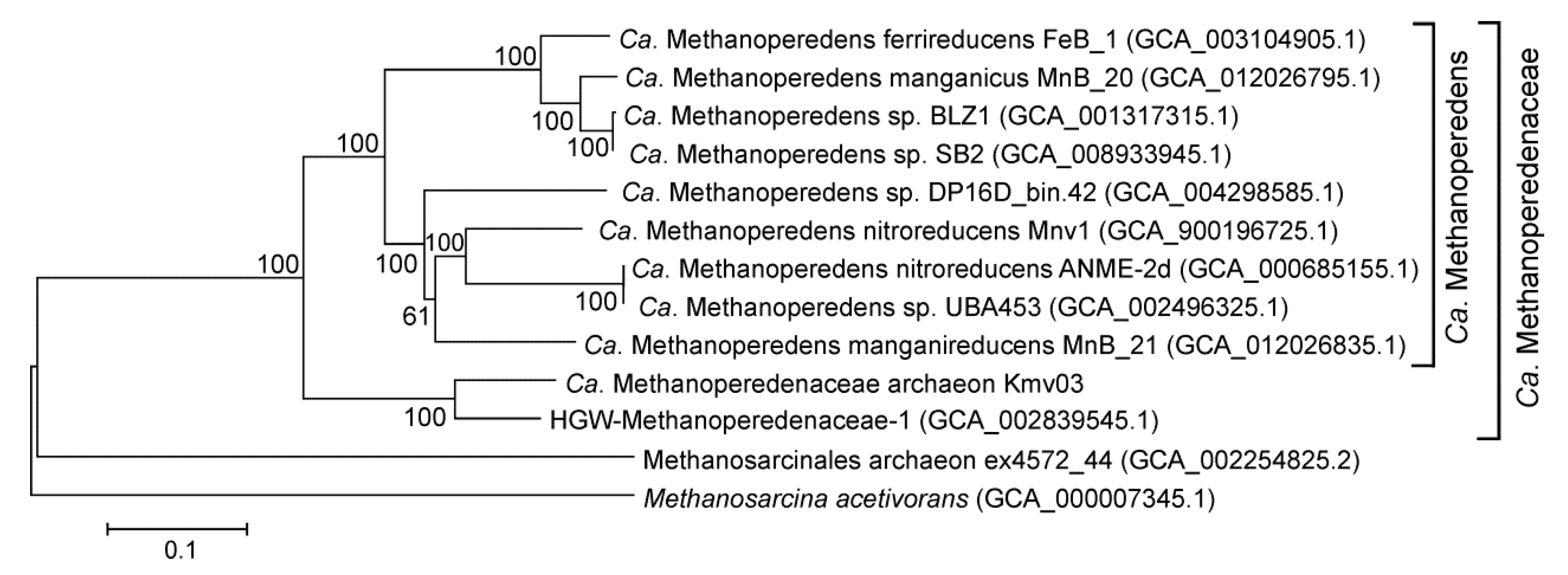
3.6. Bacteria and Archaea Involved in the Methane Cycle

Three archaeal MAGs belonged to known ANME lineages. The most abundant MAG, Kmv03, was assigned to the family Ca. Methanoperedenaceae, also known as ANME-2d group. These archaea could couple anaerobic oxidation of methane to the reduction of nitrate (Ca. Methanoperedens nitroreducens, [63]), Fe3+ (Ca. M. ferrireducens, [64]) or Mn4+ (Ca. M. manganicus and Ca. M. manganireducens [65]). The Kmv03 archaeon is most closely related (83% AAI) to Ca. Methanoperedenaceae archaeon HGW-Methanoperedenaceae-1, recovered from the metagenome of a groundwater sample collected at depths of 140–250 m in Japan [66]. Together these two MAGs formed a distinct genus-level lineage in the family Ca. Methanoperedenaceae, which was second to the previously described Ca. Methanoperedens [63] (Figure 3).
The Kmv03 genome encoded a complete reverse methanogenesis pathway and related energy converting enzymes, including the cytoplasmic and membrane-bound CoB-CoM heterodisulfide reductase, F420H2 dehydrogenase, sodium-translocating methyltransferase complex (Mtr), and V-type ATPase. The nitrate reductase complex, responsible for nitrate reduction coupled to AOM in Ca. M. nitroreducens [67], was not encoded in the Kmv03 genome. Therefore, Kmv03 likely use electron acceptors other than nitrate. The ability of Ca. M. ferrireducens, Ca. M. manganicus, and Ca. M. manganireducens to reduce insoluble Fe(III) or Mn(IV) oxides was attributed to MHCs [65,68]. Analysis of the Kmv03 genome revealed 18 MHCs containing up to 22 hemes, of which 18 cytochromes were predicted to contain an N-terminal secretion signal. In particular, we identified a gene cluster containing two pentaheme c-type cytochromes, cytochrome c with a single heme, two membrane proteins, and two membrane-integral cytochrome b proteins. All three cytochrome c proteins, besides the N-terminal secretion sequence, contained C-terminal transmembrane helices, indicating they were localized on the external side of the cell membrane. In addition to MHCs, archaeal flagellar-like conductive structures were proposed to be involved in the long-distance transfer of electrons between ANME archaea and their sulfate-reducing partners [69] or metal oxides [65]. Kmv03 contained genes involved in the formation of the archaellum, including two flaB genes encoding for archaellin. Since MHCs and archaella in different ANME lineages could be involved in the electron transfer to their syntrophic partners or insoluble electron acceptors [65,68,69], we could not predict which function was relevant for Ca. Methanoperedenaceae Kmv03 archaeon. However, the abundance of potential metal-reducing bacteria carrying MHCs in the microbial community and the minor fraction of sulfate reducers favors the hypothesis that metal oxides are used as electron acceptors for AOM.
Two other, less abundant ANME lineages, ANME-2a-2b of the Methanosarcinales and ANME-1, were represented by MAGs Kmv04 and Kmv05, respectively. Both of these ANME groups are known to be associated with sulfate-reducing Deltaproteobacteria [9,70], but the latter were not identified by 16S rRNA profiling or among the MAGs.
Most of bacterial methanotrophs were represented by MAG Kmv24, assigned to the genus Methyloprofundus, with 72.2% AAI to Methyloprofundus sedimenti. Analysis of the Kmv24 genome revealed the presence of both soluble and particulate methane monooxygenases, methanol dehydrogenase, and downstream enzymes required for the metabolism of formaldehyde. Three cytochrome c oxidases were identified, while enzymes for dissimilatory reduction of nitrate and other nitrogen compounds were absent. All of these traits were consistent with methanotrophic lifestyle. Two other MAGs, Kmv25 and Kmv26, represented well-characterized groups of aerobic methanotrophic bacteria and were assigned to the genera Methylomicrobium and Methylophaga, respectively.

3.7. Iron-Reducing Deltaproteobacteria

Deltaproteobacteria, according to the 16S rRNA profiling, only accounted for 0.34% of the community and belonged to the order Desulfuromonadales. Nevertheless, two high-quality MAGs, Kmv15 and Kmv16, were obtained and assigned to Desulfuromonadales. Although many members of this order are sulfate-reducers, analysis of the Kmv15 genome revealed the absence of a dissimilatory sulfate-reduction pathway. The hallmark of this genome is the presence of a complete pathway for beta-oxidation of fatty acids, including fatty-acid-CoA ligases, two copies of fadN-fadA-fadE operon, propionyl-CoA carboxylase, methylmalonyl-CoA mutase, and methylmalonyl-CoA epimerase. Oxidation of lactate to pyruvate is probably enabled by a multi-subunit lactate dehydrogenase belonging to the CCG family, which were found in the genomes of various Deltaproteobacteria, particularly in Desulfovibrio vulgaris [71]. Fatty acids and lactate are most likely the main substrates, since no genes encoding extracellular glycoside hydrolases and proteases were found.
Besides fermentation, Kmv15 bacterium is capable of anaerobic respiration. Its genome encoded a molybdopterin family respiratory arsenate reductase, cytochrome c nitrite reductase, and more than a dozen of MHCs with N-terminal secretion signals. These cytochromes could be necessary to contact the insoluble electron acceptors and probably enable dissimilatory Fe(III) reduction, as reported in Geobacter species [55]. Adhesion to Fe(III) minerals could also be facilitated by the type IV pili encoded by the Kmv15 genome. Hydrogen could be used as an electron donor, as indicated by the presence of two group 1 respiratory H2-uptake [NiFe] hydrogenases. Three membrane-linked complexes, NADH-ubiquinone oxidoreductase, an electron transport Rnf complex, and Na+-translocating NADH-quinone reductase, can generate a transmembrane ion gradient, which can be employed by F0F1 ATP synthase for ATP production. Overall, the genome analysis revealed that Kmv15 is an organotrophic bacterium specialized in utilizing fatty acids to perform fermentation and anaerobic respiration with arsenate, nitrite, and Fe(III).
The second MAG, Kmv16, was assigned in the GTDB taxonomy to another candidate family of Desulfuromonadales. Like Kmv15, the Kmv16 genome lacked a dissimilatory sulfate reduction pathway, and contained genes for beta-oxidation of fatty acids, respiratory arsenate reductase, multiheme cytochromes c, NADH-ubiquinone oxidoreductase, Rnf complex, sodium-translocating NADH-quinone reductase, group 1 respiratory [NiFe] hydrogenases, and L-lactate dehydrogenase. The cytochrome c nitrite reductase was not found, but respiratory nitrate reductase was present. Contrary to Kmv15, Kmv15 bacterium possesses an aerobic respiratory chain with two cytochrome c oxidases: caa3 and cbb3 types. These oxidases vary in their affinities for oxygen and could enable respiration under fluctuating oxygen concentrations.

3.8. Bathyarchaeota

The archaea of the uncultured candidate phylum Bathyarchaeota (recognized as a class of the phylum Crenarchaeota in the GTDB taxonomy) accounted for almost 10% of all the 16S rRNA gene sequences. Analysis of Bathyarchaeota genomes revealed that these organisms are organoheterotrophs that possibly utilize various proteinaceous substrates and polysaccharides of plant origin [72]. Their genomes encoded the Wood-Ljungdahl pathway of autotrophic carbon fixation. Depending on the environmental conditions, Bathyarchaeota can use this pathway for acetogenesis or in reverse, using acetate to form H2 and CO2 [73]. Genomes of some members of Bathyarchaeota contain Mcr-like genes, and it was suggested that they perform methyl-dependent hydrogenotrophic methanogenesis [74]. However, it was later proposed that these genes are probably involved in the oxidation of short-chain alkanes rather than methanogenesis [75]. Assembly of two Bathyarchaeota MAGs with an estimated completeness of 86% (Kmv01) and 89% (Kmv02) enabled to get insights into the metabolic potential of these archaea.
According to GTDB taxonomy, these MAGs were assigned to families UBA233 and BA1 of the order B26-1. Notably, two potential methanogenic members of the Bathyarchaeota, Ca. Bathyarchaeota archaeon BA1 and Ca. Bathyarchaeota archaeon BA2, belonging to the BA1 family, were found in the formation waters from a coalbed methane well [74]. Kmv02 and BA2 archaeons likely belong to the same genus sharing a 74% AAI. The genome size of Kmv02 (1,674,670 bp) was similar to that of BA1 (1,931,714 bp) and BA2 (1,455,689 bp). Meanwhile, Kmv01 had a smaller genome (1,046,189 bp). Considering the close phylogenetic relatedness of Kmv02 and BA1/BA2 archaea, the absence of methyl-coenzyme M reductase genes in the Kmv02 genome was rather unexpected. The Mtr complex was also missing. The second MAG, Kmv01, also lacked all mcr and mtr genes. Considering that these MAGs were estimated to be 86% and 89% complete, the probability that mcr and mtr genes were missed in the assemblies by chance is unlikely; therefore, both bathyarchaeons are probably not involved in methanogenesis or short-chain alkane oxidation dependent on Mcr-like proteins.
The search for carbohydrate-active enzymes in the genome of Bathyarchaeota Kmv02 revealed GH57 family alpha-amylase and alpha-mannosidase, and GH1 family beta-galactosidase/beta-glucosidase, all of which lacked N-terminal signal peptides, and several ABC-type sugar transporters. The genome also encoded ABC-type peptide transporters, peptidases, aminotransferases, and 2-oxoacid ferredoxin oxidoreductases, enabling fermentation of proteinaceous substrates. The genome encoded a near-complete Embden–Meyerhof glycolysis pathway and enzymes required for gluconeogenesis (i.e., fructose-1,6-bisphosphatase and phosphoenolpyruvate synthase). In addition, the beta-oxidation pathway for fatty acids utilization was present, including long-chain-fatty-acid-CoA ligases, enoyl-CoA hydratases, 3-ketoacyl-CoA thiolases, and acyl-CoA dehydrogenases. Therefore, the Kmv02 archaeon might utilize a wide range of organic substrates, but it cannot degrade complex polymers.
Pyruvate produced in fermentation pathways could be converted to formate and acetyl-CoA by pyruvate formate-lyase, or oxidized by pyruvate:ferredoxin oxidoreductase. The acetyl-CoA produced may either enter the Wood–Ljungdahl pathway, or it could be oxidized to acetate with ATP production by acetyl-CoA synthetase. Four [NiFe] hydrogenases were identified in the Bathyarchaeota Kmv02 genome. The first is group 3c heterodisulfide reductase-linked complex (MvhADG-HdrABC) that could bifurcate electrons from H2 to heterodisulfide (CoM-S-S-CoB) and ferredoxin [76]. The second is cytoplasmic group 3b cofactor-coupled bidirectional sulfhydrogenase. Together these hydrogenases could re-oxidize reduced cofactors generated in fermentation reactions. In addition, there are two group 4g H2-evolving membrane-linked hydrogenases that may form respiratory complexes that couple ferredoxin oxidation with proton reduction and translocate protons across the membrane. Usually, the generated proton motive force is coupled to ATP synthesis, but the Kmv02 genome lacked genes encoding membrane-bound ATP synthases. The ATP synthases were not found also in the genome of Kmv01. The transmembrane proton gradient is probably only utilized for transport purposes. Consistently with fermentative heterotrophic lifestyle, both Bathyrchaeota genomes lacked genes for aerobic and anaerobic respiration.

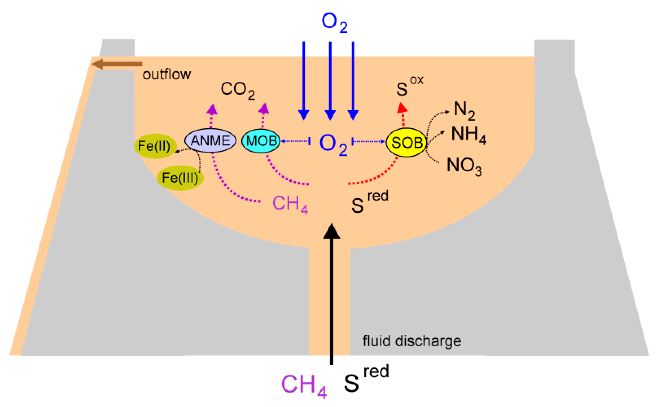
3.9. An overview of Microbial Processes in the Mud Volcano

Fluids and gases discharged from mud volcanoes are thought to originate from deep horizons and provide an abundant source of electron donors for microbial growth. Reaching the surface, the reduced mud fluids are exposed to atmospheric oxygen, which provides conditions for the proliferation of microorganisms with a wide range of oxygen requirements and metabolic capabilities (Figure 4). This facilitates the energy and material exchange at the oxic–anoxic interface [77]. In some mud volcanoes, such interactions could lead to geochemical and metabolic stratification forming the upper sulfate-rich oxic and lower methane-rich anoxic zones, harboring distinct bacterial and archaeal communities [14]. For example, in a methane-rich mud volcano in southwestern Taiwan, oxygen penetration was limited to the upper 4 mm layer of the fluids and counteracted by the oxidation of sulfide, methane, and organic matter [16]. However, in the mud volcano we studied, the active release of gases and bubbling created conditions for mud mixing in the crater, at least at the depth from which the samples have been acquired (10–20 cm).
Metagenomic analysis of the mud sample revealed that the microbial community was dominated by chemolithoautotrophic sulfur-oxidizing Campylobacterota and Gammaproteobacteria. Altogether the sulfur-oxidizing bacteria accounted for about 55% of the 16S rRNA genes sequences. On the contrary, sulfate reducers were found in minor amounts and probably do not play an important role in the sulfur cycle in the mud pool. Members of the Campylobacterota appeared to be metabolically versatile being capable of using both sulfur compounds and molecular hydrogen as electron donors. In addition to aerobic respiration under microaerophilic conditions, they had the genetic capacity for anaerobic growth using various electron acceptors, including nitrate, nitrite, nitrogen oxides, arsenate, and sulfur compounds. The presence of about 200 µM of nitrate should enable the growth of these bacteria in the anoxic zones. The sulfur-oxidizing members of the Gammaproteobacteria were predicted to only oxidize sulfur compounds under aerobic conditions.
Availability of methane among the emitted gases and the presence of various electron acceptors could support the growth of methanotrophs (Figure 4). Consistently, we found aerobic methanotrophs, gamma-proteobacteria of the order Methylococcales, and ANME archaea. The latter can inhabit local anaerobic niches, for example, ones associated with clay particles and the walls of the crater, or be delivered from deeper anaerobic zones with the discharged fluids. ANME archaea are typical inhabitants of microbial communities associated with mud volcanoes [18]. Molecular studies of a mud volcano in eastern Taiwan revealed that the ANME-2a/2b group was most abundant among archaea, and it was suggested that they form syntrophic consortia with Desulfuromonadales to facilitate electron transport during AOM [18]. Both of these two lineages were identified in our study, but only in minor amounts. The ANME community was dominated by a novel species of an ANME-2d group, Ca. Methanoperedenaceae Kmv03. Members of the Ca. Methanoperedenaceae can couple AOM to the reduction of nitrate, Fe(III), or Mn(IV) [63,64,65] without a syntrophic partner. Although nitrate was available in the mud breccia, Ca. Methanoperedenaceae Kmv03 archaeon lacked nitrate reductase and most likely relies on the reduction of metal oxides. The reduction of insoluble metal minerals could be an essential process in the mud volcano community. The genetic capacity for this process has been identified in the genomes of various other community members, such as Sulfurospirillum, and two Desulfuromonadales species.
Organic matter produced by autotrophic sulfur-oxidizing bacteria and methanotrophs could support the heterotrophic part of the community comprising of both aerobic organotrophs (i.e., Marinospirillum, Marinobacter, Halomonas, Rhodobacteraceae sp.) and fermentative organisms. According to the 16S rRNA profiling, Bathyarchaeota was the most abundant among the second group. Like ANME archaea, Bathyarchaeota probably occupies local anaerobic environments.
The fluids discharged from mud volcanoes are expected to deliver microorganisms from the deep subsurface to the surface where they became mixed with those actively proliferating in the mud breccia. Two groups of presumably thermophilic sulfate reducers indigenous to the deep subsurface have been detected: members of the order Thermodesulfovibrionales (Nitrospirae) and the genus Ca. Desulforudis (Firmicutes). Thermodesulfovibrio species are widespread in different hydrothermal habitats, including the deep thermal aquifers [78], while Ca. Desulforudis have been detected exclusively in the deep subsurface [60,61,62]. Interestingly, other typical members of the microbial communities of the deep subsurface ecosystems, thermophilic methanogenic archaea [79], were not found. This might be explained by the source of the fluids. Probably, the fluids originate from the sulfate-rich subsurface site with predominantly sulfate-reducing microbial population such as South African deep subsurface site where Ca. Desulforudis audaxviator formed a single-species ecosystem [60]. Sulfide produced by sulfate reducers in the deep subsurface is transferred to the surface when fluid is discharged and supports the development of the sulfur-oxidizing microbial community in the mud breccia.
Overall the results obtained suggest that reduced sulfur compounds and methane delivered from the deep subsurface support the development of chemolithoautotrophic microbial community using various electron acceptors for respiration.

Supplementary Materials

The following are available online at https://www.mdpi.com/2076-2607/8/9/1333/s1, Table S1: Relative abundance and taxonomic classification of OTUs; Table S2: Main characteristics of MAGs obtained in this study.

Author Contributions

Conceptualization, N.V.R.; investigation, A.V.M., V.V.K. and A.V.B.; resources, A.V.M. and N.V.R.; data curation, A.V.B. and N.V.R.; writing—original draft preparation, N.V.R.; writing—review and editing, N.V.R.; supervision, N.V.R.; funding acquisition, N.V.R. All authors have read and agreed to the published version of the manuscript.

Funding

This work was partly funded by the Russian Science Foundation (grant 19-14-00245).

Acknowledgments

This work was performed using the scientific equipment of the Core Research Facility ‘Bioengineering’ (Research Center of Biotechnology RAS).

Conflicts of Interest

The authors declare no conflict of interest.

References

  1. Mazzini, A.; Etiope, G. Mud volcanism: An updated review. Earth Sci. Rev. 2017, 168, 81–112. [Google Scholar] [CrossRef] [Green Version]
  2. Dimitrov, L.I. Mud volcanoes—The most important pathway for degassing deeply buried sediments. Earth Sci. Rev. 2002, 59, 49–76. [Google Scholar] [CrossRef]
  3. Kopf, A.J. Significance of mud volcanism. Rev. Geophys. 2002, 40, 1–52. [Google Scholar] [CrossRef] [Green Version]
  4. Kioka, A.; Ashi, J. Episodic massive mud eruptions from submarine mud volcanoes examined through topographical signatures. Geophys. Res. Lett. 2015, 42, 8406–8414. [Google Scholar] [CrossRef] [Green Version]
  5. Mazzini, A.; Svensen, H.; Planke, S.; Guliyev, I.; Akhmanov, G.G.; Fallik, T.; Banks, D. When mud volcanoes sleep: Insight from seep geochemistry at the Dashgil mud volcano, Azerbaijan. Mar. Pet. Geol. 2009, 26, 1704–1715. [Google Scholar] [CrossRef]
  6. Etiope, G.; Lassey, K.R.; Klusman, R.W.; Boschi, E. Reappraisal of the fossil methane budget and related emission from geologic sources. Geophys. Res. Lett. 2008, 35, L09307. [Google Scholar] [CrossRef]
  7. Martinez, R.J.; Mills, H.J.; Story, S.; Sobecky, P.A. Prokaryotic diversity and metabolically active microbial populations in sediments from an active mud volcano in the Gulf of Mexico. Environ. Microbiol. 2006, 8, 1783–1796. [Google Scholar] [CrossRef]
  8. Niemann, H.; Lösekann, T.; de Beer, D.; Elvert, M.; Nadalig, T.; Knittel, K.; Amann, R.; Sauter, E.J.; Schlüter, M.; Klages, M.; et al. Novel microbial communities of the Haakon Mosby mud volcano and their role as methane sink. Nature 2006, 443, 854–858. [Google Scholar] [CrossRef]
  9. Lösekann, T.; Knittel, K.; Nadalig, T.; Fuchs, B.; Niemann, H.; Boetius, A.; Amann, R. Diversity and abundance of aerobic and anaerobic methane oxidizers at the Haakon Mosby mud volcano, Barents Sea. Appl. Environ. Microbiol. 2007, 73, 3348–3362. [Google Scholar] [CrossRef] [Green Version]
  10. Klasek, S.A.; Torres, M.E.; Loher, M.; Bohrmann, G.; Pape, T.; Colwell, F.S. Deep-Sourced Fluids from a Convergent Margin Host Distinct Subseafloor Microbial Communities That Change Upon Mud Flow Expulsion. Front. Microbiol. 2019, 10, 1436. [Google Scholar] [CrossRef]
  11. Coelho, F.J.; Louvado, A.; Domingues, P.M.; Cleary, D.F.; Ferreira, M.; Almeida, A.; Cunha, M.R.; Cunha, Â.; Gomes, N.C. Integrated analysis of bacterial and microeukaryotic communities from differentially active mud volcanoes in the Gulf of Cadiz. Sci. Rep. 2016, 6, 35272. [Google Scholar] [CrossRef] [PubMed] [Green Version]
  12. Hoshino, T.; Toki, T.; Ijiri, A.; Morono, Y.; Machiyama, H.; Ashi, J.; Okamura, K.; Inagaki, F. Atribacteria from the Subseafloor Sedimentary Biosphere Disperse to the Hydrosphere through Submarine Mud Volcanoes. Front. Microbiol. 2017, 8, 1135. [Google Scholar] [CrossRef] [PubMed]
  13. Alain, K.; Holler, T.; Musat, F.; Elvert, M.; Treude, T.; Krüger, M. Microbiological investigation of methane- and hydrocarbon-discharging mud volcanoes in the Carpathian Mountains, Romania. Environ. Microbiol. 2006, 8, 574–590. [Google Scholar] [CrossRef] [PubMed]
  14. Cheng, T.W.; Chang, Y.H.; Tang, S.L.; Tseng, C.H.; Chiang, P.W.; Chang, K.T.; Sun, C.H.; Chen, Y.G.; Kuo, H.C.; Wang, C.H.; et al. Metabolic stratification driven by surface and subsurface interactions in a terrestrial mud volcano. ISME J. 2012, 6, 2280–2290. [Google Scholar] [CrossRef]
  15. Wrede, C.; Brady, S.; Rockstroh, S.; Dreier, A.; Kokoschka, S.; Heinzelmann, S.M.; Heller, C.; Reitner, J.; Taviani, M.; Daniel, R.; et al. Aerobic and anaerobic methane oxidation in terrestrial mud volcanoes in the Northern Apennines. Sediment. Geol. 2012, 263–264, 210–219. [Google Scholar] [CrossRef]
  16. Lin, Y.T.; Tu, T.H.; Wei, C.L.; Rumble, D.; Lin, L.H.; Wang, P.L. Steep redox gradient and biogeochemical cycling driven by deeply sourced fluids and gases in a terrestrial mud volcano. FEMS Microbiol. Ecol. 2018, 94. [Google Scholar] [CrossRef]
  17. Ren, G.; Ma, A.; Zhang, Y.; Deng, Y.; Zheng, G.; Zhuang, X.; Zhuang, G.; Fortin, D. Electron acceptors for anaerobic oxidation of methane drive microbial community structure and diversity in mud volcanoes. Environ. Microbiol. 2018, 20, 2370–2385. [Google Scholar] [CrossRef] [Green Version]
  18. Tu, T.H.; Wu, L.W.; Lin, Y.S.; Imachi, H.; Lin, L.H.; Wang, P.L. Microbial Community Composition and Functional Capacity in a Terrestrial Ferruginous, Sulfate-Depleted Mud Volcano. Front. Microbiol. 2017, 8, 2137. [Google Scholar] [CrossRef]
  19. Wang, P.L.; Chiu, Y.P.; Cheng, T.W.; Chang, Y.H.; Tu, W.X.; Lin, L.H. Spatial variations of community structures and methane cycling across a transect of Lei-Gong-Hou mud volcanoes in eastern Taiwan. Front. Microbiol. 2014, 5, 121. [Google Scholar] [CrossRef] [Green Version]
  20. Schulze-Makuch, D.; Haque, S.; Beckles, D.; Schmitt-Kopplin, P.; Harir, M.; Schneider, B.; Stumpp, C.; Wagner, D.A. A chemical and microbial characterization of selected mud volcanoes in Trinidad reveals pathogens introduced by surface water and rain water. Sci. Total Environ. 2020, 707, 136087. [Google Scholar] [CrossRef]
  21. Boetius, A.; Ravenschlag, K.; Schubert, C.J.; Rickert, D.; Widdel, F.; Gieseke, A.; Amann, R.; Jørgensen, B.B.; Witte, U.; Pfannkuche, O. A marine microbial consortium apparently mediating anaerobic oxidation of methane. Nature 2000, 407, 623–626. [Google Scholar] [CrossRef] [PubMed]
  22. Raghoebarsing, A.A.; Pol, A.; van de Pas-Schoonen, K.T.; Smolders, A.J.; Ettwig, K.F.; Rijpstra, W.I.; Schouten, S.; Damsté, J.S.; Op den Camp, H.J.; Jetten, M.S.; et al. A microbial consortium couples anaerobic methane oxidation to denitrification. Nature 2006, 440, 918–921. [Google Scholar] [CrossRef] [PubMed]
  23. Beal, E.J.; House, C.H.; Orphan, V.J. Manganese- and iron-dependent marine methane oxidation. Science 2009, 325, 184–187. [Google Scholar] [CrossRef] [Green Version]
  24. Ettwig, K.F.; Butler, M.K.; Le Paslier, D.; Pelletier, E.; Mangenot, S.; Kuypers, M.M.; Schreiber, F.; Dutilh, B.E.; Zedelius, J.; de Beer, D.; et al. Nitrite-driven anaerobic methane oxidation by oxygenic bacteria. Nature 2010, 464, 543–548. [Google Scholar] [CrossRef] [PubMed] [Green Version]
  25. Akhmetjanov, A.; Akhmanov, G.; Krylov, O.; Basov, E.; Kozlova, E.; Stadnitskaya, A. Mud volcanoes of the Kerch Peninsula: General review. MarinF (UNESCO) 1996, 100, 23–24. [Google Scholar]
  26. Frey, B.; Rime, T.; Phillips, M.; Stierli, B.; Hajdas, I.; Widmer, F.; Hartmann, M. Microbial diversity in European alpine permafrost and active layers. FEMS Microbiol. Ecol. 2016, 92, fiw018. [Google Scholar] [CrossRef] [Green Version]
  27. Magoč, T.; Salzberg, S.L. FLASH: Fast length adjustment of short reads to improve genome assemblies. Bioinformatics 2011, 27, 2957–2963. [Google Scholar] [CrossRef]
  28. Edgar, R.C. Search and clustering orders of magnitude faster than BLAST. Bioinformatics 2010, 26, 2460–2461. [Google Scholar] [CrossRef] [Green Version]
  29. Rognes, T.; Flouri, T.; Nichols, B.; Quince, C.; Mahé, F. VSEARCH: A versatile open source tool for metagenomics. PeerJ 2016, 4, e2584. [Google Scholar] [CrossRef]
  30. Martin, M. Cutadapt removes adapter sequences from high-throughput sequencing reads. EMBnet. J. 2011, 17, 10–12. [Google Scholar] [CrossRef]
  31. Sickle v.1.33. Available online: https://github.com/najoshi/sickle (accessed on 30 August 2020).
  32. Bankevich, A.; Nurk, S.; Antipov, D.; Gurevich, A.A.; Dvorkin, M.; Kulikov, A.S.; Lesin, V.M.; Nikolenko, S.I.; Pham, S.; Prjibelski, A.D.; et al. SPAdes: A new genome assembly algorithm and its applications to single-cell sequencing. J. Comput. Biol. 2012, 19, 455–477. [Google Scholar] [CrossRef] [PubMed] [Green Version]
  33. Alneberg, J.; Bjarnason, B.S.; de Bruijn, I.; Schirmer, M.; Quick, J.; Ijaz, U.Z.; Lahti, L.; Loman, N.J.; Andersson, A.F.; Quince, C. Binning metagenomic contigs by coverage and composition. Nat. Methods 2014, 11, 1144–1146. [Google Scholar] [CrossRef] [PubMed]
  34. Kang, D.D.; Froula, J.; Egan, R.; Wang, Z. MetaBAT, an efficient tool for accurately reconstructing single genomes from complex microbial communities. PeerJ 2015, 3, e1165. [Google Scholar] [CrossRef] [Green Version]
  35. Parks, D.H.; Imelfort, M.; Skennerton, C.T.; Hugenholtz, P.; Tyson, G.W. CheckM: Assessing the quality of microbial genomes recovered from isolates, single cells, and metagenomes. Genome Res. 2015, 25, 1043–1055. [Google Scholar] [CrossRef] [Green Version]
  36. Parks, D.H.; Chuvochina, M.; Waite, D.W.; Rinke, C.; Skarshewski, A.; Chaumeil, P.A.; Hugenholtz, P. A standardized bacterial taxonomy based on genome phylogeny substantially revises the tree of life. Nat. Biotechnol. 2018, 36, 996–1004. [Google Scholar] [CrossRef] [PubMed]
  37. Tatusova, T.; DiCuccio, M.; Badretdin, A.; Chetvernin, V.; Nawrocki, E.P.; Zaslavsky, L.; Lomsadze, A.; Pruitt, K.D.; Borodovsky, M.; Ostell, J. NCBI prokaryotic genome annotation pipeline. Nucleic Acids Res. 2016, 44, 6614–6624. [Google Scholar] [CrossRef]
  38. Brettin, T.; Davis, J.J.; Disz, T.; Edwards, R.A.; Gerdes, S.; Olsen, G.J.; Olson, R.; Overbeek, R.; Parrello, B.; Pusch, G.D.; et al. RASTtk: A modular and extensible implementation of the RAST algorithm for building custom annotation pipelines and annotating batches of genomes. Sci. Rep. 2015, 5, 8365. [Google Scholar] [CrossRef] [Green Version]
  39. Rodriguez-R, L.M.; Konstantinidis, K.T. The enveomics collection: A toolbox for specialized analyses of microbial genomes and metagenomes. PeerJ Prepr. 2016, 4, e1900v1. [Google Scholar]
  40. Guindon, S.; Dufayard, J.F.; Lefort, V.; Anisimova, M.; Hordijk, W.; Gascuel, O. New algorithms and methods to estimate maximum-likelihood phylogenies: Assessing the performance of PhyML 3.0. Syst. Biol. 2010, 59, 307–321. [Google Scholar] [CrossRef] [Green Version]
  41. Inagaki, F.; Takai, K.; Kobayashi, H.; Nealson, K.H.; Horikoshi, K. Sulfurimonas autotrophica gen. nov., sp. nov., a novel sulfur-oxidizing epsilon-proteobacterium isolated from hydrothermal sediments in the Mid-Okinawa Trough. Int. J. Syst. Evol. Microbiol. 2003, 53 Pt 6, 1801–1805. [Google Scholar] [CrossRef] [Green Version]
  42. Han, Y.; Perner, M. The globally widespread genus Sulfurimonas: Versatile energy metabolisms and adaptations to redox clines. Front. Microbiol. 2015, 6, 989. [Google Scholar] [CrossRef] [PubMed] [Green Version]
  43. van der Stel, A.X.; Wösten, M.M.S.M. Regulation of Respiratory Pathways in Campylobacterota: A Review. Front. Microbiol. 2019, 10, 1719. [Google Scholar] [CrossRef] [PubMed] [Green Version]
  44. Brinkhoff, T.; Muyzer, G. Increased species diversity and extended habitat range of sulfur-oxidizing Thiomicrospira spp. Appl. Environ. Microbiol. 1997, 63, 3789–3796. [Google Scholar] [CrossRef] [Green Version]
  45. Sorokin, D.Y.; Tourova, T.P.; Kolganova, T.V.; Sjollema, K.A.; Kuenen, J.G. Thioalkalispira microaerophila gen. nov., sp. nov., a novel lithoautotrophic, sulfur-oxidizing bacterium from a soda lake. Int. J. Syst. Evol. Microbiol 2002, 52 Pt 6, 2175–2182. [Google Scholar]
  46. Tavormina, P.L.; Hatzenpichler, R.; McGlynn, S.; Chadwick, G.; Dawson, K.S.; Connon, S.A.; Orphan, V.J. Methyloprofundus sedimenti gen. nov., sp. nov., an obligate methanotroph from ocean sediment belonging to the ‘deep sea-1’ clade of marine methanotrophs. Int. J. Syst. Evol. Microbiol. 2015, 65 Pt 1, 251–259. [Google Scholar] [CrossRef]
  47. Zhang, W.; Xue, Y.; Ma, Y.; Grant, W.D.; Ventosa, A.; Zhou, P. Marinospirillum alkaliphilum sp. nov., a new alkaliphilic helical bacterium from Haoji soda lake in Inner Mongolia Autonomous Region of China. Extremophiles 2002, 6, 33–37. [Google Scholar] [CrossRef] [PubMed]
  48. Wang, X.M.; Meng, X.; Du, Z.Z.; Du, Z.J.; Mu, D.S. Marinospirillum perlucidum sp. nov., a novel helical bacterium isolated from a sea cucumber culture pond. Int. J. Syst. Evol. Microbiol. 2020, 70, 576–581. [Google Scholar] [CrossRef] [PubMed]
  49. Zhang, Y.; Zhong, X.C.; Xu, W.; Lu, D.C.; Zhao, J.X.; Du, Z.J. Marinobacter vulgaris sp. nov., a moderately halophilic bacterium isolated from a marine solar saltern. Int. J. Syst. Evol. Microbiol. 2020, 70, 450–456. [Google Scholar] [CrossRef] [PubMed]
  50. Kim, K.K.; Lee, J.S.; Stevens, D.A. Microbiology and epidemiology of Halomonas species. Future Microbiol. 2013, 8, 1559–1573. [Google Scholar] [CrossRef]
  51. Sievert, S.M.; Scott, K.M.; Klotz, M.G.; Chain, P.S.; Hauser, L.J.; Hemp, J.; Hügler, M.; Land, M.; Lapidus, A.; Larimer, F.W.; et al. Genome of the epsilonproteobacterial chemolithoautotroph Sulfurimonas denitrificans. Appl. Environ. Microbiol. 2008, 74, 1145–1156. [Google Scholar] [CrossRef] [Green Version]
  52. Lahme, S.; Callbeck, C.M.; Eland, L.E.; Wipat, A.; Enning, D.; Head, I.M.; Hubert, C.R.J. Comparison of sulfide-oxidizing Sulfurimonas strains reveals a new mode of thiosulfate formation in subsurface environments. Environ. Microbiol. 2020, 22, 1784–1800. [Google Scholar] [CrossRef] [PubMed]
  53. Preisig, O.; Zufferey, R.; Thöny-Meyer, L.; Appleby, C.A.; Hennecke, H. A high-affinity cbb3-type cytochrome oxidase terminates the symbiosis-specific respiratory chain of Bradyrhizobium japonicum. J. Bacteriol. 1996, 178, 1532–1538. [Google Scholar] [CrossRef] [PubMed] [Green Version]
  54. Ross, D.E.; Marshall, C.W.; May, H.D.; Norman, R.S. Comparative Genomic Analysis of Sulfurospirillum cavolei MES Reconstructed from the Metagenome of an Electrosynthetic Microbiome. PLoS ONE 2016, 11, e0151214. [Google Scholar] [CrossRef] [PubMed]
  55. Shi, L.; Squier, T.C.; Zachara, J.M.; Fredrickson, J.K. Respiration of metal (hydr)oxides by Shewanella and Geobacter: A key role for multihaem c-type cytochromes. Mol. Microbiol. 2007, 65, 12–20. [Google Scholar] [CrossRef] [Green Version]
  56. Goris, T.; Schubert, T.; Gadkari, J.; Wubet, T.; Tarkka, M.; Buscot, F.; Adrian, L.; Diekert, G. Insights into organohalide respiration and the versatile catabolism of Sulfurospirillum multivorans gained from comparative genomics and physiological studies. Environ. Microbiol. 2014, 16, 3562–3580. [Google Scholar] [CrossRef]
  57. Scott, K.M.; Sievert, S.M.; Abril, F.N.; Ball, L.A.; Barrett, C.J.; Blake, R.A.; Boller, A.J.; Chain, P.S.; Clark, J.A.; Davis, C.R.; et al. The genome of deep-sea vent chemolithoautotroph Thiomicrospira crunogena XCL-2. PLoS Biol. 2006, 4, e383. [Google Scholar] [CrossRef]
  58. Watsuji, T.O.; Hada, E.; Miyazaki, M.; Ichimura, M.; Takai, K. Thiomicrospira hydrogeniphila sp. nov., an aerobic, hydrogen- and sulfur-oxidizing chemolithoautotroph isolated from a seawater tank containing a block of beef tallow. Int. J. Syst. Evol. Microbiol. 2016, 66, 3688–3693. [Google Scholar] [CrossRef]
  59. Henry, E.A.; Devereux, R.; Maki, J.S.; Gilmour, C.C.; Woese, C.R.; Mandelco, L.; Schauder, R.; Remsen, C.C.; Mitchell, R. Characterization of a new thermophilic sulfate-reducing bacterium Thermodesulfovibrio yellowstonii, gen. nov. and sp. nov.: Its phylogenetic relationship to Thermodesulfobacterium commune and their origins deep within the bacterial domain. Arch. Microbiol. 1994, 161, 62–69. [Google Scholar] [CrossRef]
  60. Chivian, D.; Brodie, E.L.; Alm, E.J.; Culley, D.E.; Dehal, P.S.; DeSantis, T.Z.; Gihring, T.M.; Lapidus, A.; Lin, L.H.; Lowry, S.R.; et al. Environmental genomics reveals a single-species ecosystem deep within Earth. Science 2008, 322, 275–278. [Google Scholar] [CrossRef] [Green Version]
  61. Karnachuk, O.V.; Frank, Y.A.; Lukina, A.P.; Kadnikov, V.V.; Beletsky, A.V.; Mardanov, A.V.; Ravin, N.V. Domestication of previously uncultivated Candidatus Desulforudis audaxviator from a deep aquifer in Siberia sheds light on its physiology and evolution. ISME J. 2019, 13, 1947–1959. [Google Scholar] [CrossRef]
  62. Jungbluth, S.P.; Glavina Del Rio, T.; Tringe, S.G.; Stepanauskas, R.; Rappé, M.S. Genomic comparisons of a bacterial lineage that inhabits both marine and terrestrial deep subsurface systems. PeerJ 2017, 5, e3134. [Google Scholar] [CrossRef] [Green Version]
  63. Haroon, M.F.; Hu, S.; Shi, Y.; Imelfort, M.; Keller, J.; Hugenholtz, P.; Yuan, Z.; Tyson, G.W. Anaerobic oxidation of methane coupled to nitrate reduction in a novel archaeal lineage. Nature 2013, 500, 567–570. [Google Scholar] [CrossRef] [PubMed]
  64. Ettwig, K.F.; Zhu, B.; Speth, D.; Keltjens, J.T.; Jetten, M.S.M.; Kartal, B. Archaea catalyze iron-dependent anaerobic oxidation of methane. Proc. Natl. Acad. Sci. USA 2016, 113, 12792–12796. [Google Scholar] [CrossRef] [Green Version]
  65. Leu, A.O.; Cai, C.; McIlroy, S.J.; Southam, G.; Orphan, V.J.; Yuan, Z.; Hu, S.; Tyson, G.W. Anaerobic methane oxidation coupled to manganese reduction by members of the Methanoperedenaceae. ISME J. 2020, 14, 1030–1041. [Google Scholar] [CrossRef] [Green Version]
  66. Hernsdorf, A.W.; Amano, Y.; Miyakawa, K.; Ise, K.; Suzuki, Y.; Anantharaman, K.; Probst, A.; Burstein, D.; Thomas, B.C.; Banfield, J.F. Potential for microbial H2 and metal transformations associated with novel bacteria and archaea in deep terrestrial subsurface sediments. ISME J. 2017, 11, 1915–1929. [Google Scholar] [CrossRef] [PubMed]
  67. Arshad, A.; Speth, D.R.; de Graaf, R.M.; Op den Camp, H.J.; Jetten, M.S.; Welte, C.U. A Metagenomics-Based Metabolic Model of Nitrate-Dependent Anaerobic Oxidation of Methane by Methanoperedens-Like Archaea. Front. Microbiol. 2015, 6, 1423. [Google Scholar] [CrossRef] [Green Version]
  68. Cai, C.; Leu, A.O.; Xie, G.J.; Guo, J.; Feng, Y.; Zhao, J.X.; Tyson, G.W.; Yuan, Z.; Hu, S. A methanotrophic archaeon couples anaerobic oxidation of methane to Fe(III) reduction. ISME J. 2018, 12, 1929–1939. [Google Scholar] [CrossRef] [PubMed]
  69. Krukenberg, V.; Riedel, D.; Gruber-Vodicka, H.R.; Buttigieg, P.L.; Tegetmeyer, H.E.; Boetius, A.; Wegener, G. Gene expression and ultrastructure of meso- and thermophilic methanotrophic consortia. Environ. Microbiol. 2018, 20, 1651–1666. [Google Scholar] [CrossRef] [Green Version]
  70. Michaelis, W.; Seifert, R.; Nauhaus, K.; Treude, T.; Thiel, V.; Blumenberg, M.; Knittel, K.; Gieseke, A.; Peterknecht, K.; Pape, T.; et al. Microbial reefs in the Black Sea fueled by anaerobic oxidation of methane. Science 2002, 297, 1013–1015. [Google Scholar] [CrossRef]
  71. Pereira, I.A.C.; Haveman, S.A.; Voordouw, G. Biochemical, genetic and genomic characterization of anaerobic electron transport pathways in sulphate-reducing delta-proteobacteria. In Sulphate–Reducing Bacteria: Environmental and Engineered Systems; Barton, L.L., Hamilton, W.A.A., Eds.; Cambridge University Press: Cambridge, UK, 2007; pp. 215–240. [Google Scholar]
  72. Lazar, C.S.; Baker, B.J.; Seitz, K.; Hyde, A.S.; Dick, G.J.; Hinrichs, K.U.; Teske, A.P. Genomic evidence for distinct carbon substrate preferences and ecological niches of Bathyarchaeota in estuarine sediments. Environ. Microbiol. 2016, 18, 1200–1211. [Google Scholar] [CrossRef]
  73. He, Y.; Li, M.; Perumal, V.; Feng, X.; Fang, J.; Xie, J.; Sievert, S.M.; Wang, F. Genomic and enzymatic evidence for acetogenesis among multiple lineages of the archaeal phylum Bathyarchaeota widespread in marine sediments. Nat. Microbiol. 2016, 1, 16035. [Google Scholar] [CrossRef] [PubMed]
  74. Evans, P.N.; Parks, D.H.; Chadwick, G.L.; Robbins, S.J.; Orphan, V.J.; Golding, S.D.; Tyson, G.W. Methane metabolism in the archaeal phylum Bathyarchaeota revealed by genomecentric metagenomics. Science 2015, 350, 434–438. [Google Scholar] [CrossRef] [PubMed] [Green Version]
  75. Borrel, G.; Adam, P.S.; McKay, L.J.; Chen, L.X.; Sierra-Garcıa, I.N.; Sieber, C.M.K.; Letourneur, Q.; Ghozlane, A.; Andersen, G.L.; Li, W.J.; et al. Wide diversity of methane and short-chain alkane metabolisms in uncultured archaea. Nat. Microbiol. 2019, 4, 603–613. [Google Scholar] [CrossRef] [PubMed]
  76. Buckel, W.; Thauer, R.K. Energy conservation via electron bifurcating ferredoxin reduction and proton/Na(+) translocating ferredoxin oxidation. Biochim. Biophys. Acta 2013, 1827, 94–113. [Google Scholar] [CrossRef] [Green Version]
  77. Thomson, J.; Higgs, N.C.; Croudace, I.W.; Colley, S.; Hydes, D.J. Redox zonation of elements at an oxic/post-oxic boundary in deep-sea sediments. Geochim. Cosmochim. Acta 1993, 57, 579–595. [Google Scholar] [CrossRef]
  78. Frank, Y.A.; Kadnikov, V.V.; Lukina, A.P.; Banks, D.; Beletsky, A.V.; Mardanov, A.V.; Sen’kina, E.I.; Avakyan, M.R.; Karnachuk, O.V.; Ravin, N.V. Characterization and Genome Analysis of the First Facultatively Alkaliphilic Thermodesulfovibrio Isolated from the Deep Terrestrial Subsurface. Front. Microbiol. 2016, 7, 2000. [Google Scholar] [CrossRef]
  79. Moser, D.P.; Gihring, T.M.; Brockman, F.J.; Fredrickson, J.K.; Balkwill, D.L.; Dollhopf, M.E.; Lollar, B.S.; Pratt, L.M.; Boice, E.; Southam, G.; et al. Desulfotomaculum and Methanobacterium spp. dominate a 4- to 5-kilometer-deep fault. Appl. Environ. Microbiol. 2005, 71, 8773–8783. [Google Scholar] [CrossRef] [Green Version]
Figure 1. Field images of the Bulganak mud volcano area (left) and the studied mud volcano (right).
Microorganisms 08 01333 g001
Figure 2. The relative abundance of taxonomic groups of microorganisms according to 16S rRNA gene profiling.
Microorganisms 08 01333 g002
Figure 3. Phylogenetic placement of the Ca. Methanoperedeaceae archaeon Kmv03. The genome tree was inferred using the maximum-likelihood method with a concatenated set of 122 archaeal-specific marker genes. GenBank assembly accession numbers are shown in parentheses after the genome names. The genome of Methanosarcina acetivorans was used to root the tree.
Microorganisms 08 01333 g003
Figure 4. Microbial processes related to sulfur and methane cycling in the mud volcano. SOB, bacteria performing aerobic and/or anaerobic oxidation of sulfur compounds; MOB, aerobic methanotrophic bacteria.
Microorganisms 08 01333 g004
Table 1. Physical and chemical characteristics of the water extracted from the mud sample.
Parameter (Unit)ValueParameter (Unit)Value
Temperature (°C)16Sr (mg L−1)2.4
pH7.43Li (mg L−1)1.9
Eh, mV−123Ba (mg L−1)1.0
Na (mg L−1)4165Fe (mg L−1)0.4
B (mg L−1)337As (mg L−1)0.06
K (mg L−1)26Cl (mg L−1)1865
Ca (mg L−1)58NO3 (mg L−1)12.0
Mg (mg L−1)23PO43− (mg L−1)8.3
Si (mg L−1)17SO42− (mg L−1)6.1

