Article

Probiotic Effects of a Novel Strain, Acinetobacter KU011TH, on the Growth Performance, Immune Responses, and Resistance against Aeromonas hydrophila of Bighead Catfish (Clarias macrocephalus Günther, 1864)

by
Anurak Bunnoy
1,
Uthairat Na-Nakorn
2 and
Prapansak Srisapoome
1,*
1
Laboratory of Aquatic Animal Health Management, Department of Aquaculture, Faculty of Fisheries, Kasetsart University, 50 Paholayothin Rd, Ladyao, Chatuchak, Bangkok 10900, Thailand
2
Laboratory of Aquatic Animal Genetics, Department of Aquaculture, Faculty of Fisheries, Kasetsart University, 50 Paholayothin Rd, Ladyao, Chatuchak, Bangkok 10900, Thailand
*
Author to whom correspondence should be addressed.
Microorganisms 2019, 7(12), 613; https://doi.org/10.3390/microorganisms7120613
Submission received: 19 September 2019 / Revised: 11 October 2019 / Accepted: 23 November 2019 / Published: 25 November 2019

Abstract

:
In the present study, the novel probiotic strain Acinetobacter KU011TH with an evident lack of pathogenicity in catfish was experimented. Three practical administration routes, namely, feed additive (FD), water-soluble additive (SOL), and a combination route (FD+SOL), were applied in two sizes of catfish. After 120 days of FD+SOL administration, catfish fingerlings (15 g) exhibited a significant improvement in all tested growth performance parameters. For 15- and 30-day applications at the juvenile stage (150 g), phagocytic activity, phagocytic index, lysozyme activity, respiratory burst activity, alternative complement pathway, and bactericidal activity were significantly increased. Furthermore, probiotic-administered bighead catfish exhibited an upregulated expression of several immune-related genes in tested organs. Significant colonization by Acinetobacter KU011TH in rearing water and on skin and gills was observed among experimental groups. Histological analysis clearly indicated enhanced physical characteristics of skin mucosal immunity in the treated groups. No histopathological changes in the gills, skin, intestine or liver were observed among the fish groups. Interestingly, after challenge with Aeromonas hydrophila, the survival rates of the treated groups were significantly higher than those of the controls. In conclusion, the novel probiont Acinetobacter KU011TH provides a potent strategy for improvement in growth and disease resistance, which is an important steppingstone for sustaining catfish aquaculture.

1. Introduction

In Thailand, catfish farming has been growing at an impressive rate in recent decades. Catfish production is the second largest fish aquaculture industry, after that of Nile tilapia (Oreochromis niloticus), with an average of approximately 17,900 million tons produced per year since 1990, reaching maximal production at 159,314 million tons in 2004 and subsequently decreasing to 107,730 metric tons in 2017 (Food and Agriculture Organization, FAO; http://www.fao.org/statistics/en/;1-12-2018) [1]. The bighead catfish (Clarias macrocephalus Günther, 1864), known as broadhead catfish or “pla-duk-uey” in Thai, is an economically important cultured species in Southeast Asia, especially Thailand. This species provides high-quality meat with unique flavors. Consequently, female C. macrocephalus fish have been used for producing hybrid catfish (C. macrocephalus x Clarias gariepinus), known as “pla-duk-big-uey” [2]. Specifically, hybrid catfish have demonstrated many commercially desirable characteristics, such as rapid growth; increased feed conversion, resistance to infectious diseases, and overall survival, as well as their tolerance to a wide range of environmental conditions. As a consequence, these fish have attained a remarkable market value in production and export among the fish and fishery products of Thailand [2,3]. Unfortunately, a main problem of the hybrid parent species, bighead catfish, is high susceptibility to infectious diseases and sensitivity to various environmental conditions [4]. A dramatic decrease in the population of these fish has become a serious issue in hybrid catfish farming and production. The recent extensive demands and increased output of catfish are likely to require an intensive aquaculture system. Intensively farmed catfish are occasionally subjected to stressful conditions, leading to increased susceptibility to pathogens, especially Aeromonas hydrophila and Flavobacterium columnare, which also affect a wide variety of freshwater fish species [3,5]. Accordingly, disease outbreaks continue to be a major problem for the sustainable development of aquaculture industries. Traditionally, antibiotics and chemicals have been the most common and extensively used therapies to treat and control diseases of infected aquatic animals [6]. Nevertheless, the adverse effects of these substances have been a concern for many years because the excessive usage of antibiotics and chemicals could lead to drug-resistant pathogens, food and environmental contamination and the continuously rising cost of disease treatment [2,3]. Consequently, interest in alternative disease control strategies, including nonspecific immunostimulants, vaccines, nutritional supplements, phytochemical products, and probiotics, has been increasing in the global aquaculture industry during the last decade [7].
Probiotics, defined by Fuller [8] as “a live microbial feed supplement which beneficially affects the host animal by improving its microbial balance,” beneficially affect the host by enhancing growth performance, stimulating the immune system and improving water quality in aquatic environments [9]. Various microorganism species have been reported and introduced as probiotics in the aquaculture industry, including Gram-negative bacteria such as those of the genera Aeromonas, Enterobacter, Pseudoalteromonas, Pseudomonas, Rhodopseudomonas, and Vibrio, and Gram-positive bacteria such as those of the genera Bacillus, Enterococcus, Lactobacillus, Lactococcus, Microbacterium, Micrococcus, Streptococcus, and Streptomyces [10,11]. However, the isolation and efficacy of probiotic microorganisms that originated from bighead catfish (C. macrocephalus Günther, 1864) have not been reported thus far.
Acinetobacter KU011TH is a microorganism that was successfully isolated from the skin mucus of bighead catfish (C. macrocephalus Günther, 1864), and then, genome sequence analysis classified it as a novel bacterial species of the genus Acinetobacter [12]. Acinetobacter KU011TH is a Gram-negative coccobacillus that is immotile, aerobic, catalase positive, oxidase negative, and nonhemolytic and does not form spores. This species has the ability to grow under a wide range of environmental conditions, at temperatures ranging from 4–41 °C, in pH values ranging from 3.0–10.0, and in the presence of 0–10 (w/v) NaCl. Optimal growth of the strain was observed at 30–32 °C, in pH 7.0–8.0 and in the presence of 1.0% (w/v) NaCl. Interestingly, Acinetobacter KU011TH exhibits a wide range of efficacy in antagonistic activity against various freshwater fish pathogens in agar dot-spot and coculture assays, including against A. hydrophila, F. columnare, Flectobacillus roseus, Streptococcus agalactiae, Staphylococcus warneri and Edwardsiella tarda. Additionally, the strain could also grow in saline conditions, with potent activity against marine fish pathogens, including Vibrio alginolyticus, Vibrio harveyi, Vibrio parahaemolyticus AHPND, and Vibrio vulnificus [12].
Therefore, the aim of this study was to investigate the efficacy of Acinetobacter KU011TH on growth performance, innate immune responses, skin mucosal characteristics, immune-related gene expression and disease resistance in bighead catfish. The data obtained from this research could be further applied by replacing antibiotics used in the catfish aquaculture industry.

2. Materials and Methods

2.1. Cultivation and Preparation of the Probiotic Bacterium

The Acinetobacter KU011TH bacterium was obtained from the Laboratory of Aquatic Animal Health Management, Department of Aquaculture, Faculty of Fisheries, Kasetsart University, Thailand. This bacterium was entirely described in terms of both genomic and phenotypic information in [12]. The bacterium was routinely grown on plate count agar (PCA, HiMedia Laboratories) at pH 7.3 and incubated at 32 °C for 18–24 h. After incubation, a purified single colony was selected, cultured in a sterilized 50-mL tube in nutrient broth (NB) medium containing 0.5% tryptone, 0.3% yeast extract and 1.0% dextrose/glucose at pH 7.3 and incubated at 32 °C for 18–24 h. Bacterial pellets were obtained by centrifugation at 2500 rpm for 10 min. Bacterial cells were then washed twice in 0.85% NaCl. The bacterial cell density was measured using a spectrophotometer (Thermo Fisher Scientific, MA, USA), monitoring the optical density at 600 nm. Microscopic examinations of colony and cell morphologies were performed to confirm the purity of the strains before all experiments.

2.2. Analyses of Probiotic Shelf Life and Its Efficacy Against Fish Pathogens

To determine how long the probiotic Acinetobacter KU011TH maintained viable efficacy when preserved at 4 °C for further practical application, a study of bacterial shelf life and its efficacy against the pathogen A. hydrophila was carried out prior to in vitro trials. Bacterial cells were prepared as described above. Briefly, bacterial cells were suspended in 0.85% (w/v) NaCl at approximately 1 × 10⁷ colony-forming units (CFU)/mL, and then, one milliliter of bacterial suspension was transferred to thirty different 1.5-mL tubes. All bacteria-containing tubes were then refrigerated at 4 °C for ten weeks of trials. The shelf life and viable efficacies of the probiotics were measured weekly using bacterial colony counting on PCA plates at 24 h [12]. Additionally, the efficacy of the probiotic against the pathogen A. hydrophila was assessed using an agar dot-spot assay. The diameters of clear zones around the probiotic bacteria were measured in centimeters [12]. All tests were performed weekly, with three replications. The viable cell counts and clear-zone diameters were compared to those at day 0 (control) of the experiment.

2.3. Safety and Pathogenicity Assessments of Acinetobacter KU011TH on C. macrocephalus

Whether the novel bacterial strain was nonpathogenic to the host bighead catfish was assessed through median lethal dose (LD50) and median lethal concentration (LC50) toxicity tests by injection and immersion strategies, respectively, according to [13]. A pathogenicity test was conducted on healthy catfish weighing 100 ± 5 g to determine the LD50 and LC50 of Acinetobacter KU011TH. After acclimatization, fish were randomly divided into nine different tanks (10 fish/tank) with three replications for LD50 or LC50 tests. Acinetobacter KU011TH suspensions were prepared as described above. For the LD50 toxicity assay, bacterial suspensions were intraperitoneally injected (0.1 mL of 1 × 10², 104, 106, 108, 1010, 1012, 1014 or 1016 CFU/mL Acinetobacter KU011TH), and a control group was injected with 0.1 mL of 0.85% NaCl. For the LC50 toxicity assay, fish were immersed with bacterial suspensions at 1 × 10², 104, 106, 108, 1010, 1012, 1014 and 1016 CFU/mL Acinetobacter KU011TH. After exposure, cumulative mortality and clinical signs were monitored at 12-h intervals for 15 days. Histopathological examination of treated and control fish was also performed on the target organs, including the liver, spleen, head kidney and gastrointestinal (GI) tract, to confirm safety at the cellular level. Feeding was optimized during the experiment.

2.4. Preparation of the Acinetobacter KU011TH-Supplemented Diets and Water-Soluble Bacteria

A bacterial suspension was prepared for two practical uses, namely for diet supplementation and for water-soluble administration, to determine the efficacy of Acinetobacter KU011TH in diet supplementation, water-soluble administration, and the combination of the two administration routes in bighead catfish culture conditions.
For the diet supplementation of bacteria, three different concentrations (1 × 10⁵, 1 × 10⁷ and 1 × 10⁹ CFU/mL) in a total volume of 200 mL were also prepared as described above. Bacterial suspensions were separately mixed with 1.0 kg of commercial diet (Betagro Ltd., Thailand) and slowly mixed in a mixer, resulting in 1 × 10⁵, 1 × 10⁷, and 1 × 10⁹ CFU/kg diet for the respective diet supplementation tests. The mixed diets were then coated with 2.0% (v/w) sterile squid oil. Then, the mixed diets were allowed to air dry at room temperature for 2 h to remove the excess moisture from the diets and stored at 4 °C for all experimental use. The proximate compositions of the diets were analyzed by Betagro Ltd., Thailand. The major compositions are 40% crude protein, 12% moisture, 4% crude fat, 4% crude fiber, 1.0% amino acids, 1.5% minerals and 1.0% vitamins. No significant differences of each nutritional profile among tested diets were observed.
The preparation of a bacterial suspension for diet supplementation and for water-soluble administration was routinely performed at three-day intervals. To ensure appropriate probiotic levels in the diet, the retention and variability of the supplemented bacteria in the diet were monitored on day 3 after feed mixing by counting colonies on PCA plates as described above.
For water-soluble administration, three different concentrations (1 × 10⁶, 1 × 10⁷ and 1 × 10⁸ CFU/mL) of the probiotic Acinetobacter KU011TH in a total volume of 200 mL were prepared as described above. Bacterial suspensions were then directly administered into the rearing water in fish tanks containing 200 L of aerated water, resulting in final concentrations of 1 × 10³, 1 × 10⁴ and 1 × 10⁵ CFU/mL for the respective water-soluble administration tests.

2.5. Experimental Fish and Conditions

Healthy bighead catfish (C. macrocephalus Günther, 1864) fingerlings weighing 15 g and juveniles weighing approximately 150 g were obtained from the Department of Aquaculture, Faculty of Fisheries, Kasetsart University, Thailand. The fingerling and adult catfish were assessed with the same procedures but based on different trial parameters by monitoring growth (120-day trial) and immune responses (30-day trial), respectively. Catfish were acclimatized in a quarantine tank with aerated freshwater at a temperature of 28 ± 2 °C and pH of 7 ± 1 for 15 days before the experiment trial, during which their health conditions were monitored. The photoperiod was 12 h light:12 h dark. The fish were fed a commercial diet without probiotics (Betagro Ltd., Thailand) twice a day at a rate of 2% of their body weight. Uneaten feeds and unwanted sediment were regularly removed.
After the acclimatization period, fish were removed to avoid microbial contamination of the fish and water before initiation of the probiotic experimental trials; the catfish were immersed in a 100-ppm formalin solution for one minute and then randomly separated into 250-L tanks containing 200 L of water. The temperature, dissolved oxygen content and pH of the rearing water were measured weekly using an EcoSense EC300M, EcoSense DO200A and EcoSense pH100M (YSI Environmental, USA), respectively. Chemical parameters, such as ammonia, nitrite and nitrate levels, were also measured using commercial kits (Advance Pharma, Bangkok, Thailand) according to the manufacturer’s instructions.
The experimental procedures performed with aquatic animals were carried out in accordance with the Ethical Principles and Guidelines for the Use of Animals National Research Council of Thailand for the care and use of animals for scientific purposes. The protocol was approved by the Animal Ethics Committee, Kasetsart University, Thailand (ethics ID: ACKU61-FIS-004).

2.6. Effects of Acinetobacter KU011TH Application on Growth Performance after 120 Days of Trial and on Innate Immune Responses, Immune-Related Gene Expression, and Skin Physical Characteristics of C. macrocephalus after 30 Days of Trial

2.6.1. Experimental Designs

In this experiment, for ideal results, fish were separated based on the two main purposes, with different trial parameters, but with the same experimental procedures: 1) fingerling fish weighing 15 g were used to determine the effect of the Acinetobacter KU011TH bacterium on growth for a 120-day trial, and 2) juvenile fish weighing 150 g were used to determine the effect of the bacterium on fish immune responses for a 30-day trial.
A completely randomized design (CRD) of all trials with three replicates was performed. Acclimatized healthy fish were assigned to ten experimental groups. Fish in group 1 (control) were treated with only commercial (control) diet without administration with probiotic bacteria in the diet or rearing water. Fish in groups 2–4 (FD5, FD7 and FD9) were subjected to only diet supplementation with Acinetobacter KU011TH at 1 × 10³, 1 × 10⁵ and 1 × 10⁷ CFU/kg diet, respectively, without administration of probiotic bacteria in the rearing water. Fish in groups 5–7 (SOL3, SOL4 and SOL5) were treated with commercial (control) diet and administration only the water-soluble form of the probiotic bacterial suspension via the rearing water at 1 × 10³, 1 × 10⁴ and 1 × 10⁵ CFU/mL rearing water, respectively. Fish in groups 8–10 (FD5+SOL3, FD7+SOL3 and FD9+SOL3) were combinatorially treated with Acinetobacter KU011TH via diet supplementation with 1 × 10³, 1 × 10⁵ and 1 × 10⁷ CFU/kg diet, respectively, and with the water-soluble form of the probiotic bacteria via the rearing water at 1 × 10³ CFU/mL water. All experimental groups contained 60 fish, with three replicates (20 fish/replicate), and were reared in 200-L aerated-water tanks. Approximately 30% of the water in all the tanks was exchanged weekly. Water-soluble bacterial suspensions with different concentrations were directly added to 200 L of rearing water in the tanks of groups 4–10 (SOL3, SOL4, SOL5, FD5+SOL3, FD7+SOL3 and FD9+SOL3) in the respective trials, as described above. For the control group, an equal volume of a 0.85% NaCl solution was added instead of water-soluble bacterial suspensions. Soluble bacterial suspensions were added at three-day intervals until the end of the experimental period. Additionally, fish were manually fed with the control diet or different supplemented diets twice a day, with a feeding rate of 2.0% body weight for the entire experimental period. The daily feed rate was adjusted every seven days by random weighing.

2.6.2. Collection of Peripheral Blood Lymphocytes (PBLs) and Fish Tissues

The effects of Acinetobacter KU011TH on innate humoral and cellular immune responses were assessed over 30 days, and sampling was performed three times: before starting the experiment (day 0), in the middle of the experiment (day 15) and at the end of experimental trials (day 30). Nine fish per treatment (three fish per tank) at each sampling time were weighed and collected to determine the innate immune parameters and immune-related gene expression levels and to quantify colonization by Acinetobacter KU011TH of the rearing water, gills, skin and intestine at 0, 15, and 30 days. Moreover, measurement of the skin mucus cells of catfish and histopathology of the gill, skin, liver, and intestine tissues was conducted once at 30 days.
One milliliter of blood was collected from the caudal vein of each fish with a 1.0-mL syringe. Five hundred microliters of blood from each fish was transferred to 1.5-mL microcentrifuge tubes without anticoagulant solution and containing an anticoagulant solution (heparin) to collect the serum and PBLs, respectively. The non-anticoagulant tubes were kept at room temperature for 2 h and then centrifuged at 3000 × g for 10 min. The obtained serum was stored at −80 °C for further use. For tubes containing five hundred microliters of anticoagulant, PBLs were separated from other blood components using the Ficoll method [14]. The purified PBLs were resuspended in phosphate-buffered saline (PBS; pH 7.4) and transferred to a 1.5-mL microcentrifuge tube at 4 °C for further experiments.
Approximately 25 mg of the gill, liver and head kidney tissues of each fish were aseptically collected and immediately preserved in 1.0 mL of NucleoZOLTM reagent (Clontech Laboratories, CA, USA). Following the recommended protocol, mRNAs of these tissues were extracted and reverse transcribed to synthesize first strand cDNAs to determine immune-related gene expression. For DNA extraction and quantification of Acinetobacter KU011TH colonization in target organs and rearing water, DNA isolation was conducted by collecting approximately 25 mg of the gill, skin and head kidney tissues of each fish and 1 mL of rearing water from each tank. DNA extraction from tissues and water was performed using DNAzol reagent (Thermo Fisher, CA, USA) and the GeneJET Genomic DNA Purification Kit (Thermo Fisher, CA, USA) according to the manufacturer’s instructions. Additionally, small pieces of gills, skin, intestine and liver were also collected, fixed in a 10% (v/v) buffered formalin solution and processed for histological assessment [15].

2.6.3. Effects of Acinetobacter KU011TH on the Growth Performance of Bighead Catfish at 120 Days

The effects of the Acinetobacter KU011TH bacterium on growth performance were assessed using fingerling fish weighing 15.0 ± 2.0 g with a body length of 7.0 ± 3.0 cm at the beginning of a 120-day trial. The growth performances of all treatment groups were based on their body weight and length data. Thirty fish from each treatment group (10 fish per tank) were randomly monitored for growth performance parameters by weighing body weight and measuring the total length monthly. Growth performances were reported as 1) absolute growth rate (AGR), including total weight gain (WG) or length gain (TLG), and average daily growth rate (ADG), 2) relative growth rate (RGR), and 3) specific growth rate (SGR). Moreover, the feed conversion ratio (FCR) was also measured in all experimental trials. All calculations of growth were performed using the following methods as described by [16]:
WG or TLG (g or cm /120 days) = Wt -Wi
ADG (g or cm / day) = (Wt -Wi) / t
RGR (% in 120 days) = (Wt -Wi) / Wi × 100
SGR (% / day) = log (Wt) – log (Wi) / t × 100
FCR = amount of total feed given / WG
where Wt is the final weight/length, Wi is the initial weight/length, and t is the duration of the trial (120 days).
The amount of diet consumed was monitored by drying and weighing daily excess feed. Daily feeding rations were adjusted every 15 days by randomly batch weighing experimental fish.

2.6.4. Effects of Acinetobacter KU011TH on the Innate Immune Response of Bighead Catfish

Lysozyme Activity Analysis

The lysozyme activity of the serum was determined according to the method of [17] based on lysis of Micrococcus lysodeikticus (Sigma, MO, USA). The units of lysozyme activity were calculated as units/mL enzyme = [(Δ A540 sample - Δ A540 blank) × dilution factor]/ (0.001) (0.1)

Respiratory Burst Reaction Analysis

The respiratory burst activity of phagocytes was measured using nitroblue tetrazolium (NBT) dye reduction to quantitate superoxide anion, which is one of the first reactive oxygen intermediates (ROIs) produced by phagocytes. The NBT reaction was conducted following methods described by [18]. Color development of the NBT dye reactions was monitored at 650 nm.

Hemolytic Activity by the Alternative Complement Pathway (ACH50) Assay

Hemolytic activity was determined using the alternative complement pathway (ACH50) assay following the method described by [19], using sheep red blood cells (ShRBCs) as target cells for hemolysis. Serially diluted plasma of each fish sample was incubated with ShRBCs (1 × 10⁸ cells/mL) in round-bottomed 96-well plates at 32 °C for 3 h. Then, 50 μL of the supernatants was transferred to new flat-bottomed 96-well plates after centrifugation at 2500 rpm for 5 min, and the absorbance of the supernatant was measured at 540 nm. Incubation of ShRBCs with distilled water was performed as a positive control (100% lysis), and ShRBCs without serum were used as blanks or negative controls (0% lysis). The reciprocal of the serum dilution that caused 50% lysis of ShRBCs was designated ACH50. The results are presented as ACH50 units/mL fish serum based on the value of K, which was obtained from Y/(1-Y), where the value of Y was defined as the ratio of adjusted serum hemolysis to complete hemolysis against the reciprocal of the serum dilution as follows:
ACH50 units/mL = 1/K x reciprocal of the serum dilution × 0.2
where K is the amount of serum (mL) that caused 50% lysis, and 0.2 is the correction factor of the original method.

Bactericidal Activity Analysis

A septicemia-causing pathogenic bacterium of catfish, A. hydrophila, was used to determine the bactericidal activity of the serum samples. Purified bacteria were grown on tryptic soy agar (TSA) plates at 32 °C for 18 h. A single colony from the incubated plate was then subcultured in 50 mL of tryptic soy broth (TSB). Cell pellets were obtained by centrifugation at 2500 rpm for 10 min. A bacterial suspension with a concentration of 1 × 10³ CFU/mL was prepared and adjusted by measuring the absorbance at 600 nm. The bactericidal activity of the experimental fish serum was assessed by determining the survival of bacteria after incubation with serum [20]. Briefly, one hundred microliters of fish serum were cultured with 100 μL of a bacterial suspension of A. hydrophila in flat-bottomed 96-well plates and incubated at 32 °C for 1 h prior to the enumeration of A. hydrophila survival by plate counting on TSA plates after 24 h. The bactericidal activity of fish serum was expressed as the survival of A. hydrophila after exposure to fish serum relative to that of an experimental control group. Samples without bacteria and samples without serum were used as blanks (negative control) and positive controls (100% survival or 0% bactericidal activity), respectively.

Phagocytic Activity Analysis

The phagocytic activity of blood leukocytes was determined using a previously described method [21,22] with some modifications. Briefly, 200 μL of PBLs at approximately 1 × 10⁸ cells/mL for each sample were loaded onto 22 × 22 mm cover glass slides with three replicates and allowed to adhere on the glass surface for 2 h. Cells were washed with RPMI twice to eliminate unadhered cells. An approximately 1 × 10⁷ particle/mL latex bead (Sigma-Aldrich, USA) suspension was then loaded onto the adhered cells on the cover glass slide. Excessive bead particles were washed with RPMI twice at 2 h. Cells were stained with a Dip-Quick staining kit (VR Bioscience, Thailand) and analyzed under a microscope, counting 300 cells in total. Phagocytic activity (PA) and phagocytic index (PI) were calculated using the following formula:
PA = (number of phagocytic cells/number of total cells count) × 100.
PI = (number of total bead particles/number of total phagocytic cells) × 100.

2.6.5. Effects of Acinetobacter KU011TH on Immune-Related Gene Expression of Bighead Catfish

Total RNA from three organs, including the gills, liver and skin, was extracted by homogenization in a cell disrupter (MP FastPrep 24, CA, USA) using 1.0 mL of NucleoZOLTM reagent (Clontech Laboratories, CA, USA) according to the manufacturer’s protocol. Total RNA concentration was determined using a NanoDropTM spectrophotometer (Thermo Fisher Scientific, MA, USA). One microliter of 1000 ng/μL total RNA was then used as a template to synthesize first-strand cDNA using the ReverTra Ace qPCR RT Master Mix with gDNA Remover Kit (TOYOBO, Japan) according to the manufacturer’s protocol. The product of the first-strand cDNA synthesis was stored at −80 °C for further experiments.
Immune-related genes of the innate immune response, namely, alpha-2-macroglobulin (A2M), CC chemokine (CC), C3 complement (C3), lysozyme C (LyzC), myeloperoxidase (MPO), nuclear factor kappa B (NF-kB) and bactericidal/permeability-increasing protein (BPIP), were used to assess immune-related gene expression [3,22,23,24,25,26,27]. The primers used in the study were designed and obtained from the transcriptome project of liver tissues of the bighead catfish, C. macrocephalus (accession number SRS1284019). The primers used in the present study are listed in Table 1.
The quantification of immune-related gene expression was performed using quantitative reverse transcriptase PCR (qRT-PCR) assays. A qRT-PCR assay was performed with Brilliant III Ultra-Fast SYBR Green (Agilent, CA, USA) in an Mx3005P QPCR Systems instrument (Agilent, CA, USA). The qPCRs were optimized in 20-μL reactions containing 10 μL of 2 × SYBR Green QPCR Master Mix, 2 μL of 0.5 mM forward and reverse primers, and 1 μL of cDNA template, adjusting the reaction mixture with distilled water to a final volume of 20 μL. The qPCR cycling conditions included initial conditions of one cycle of 95 °C for 5 min; 40 cycles of 95 °C for 30 s, 55 °C for 30 s, and 72 °C for 90 s; and a final extension at 72 °C for 10 min. Triplicate qRT-PCRs were carried out for each sample. The β-actin gene was used as the housekeeping gene to standardize the results by eliminating variation in mRNA and cDNA quantity and quality. The relative expression of immune-related genes in catfish tissues at different time points was calculated using 2-ΔΔCT analysis according to the protocol of [28]. The level of statistical significance between the control and treatment groups was indicated as * (p < 0.05) and ** (p < 0.01) using Student’s t-test.

2.6.6. Quantification of Acinetobacter KU011TH in the Rearing Water, Skin, Gills and Intestine of Bighead Catfish

To enumerate the Acinetobacter KU011TH in the experimental systems, genomic DNA of strain KU011TH in target organs and rearing water was obtained as described above. Quantification of bacterial numbers was performed using a qPCR assay. Specific primers for the DNA gyrase subunit B (gyrB) gene of Acinetobacter KU011TH were obtained from its whole-genome sequence (accession number MG950236) and are listed in Table 1.
Samples were subjected to a qPCR assay using Brilliant III Ultra-Fast SYBR Green (Agilent, CA, USA) in an Mx3005P QPCR Systems instrument (Agilent, CA, USA). The qPCRs were previously described. The specificity and efficiency of the primers were evaluated by conventional PCR and real-time PCR, respectively. Using the above optimized real-time PCR amplification conditions, a standard curve for quantification of Acinetobacter KU011TH was prepared using 10-fold DNA serial dilutions from 1 × 10² to 1 × 10¹⁰ copies/μL. The threshold cycle (CT) was measured for each qPCR. The obtained CT values of the samples were then plotted against the number of microorganisms. The resulting values (DNA copy numbers) were calculated from the standard equation for a target strain. All tests and qPCRs were run in triplicate.

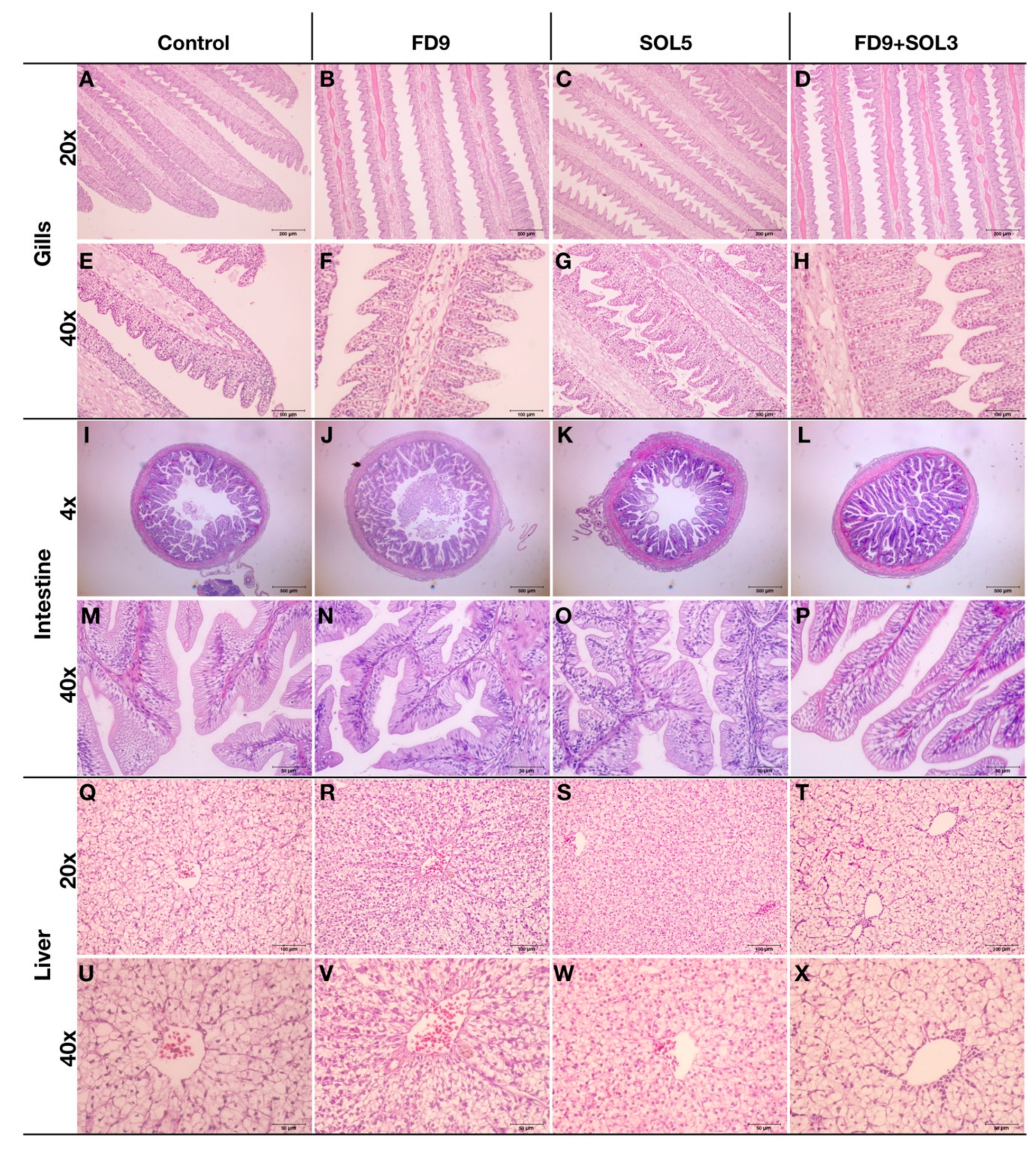
2.6.7. Effects of Acinetobacter KU011TH on the Histopathology of the Gills, Skin, Intestine and Liver of Bighead Catfish

After euthanasia, three fish per treatment were dissected, and their gills, skin, and middle intestine were collected and fixed in a 10% buffered formalin solution for 24 h. After fixation, the samples were cross-sectioned, dehydrated and embedded in paraffin blocks according to standard histological procedures [15,29]. Three-micron thick sections were stained for histological purposes with hematoxylin and eosin (H&E). The histopathology of the tissues was examined with a light microscope with DP2-BSW live imaging software (Olympus, MA, USA).

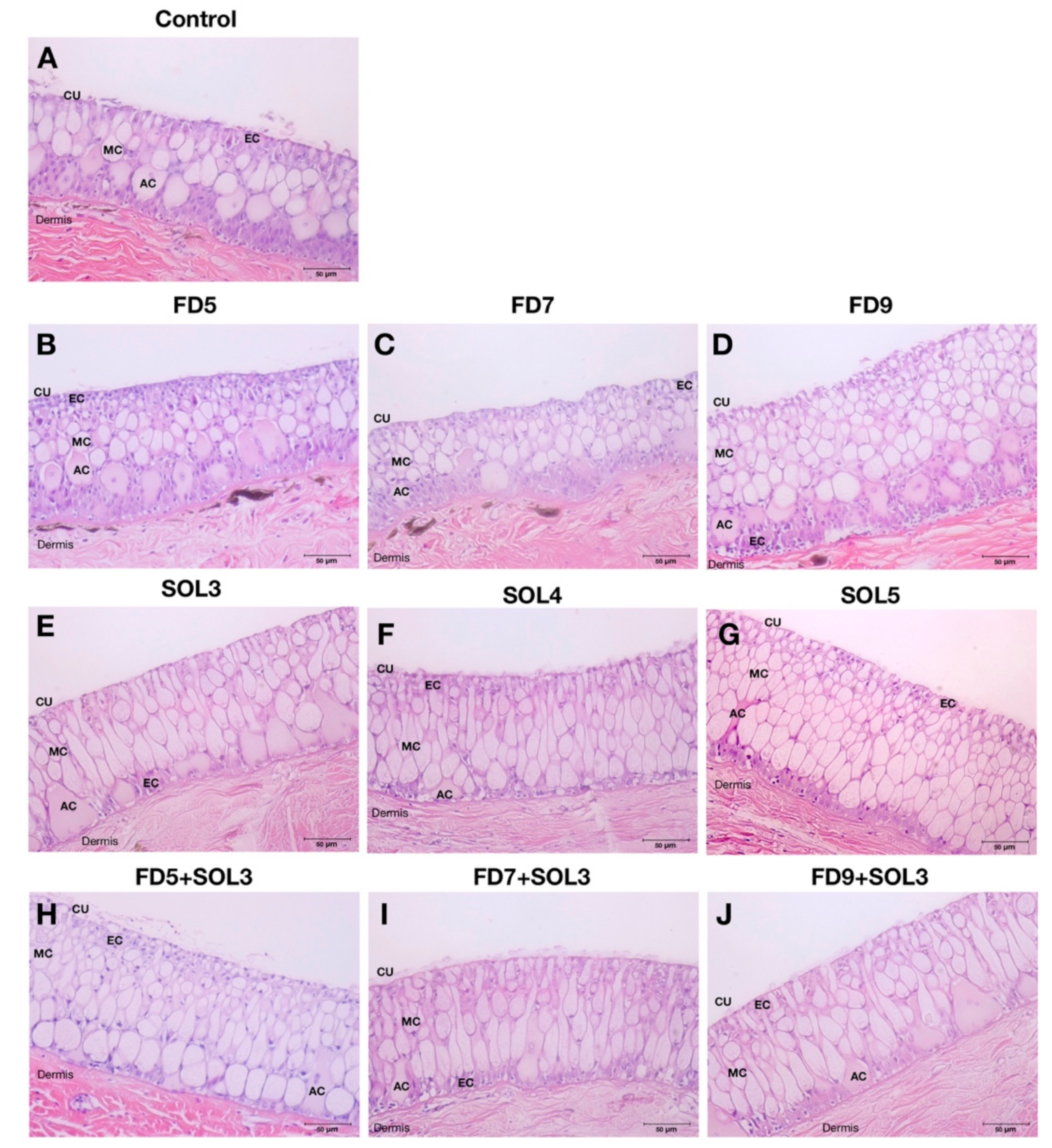
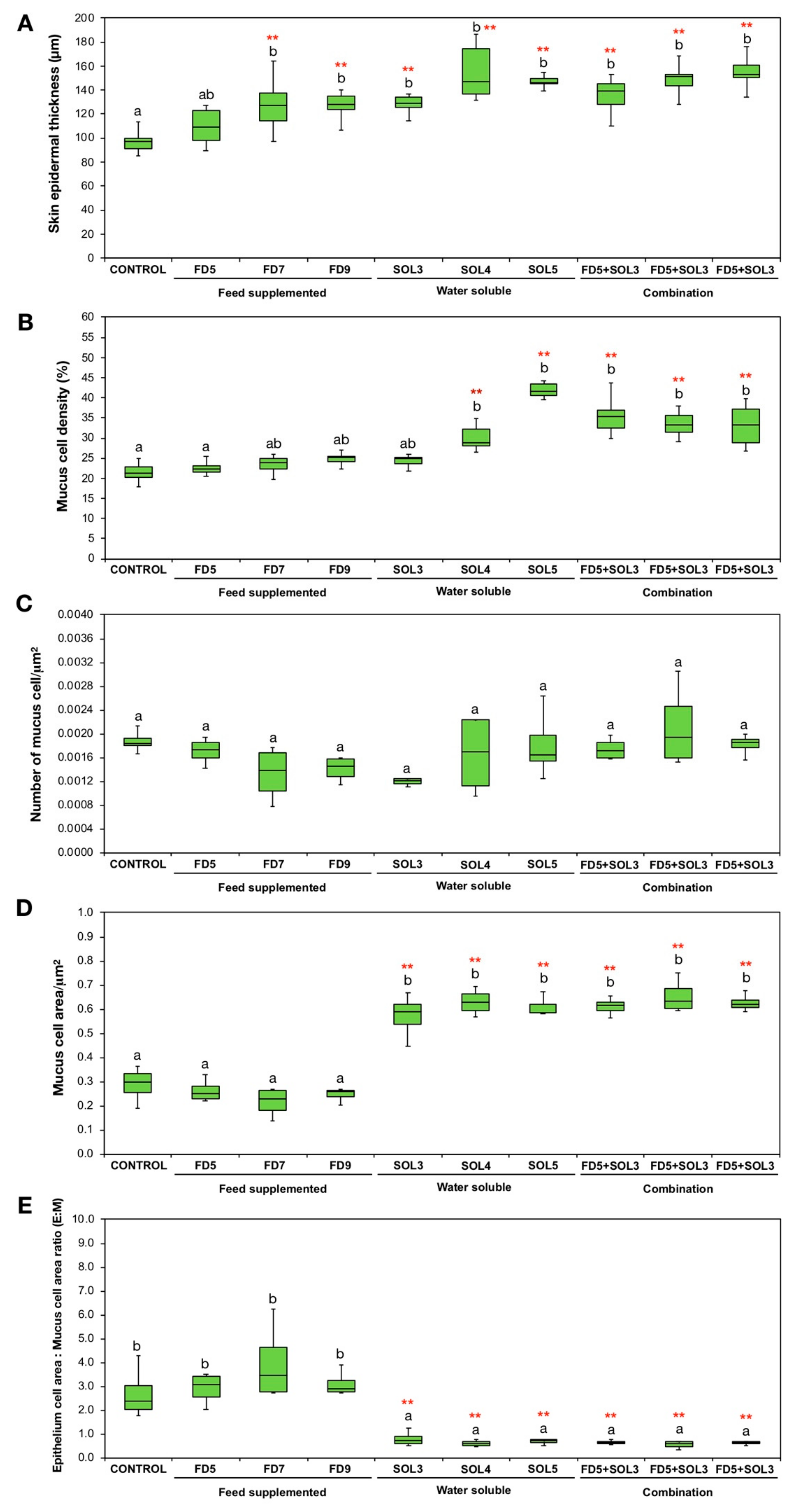
2.6.8. Effects of Acinetobacter KU011TH on the Skin Epithelium and Skin Mucus Cells of Bighead Catfish

The skin epithelium and skin mucus cells were analyzed using the obtained skin histological sections of 3 different fish in each treatment at day 30 after application. The skin histological images of each sample were subjected to analysis of skin mucosal tissues, which are described in [30]. The skin thickness and areas of interest were randomly outlined on skin epithelial tissues for up to three different areas per section using Olympus live imaging software (DP2-BSW, Olympus, MA, USA). Skin epithelial thickness (μm), number of mucous cells/μm2 epithelial area, mucus cell areas/μm2 epithelial area, mucus cell density in the epithelium (%), and the ratio of epithelial cell areas to mucus cell areas (E:M) were obtained using the Materials Image Processing and Automated Reconstruction (MIPAR™) software [31].

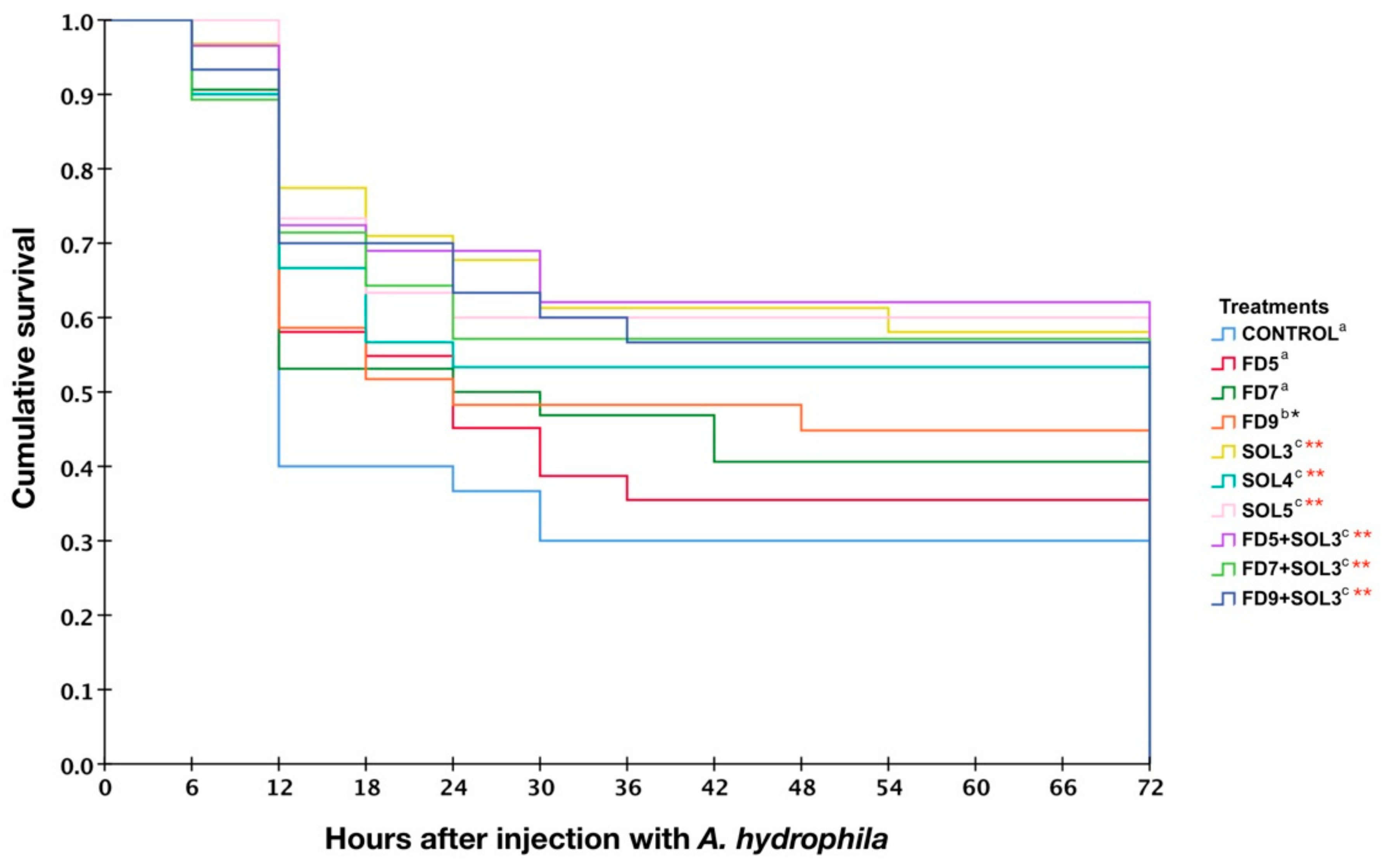
2.6.9. Effects of Acinetobacter KU011TH on Disease Resistance of Bighead Catfish against A. hydrophila

At the end of the experimental trial period (day 30), thirty fish from each treatment (ten fish per tank) were randomly separated for a challenge test with A. hydrophila. A virulent pathogenic bacterium, A. hydrophila AQH0018, was prepared as described above. A. hydrophila was identified based on bacterial morphology and biochemical properties. The preliminary pathogenicity of virulent A. hydrophila toward C. macrocephalus was assessed to validate the optimum dose for the experimental challenge trials. The seven-day median lethal dose (LD50) was assessed by intraperitoneal injection with 0.1 mL of graded doses of A. hydrophila at 1 × 10⁵, 106, 107, 108 and 109 CFU/mL into 10 fish. The LD50 of virulent A. hydrophila toward C. macrocephalus was approximately 1 × 10⁶ CFU/mL. For the infection experiment, the obtained optimal dose was used for the challenge test in the trial. All fish were intraperitonially injected with 0.1 mL of virulent A. hydrophila at 1 × 10⁶ CFU/mL. The mortality was recorded every 6 h for up to 15 days post challenge. Re-isolation of A. hydrophila from moribund fish was performed to verify the cause of the death in fish using a standard diagnostic protocol [32].

2.6.10. Statistical and Data Analysis

The obtained growth performance, innate immune response, bacterial colonization, and skin mucosal immunity data were obtained for three replicates, and the results are expressed as the mean ± standard error of the mean (SEM). Data were statistically analyzed by one-way analysis of variance (ANOVA) and Duncan’s new multiple range test (DMRT) were used to determine differences among groups at days 15 and 30 of the trials, respectively. All statistical analyses were performed using Statistical Package for Social Science (SPSS for Mac version 24.0, Chicago, IL, USA). The level of statistical significance between the control and treatment groups at different times checked was indicated as * (p < 0.05) and ** (p < 0.01) using Student’s t-test.
Survival analysis of bighead catfish in each group was calculated using the Kaplan-Meier method [33]. Cumulative survival plots were generated using SPSS for Mac (version 24.0, Chicago, IL, USA). The level of statistical significance between the control and treatment groups was indicated as * (p < 0.05) and ** (p < 0.01) using Student’s t-test.

3. Results

3.1. Analysis of the Bacterial Shelf Life of the Probiotic and its Efficacy Against Pathogens

The shelf life and efficacy of Acinetobacter KU011TH against A. hydrophila after preservation at 4 °C for ten weeks are presented in Table 2. The total viable bacterial counts were not significantly decreased from the first week until the fourth week, with an approximate decrease of 3.15% to 4.87% (p > 0.05). After four weeks of preservation, the viable bacterial counts decreased slightly by 21.17%, 34.22%, 45.69%, 49.29%, 46.28% and 52.63% from weeks 5 to 10, respectively, compared to those of the control (week 0). However, there were no significant differences in the bacterial numbers from weeks 7 to 10 (p > 0.05). Additionally, the efficacy of Acinetobacter KU011TH against the pathogen A. hydrophila each week was not significantly different in terms of inhibitory clearance zone diameters compared to that of the control (week 0) (p > 0.05).

3.2. Safety and Pathogenicity Assessments of Acinetobacter KU011TH on C. macrocephalus

Upon isolation and characterization of the novel bacterial species Acinetobacter KU011TH from the skin mucus of bighead catfish (C. macrocephalus), the pathogenicity of the host was unsuccessfully assessed through LD50 and LC50 values in this study. Mortality, clinical and pathological features of swimming behavior and skin and gill appearances were not observed over the entire period in either control or treated fish during the trials. The values of LD50 and LC50 could not be obtained for the Acinetobacter KU011TH bacterium, even when the bacteria were applied at 1 × 10¹⁶ CFU/mL. Indeed, no histopathological changes were observed in any of the groups of fish (data not shown). Notably, the novel strain was proven to be a nonpathogenic bacterium toward the host, which warrants further study.

3.3. Effect of a Probiotic Bacterium on Growth Performance in a 120-Day Trial

The effects of Acinetobacter KU011TH on the growth performance of C. macrocephalus are presented in Table 3. After a 120-day trial, fish treated with only a combination of an Acinetobacter KU011TH-supplemented diet at 1 × 10⁹ CFU/kg diet and water-soluble bacteria at 1 × 10³ CFU/mL (FD9+SOL3) showed a significant increase in WG, ADG, RGR, SGR and FCR for body weight compared with those of control groups (p < 0.05). However, there were no significant differences in growth parameters among the control, FD5, FD7, FD9, SOL3, SOL4, SOL5, FD5+SOL3 and FD7+SOL3 groups after the 120-day trial. Additionally, no significant difference was observed in the TLG, ADG, RGR, and SGR for body length in any of the treatments. The fish survival rate after the 120-day trial period was not significantly different among the groups, with averages of 80.24 ± 5.67% to 85.97 ± 6.77% (data not shown).

3.4. Effect of a Probiotic Bacterium on Innate Immune Responses, Immune-Related Gene Expression, and Skin Characteristics of C. macrocephalus

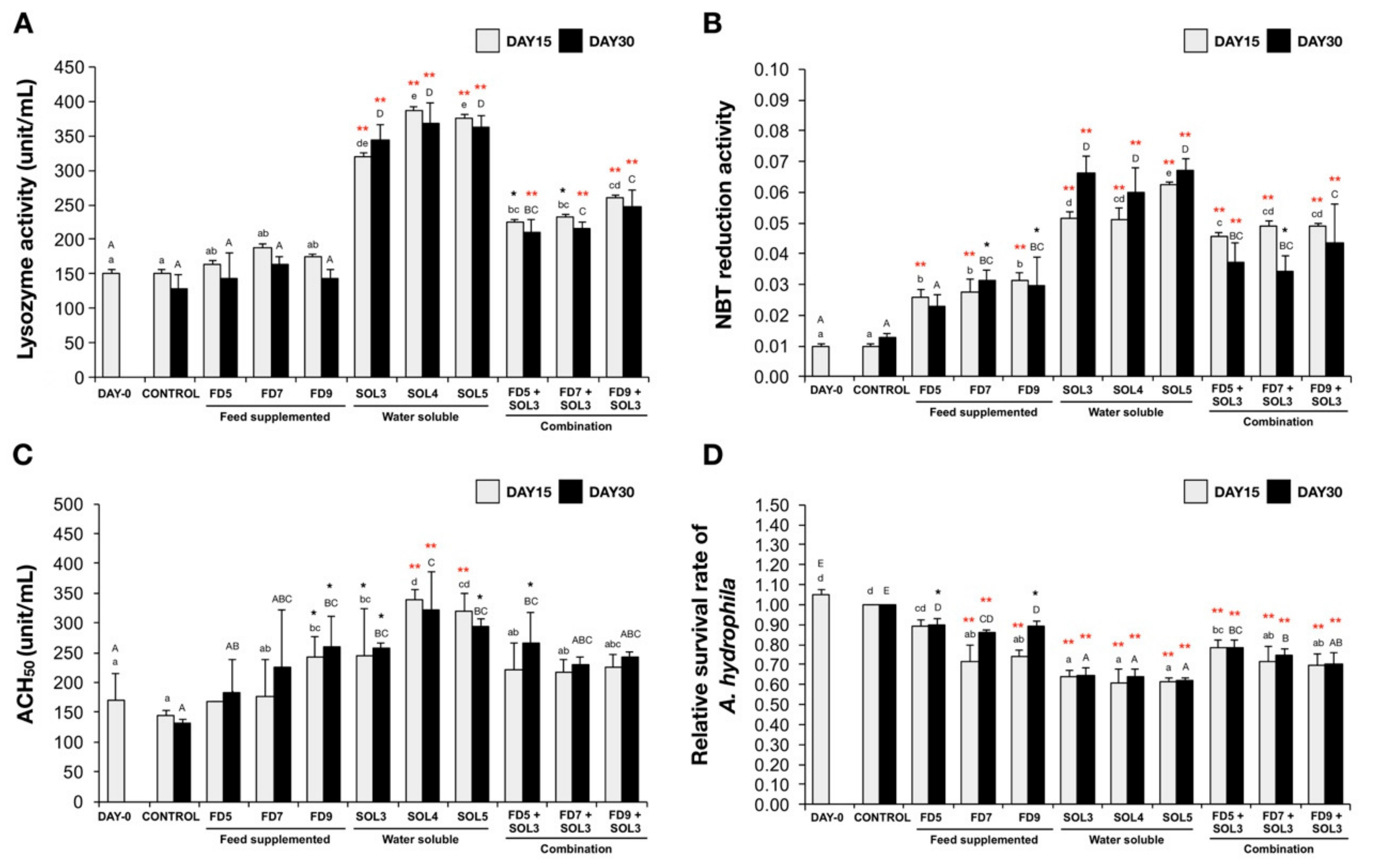
3.4.1. Humoral and Cellular Immune Responses of Innate Immunity

The effects of Acinetobacter KU011TH on innate immune responses, including the PA, lysozyme activity, respiratory burst reaction, ACH50 and bactericidal activity, of C. macrocephalus are presented in Figure 1 and Figure 2. The lysozyme activity of fish administered water-soluble bacteria and the combination with Acinetobacter KU011TH-supplemented diets in the SOL3, SOL4, SOL5, FD5+SOL3, FD7+SOL3 and FD9+SOL3 groups was significantly higher than that of the fish in the control group (p < 0.05 or p < 0.01), but the lysozyme activity of fish in the FD5, FD7 and FD9 groups was not significantly different from that of the fish in control group (p > 0.05) after 15 and 30 days of the trial.
Notably, the highest lysozyme activity was also observed in the groups of fish treated with water-soluble bacteria (SOL), namely, SOL3, SOL4 and SOL5, increasing the lysozyme activity by approximately 2.4-, 2.5-, and 2.6-fold, respectively, compared to that of the fish in the control group at both 15 and 30 days of the trial (Figure 1A).
The respiratory burst (NBT) reaction of all the treatment groups had a statistically significant increase when compared to that of the control groups after 15 and 30 days of the trial (p < 0.05 or p < 0.01), but no significant difference in the respiratory burst reaction was observed in only the FD5 group after 30 days of the trial compared to that of the control group (p > 0.05). The groups of fish treated with water-soluble bacteria (SOL), namely, SOL3, SOL4 and SOL5, exhibited the highest NBT reductions of 3.1-, 2.8, and 3.2-fold, respectively, compared to those of the control group at both 15 and 30 days of the trial (Figure 1B).
The ACH50 levels of fish in the FD9, SOL3, SOL4 and SOL5 groups were significantly higher than those of fish in the control groups after 15 days of the trial (p < 0.05 or p < 0.01). Moreover, the ACH50 levels of fish in the FD9, SOL3, SOL4, SOL5 and FD5+SOL3 groups were also significantly higher than those of fish in the control groups after 30 days of the trial (p < 0.05 or p < 0.01). There was no significant difference in two feed supplementation groups (FD5 and FD7) compared to the control groups (p > 0.05) (Figure 1C).
The serum bactericidal activities of all experimental groups, with the exception of the FD5 group and only day 15, produced a significant decrease in the relative survival rate of A. hydrophila in the bactericidal assay compared to that of the control group after 15 and 30 days of the trial (p < 0.05 or p < 0.01). Notably, the greatest decrease in A. hydrophila abundance was observed in the groups of fish treated with water-soluble bacteria (SOL), namely, SOL3, SOL4 and SOL5, compared to the control, diet supplementation and combination groups (Figure 1D).
The PAs of all experimental treatment groups are illustrated in Figure 2A–K. The PAs of all treatment groups (with the exception of FD5 at day 30) were significantly higher than those of the control groups after 15 and 30 days of the trial (p < 0.05 or p < 0.01) (Figure 2L). Additionally, the PI was significantly increased in the FD7, FD9, SOL3, SOL4, SOL5, FD5+SOL3, FD7+SOL3 and FD9+SOL3 groups after 15 days of the trial and remained increased in the FD9, SOL3, SOL4, SOL5, FD5+SOL3, FD7+SOL3, and FD9+SOL3 groups after 30 days of the trial (p < 0.05 or p < 0.01). The increase in PI for the groups of fish treated with water-soluble bacteria (SOL), namely, SOL3, SOL4 and SOL5, appeared to produce the highest value of the PI in this experiment (Figure 2M).

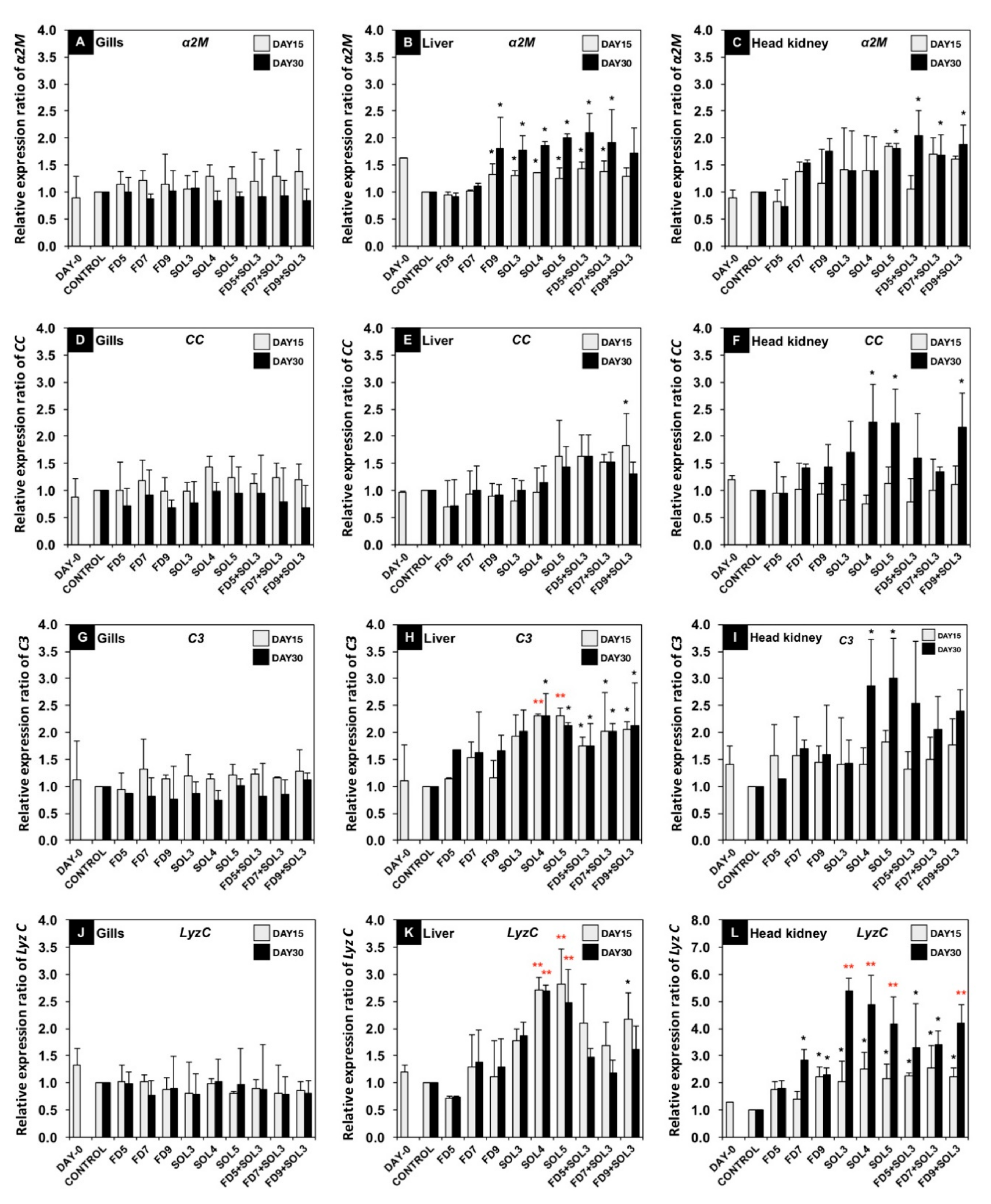
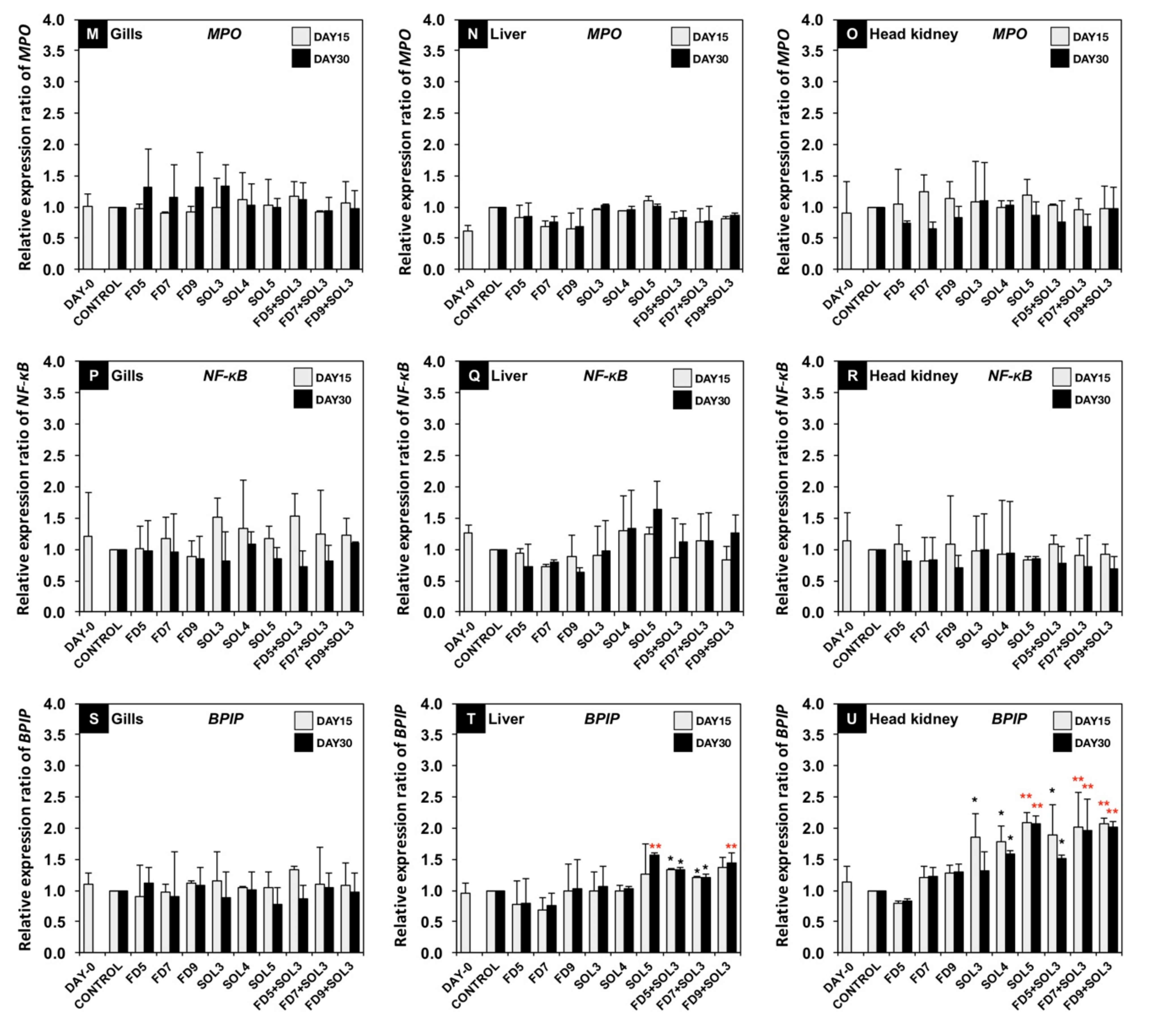
3.4.2. mRNA Expression of Immune-Related Genes

The relative gene expression levels of the immune-related genes α2M, CC, C3, LyzC, MPO, NF-kB and BPIP in the gills, liver and head kidneys at 15 and 30 days of the trial are shown in Figure 3.
α2M gene expression in the liver was significantly upregulated in the treatment groups FD9, SOL3, SOL4, SOL5, FD5+SOL3 and FD7+SOL3 at both 15 and 30 days of the trial (p < 0.05). However, only the SOL5, FD5+SOL3, FD7+SOL3, and FD9+SOL3 groups exhibited significant upregulation of the α2M gene in the head kidney after 30 days of the trial compared to the control groups (p < 0.05). No significant differences in α2M gene expression in gills were detected among all groups after 15 and 30 days of the trial (Figure 3A–C).
CC gene expression in the FD9+SOL3 group at 15 days of the trial and in the SOL4, SOL5, and FD9+SOL3 groups at 30 days of the trial exhibited a significant increase in the liver and head kidney, respectively, compared to that in the control groups (p < 0.05). However, there were no significant differences in CC gene expression in gills among all treatment groups at 15 and 30 days of the trial (Figure 3D–F). Expression of the C3 gene in the liver was significantly upregulated in the SOL4 SOL5, FD5+SOL3, FD7+SOL3 and FD9+SOL3 groups at 15 and 30 days of the trial compared to that in the control groups (p < 0.05 or p < 0.01). The highest significant upregulation of expression of the C3 gene was found in the head kidneys of the SOL4 and SOL5 groups at 30 days of the trial compared to that in the head kidneys of the control groups (p < 0.05). No significant differences in the expression of the C3 gene in gills were detected among all groups at 15 and 30 days of the trial (Figure 3G–I).
The highest upregulation of expression level of the LyzC gene was observed in the livers of the SOL4 and SOL5 groups at 15 and 30 days of the trial compared to that in the livers of the control groups (p < 0.05 or p < 0.01). Additionally, the levels were significantly increased in the head kidneys of the FD7, FD9, SOL3, SOL4, SOL5, FD5+SOL3, FD7+SOL3 and FD9+SOL3 groups at 15 days of the trial compared to those in the head kidneys of the control groups (p < 0.05 or p < 0.01). Furthermore, there were also significant increases in the FD9, SOL3, SOL4, SOL5, FD5+SOL3, FD7+SOL3 and FD9+SOL3 groups at 30 days of the trial compared to the control groups (p < 0.05). There were no significant differences in LyzC gene expression in gills among all treatment groups at 15 and 30 days of the trial (Figure 3J–L).
There were no significant differences in MPO and NF-kB gene expression in the gills, liver, or head kidney among all groups compared to the control groups at 15 and 30 days of the trial (p > 0.05) (Figure 3M–R). However, the expression of the BPIP gene was significantly upregulated in the livers of the SOL5, FD5+SOL3, FD7+SOL3 and FD9+SOL3 groups and in the head kidneys of the SOL4, SOL5, FD5+SOL3, FD7+SOL3 and FD9+SOL3 groups (p < 0.05 or p < 0.01). No significant differences in BPIP gene expression in the gills were observed among all groups at 15 and 30 days of the trial compared to that in the gills of the control groups (p > 0.05) (Figure 3S–U).

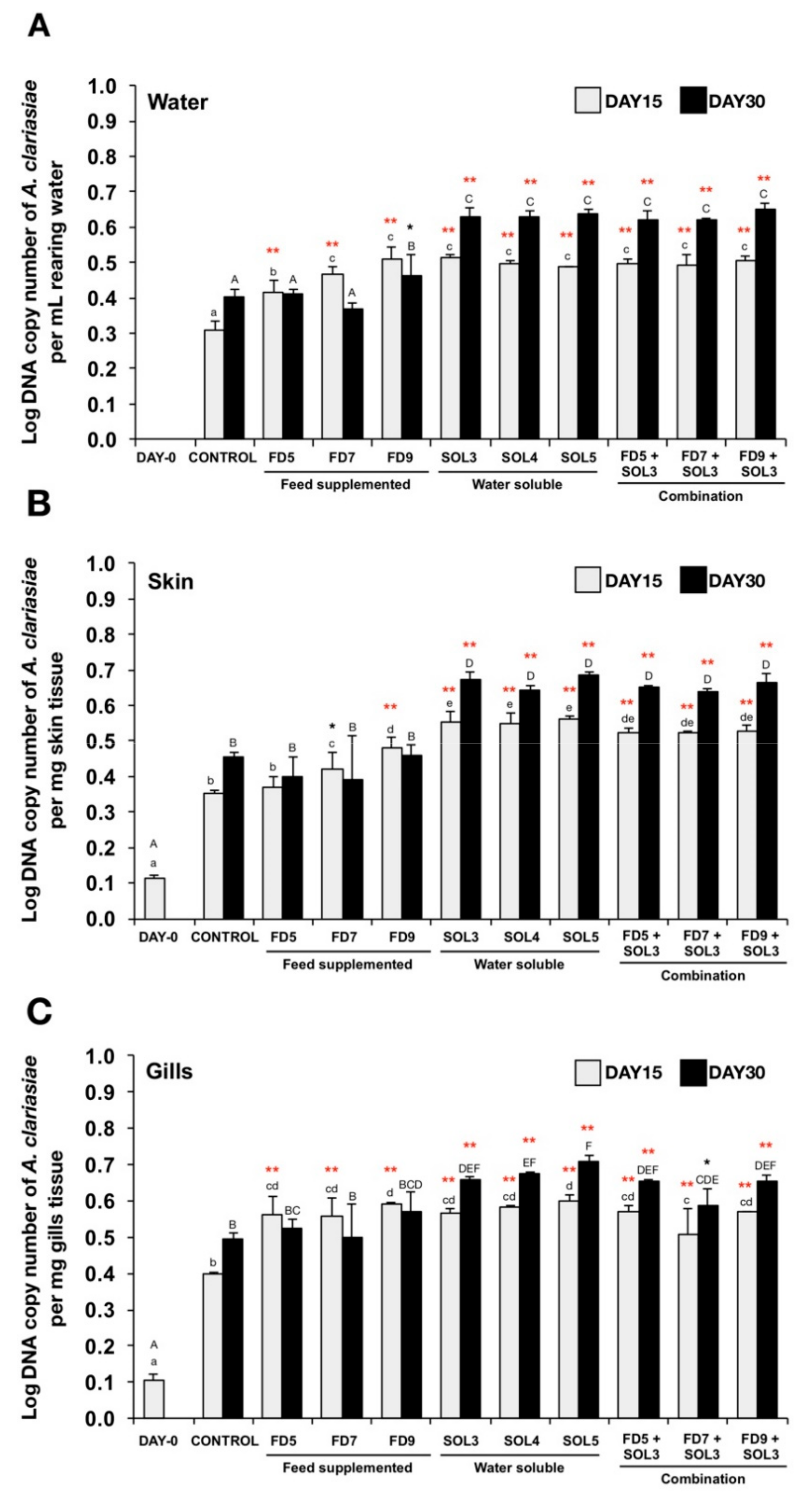
3.4.3. Quantification of Acinetobacter KU011TH in Water, Skin, Gills and Intestine

Bacterial colonization by Acinetobacter KU011TH in water, skin and gills (Figure 4A–C, respectively) was significantly higher in all the treatment groups at 15 days (except FD5 group) of the trial compared to that in the control groups (p < 0.05 or p < 0.01). After 30 days of the trial, the significant increase of bacterial colonization was observed in FD9, SOL3, SOL4, SOL5, FD5+SOL3, FD7+SOL3 and FD9+SOL3 groups for water. In addition, the significant increase of bacterial colonization was also observed in SOL3, SOL4, SOL5, FD5+SOL3, FD7+SOL3, and FD9+SOL3 groups for the skin and gills.
No bacterial DNA copies of Acinetobacter KU011TH were detected in the intestine of any experimental group.

3.4.4. Skin Epithelial and Mucus Cells

The effects of Acinetobacter KU011TH on skin mucus cells of C. macrocephalus are presented in Figure 5 and Figure 6. After 30 days of the trial period, there was a significant increase in epidermal skin thickness in the experimental groups FD7, SOL3, SOL4, SOL5, FD5+SOL3, FD7+SOL3, and FD9+SOL3 compared to the control groups (p < 0.01), but no significant difference was observed between the FD5 and control groups (p > 0.05) (Figure 6A). The increases in mucus cell density in the skin epithelium were highly significant in the SOL4, SOL5, FD5+SOL3, FD7+SOL3, and FD9+SOL3 groups compared to those in the control groups (p < 0.01) (Figure 6B). No significant differences in the number of mucus cells per square micrometer of skin epithelium were detected among all the treatment groups (p > 0.05) (Figure 6C). Nevertheless, the mucus cell area per square micrometer of skin epithelium and E:M ratio showed highly significant differences in the SOL3, SOL4, SOL5, FD5+SOL3, FD7+SOL3, and FD9+SOL3 groups compared to those in the control groups (p < 0.01). No significant differences were observed among the control, FD5, FD7, and FD9 groups (p > 0.05) (Figure 6D–E).

3.4.5. Histopathology of the Skin, Gills, Intestine and Liver

The effects of Acinetobacter KU011TH on histopathological changes in the skin, gills, head kidney and liver are presented in Figure 5 and Figure 7. After 30 days of the trial, no histological changes in skin, gills, intestine, and liver tissues were observed upon examination, which revealed neither clinical signs nor pathological lesions in all treatment groups compared to the control groups. Notably, the intestinal sections of all catfish in the FD9+SOL3 group showed enlargement of the intestinal mucosal epithelium through organization of villi mucosal folds compared to those of the treatment groups.

3.4.6. Resistance against A. hydrophila

The survival rates of C. macrocephalus after challenge with A. hydrophila are shown in Figure 8. After injection, rapid mortality was observed from h 12 to 30, stabilizing at hr 54 until the end of observation at day 15. The disease resistance of fish administered Acinetobacter KU011TH against A. hydrophila demonstrated that the survival of fish in groups FD9, SOL3, SOL4, SOL5, FD5+SOL3, FD7+SOL3, and FD9+SOL3 was significantly higher than that in the control, FD5, and FD7 groups (p < 0.05 or p < 0.01). The survival rates of the FD9, SOL3, SOL4, SOL5, FD5+SOL3, FD7+SOL3 and FD9+SOL3 groups were 48.33 ± 2.36%, 56.70 ± 0.63%, 53.13 ± 4.42%, 58.81 ± 7.74%, 57.69 ± 5.44%, 57.14 ± 10.10%, and 56.67 ± 4.71%, respectively. Meanwhile, the survival rates of the control, FD5, and FD7 groups were 30.00 ± 4.71%, 25.63 ± 7.95%, and 34.76 ± 11.45%, respectively.

4. Discussion

The genus Acinetobacter is currently comprised of fifty-five distinct species that have been originally isolated from various sources, such as human clinical specimens, invertebrate and vertebrate animals, vegetables, soil and water [34,35]. Although research on the genus Acinetobacter has been particularly focused on some species that have effects on human health, especially A. baumannii, the beneficial effects of most of the remaining species on their hosts have never been addressed to date. The present study provides strong evidence of a novel probiotic candidate of the Acinetobacter genus, Acinetobacter KU011TH, which has been proven to be safe and has numerous beneficial effects on its host, C. macrocephalus. Administration of Acinetobacter KU011TH to C. macrocephalus led to a significant increase in growth performance, innate immune responses, skin physical immunity, immune-related gene expression, and disease resistance against the A. hydrophila pathogen. This study is the first report of the application of the genus Acinetobacter as a potential probiotic in vertebrates.
The importance of toxicity and pathogenicity assessments of applied probiotics has been recognized, and these assessments are required for applications in hosts or living organisms. The safety assessment of Acinetobacter KU011TH probiotics in the scaleless host catfish has scientifically been performed thoroughly by both LD50 and LC50 determination. No mortality, clinical or pathological features of swimming behavior and changes in skin and gill appearances were observed during the trials. Thus far, the strain is considered “generally recognized as safe” (GRAS) according to the United States Food and Drug Administration (FDA) designation [36]. The results suggest that the strain is safe for the host and can be applied as a potential probiotic for further study.
The application of probiotics in the aquaculture industry has been successfully achieved in various aquatic animals, especially fish and shellfish [10,16,23]. Moreover, various factors associated with probiotic administration, such as probiotic sources, administration methods, doses, and durations, have been established. Several microorganisms that are normally found in the digestive tracts, skin, gonads and culture systems, such as rearing water and sediment, are often used as probiotics in the aquaculture industry [10,16,37]. Potential probiotics could be administered via the feed as feed additives, directly into the rearing water or as a combination of feed supplementation and administration via rearing water [10,26,38]. Furthermore, a wide range of doses has been commonly applied, such as 1 × 10⁵ to 1 × 10⁹ CFU/g feed and 1 × 10⁵ to 1 × 10⁹ CFU/mL water for feed and rearing water additives, respectively [26,37,39,40]. Probiotics could be practically administered over a wide range of time periods, from two weeks up to eight months [10,16,23,37,41,42]. Unfortunately, there is no evidence to prove which application methods are the best strategies for aquatic animals. Therefore, understanding the nature of probiotic strains and host specificity along with the modes of action may lead to optimization of the appropriate administration methods that can help provide favorable benefits to the hosts.
The viable shelf life of Acinetobacter KU011TH stored at 4 °C was stable during weeks 1–4 but seemingly decreased at week five and stabilized during weeks 7–10. At the end of the experiment, the bacterial concentrations were recorded and were seen to be clearly decreased by approximately 50%. However, the efficacy of Acinetobacter KU011TH in the control of A. hydrophilia remained strong compared to that of any previous storage time. This result suggested that important components used to inhibit the target pathogen were functionally maintained in all remaining bacterial cells, even though half of them were sacrificed. Therefore, further study and validation of powerful preservation methods are required to prolong shelf life for full-scale industrial application.
Growth performance enhancement is the most promising beneficial effect of probiotic administration on the host because it may lead to enhanced appetite and increased growth. According to our findings, a long-term 120-day administration of Acinetobacter KU011TH via a combination of feed and water at concentrations of 1 × 10⁹ CFU/kg diet and 1 × 10³ CFU/mL, respectively, led to significantly increased WG, ADG, RGR and SGR and decreased FCR for body weight compared to those of the control groups (p < 0.05). These improvements could be attributed to the production of exogenous enzymes involved in feed utilization by the strain. In addition, perhaps the strain produces some essential vitamins for growth promotion or may produce enzymes involved in feed utilization [26,43]. Similar to our study, administration of several probiotic bacteria has been proven to promote growth in several fish species, including channel catfish (Ictalurus punctatus), striped catfish (Pangasianodon hypophthalmus), pla-mong (Pangasius bocourti), grouper (Epinephelus coioides), rainbow trout (Oncorhynchus mykiss), and starry flounder (Platichthys stellatus) [24,25,26,27,44,45,46,47].
In the long-term experimental trial, based on the obtained information, application of Acinetobacter KU011TH as probiotics for catfish rearing was obviously found to increase the catfish growth rate solely upon administration via a combination of water and diet supplementation (FD9 + SOL3). However, colonization by Acinetobacter KU011TH in this study was not detected in the intestines of the treated groups or the control group by qPCR. The results suggest that the probiotic bacterium cannot survive and colonize during passage through the GI tract. Indeed, this bacterium can be destroyed by the acidity, digestive enzymes and lack of oxygen in the GI tract, which is not the original source of this bacterium. Nevertheless, the results also suggest that the strain may influence GI tract performance by sacrificing itself and providing some important nutrients for the host and its microbiota. Their interactions could extensively promote GI mucosal cell proliferation to give rise to all cell lineages, such as epithelial or goblet cells, and enhance mucosal barrier function, as found in a previous study [48,49,50] and in this study in the FD9+SOL3 fish treatment group (Figure 7L,P). This effect must be due to unknown important factors that improve the optimal balance of the gut microbiome and nutrient utilization of both the host and microorganisms, which consequently influences and elevates host growth eventually. The results suggest that this bacterium is a beneficial normal flora microorganism found in the body and in the environment surrounding the catfish.
Modulation of the immune system is an important characteristic of probiotic bacteria that could enhance innate immune responses in both humoral and cellular defense mechanisms that act as a first line of protection against detrimental substances or pathogens [23,41]. Probiotics interact with humoral defense mechanisms by enhancing various serum enzyme activities associated with bacteriolysis against Gram-positive and Gram-negative bacteria, such as lysozyme, respiratory burst, as well as complement and bactericidal activities. In addition, probiotics could also actively stimulate innate cellular defenses through immune cells, such as phagocytic cells, including monocytes, macrophages and neutrophils, to enhance innate immune responses [23]. In our findings, the probiotic bacteria could markedly increase both innate humoral and cellular defenses through lysozyme activity, respiratory burst activity, ACH50, serum bactericidal activity, PA and PI in fish administered Acinetobacter KU011TH through feed, water or a combination of the two routes. Moreover, the Acinetobacter KU011TH probiotics demonstrated upregulatory effects on the expression of the α2M, CC, C3, LyzC and BPIP genes in catfish livers and head kidneys [24].
There are associations with the regulation of nonspecific humoral factor responses, which are also regarded as biological markers for determining fish health [22,23]. In general, the most effective strategy for activating immune responses by probiotic bacterium administration in previous reports was typically found to be administration via water, a combination of feed and water, or feed [26,38,51,52]. However, most of the studied probiotics exhibited the highest efficacy via only feeding with probiotic-supplemented diets because most of these probiotics were spore-forming organisms that were originally isolated from the digestive tracts of fish. In contrast, the Acinetobacter KU011TH probiotic bacterium was originally isolated from the skin mucus of bighead catfish and can be beneficial to the host in all practical applications in the aquaculture industry. Although the mode of action could not be elucidated in this study, the most likely mechanism can be hypothesized. Lipopolysaccharide (LPS) and peptidoglycan (PGN) are classified as abundant pathogen-associated molecular patterns (PAMPs) found on the cell surfaces of Gram-positive and Gram-negative bacteria, respectively [53]. LPS and PGN from probiotic bacteria have been widely reported as immunostimulants in several aquatic animals and successfully enhance innate humoral and cellular immune responses. The current study suggests that Acinetobacter KU011TH provides LPS to activate and improve innate immune responses in both humoral and cellular defenses in bighead catfish. Such probiotic water additive-induced innate immunity could effectively increase the interactions between PAMPs and nonspecific immune receptors by triggering fish mucosal immunity, such as in the gills, nares and skin, which are the primary surface organs that are under constant exposure to the environment [54]. Indeed, the interaction can also increase the interactions of the intestinal epithelium and immune cells in the GI tract via accidental ingestion of bacteria from the surrounding water, resulting in local activation of the fish innate immune system.
Recently, immunomodulatory features of probiotics in aquatic animals were suggested by many studies that be associated with mucosal immunity, which plays a key role in the first-line defense against harmful pathogens and is thus considered to be the most active immunological site in fish [25,54]. Mucosal immunity of fish is driven by mucosa-associated lymphoid tissues (MALTs). These sites are divided into gut-associated lymphoid tissues (GALTs), skin-associated lymphoid tissues (SALTs), gill-associated lymphoid tissues (GIALTs) and, recently, nasopharynx-associated lymphoid tissues (NALTs) [54]. Moreover, colonization by probiotic bacteria represents the persistence of living microbial cells that adhere to favorable surfaces, including in rearing water and mucosal surfaces, such as the gut, skin and gills. The current study also demonstrated that a probiotic bacterium, Acinetobacter KU011TH, could colonize rearing water and the skin, and gills but not the intestine. This finding suggested that Acinetobacter KU011TH is a probiotic microbe that influences MALTs, especially the SALT, of the scaleless bighead catfish. Acinetobacter KU011TH affects the host by maintenance of the microbiota and mucosal surface and reinforcement of immunity. The administration of rich, potent probiotics to the mucosal surface has become a prominent mode of immunomodulation and control of infectious disease in aquatic animals [25,54,55,56].
In fish, skin and skin mucus cells are directly influenced by SALTs, which are classified as a first-line defense of innate immune responses to both physical and chemical approaches. Fish skin cells are typically localized with a large number of different nonspecific immune cells [57]. However, the skin of all fish species is constructed similarly to that of other vertebrates, consisting of two basic layers, namely, the epidermis and dermis. Indeed, the mucus cells, club cells, alarm cells and sensory cells are abundantly embedded in the epidermal layer [57]. Recently, mucus cells have been reported as the major immunocompetent cells that contain a rich assortment of biologically active components, especially antimicrobial proteins (AMPs), such as alkaline phosphatase, cathepsin B, complement, transferrin, lysozyme, hepcidin, cathelicidins, apolipoproteins, piscidin, pleurocidin and C-reactive protein [58,59]. During the last decade, it has been reported that several types of AMPs, including H2A, H1, H6, cathelicidins and piscidin, are produced by skin mucus cells of rainbow trout (Oncorhynchus mykiss) [55,56]. Additionally, the skin mucus of Clarias batrachus exhibited strong inhibitory activity against five pathogenic bacteria, namely Escherichia coli, A. hydrophila, Pseudomonas aeruginosa, Vibrio fischeri and Vibrio anguillarum, on agar plates [60]. Several recent studies have demonstrated that enhancement of SALT performance leads to increased skin mucus levels, skin AMP levels, skin epidermal thickness, number of mucus cells and mucus cell areas in fish fed a probiotic-supplemented diet, such as angelfish (Pterophyllum scalare), common carp (Cyprinus carpio), European sea bass (Dicentrarchus labrax) and Atlantic salmon (Salmo salar L.) [30,40,61,62]. Similar to the results of our study, significantly increased epidermal skin thickness, mucus cell density, and mucus cell areas and decreased E:M ratios were observed in fish administered Acinetobacter KU011TH through feeding, as a water additive and their combination for 30 days. It has been suggested that the probiotic Acinetobacter KU011TH may exert an influence on the SALT, GALT, NALT and GIALT performances of bighead catfish, but the mechanisms involved remain unknown.
Notably, histological analyses of the skin and intestine strongly confirmed the histoarchitectural performance of SALTs in all treatment groups and GIALTs in the FD9+SOL3 group compared to those in the control group. Furthermore, probiotic Acinetobacter KU011TH administration was associated with enhanced innate immune responses and increased skin physical immunity without negative effects on the histoarchitectures of the target tissue organs, including the skin, gills, intestine and liver, of fish administered the probiotic for 30 days.
The final proof of the magnitude of the effect of the probiotic bacteria on aquatic animals was performed by health assessments through challenge tests with specific harmful pathogens that cause infectious diseases. Fish typically respond to any pathogens via the entire innate and adaptive immune responses with humoral and cellular mechanisms to contain infectious diseases [63]. The initial responses are primarily driven by nonspecific humoral factors, including growth-inhibiting substances, antimicrobial peptides, complement peptides, and nonspecific cellular responses, such as phagocytosis. Furthermore, in certain infectious disease outbreaks, strong potential responses [37] associated with adaptive immune responses are required, which are driven by mainly immunoglobulins [64]. In accordance with our finding, an increased survival rate after challenge with A. hydrophila was observed in fish administered Acinetobacter KU011TH in all experimental treatment groups. Our findings suggest that administration of Acinetobacter KU011TH by feeding, as water additive and their combination activates fish immunity through several mechanisms of both innate and adaptive immune responses, leading to disease resistance against A. hydrophila. Furthermore, previous studies of probiotic administration in fish have shown successfully increased survival rates after challenge with pathogens in several fish, including O. niloticus, O. mykiss and Labeo rohita. These administered probiotics were also found to enhance innate immune responses, including respiratory burst, lysozyme and phagocytic activities [39,43,65].
Among application routes, water supplementation with 1 × 10³–105 CFU/mL Acinetobacter KU011TH was the most predominant method that strongly elevated both innate immune parameters and immune-gene e × pression. Feed supplementation with 1 × 10⁵−107 CFU/mL probiotic was less effective than the other routes but exhibited significant differences compared to the control group. Surprisingly, the combination of the water- (1 × 10³ CFU/mL) and feed-supplemented (1 × 10⁵–109 CFU/kg diet) routes did not have a synergistic effect over the others, but the effect remained stronger than that of the control and feed-supplemented groups. When considered together with pathogen challenge, the data clearly showed that the combination of water supplemented with 103 CFU/mL and feed supplemented with1 × 103, 1 × 10⁵ and1 × 107 CFU/kg diet effectively enhanced catfish resistance against the harmful pathogen. These results suggested that water-soluble application of the viable probiotic at concentrations ranging from 1 × 10³–105 CFU/mL at three-day intervals for 30 days is sufficient to elevate the catfish immune system to safeguard the fish from A. hydrophila infection.
Additionally, supplementation with Acinetobacter KU011TH via both rearing water and feed in the short trial clearly demonstrated that all skin characteristic parameters were significantly improved compared to those of the control and feed-supplemented probiotic treatment groups. This result indicated that adding this bacterium to fish feed did not lead to skin alteration, and no synergistic effect of the combinatorial application of this bacterium was observed. The improvement of skin characteristics may also greatly induce first-line defense mechanisms to effectively protect catfish from several contaminating pathogens in water. Furthermore, the impenetrability of mucosal immunity at the gills, nares, and skin of the scaleless fish, which is crucial for preventing invasion by all pathogens from the external environment, may be properly strengthened after probiotic application. The use of this probiotic will be a crucial and novel application for disease prophylaxis strategies during walking catfish aquaculture.

5. Conclusions

The importance of the achievements from the use of probiotics in catfish cultures has been established in this study. Acinetobacter KU011TH is a novel probiotic bacterial species in the genus Acinetobacter that was originally isolated from the skin mucus of bighead catfish. From previous research, this strain exhibited high efficacy against several pathogenic bacteria of freshwater fish in vitro, such as A. hydrophila, F. columnare, F. roseus, S. agalactiae, S. warneri and E. tarda [12]. To date, Acinetobacter KU011TH has appreciably enhanced catfish health performance, increasing growth parameters, innate humoral and cellular responses, immune-related gene expression, skin physical defense and disease resistance. Additionally, Acinetobacter KU011TH has been proven to be a nonpathogenic probiotic toward C. macrocephalus and does not negatively affect any histopathological changes in the target organs. Acinetobacter KU011TH in the catfish aquaculture industry could be practically applied as an additive in rearing water or as a combination of water additive and feed additive for enhanced benefits. Certainly, among the probiotic candidates for aquatic animals suggested during this decade, Acinetobacter KU011TH could be a noteworthy species with applications in the catfish industry, achieving a reduction in disease outbreaks and antibiotic application, which have been longstanding issues for the aquaculture industry worldwide.

Author Contributions

A.B. designed and conducted the experiments and wrote the manuscript. U.N. revised the manuscript. P.S. designed the experiments and revised the manuscript. All authors listed have read and approved the manuscript for publication.

Funding

This work was financially supported by the Thailand Research Fund (TRF) and the Betagro Science Center, Thailand. This study is part of the project entitled “Genetics and Biotechnology for Improvement of Aquatic Animal Production” (contract no. DPG5980003) awarded to Professor Uthairat Na-Nakorn under the “Distinguished Research Professor 2016 Award.”

Acknowledgments

We would also like to thank the Graduate School at Kasetsart University, Thailand, for partial financial support of the student who is the first author of this article.

Conflicts of Interest

The authors declare that this research was conducted in the absence of any commercial or financial relationship that could be construed as a potential conflict of interest.

References

  1. Food and Agriculture Organization of the United Nations (FAO). Fishstat Plus Rome 2017. In: FAO Fisheries and Aquaculture Department. Rome. Updated 14 September 2017. Available online: http://www.fao.org/fishery/statistics/software/fishstat/en (accessed on 1 December 2018).
  2. Na-Nakorn, U.; Kamonrat, W.; Ngamsiri, T. Genetic diversity of walking catfish, Clarias macrocephalus, in Thailand and evidence of genetic introgression from introduced farmed C. gariepinus. Aquaculture 2004, 240, 145–163. [Google Scholar] [CrossRef]
  3. Chatchaiphan, S.; Srisapoome, P.; Kim, J.H.; Devlin, R.H.; Na-Nakorn, U. De novo transcriptome characterization and growth-related gene expression profiling of diploid and triploid bighead catfish (Clarias macrocephalus Gunther, 1864). Mar. Biotechnol. (Ny) 2017, 19, 36–48. [Google Scholar] [CrossRef] [PubMed]
  4. Suanyuk, N.; Rogge, M.; Thune, R.; Watthanaphiromsakul, M.; Champhat, N.; Wiangkum, W. Mortality and pathology of hybrid catfish, Clarias macrocephalus (Gunther) x Clarias gariepinus (Burchell), associated with Edwardsiella ictaluri infection in southern Thailand. J. Fish. Dis. 2014, 37, 385–395. [Google Scholar] [CrossRef] [PubMed]
  5. Na-Nakorn, U.; Chantsawang, S.; Tarnchalanukit, W. Response to mass selection for disease resistance in walking catfish, Clarias macrocephalus. J. Appl. Aquac. 1995, 4, 65–74. [Google Scholar] [CrossRef]
  6. Hoseinifar, S.H.; Dadar, M.; Ringø, E. Modulation of nutrient digestibility and digestive enzyme activities in aquatic animals: The functional feed additives scenario. Aquac. Res. 2017, 48, 3987–4000. [Google Scholar] [CrossRef]
  7. Nawaz, A.; Bakhsh javaid, A.; Irshad, S.; Hoseinifar, S.H.; Xiong, H. The functionality of prebiotics as immunostimulant: Evidences from trials on terrestrial and aquatic animals. Fish Shellfish Immunol. 2018, 76, 272–278. [Google Scholar] [CrossRef]
  8. Fuller, R. Probiotics in man and animals. J. Appl. Bacteriol. 1989, 66, 365–378. [Google Scholar]
  9. Merrifield, D.L.; Dimitroglou, A.; Foey, A.; Davies, S.; Baker, R.T.M.; Bøgwald, J.; Castex, M.; Ringø, E. The current status and future focus of probiotic and prebiotic applications for salmonids. Aquaculture 2010, 302, 1–18. [Google Scholar] [CrossRef]
  10. Martínez Cruz, P.; Ibáñez, A.L.; Monroy Hermosillo, O.A.; Ramírez Saad, H.C. Use of probiotics in aquaculture. ISRN Microbiol. 2012, 2012, 916845. [Google Scholar] [CrossRef]
  11. Van Doan, H.; Hoseinifar, S.H.; Ringø, E.; Ángeles Esteban, M.; Dadar, M.; Dawood, M.A.O.; Faggio, C. Host-Associated Probiotics: A Key Factor in Sustainable Aquaculture. Rev. Fish. Sci. Aquac. 2019, 1–27. [Google Scholar] [CrossRef]
  12. Bunnoy, A.; Na-Nakorn, U.; Kayansamruaj, P.; Srisapoome, P. Acinetobacter strain KUO11TH, a unique organism related to Acinetobacter pittii and isolated from the skin mucus of healthy bighead catfish and its efficacy against several fish pathogens. Microorganisms 2019, 7, 459. [Google Scholar] [CrossRef] [PubMed]
  13. Chinedu, E.; Arome, D.; Ameh, F.S. A new method for determining acute toxicity in animal models. Int. J. Toxicol. 2013, 20, 224–226. [Google Scholar] [CrossRef] [PubMed]
  14. Boyum, A. Isolation of mononuclear cells and granulocytes from human blood. Isolation of monuclear cells by one centrifugation, and of granulocytes by combining centrifugation and sedimentation at 1 g. Scand. J. Clin. Lab. Invest. Suppl. 1968, 97, 77–89. [Google Scholar] [PubMed]
  15. Bernet, D.; Schmidt, H.; Meier, W.; Burkhardt-Holm, P.; Wahli, T. Histopathology in fish: proposal for a protocol to assess aquatic pollution. J. Fish. Dis. 1999, 22, 25–34. [Google Scholar] [CrossRef]
  16. Lugert, V.; Thaller, G.; Tetens, J.; Schulz, C.; Krieter, J. A review on fish growth calculation: multiple functions in fish production and their specific application. Rev. Aquac. 2016, 8, 30–42. [Google Scholar] [CrossRef]
  17. Ito, Y.; Yamada, H.; Imoto, T. Colorimetric assay for lysozyme using Micrococcus luteus labeled with a blue dye, Remazol brilliant blue R, as a substrate. Chem. Pharm. Bull. (Tokyo) 1992, 40, 1523–1526. [Google Scholar] [CrossRef] [PubMed]
  18. Segal, A.W.; Levi, A.J. Cell damage and dye reduction in the quantitative nitroblue tetrazolium (NBT) test. Clin. Exp. Immunol. 1975, 19, 309–318. [Google Scholar]
  19. Nayak, S.; Portugal, I.; Zilberg, D. Analyzing complement activity in the serum and body homogenates of different fish species, using rabbit and sheep red blood cells. Vet. Immunol. Immunopathol. 2018, 199, 39–42. [Google Scholar] [CrossRef]
  20. Guo, L.; Zhang, F.; Wang, X.; Chen, H.; Wang, Q.; Guo, J.; Cao, X.; Wang, L. Antibacterial activity and action mechanism of questin from marine Aspergillus flavipes HN4-13 against aquatic pathogen Vibrio harveyi. 3 Biotech. 2019, 9, 1–14. [Google Scholar] [CrossRef]
  21. Koenigsknecht, J.; Landreth, G. Microglial phagocytosis of fibrillar beta-amyloid through a beta1 integrin-dependent mechanism. J. Neurosci. 2004, 24, 9838–9846. [Google Scholar] [CrossRef]
  22. Panigrahi, A.; Kiron, V.; Kobayashi, T.; Puangkaew, J.; Satoh, S.; Sugita, H. Immune responses in rainbow trout Oncorhynchus mykiss induced by a potential probiotic bacteria Lactobacillus rhamnosus JCM 1136. Vet. Immunol. Immunopathol. 2004, 102, 379–388. [Google Scholar] [CrossRef] [PubMed]
  23. Hai, N.V. Research findings from the use of probiotics in tilapia aquaculture: A review. Fish Shellfish Immunol. 2015, 45, 592–597. [Google Scholar] [CrossRef] [PubMed]
  24. Hoseinifar, S.H.; Hosseini, M.; Paknejad, H.; Safari, R.; Jafar, A.; Yousefi, M.; Van Doan, H.; Torfi Mozanzadeh, M. Enhanced mucosal immune responses, immune related genes and growth performance in common carp (Cyprinus carpio) juveniles fed dietary Pediococcus acidilactici MA18/5M and raffinose. Dev. Comp. Immunol. 2019, 94, 59–65. [Google Scholar] [CrossRef] [PubMed]
  25. Liu, C.-H.; Chiu, C.-H.; Wang, S.-W.; Cheng, W. Dietary administration of the probiotic, Bacillus subtilis E20, enhances the growth, innate immune responses, and disease resistance of the grouper, Epinephelus coioides. Fish Shellfish Immunol. 2012, 33, 699–706. [Google Scholar] [CrossRef] [PubMed]
  26. Meidong, R.; Khotchanalekha, K.; Doolgindachbaporn, S.; Nagasawa, T.; Nakao, M.; Sakai, K.; Tongpim, S. Evaluation of probiotic Bacillus aerius B81e isolated from healthy hybrid catfish on growth, disease resistance and innate immunity of Pla-mong Pangasius bocourti. Fish Shellfish Immunol. 2018, 73, 1–10. [Google Scholar] [CrossRef]
  27. Truong Thy, H.T.; Tri, N.N.; Quy, O.M.; Fotedar, R.; Kannika, K.; Unajak, S.; Areechon, N. Effects of the dietary supplementation of mixed probiotic spores of Bacillus amyloliquefaciens 54A, and Bacillus pumilus 47B on growth, innate immunity and stress responses of striped catfish (Pangasianodon hypophthalmus). Fish Shellfish Immunol. 2017, 60, 391–399. [Google Scholar] [CrossRef]
  28. Livak, K.J.; Schmittgen, T.D. Analysis of relative gene expression data using real-time quantitative PCR and the 2(-Delta Delta C(T)) method. Methods 2001, 25, 402–408. [Google Scholar] [CrossRef]
  29. Fischer, A.H.; Jacobson, K.A.; Rose, J.; Zeller, R. Hematoxylin and eosin staining of tissue and cell sections. Csh. Protoc. 2008, 49, 86. [Google Scholar] [CrossRef]
  30. Torrecillas, S.; Montero, D.; Caballero, M.J.; Pittman, K.A.; Custodio, M.; Campo, A.; Sweetman, J.; Izquierdo, M. Dietary mannan oligosaccharides: counteracting the side effects of soybean meal oil inclusion on European sea bass (Dicentrarchus labrax) gut health and skin mucosa mucus production? Front. Immunol. 2015, 6, 397. [Google Scholar] [CrossRef]
  31. Sosa, J.M.; Huber, D.E.; Welk, B.; Fraser, H.L. Development and application of MIPAR™: a novel software package for two- and three-dimensional microstructural characterization. IMMI 2014, 3, 10. [Google Scholar] [CrossRef]
  32. Kent, M.L.; Feist, S.W.; Harper, C.; Hoogstraten-Miller, S.; Law, J.M.; Sánchez-Morgado, J.M.; Tanguay, R.L.; Sanders, G.E.; Spitsbergen, J.M.; Whipps, C.M. Recommendations for control of pathogens and infectious diseases in fish research facilities. Comp. Biochem. Physiol. Toxicol. Pharm. 2009, 149, 240–248. [Google Scholar] [CrossRef] [PubMed]
  33. Bewick, V.; Cheek, L.; Ball, J. Statistics review 12: survival analysis. Crit Care 2004, 8, 389–394. [Google Scholar] [CrossRef] [PubMed]
  34. Kim, P.S.; Shin, N.R.; Kim, J.Y.; Yun, J.H.; Hyun, D.W.; Bae, J.W. Acinetobacter apis sp. nov., isolated from the intestinal tract of a honey bee, Apis mellifera. J. Microbiol. 2014, 52, 639–645. [Google Scholar] [CrossRef] [PubMed]
  35. Liu, S.; Wang, Y.; Ruan, Z.; Ma, K.; Wu, B.; Xu, Y.; Wang, J.; You, Y.; He, M.; Hu, G. Acinetobacter larvae sp. nov., isolated from the larval gut of Omphisa fuscidentalis. Int. J. Syst. Evol. Microbiol. 2017, 67, 806–811. [Google Scholar] [CrossRef]
  36. O’Sullivan, M.G.; Thornton, G.; O’Sullivan, G.C.; Collins, J.K. Probiotic bacteria: myth or reality? Trends Food Sci. Technol. 1992, 3, 309–314. [Google Scholar] [CrossRef]
  37. Brunt, J.; Austin, B. Use of a probiotic to control lactococcosis and streptococcosis in rainbow trout, Oncorhynchus mykiss (Walbaum). J. Fish. Dis. 2005, 28, 693–701. [Google Scholar] [CrossRef]
  38. Salinas, I.; Cuesta, A.; Esteban, M.A.; Meseguer, J. Dietary administration of Lactobacillus delbrueckii and Bacillus subtilis, single or combined, on gilthead seabream cellular innate immune responses. Fish Shellfish Immunol. 2005, 19, 67–77. [Google Scholar] [CrossRef]
  39. Aly, S.M.; Abdel-Galil Ahmed, Y.; Abdel-Aziz Ghareeb, A.; Mohamed, M.F. Studies on Bacillus subtilis and Lactobacillus acidophilus, as potential probiotics, on the immune response and resistance of Tilapia nilotica (Oreochromis niloticus) to challenge infections. Fish Shellfish Immunol. 2008, 25, 128–136. [Google Scholar] [CrossRef]
  40. Azimirad, M.; Meshkini, S.; Ahmadifard, N.; Hoseinifar, S.H. The effects of feeding with synbiotic (Pediococcus acidilactici and fructooligosaccharide) enriched adult Artemia on skin mucus immune responses, stress resistance, intestinal microbiota and performance of angelfish (Pterophyllum scalare). Fish Shellfish Immunol. 2016, 54, 516–522. [Google Scholar] [CrossRef]
  41. Irianto, A.; Austin, B. Use of probiotics to control furunculosis in rainbow trout, Oncorhynchus mykiss (Walbaum). J. Fish. Dis. 2002, 25, 333–342. [Google Scholar] [CrossRef]
  42. Aly, S.M.; Mohamed, M.F.; John, G. Effect of probiotics on the survival, growth and challenge infection in Tilapia nilotica (Oreochromis niloticus). Aquac. Res. 2008, 39, 647–656. [Google Scholar] [CrossRef]
  43. Nayak, S.K.; Swain, P.; Mukherjee, S.C. Effect of dietary supplementation of probiotic and vitamin C on the immune response of Indian major carp, Labeo rohita (Ham.). Fish Shellfish Immunol. 2017, 23, 892–896. [Google Scholar] [CrossRef] [PubMed]
  44. Thurlow, C.M.; Williams, M.A.; Carrias, A.; Ran, C.; Newman, M.; Tweedie, J.; Allison, E.; Jescovitch, L.N.; Wilson, A.E.; Terhune, J.S.; et al. Bacillus velezensis AP193 exerts probiotic effects in channel catfish (Ictalurus punctatus) and reduces aquaculture pond eutrophication. Aquaculture 2019, 503, 347–356. [Google Scholar] [CrossRef]
  45. Mohammadian, T.; Nasirpour, M.; Tabandeh, M.R.; Heidary, A.A.; Ghanei-Motlagh, R.; Hosseini, S.S. Administrations of autochthonous probiotics altered juvenile rainbow trout Oncorhynchus mykiss health status, growth performance and resistance to Lactococcus garvieae, an experimental infection. Fish Shellfish Immunol. 2019, 86, 269–279. [Google Scholar] [CrossRef] [PubMed]
  46. Park, Y.; Moniruzzaman, M.; Lee, S.; Hong, J.; Won, S.; Lee, J.M.; Yun, H.; Kim, K.-W.; Ko, D.; Bai, S.C. Comparison of the effects of dietary single and multi-probiotics on growth, non-specific immune responses and disease resistance in starry flounder, Platichthys stellatus. Fish Shellfish Immunol. 2016, 59, 351–357. [Google Scholar] [CrossRef] [PubMed]
  47. Van Doan, H.; Hoseinifar, S.H.; Khanongnuch, C.; Kanpiengjai, A.; Unban, K.; Van Kim, V.; Srichaiyo, S. Host-associated probiotics boosted mucosal and serum immunity, disease resistance and growth performance of Nile tilapia (Oreochromis niloticus). Aquaculture 2018, 491, 94–100. [Google Scholar] [CrossRef]
  48. Butel, M.J. Probiotics, gut microbiota and health. Med. Mal. Infect. 2014, 44, 1–8. [Google Scholar] [CrossRef]
  49. Di Gioia, D.; Aloisio, I.; Mazzola, G.; Biavati, B. Bifidobacteria: their impact on gut microbiota composition and their applications as probiotics in infants. Appl. Microbiol. Biotechnol. 2014, 98, 563–577. [Google Scholar] [CrossRef]
  50. Vieira, A.T.; Teixeira, M.M.; Martins, F.S. The role of probiotics and prebiotics in inducing gut immunity. Front. Immunol. 2013, 4, 445. [Google Scholar] [CrossRef]
  51. Sun, Y.Z.; Yang, H.L.; Ma, R.L.; Lin, W.Y. Probiotic applications of two dominant gut Bacillus strains with antagonistic activity improved the growth performance and immune responses of grouper Epinephelus coioides. Fish Shellfish Immunol. 2010, 29, 803–809. [Google Scholar] [CrossRef]
  52. Ramesh, D.; Souissi, S. Effects of potential probiotic Bacillus subtilis KADR1 and its subcellular components on immune responses and disease resistance in Labeo rohita. Aquac. Res. 2018, 49, 367–377. [Google Scholar] [CrossRef]
  53. Silva-Gomes, S.; Decout, A.; Nigou, J. Pathogen-associated molecular patterns (PAMPs). In Encyclopedia of Inflammatory Diseases; Parnham, M., Ed.; Springer Basel: Basel, Switzerland, 2015; pp. 1–16. [Google Scholar]
  54. Lazado, C.C.; Caipang, C.M.A. Mucosal immunity and probiotics in fish. Fish Shellfish Immunol. 2014, 39, 78–89. [Google Scholar] [CrossRef] [PubMed]
  55. Fernandes, J.M.; Kemp, G.D.; Molle, M.G.; Smith, V.J. Anti-microbial properties of histone H2A from skin secretions of rainbow trout, Oncorhynchus mykiss. Biochem. J. 2002, 368, 611–620. [Google Scholar] [CrossRef] [PubMed] [Green Version]
  56. Noga, E.J.; Silphaduang, U. Piscidins: a novel family of peptide antibiotics from fish. Drug News Perspect. 2003, 16, 87–92. [Google Scholar] [CrossRef]
  57. Rakers, S.; Gebert, M.; Uppalapati, S.; Meyer, W.; Maderson, P.; Sell, A.F.; Kruse, C.; Paus, R. ‘Fish matters’: the relevance of fish skin biology to investigative dermatology. Exp. Derm. 2010, 19, 313–324. [Google Scholar] [CrossRef]
  58. Rakers, S.; Niklasson, L.; Steinhagen, D.; Kruse, C.; Schauber, J.; Sundell, K.; Paus, R. Antimicrobial peptides (AMPs) from fish epidermis: perspectives for investigative dermatology. J. Investig. Derm. 2013, 133, 1140–1149. [Google Scholar] [CrossRef] [Green Version]
  59. Subramanian, S.; MacKinnon, S.L.; Ross, N.W. A comparative study on innate immune parameters in the epidermal mucus of various fish species. Comp. Biochem. Physiol. B Biochem. Mol. Biol. 2007, 148, 256–263. [Google Scholar] [CrossRef]
  60. Loganathan, K.; Muniyan, M.; Arul Prakash, A.; Senthil Raja, P.; Prakash, M. Studies on the role of mucus from Clarias batrachus (Linn) against selected microbes. IJPA 2011, 2, 202–206. [Google Scholar]
  61. Modanloo, M.; Soltanian, S.; Akhlaghi, M.; Hoseinifar, S.H. The effects of single or combined administration of galactooligosaccharide and Pediococcus acidilactici on cutaneous mucus immune parameters, humoral immune responses and immune related genes expression in common carp (Cyprinus carpio) fingerlings. Fish Shellfish Immunol. 2017, 70, 391–397. [Google Scholar] [CrossRef]
  62. Pittman, K.; Pittman, A.; Karlson, S.; Cieplinska, T.; Sourd, P.; Redmond, K.; Ravnoy, B.; Sweetman, E. Body site matters: an evaluation and application of a novel histological methodology on the quantification of mucous cells in the skin of Atlantic salmon, Salmo salar L. J. Fish. Dis. 2013, 36, 115–127. [Google Scholar] [CrossRef]
  63. Ellis, A.E. Immunity to bacteria in fish. Fish Shellfish Immunol. 1999, 9, 291–308. [Google Scholar] [CrossRef]
  64. Kurath, G.; Adams, A. Foreword: pathogens and immune responses of fish and reptiles. Vet. Res. 2011, 42, 101. [Google Scholar] [CrossRef] [Green Version]
  65. Brunt, J.; Newaj-Fyzul, A.; Austin, B. The development of probiotics for the control of multiple bacterial diseases of rainbow trout, Oncorhynchus mykiss (Walbaum). J. Fish. Dis. 2007, 30, 573–579. [Google Scholar] [CrossRef]
Figure 1. Effect of Acinetobacter KU011TH administration on the humoral and cellular innate immune responses of bighead catfish. Lysozyme activity (A), respiratory burst activity (B), ACH50 (C) and bactericidal activity (D) values are presented as the mean ± SEM (n = 9). Lowercase and uppercase letters indicate differences among treatment groups at 15 and 30 days of the trial, respectively. The levels of statistical significance between the control and treatment groups are indicated by * (p < 0.05) or ** (p < 0.01).
Microorganisms 07 00613 g001
Figure 2. Microscopic images of phagocytosis assays show the adhered and engulfed bead particles of phagocytic cells of non- Acinetobacter KU011TH-administered fish (AB) and fish administered Acinetobacter KU011TH through feed (CE), water (FH) or a combination (IK). Phagocytic activity (L) and phagocytic index (M) values of phagocytes of bighead catfish (C. macrocephalus) are presented as the mean ± SEM (n = 9). Lowercase and uppercase letters indicate significant differences among treatment groups at 15 and 30 days of the trial, respectively. The levels of statistical significance between the control and treatment groups are indicated by * (p < 0.05) or ** (p < 0.01).
Microorganisms 07 00613 g002
Figure 3. mRNA expression levels of immune-related genes, including A2M (AC), CC (DF), C3 (GI), LyzC (JL), MPO (MO), NF-kB (PR) and BPIP (SU), in the gills (A, D, G, J, M, P, S), liver (B, E, H, K, N, Q, T) and head kidney (C, F, I, L, O, R, U) of bighead catfish. The levels of statistical significance between the control and treatment groups are indicated by * (p < 0.05) or ** (p < 0.01) (n = 9).
Microorganisms 07 00613 g003aMicroorganisms 07 00613 g003b
Figure 4. Determination of bacterial colonization using qPCR analysis of Acinetobacter KU011TH in rearing water (A), skin (B) and gills (C) of bighead catfish. Data are presented as the mean ± SEM of log DNA copy number/mg tissue or mL water (n = 9). Lowercase and uppercase letters indicate differences among treatment groups at 15 and 30 days of the trial, respectively. The levels of statistical significance between the control and treatment groups are indicated by * (p < 0.05) or ** (p < 0.01).
Microorganisms 07 00613 g004
Figure 5. Skin histology of bighead catfish after administration of the Acinetobacter KU011TH bacterium for 30 days through feed (BD), water (EG) or a combination (HJ) compared to that of the control group (A). cu, skin cuticle; ec, epidermal cells; mc, mucus cells; ac, alarm cells.
Microorganisms 07 00613 g005
Figure 6. Skin physical characteristics exhibit by skin epithermal thickness (μm) (A), number of mucous cells/μm2 epithelial area (B), mucus cell areas/μm2 epithelial area (C), mucus cell density in the epithelium (%) (D), and the ratio of epithelial cell area to mucus cell area (E:M) (E) of bighead catfish (n = 9) after administration of the Acinetobacter KU011TH bacterium for 30 days. Lowercase and uppercase letters indicate differences among treatment groups at 15 and 30 days of the trial, respectively. The levels of statistical significance between the control and treatment groups are indicated as ** (p < 0.01).
Microorganisms 07 00613 g006
Figure 7. Histological sections of the gills (AH), intestine (IP) and liver (QX) of bighead catfish after administration of an Acinetobacter KU011TH supplemented diet with 1 × 10⁹ CFU/kg diet (FD9) (B, F, J, N, R, V), water-soluble bacteria at 1 × 10⁵ CFU/mL rearing water (SOL5) (C, G, K, O, S, W), or a combination of the Acinetobacter KU011TH supplemented diet at 1 × 10⁹ CFU/kg diet and water-soluble bacteria at 1 × 10³ CFU/mL rearing water (FD9+SOL3) (D, H, L, P, T, X) for 30 days compared to those of a control group (A, E, I, M, Q, U). The histological sections illustrate only the highest concentration of administered probiotics in each experimental treatment group since there were no significant differences among the other groups.
Microorganisms 07 00613 g007
Figure 8. Survival analysis of bighead catfish administered Acinetobacter KU011TH for 30 days after challenge with A. hydrophila. Data and survival plots were examined using the Kaplan-Meier method. Letter superscripts indicate that the level of statistical significance between the control group and treatment group is p < 0.05. The levels of statistical significance between the control and treatment groups are indicated as * (p < 0.05) or ** (p < 0.01) (n = 30).
Microorganisms 07 00613 g008
Table 1. Primers used for quantification of Acinetobacter KU011TH colonization and immune-related gene expression by qPCR and qRT-PCR of bighead catfish.
GenePrimer NamesNucleotide Sequences (5’-3’)Amplicon Size (bp)Tm (°C)Accession Number
Acinetobacter KU011TH gyrBAc_gyrB_F
Ac_gyrB_R
GGCGTGCGTATTGTTTTACGTGAT
CAATACCGTTTTCTGTATCTGCGG
15459MG950236
Alpha-2-macroglobulin (A2M)Cm_A2M_F
Cm_A2M_R
TACTTCTCTACAATGCCCCTACAC
GATCGGCTATGAACCCTGATAAGA
19260SRS1284019
CC chemokine (CC)Cm_CC_F
Cm_CC_R
TCTTAACAATGAAGCCTTGCTGTG
ACATGAAGATGAACCGTGTGTTTT
19659
C3 complement (C3)Cm_C3_F
Cm_C3_R
GAGCAAATACTTTGGCAACATACG
GTGAGGCTATCCAACACATTCAGA
13560
Lysozyme C (LyzC)Cm_LYZc_F
Cm_LYZc_R
TGCTAAACAGTATGATCGGTGTGA
TATCTGGAAAATGCCGTAGTCTGT
17860
Myeloperoxide (MPO)Cm_MPO_F
Cm_MPO_
R
ACTTCCTCAACTGCAGAAGTATCC
GAGGCTTCTCATTACTGGCTGAT
12460
Nuclear factor kappa B (NF-κB)Cm_NFKb_F
Cm_NFKb_R
CGTACCCTCAGGTTAAGATCTGTC
TACCAAGGTTAGGGAAACTGATGG
18360
Bactericidal/permeability-increasing protein (BPIP)Cm_BPIP_F
Cm_BPIP_R
TCGACTGAACACAAAGACATTTGG
GCTGAATAGCGTAAGCAGTAATGG
17059
Beta-actin (β-actin)Cm_ βActin_F
Cm_ βActin _R
GTCCGTGACATCAAGGAGAAGCTC
GGACTCCATACCCAGGAAAGATGG
18959
Table 2. Bacterial shelf life analyses and efficacy of the probiotic Acinetobacter KU011TH activity against the pathogen A. hydrophila after storage at 4 °C for ten weeks.
WeekTotal Viable Count
( × 107 CFU/mL)
Percentage Change *Clearance Zone Diameter (cm)
0 (control)6.77 ± 0.28 d01.76 ± 0.03 a
16.49 ± 0.42 d−4.19%1.73 ± 0.03 a
26.56 ± 0.38 d−3.15%1.69 ± 0.09 a
36.44 ± 0.23 d−4.87%1.66 ± 0.03 a
46.46 ± 0.42 d- 4.53%1.66 ± 0.05 a
55.34 ± 0.22 c−21.17%1.68 ± 0.04 a
64.45 ± 0.29 b−34.22%1.74 ± 0.10 a
73.68 ± 0.50 a−45.69%1.67 ± 0.06 a
83.43 ± 0.03 a−49.29%1.66 ± 0.03 a
93.64 ± 0.22 a−46.28%1.72 ± 0.05 a
103.21 ± 0.04 a−52.63%1.70 ± 0.04 a
Data are expressed as the mean ± SEM. Letters in the same column indicate that the level of statistical significance between week 0 (control) samples and the tested samples in different time intervals is p < 0.05. *, percentage changes in the viable bacterial count in each week were calculated compared to those in week 0 (control).
Table 3. Of various administration routes of Acinetobacter KU011TH on the growth performance of bighead catfish at 120 days (n = 30).
Growth Performance ParametersExperimental Groups
ControlProbiotic-Supplemented Diet (FD)Water-Soluble Probiotics (SOL)Combination of FD and SOL
FD5FD7FD9SOL3SOL4SOL5FD5 + SOL3FD7 + SOL3FD9 + SOL3
1. Body weight analysis
1.1 AGR: WG (g /120 days)19.45 ± 0.57 a23.71 ± 1.85 a24.48 ± 1.24 a23.57 ± 5.48 a21.99 ± 4.13 a21.51 ± 4.06 a23.16 ± 4.17 a24.34 ± 1.26 a22.16 ± 0.81 a25.19 ± 0.19 b
1.2 AGR: ADG (g/day)0.16 ± 0.00 a0.20 ± 0.02 a0.20 ± 0.01 a0.20 ± 0.05 a0.18 ± 0.03 a0.18 ± 0.03 a0.19 ± 0.03 a0.20 ± 0.01 a0.18 ± 0.01 a0.24 ± 0.00 b
1.3 RGR (% in 120 days)177.37 ± 2.25 a205.23 ± 14.48 ab224.61 ± 0.56 ab209.72 ± 36.41 ab207.46 ± 45.14 ab195.17 ± 32.34 ab210.55 ± 35.08 ab219.20 ± 0.66 ab202.55 ± 12.83 ab240.53 ± 7.06 b
1.4 SGR (%/day)0.369 ± 0.003 a0.404 ± 0.017 ab0.426 ± 0.001 ab0.408 ± 0.043 ab0.405 ± 0.053 ab0.391 ± 0.040 ab0.409 ± 0.041 ab0.420 ± 0.001 ab0.400 ± 0.015 ab0.443 ± 0.008 b
1.5 FCR 3.09 ± 0.09 a2.74 ± 0.20 a2.75 ± 0.12 a2.62 ± 0.6 a2.78 ± 0.52 a2.84 ± 0.54 a2.63 ± 0.47 a2.77 ± 0.13 a2.71 ± 0.10 a2.38 ± 0.02 b
2. Body length analysis
2.1 AGR: TLG (cm/120 days)12.65 ± 0.39 a10.78 ± 0.68 a10.09 ± 1.71 a12.57 ± 2.17 a11.13 ± 0.03 a10.82 ± 0.45 a14.16 ± 2.52 a12.55 ± 0.48 a11.71 ± 0.28 a12.00 ± 0.18 a
2.2 AGR: ADG (cm/day)0.11 ± 0.00 a 0.09 ± 0.01 a0.08 ± 0.01 a0.10 ± 0.02 a0.09 ± 0.00 a0.09 ± 0.00 a0.12 ± 0.02 a0.10 ± 0.00 a0.10 ± 0.00 a0.10 ± 0.00 a
2.3 RGR (% in 120 days)169.04 ± 2.16 a159.21 ± 15.38 a160.03 ± 15.93 a163.78 ± 42.03 a1520.94 ± 5.61 a139.46 ± 8.98 a168.04 ± 37.60 a147.89 ± 16.04 a151.82 ± 9.92 a155.94 ± 2.05 a
2.4 SGR (%/day)0.36 ± 0.00 a0.28 ± 0.03 a0.28 ± 0.03 a0.35 ± 0.06 a0.32 ± 0.01 a0.32 ± 0.01 a0.38 ± 0.05 a0.33 ± 0.02 a0.33 ± 0.01 a0.34 ± 0.00 a
Data are expressed as mean ± SEM. Data in the same rows with different letters indicate that the level of statistical significance between the control and treatment groups is p < 0.05.

