1. Introduction
The marine red algal genus
Gelidium Lamouroux is economically renowned as a primary raw material for industrial agar extraction [
1]. Beyond traditional hydrocolloid utility, recent molecular evidence indicates that
Gelidium biomass contains a dense matrix of non-digestible sulfated galactans, low-molecular-weight agaro-oligosaccharides (AOS), bromophenols, polyphenols, and mycosporine-like amino acids (MAAs) [
2]. Because human digestive enzymes cannot hydrolyze marine galactans, these polysaccharides pass into the large bowel to serve as selective prebiotic substrates [
3]. Microbial fermentation of these glycans expands beneficial saccharolytic taxa and elevates short-chain fatty acids (SCFAs) to reinforce intestinal barrier integrity [
4]. Concurrently,
Gelidium elegans Kützing extracts have been documented to regulate mitogen-activated protein kinase (MAPK) and phosphoinositide 3-kinase/protein kinase B pathways, which modulate metabolic inflammation and attenuate downstream oxidative stress [
5]. This review synthesizes these dual-target mechanisms, bridging prebiotic gut remodeling and dermal protection through the systemic framework of the gut–skin axis [
6]. We evaluate current green extraction technologies, detail molecular signaling cascades, and outline translational perspectives for developing
Gelidium-based nutricosmetics and marine therapeutics [
7].
2. Literature Search Methodology
To ensure academic transparency, reproducibility, and rigorous data selection, this review combines elements of a narrative synthesis with a structured, systematic search framework. The literature search was executed across three primary electronic databases: PubMed/MEDLINE, Scopus, and the Web of Science Core Collection. The search timeline was limited to peer-reviewed articles published up to June 2026, with an emphasis on original research papers, molecular mechanistic studies, and high-impact reviews from the past ten years.
2.1. Search Strategy and Boolean Matrix
The electronic search utilized a precise combination of Medical Subject Headings (MeSH) terms and uncontrolled keywords. The primary search string was constructed using the following Boolean matrix: (“Gelidium” OR “Gelidium amansii” OR “Gelidium elegans” OR “Gelidium sesquipedale”) AND (“gut microbiota” OR “gut microbiome” OR “prebiotics” OR “short-chain fatty acids” OR “intestinal barrier” OR “skin” OR “photoaging” OR “matrix metalloproteinases” OR “melanogenesis” OR “tyrosinase” OR “atopic dermatitis” OR “gut-skin axis”).
2.2. Inclusion and Exclusion Criteria
To refine the body of literature, strict eligibility criteria were applied to the retrieved documents:
Inclusion Criteria: (1) Peer-reviewed original research articles investigating the chemical isolation, extraction, or characterization of Gelidium species; (2) In vitro, in vivo (animal models), or clinical studies examining the physiological effects of Gelidium raw extracts, fractions, or isolated compounds on gastrointestinal parameters or cutaneous tissues; (3) Articles detailing the molecular signaling pathways (e.g., NF-κB, MAPK, MITF) activated or suppressed by Gelidium bioactives; (4) Academic literature explicitly discussing marine-derived compounds within the context of the gut–skin axis.
Exclusion Criteria: (1) Non-peer-reviewed literature, including conference abstracts, patents, dissertations, and preprints; (2) Studies focusing exclusively on ecological, environmental, or purely agricultural cultivation aspects of Gelidium without biomedical, nutritional, or cosmeceutical applications; (3) Studies utilizing generic, non-identified “red seaweed extracts” where the specific inclusion of Gelidium could not be verified; (4) Articles published in languages other than English without available validated translations.
2.3. Screening Framework and PRISMA-Style Flow
The initial database interrogation yielded a total of 412 records. After executing automated and manual deduplication routines, 285 unique records remained. Phase I screening involved a blind, independent title and abstract review conducted by two independent reviewers, resulting in the exclusion of 142 records due to irrelevance or mismatch with the core inclusion criteria. Phase II screening entailed full-text evaluation of the remaining 143 articles. During this stage, 23 articles were excluded because they lacked specific molecular endpoints or did not isolate the Gelidium genus clearly. Ultimately, high-quality peer-reviewed articles were selected as the primary foundation for this comprehensive review, providing a robust, data-driven synthesis of the genus.
3. Chemical Composition of Gelidium
The clinical and cosmeceutical efficacy of
Gelidium species is entirely dictated by the structural configuration and concentration of its diverse biomolecules. The cellular architecture of
Gelidium can be divided into structural cell wall polysaccharides and specialized intracellular secondary metabolites. The global distribution, dominant bioactive fractions, and primary core molecular targets of major commercial
Gelidium species are systematically collated in
Table 1.
3.1. Structural Galactans: Agarose and Agaropectin
The principal chemical component of
Gelidium, comprising up to 30–50% of its dry biomass, is agar, a high-molecular-weight hydrophilic galactan [
10]. From a chemical perspective, agar is not a homogenous polymer but a complex mixture of two primary fractions: agarose and agaropectin [
11].
Agarose represents the neutral, gelling fraction. It is a linear polymer characterized by alternating units of β-D-galactopyranose and 3,6-anhydro-α-L-galactopyranose linked via alternating β-(1 → 4) and α-(1 → 3) glycosidic bonds [
12]. This specific stereochemical arrangement allows the polymer chains to spontaneously form stable, left-handed double helices at room temperature, aggregating into an insoluble three-dimensional microporous gel network.
In contrast, agaropectin is the non-gelling, acidic, and heavily modified fraction [
11]. While it shares the same basic oligosaccharide backbone as agarose, it is randomly substituted with varying degrees of sulfate esters, D-glucuronic acid residues, and cyclic pyruvate ketals [
13]. The presence of these negatively charged sulfate and carboxyl groups interrupts linear helical stacking, drastically reducing gelling capability and vastly enhancing its biochemical interactivity with human cellular membranes and the metabolic machinery of the gut microbiota.
3.2. Agaro-Oligosaccharides
While native agar possesses remarkable physical properties, its immense molecular weight severely restricts its absorption across cellular membranes and dampens its metabolic utility in biological systems [
12]. Consequently, significant research has focused on AOS, which are low-molecular-weight degradation products generated through controlled chemical or enzymatic cleavage of
Gelidium agarose [
4]. AOS are categorized based on their terminal sugar residues. The agaro-series oligosaccharides possess a D-galactose residue at the reducing terminus, whereas the neoagaro-series oligosaccharides (NAOS) possess a 3,6-anhydro-L-galactose residue at the reducing terminus [
14]. NAOS, particularly neoagarobiose, neoagarotetraose, and neoagarohexaose, exhibit superior water solubility, exceptional stability, and greater physicochemical accessibility, which may enhance their biological utilization by deep colonic microflora and facilitate their bioavailability.
3.3. Phenolic and Lipophilic Metabolites
Beyond structural carbohydrates,
Gelidium synthesizes highly specialized secondary metabolites to neutralize the oxidative stress induced by continuous exposure to solar ultraviolet (UV) radiation and fluctuating saline concentrations in marine ecosystems [
15]. Chief among these are marine bromophenols and polyphenolic compounds [
2]. Bromophenols isolated from
Gelidium feature benzene rings substituted with bromine and hydroxyl groups, a unique configuration that imparts exceptional electron-donating properties [
2]. Additionally,
Gelidium contains functional lipophilic pigments, including carotenoids (predominantly lutein and beta-carotene) as well as chlorophyll-a, all of which possess intrinsic radical-scavenging capabilities [
15].
3.4. Phycobiliproteins and Mycosporine-like Amino Acids
The intracellular matrix of
Gelidium is highly enriched with phycobiliproteins, which serve as accessory light-harvesting water-soluble proteins [
16]. These include Phycoerythrin, which gives the algae its vibrant red hues, and Phycocyanin [
16]. Structurally, these proteins contain covalently bound open-chain tetrapyrrole chromophores (bilins) that act as potent, non-toxic free radical terminators [
16]. Furthermore, like many Rhodophyta species,
Gelidium contains low-molecular-weight MAAs, including shinorine and porphyra-334 derivatives [
17]. Structurally composed of a cyclohexenone or cyclohexenimine chromophore conjugated with amino acid residues, MAAs absorb ultraviolet radiation across the UVA and UVB ranges (310–360 nm) and dissipate photochemical energy thermally, serving as biological photoprotectants against solar irradiation [
17].
4. Extraction Technologies
To isolate these intracellular and structural biomolecules from the tough, fibrillar cell wall matrix of
Gelidium without inducing structural denaturation, conventional organic solvent extraction has been rapidly superseded by green, energy efficient, and sustainable extraction platforms [
7]. A comprehensive technological comparison of conventional versus advanced green extraction parameters and efficiencies for
Gelidium biomass is provided in
Table 2.
4.1. Enzyme-Assisted Extraction
Enzyme-Assisted Extraction (EAE) employs carbohydrate-active enzymes under mild conditions (typically 40–50 °C and physiological pH) to selectively disrupt macroalgal biomass [
19]. In this approach, cellulases and related cell-wall-degrading enzymes facilitate matrix breakdown to release intracellular phycobiliproteins and phenolics, whereas specialized marine agarases (exo- and endo-agarases) specifically depolymerize the agarose backbone into bioactive agaro-oligosaccharides. The principal advantage of EAE lies in its high catalytic specificity, which avoids harsh organic solvents, prevents toxic byproduct formation, and preserves delicate functional groups such as ester sulfates.
4.2. Ultrasound-Assisted Extraction
Ultrasound-Assisted Extraction (UAE) leverages the physical phenomenon of acoustic cavitation [
18]. When high-frequency ultrasound waves ranging from 20 to 100 kHz pass through an aqueous extraction solvent containing
Gelidium biomass, they induce the rapid formation, growth and violent collapse of microscopic cavitation bubbles [
21]. This implosion generates localized micro-jets, severe shear forces, and shockwaves that mechanically fracture thick macroalgal cell walls. UAE drastically accelerates mass transfer, reduces extraction time from hours to minutes, minimizes solvent consumption, and allows for the highly efficient extraction of thermolabile polyphenols, bromophenols, and pigments without thermal degradation.
4.3. Subcritical Water Extraction
Subcritical Water Extraction (SWE), also designated as pressurized hot water extraction, operates at elevated temperatures (typically between 100 °C and 250 °C) and sufficient pressures to maintain water in its liquid state [
20]. Under subcritical conditions, the dielectric constant (
) of water drops significantly, mimicking the properties of moderately polar organic solvents [
22]. This allows water to efficiently dissolve both polar polysaccharides and non-polar lipophilic phenolics simultaneously. Furthermore, the auto-ionization of subcritical water yields an increased concentration of hydronium ions (
), initiating a controlled auto-hydrolysis that cleaves large agar chains directly into bioavailable agaro-oligosaccharides within a single operational sequence.
5. Gut Microbiota Modulation
The upper gastrointestinal tract of humans lacks specialized endogenous glycoside hydrolases required to cleave the equatorial β-glycosidic bonds of agarose or modified links of agaropectin [
12]. Consequently,
Gelidium-derived polysaccharides and oligosaccharides function as true dietary prebiotics, passing unaltered through the stomach and small intestine to undergo extensive colonic fermentation by large bowel microflora [
4].
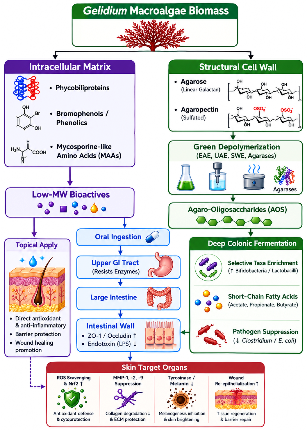
To illustrate the systemic implications of this process, the proposed mechanisms underlying the beneficial effects of
Gelidium-derived bioactive compounds on skin health through the gut–skin axis are summarized in
Figure 1.
5.1. Taxonomic Remodeling of the Microbial Consortium
Molecular profiling using 16S rRNA next-generation sequencing has confirmed that the administration of
Gelidium raw polysaccharides and AOS induce a major taxonomical remodeling of the intestinal architecture [
4].
Gelidium bioactives function as selective carbon and energy sources for specific beneficial saccharolytic bacteria that express specialized agarases or possess robust cross-feeding networks. The specific relative abundance shifts and host physiological impacts driven by
Gelidium prebiotics are detailed in
Table 3.
Regarding the enrichment of
Bifidobacterium spp., these beneficial saccharolytic taxa undergo significant numerical expansion within the colonic microflora upon
Gelidium prebiotic supplementation [
23]. Mechanistically, these microorganisms utilize saccharolytic pathways to ferment macroalgal oligosaccharide fragments, contributing to the increased production of beneficial organic acids [
23]. Furthermore, clinical supplementation with
Gelidium-derived AOS has been shown to selectively expand and activate agarose- and AOS-utilizing gut taxa, inducing specialized carbohydrate-active enzymes that facilitate glycan degradation and cross-feeding networks [
4]. Concurrently,
Gelidium fermentation contributes to the suppression of potential pathobionts, particularly limiting the overgrowth of facultative anaerobic bacteria such as
Escherichia coli [
23]. Furthermore,
Gelidium intake is associated with a modulation of the Firmicutes/Bacteroidetes (F/B) ratio, potentially supporting metabolic homeostasis through altered dietary carbohydrate utilization [
23].
5.2. SCFAs Biosynthetic Kinetics
The principal mechanism through which the
Gelidium-modulated microbiota influences host physiology is the substantial increase in SCFAs, predominantly acetate, propionate, and butyrate [
24]. When beneficial colonic taxa ferment
Gelidium AOS, they break down the galactan backbone into pyruvate via the glycolytic pathway, which is subsequently converted into volatile organic acids [
25]. The intestinal synthesis yields, host cellular receptors, and tissue-specific therapeutic outcomes of these main SCFAs are outlined in
Table 4.
Among these organic acids, butyrate acts as the definitive primary oxidative fuel for host colonocytes, supporting ATP production and cellular health [
29]. Meanwhile, propionate migrates via the portal vein to the liver, where it acts as an inhibitor of hepatocyte gluconeogenesis and cholesterol synthesis [
27]. Concurrently, acetate enters systemic arterial circulation, modulating peripheral tissue lipid profiles and serving as an important signaling ligand for metabolic regulation [
26].
5.3. Intestinal Barrier Reinforcement and Anti-Endotoxemia Mechanisms
The multi-fold increase in local SCFA concentrations directly drives structural preservation of the intestinal mucosal barrier [
28]. Butyrate binds to G-protein coupled receptors (GPR41 and GPR43) expressed on the basolateral membrane of the intestinal epithelium, activating downstream intracellular pathways that significantly upregulate the transcription and assembly of tight junction proteins, specifically Zonula Occludens-1 (ZO-1), occludin, and claudin-1 [
30].
General studies on marine and dietary polysaccharides suggest that reinforcing paracellular tight junctions against macromolecular toxins like lipopolysaccharide (LPS) provides a plausible mechanistic rationale for attenuated endotoxemia [
31]. Within this context,
Gelidium prebiotic fermentation is proposed to support systemic inflammatory homeostasis via SCFA-mediated barrier protection. Supporting local intestinal benefits,
Gelidium fractions have been demonstrated to suppress intestinal nuclear factor-kappa B (NF-κB) activation, downregulating mucosal expression of tumor necrosis factor-alpha (TNF-α), interleukin-1 beta (IL-1β), and interleukin-6 (IL-6) [
32].
6. Skin Health
Cutaneous architecture undergoes continuous degenerative degradation driven by intrinsic chronological factors and extrinsic photoaging, which is heavily mediated by solar ultraviolet (UVA/UVB) radiation [
33].
Gelidium bioactives exert targeted protective effects within both dermal and epidermal layers [
8]. The downstream cellular targets, pathways, and visible dermatological manifestations of isolated
Gelidium bioactives are summarized in
Table 5.
6.1. Cellular Antioxidant Cascades via Nrf2/HO-1
UV exposure strikes epidermal keratinocytes and dermal fibroblasts, inducing rapid intracellular accumulation of Reactive Oxygen Species (ROS), such as singlet oxygen (
), superoxide anions (
), and hydrogen peroxide (
) [
36]. These radicals initiate lipid peroxidation of cellular membranes and break down nuclear DNA [
37]. Polyphenols, bromophenols, and protein/phycobiliprotein-rich fractions isolated from
Gelidium sesquipedale (Clemente) have been demonstrated to counteract oxidative stress via direct radical scavenging, wherein phenolic hydroxyl groups and bilin chromophores neutralize free radicals and inhibit oxidative chain reactions [
9]. In broader cellular physiology, endogenous cytoprotection against sustained oxidative stress relies heavily on the Nrf2/ARE signaling pathway [
38]. Under oxidative stimulation, Nrf2 dissociates from Keap1 and translocates into the nucleus to induce cytoprotective enzymes, including heme oxygenase-1 (HO-1), superoxide dismutase (SOD), catalase (CAT), and glutathione peroxidase (GPx) [
38]. While direct
Gelidium-mediated Nrf2/ARE transactivation has been characterized in metabolic models [
5], its specific role in dermal cytoprotection is proposed as a biologically plausible mechanism supported by the radical-scavenging capacity of red algal secondary metabolites [
9,
39].
6.2. Inhibition of Matrix Metalloproteinases and Extracellular Matrix Preservation
Dermal aging is characterized by structural collapse of the extracellular matrix (ECM), composed primarily of Type I and Type III collagens and elastic fibers [
40]. Elevated ROS levels activate MAPKs—including ERK, JNK, and p38 MAPK [
41]. This signaling cascade triggers the activation of transcription factor Activator Protein-1 (AP-1), which upregulates the transcription of Matrix Metalloproteinases (MMPs) [
42]. MMP-1 initiates cleavage of structural fibrillar collagens, whereas MMP-2 and MMP-9 degrade fragmented collagen fragments and basement membrane elastin [
43]. Bioactive fractions and sulfated polysaccharides isolated from
Gelidium species directly block these degenerative pathways. Specifically, sulfated polysaccharides from
Gelidium crinale (Hare ex Turner) Gaillon significantly downregulate the expression of MMP-9 [
34], while bioactive extracts from
Gelidium amansii attenuate UVB-induced MAPK phosphorylation, effectively suppressing MMP-1 expression and preventing collagen fragmentation [
8]. This regulatory mechanism preserves dermal matrix density and attenuates wrinkle formation in preclinical photoaging models.
6.3. Anti-Melanogenic Pathways and Enzymatic Tyrosinase Blockade
Hyperpigmentation disorders stem from hyperactivation of melanogenesis within epidermal melanocytes [
44]. Tyrosinase is the key rate-limiting enzyme that catalyzes the hydroxylation of L-tyrosine to L-DOPA, and subsequent oxidation of L-DOPA to DOPAquinone [
45]. Phenolic- and bromophenol-enriched extracts from
Gelidium corneum (Hudson) J.Agardh exhibit notable photoprotective, antioxidant, and potential anti-melanogenic properties in skin cellular models [
2]. Although in vitro assays demonstrate modulation of oxidative stress and melanin-related pathways, detailed molecular investigations into direct enzymatic tyrosinase inhibition and regulatory cascades remain an area for further validation [
2].
6.4. Wound Healing Acceleration and Anti-Dermatitis Activity
Gelidium-derived polysaccharides form stable, cross-linked biomimetic hydrogels that maintain optimal moisture retention across wounded cutaneous tissue [
46]. These macroalgal matrices have been demonstrated to modulate the microenvironment of human dermal fibroblasts, thereby facilitating skin repair, controlling angiogenesis, and accelerating granulation tissue assembly [
47]. In inflammatory dermatoses, systemic or topical application of
Gelidium extracts effectively targets epidermal cellular pathways, significantly suppressing hyper-activated inflammatory responses and protecting human keratinocytes from tissue damage [
35]. Consistently, in vivo models have confirmed that treatment with
Gelidium amansii extracts suppress systemic inflammatory responses, downregulate pro-inflammatory cytokines, and attenuate tissue-specific metabolic inflammatory deviations [
33].
7. Gut–Skin Axis
The bidirectional physiological link between the gut and the skin, known as the gut–skin axis, serves as a primary conduit through which oral consumption of marine algal functional components—such as dietary fibers and polysaccharides found in Gelidium—translate into visible cutaneous benefits [
48]. This complex communication network is maintained via three interconnected pathways, categorized as immunological, metabolic, and neuroendocrine signaling axes.
7.1. Immunological Linkage and Systemic Inflammatory Reset
Under dysbiotic conditions, heightened mucosal permeability facilitates systemic translocation of gut-derived LPS and pro-inflammatory pathobionts into circulation [
48]. This systemic endotoxemia triggers a host immune cascade: circulating LPS binds to Toll-Like Receptor 4 on peripheral immune cells and cutaneous dendritic cells, driving downstream activation of the pro-inflammatory NF-κB signaling network [
49]. Consequently, elevated systemic levels of pro-inflammatory cytokines (including IL-1β, IL-6, and TNF-α) reach dermal compartments, prompting fibroblasts and keratinocytes to overexpress MMPs, accelerating ECM breakdown and exacerbating inflammatory skin disorders.
While orally administered
Gelidium-derived polysaccharides and agaro-oligosaccharides have been demonstrated to selectively enrich beneficial taxa (
Bifidobacterium) and elevate SCFA yields in experimental models [
23,
30], their capacity to blunt downstream systemic LPS translocation and cutaneous inflammation is proposed as a comprehensive mechanistic framework informed by general gut–skin axis biology [
48,
49].
While much of the mechanistic framework governing the gut–skin axis relies on preclinical animal paradigms, a growing body of human clinical trials supports the translational validity of targeted dietary microbial modulation. In broader clinical settings, targeted prebiotic and synbiotic interventions promoting colonic
Bifidobacterium have been shown to reduce disease severity and serum inflammatory biomarkers in patients with atopic dermatitis [
50]. Additionally, non-digestible marine-derived glycans can attenuate low-grade mucosal inflammation and circulating endotoxins in human subjects [
51]. Although dedicated human dermatological trials specifically utilizing
Gelidium biomass remain to be conducted, these findings provide supportive clinical evidence for the broader concept that intestinal microbiome modulation may influence systemic inflammatory homeostasis, but
Gelidium-specific clinical evidence for skin outcomes remains to be established.
7.2. Metabolic Translocation of Active Metabolites
Beyond immunological control, metabolic byproducts of
Gelidium colonic fermentation are distributed systemically to drive dermal homeostasis [
48]. Ingested
Gelidium polysaccharides escape upper gastrointestinal digestion to undergo colonic fermentation, which has been demonstrated to significantly increase total SCFA production [
23]. General physiological literature provides supporting evidence that circulating SCFAs reach the skin via cutaneous microvasculature to modulate local immune homeostasis [
48]. Although direct
Gelidium-specific cutaneous tracing remains an area of ongoing investigation, this metabolic translocation is proposed as a key systemic conduit through which oral algal bioactives support dermal resilience. Furthermore, low-molecular-weight phenolic metabolites and MAAs co-absorbed in the gut are distributed throughout dermal tissues, where they provide ongoing systemic protection against UV-induced oxidative stress and matrix degradation [
2].
8. Therapeutic Applications
The unique chemical properties of Gelidium enable diverse therapeutic applications spanning nutritional, medical, and cosmeceutical industries.
8.1. Functional Nutricosmetics and Dietary Supplements
The integration of
Gelidium biomass into functional foods and oral nutraceuticals directly aligns with the evolving paradigm of “beauty-from-within.” Oral formulations containing enzymatically degraded
Gelidium agaro-oligosaccharides serve as systemic anti-aging agents that leverage gut microbial fermentation to improve cutaneous biophysical parameters. Comprehensive academic syntheses indicate that the systematic ingestion of marine-derived prebiotic oligosaccharides effectively orchestrates the gut–skin axis to optimize cutaneous physiology. Mechanistically, these non-digestible glycan fractions promote a systemic homeostatic reset that correlates with a marked reduction in transepidermal water loss, secondary to enhanced stratum corneum hydration and the structural preservation of dermal microrelief networks [
52].
Parallel clinical evidence highlights systemic multi-organ benefits of oral
Gelidium ingestion, particularly in modulating gut-metabolic homeostasis. In 12-week RCTs involving overweight and obese individuals, daily administration of
Gelidium elegans extract (1000 mg/day) significantly reduced total body weight and visceral fat area [
53], while simultaneously alleviating functional bowel symptoms and abdominal discomfort [
54]. Additionally, short-term dietary supplementation with agaro-oligosaccharides (200 mg/day) selectively enriched colonic agarose-utilizing taxa with high gastrointestinal tolerability [
4]. Together, these translational human findings confirm that orally administered
Gelidium bioactives may have potential as dual-target functional candidates, capable of improving gastrointestinal parameters while offering preclinically supported cutaneous benefits through the gut–skin axis.
8.2. Topical Cosmeceuticals and Formulation Matrix
In the cosmeceutical field,
Gelidium-derived extracts have been increasingly investigated for use in topical formulations, including anti-photoaging preparations, photoprotective agents, and barrier-supporting matrices [
2]. Their UV-absorbing MAAs function as stable, non-toxic bio-sunscreens, replacing synthetic chemical UV filters that can cause skin irritation or environmental damage [
2]. Additionally, the film-forming properties of
Gelidium agarose serve as an exceptional natural vehicle, improving skin penetration and sustained release of other hydrophilic actives [
55].
8.3. Biomedical Devices and Advanced Wound Dressings
In regenerative medicine,
Gelidium agar-derived hydrogels are engineered into advanced biocompatible wound dressings [
56]. These hydrogels can be infused with bioactive bromophenols or antimicrobial agents to create a sterile, responsive wound environment [
57]. Their high porosity and water-holding capacity facilitate efficient absorption of wound exudates while supporting continuous gas exchange, promoting rapid tissue repair in chronic wounds [
58]. To systematically evaluate the translation of these multi-systemic mechanisms from bench to bedside, the current state of scientific evidence regarding
Gelidium bioactives and gut–skin axis components is consolidated in
Table 6.
9. Challenges and Future Perspectives
Despite the clear therapeutic potential of Gelidium species, several critical biochemical, technical, and regulatory hurdles must be systematically resolved before full commercialization can be realized.
9.1. Bioavailability and Molecular Weight Optimization
The foremost challenge lies in the high molecular weight and complex structural cross-linking of native
Gelidium polysaccharides, which limit their solubility and direct bioavailability [
59]. Future research must prioritize optimization of green, sequence-specific extraction methodologies—particularly cloning and industrial scale-up of recombinant endo-agarases—to reliably produce agaro-oligosaccharides with precise degrees of polymerization (DP 4 to 6), maximizing both prebiotic fermentation and skin penetration.
9.2. Standardization and Quality Control Metrology
As wild marine organisms,
Gelidium populations exhibit substantial seasonal, geographical, and environmental variations in chemical composition [
60]. A wild cohort harvested in the Atlantic may present an altered sulfate or polyphenol profile compared to a Pacific cohort [
61]. Establishing standardized quality control parameters, utilizing high-performance liquid chromatography-mass spectrometry fingerprinting, and setting strict baseline metrics for active marker compounds are required to ensure therapeutic consistency [
62].
9.3. Heavy Metal Bioremediation and Aquaculture Safety
Because marine macroalgae naturally bioaccumulate environmental toxins, wild-harvested
Gelidium carries an inherent risk of contamination with toxic heavy metals, including inorganic arsenic, cadmium, lead, and mercury [
63,
64]. To guarantee consumer safety and meet strict international pharmaceutical guidelines, future industrial production should transition away from wild harvesting toward highly controlled Integrated Multi-Trophic Aquaculture frameworks [
65]. These land-based or coastal aquaculture systems allow for precise regulation of water purity, nutrient input, and light exposure, ensuring sustainable cultivation of clean, ultra-pure
Gelidium biomass. Established maximum allowable thresholds, analytical metrologies, and core rationales for regulatory compliance are defined in
Table 7.
9.4. Rigorous Human Clinical Validation
Although initial clinical trials in humans have provided promising evidence regarding gut and metabolic benefits, well-designed, large-cohort, randomized, double-blind, placebo-controlled trials specifically evaluating skin-targeted parameters and metagenomic profile shifts remain limited. Future research trajectories must focus on clinical validation, utilizing advanced metagenomic sequencing and cutaneous bioengineering tools to confirm the therapeutic efficacy of Gelidium bioactives in human populations.
10. Conclusions
Gelidium Lamouroux species represent a promising marine biomaterial at the intersection of biotechnology, gastroenterology, and dermatology. This review synthesized the genus along five core domains: chemical profiling, green extraction, gut microbiota modulation, cutaneous protection, and the integrated gut–skin axis. Structurally, Gelidium yields non-digestible sulfated galactans, bioavailable AOS, antioxidant bromophenols, and photoprotective MAAs. Advanced green extraction platforms, including enzyme-assisted, ultrasound-assisted, and subcritical water extraction, offer complementary strategies for recovering polysaccharides, oligosaccharides, and selected secondary metabolites, although operational severity must be carefully optimized to minimize the thermal degradation of heat-sensitive compounds.
Mechanistically, Gelidium AOS and galactans resist upper digestion to selectively nourish colonic saccharolytic taxa (e.g., Bifidobacterium), significantly increasing SCFA production.
While Gelidium bioactives have been demonstrated to nourish beneficial saccharolytic taxa and downregulate local inflammatory cytokines, the subsequent upregulation of tight junction proteins (ZO-1, occludin, claudin-1) and attenuation of systemic LPS translocation represent a proposed, biologically plausible axis grounded in the SCFA literature. Systemically, these findings provide a plausible mechanistic rationale by which improved intestinal barrier integrity and attenuated systemic inflammatory signaling could support dermal homeostasis; however, direct Gelidium-specific gut–skin causal evidence remains limited and requires dedicated clinical validation.
Rather than a fully established clinical solution, Gelidium offers an experimentally supported strategy for systemic health via the gut–skin axis. To translate these findings into medical and nutricosmetic practice, future trajectories must focus on standardizing active marker compounds, scaling up sequence-specific recombinant endo-agarase enzymatic processing, and conducting rigorous, large-cohort human RCTs.
Author Contributions
K.-N.K.: Conceptualization, Supervision, Writing—original draft preparation. K.L.: Conceptualization, Supervision, Writing—review and editing. S.-H.L.: Formal analysis, Data curation, Visualization. S.J.: Investigation, Methodology. All authors have read and agreed to the published version of the manuscript.
Funding
This research received no external funding.
Institutional Review Board Statement
Not applicable.
Informed Consent Statement
Not applicable.
Data Availability Statement
No new data were created or analyzed in this study. Data sharing is not applicable to this article.
Conflicts of Interest
The authors declare no conflicts of interest.
References
- Sharma, A.A.K.; Baghel, R.S.; Sandhya, S.V.; Kaushik, R.; Jagtap, A.S.; Vaishnavi, B. High-quality agar polysaccharide from unexplored Gelidium micropterum Kützing biomass. Polymers 2025, 17, 3278. [Google Scholar] [CrossRef] [Scilit] [PubMed]
- Matias, M.; Martins, A.; Alves, C.; Silva, J.; Pinteus, S.; Fitas, M.; Pinto, P.; Marto, J.; Ribeiro, H.; Murray, P.; et al. New insights into the dermocosmetic potential of the red seaweed. Gelidium Corneum Antioxid. 2023, 12, 1684. [Google Scholar] [CrossRef] [Scilit] [PubMed]
- El Kaoutari, A.; Armougom, F.; Gordon, J.I.; Raoult, D.; Henrissat, B. The abundance and variety of carbohydrate-active enzymes in the human gut microbiota. Nat. Rev. Microbiol. 2013, 11, 497–504. [Google Scholar] [CrossRef] [Scilit] [PubMed]
- Hoshiba, N.; Fujii, T.; Yagasaki, R.; Ochi, T.; Shiba, K.; Takahashi, H.; Funasaka, K.; Ono, E.; Hirooka, Y.; Tochio, T.; et al. Agaro-oligosaccharide supplementation alters the gut microbiota, revealing potential agaro-oligosaccharide-utilizing taxa in healthy Japanese adults. Biomedicines 2026, 14, 1112. [Google Scholar] [CrossRef] [Scilit] [PubMed]
- Choi, J.; Kim, K.J.; Koh, E.J.; Lee, B.Y. Gelidium elegans extract ameliorates type 2 diabetes via regulation of MAPK and PI3K/Akt signaling. Nutrients 2018, 10, 51. [Google Scholar] [CrossRef] [Scilit] [PubMed]
- Andrzejczak, K.; Kucharczyk, E.; Sternak, A.; Busłowicz, T.; Ponikowska, M. Gut dysbiosis and the molecular landscape of the gut-skin axis: Comparative insights and therapeutic implications for atopic dermatitis and psoriasis. Cells 2026, 15, 594. [Google Scholar] [CrossRef] [Scilit] [PubMed]
- Guan, Y.; Guo, Y.; Lin, L.; Zhang, L.; Chen, W.; Zhao, C. Green extraction of bioactive compounds from marine macroalgae: Chemistry, pharmacological activities, and biotechnological applications. Mar. Drugs 2026, 24, 198. [Google Scholar] [CrossRef] [Scilit] [PubMed]
- Kim, H.M.; Lee, D.E.; Park, S.D.; Kim, Y.T.; Kim, Y.J.; Jeong, J.W.; Lee, J.-H.; Jang, S.S.; Chung, D.K.; Sim, J.-H. Preventive effect of fermented Gelidium amansii and Cirsium japonicum extract mixture against UVB-induced skin photoaging in hairless mice. Food Sci. Biotechnol. 2014, 23, 623–631. [Google Scholar] [CrossRef] [Scilit]
- Castejón, N.; Parailloux, M.; Izdebska, A.; Lobinski, R.; Fernandes, S.C.M. Valorization of the red algae Gelidium sesquipedale by extracting a broad spectrum of minor compounds using green approaches. Mar. Drugs 2021, 19, 574. [Google Scholar] [CrossRef] [Scilit]
- Srivastava, H.; Bisht, B.; James, J.; Malhotra, R.K.; Kurbatova, A.; Dabral, A.; Upadhyay, S.; Kumar, V. Advanced extraction technologies and functional applications of algal polysaccharides in modern food systems. Discov. Food 2025, 5, 272, Erratum in Discov. Food 2026, 6, 94. [Google Scholar] [CrossRef] [Scilit]
- Beni, B.H.; Falahati, M.; Rostamabadi, M.M.; Navid, S.; Rostamabadi, H. Agar as a natural polymer: From culture media to cutting-edge biomedical applications. Carbohydr. Polym. 2025, 368, 124100. [Google Scholar] [CrossRef] [Scilit] [PubMed]
- Wang, C.; Long, J.; Li, X.; Zhou, X.; Chen, L.; Qiu, C.; Jin, Z. Preparation of agar polysaccharides and biological activities and relationships of agar-derived oligosaccharides and monosaccharides: A review. Int. J. Biol. Macromol. 2025, 295, 139552. [Google Scholar] [CrossRef] [Scilit]
- Gonzaga, L.J.; Roa, M.E.P.; Lavecchia, R.; Zuorro, A. Unlocking marine potential: Microwave-assisted extraction of bioactive compounds from marine macroalgae. J. Environ. Chem. Eng. 2025, 13, 116858. [Google Scholar] [CrossRef] [Scilit]
- Huang, Y.; Hirose, T.; Tsai, J.M.; Hirasaka, K. The effects of agaro-oligosaccharides produced by marine bacteria (Rheinheimera sp. (HY)) possessing agarose-degrading enzymes on myotube function. Mar. Drugs 2024, 22, 515. [Google Scholar] [CrossRef] [Scilit]
- Torres, M.D.; Flórez-Fernández, N.; Domínguez, H. Integral utilization of red seaweed for bioactive production. Mar. Drugs 2019, 17, 314. [Google Scholar] [CrossRef] [Scilit]
- García-Gómez, C.; Aguirre-Cavazos, D.E.; Chávez-Montes, A.; Ballesteros-Torres, J.M.; Orozco-Flores, A.A.; Reyna-Martínez, R.; Torres-Hernández, Á.D.; González-Meza, G.M.; Castillo-Hernández, S.L.; Gloria-Garza, M.A.; et al. Phycobilins versatile pigments with wide-ranging applications: Exploring their uses, biological activities, extraction methods and future perspectives. Mar. Drugs 2025, 23, 201. [Google Scholar] [CrossRef] [Scilit]
- Vega, J.; Schneider, G.; Moreira, B.R.; Herrera, C.; Bonomi-Barufi, J.; Figueroa, F.L. Mycosporine-like amino acids from red macroalgae: UV-photoprotectors with potential cosmeceutical applications. Appl. Sci. 2021, 11, 5112. [Google Scholar] [CrossRef] [Scilit]
- Gómez Barrio, L.P.; Cabral, E.M.; Zhao, M.; Álvarez García, C.; Senthamaraikannan, R.; Padamati, R.B.; Tiwari, U.; Curtin, J.F.; Tiwari, B.K. Comparison study of an optimized ultrasound-based method versus an optimized conventional method for agar extraction, and protein co-extraction, from. Gelidium Sesquipedale Foods 2022, 11, 805. [Google Scholar] [CrossRef] [Scilit]
- Mabate, B.; Mkabayi, L.; Goddard, D.R.; Grobler, C.E.; Pletschke, B.I. Role of enzyme technologies and applied enzymology in valorising seaweed bioproducts. Mar. Drugs 2025, 23, 303. [Google Scholar] [CrossRef] [Scilit]
- Tahir, H.; Dar, A.H.; Dash, K.K.; Majeed, T.; Srivastava, S.; Singh, A.; Manzoor, M.; Sidiqi, U.S.; Ganaie, A.A. Subcritical water extraction as a green technology for the recovery of bioactive compounds from algae: A comprehensive review. Food Phys. 2025, 3, 100077. [Google Scholar] [CrossRef] [Scilit]
- Carreira-Casais, A.; Otero, P.; Garcia-Perez, P.; Garcia-Oliveira, P.; Pereira, A.G.; Carpena, M.; Soria-Lopez, A.; Simal-Gandara, J.; Prieto, M.A. Benefits and drawbacks of ultrasound-assisted extraction for the recovery of bioactive compounds from marine algae. Int. J. Environ. Res. Public Health 2021, 18, 9153. [Google Scholar] [CrossRef] [Scilit]
- Denev, P.; Ognyanov, M.; Pimpilova, M.; Teneva, D. Subcritical water extraction as a green technology for the development of standardized plant extracts for food and pharmaceutical uses. Processes 2026, 14, 1564. [Google Scholar] [CrossRef] [Scilit]
- Cui, M.; Zhang, M.; Wu, J.; Han, P.; Lv, M.; Dong, L.; Liu, K. Marine polysaccharides from Gelidium pacificum Okamura and Cereus sinensis reveal prebiotic functions. Int. J. Biol. Macromol. 2020, 164, 4381–4390. [Google Scholar] [CrossRef] [Scilit]
- Liu, S.H.; Ku, C.Y.; Chiang, M.T. Polysaccharide-rich red algae (Gelidium amansii) hot-water extracts alleviate abnormal hepatic lipid metabolism without suppression of glucose intolerance in a streptozotocin/nicotinamide-induced diabetic rat model. Molecules 2022, 27, 1447. [Google Scholar] [CrossRef] [Scilit]
- Pluvinage, B.; Grondin, J.M.; Amundsen, C.; Klassen, L.; Moote, P.E.; Xiao, Y.; Thomas, D.; Pudlo, N.A.; Anele, A.; Martens, E.C.; et al. Molecular basis of an agarose metabolic pathway acquired by a human intestinal symbiont. Nat. Commun. 2018, 9, 1043. [Google Scholar] [CrossRef] [Scilit]
- Hernández, M.A.G.; Canfora, E.E.; Jocken, J.W.E.; Blaak, E.E. The short-chain fatty acid acetate in body weight control and insulin sensitivity. Nutrients 2019, 11, 1943. [Google Scholar] [CrossRef] [Scilit]
- Yoshida, H.; Ishii, M.; Akagawa, M. Propionate suppresses hepatic gluconeogenesis via GPR43/AMPK signaling pathway. Arch. Biochem. Biophys. 2019, 672, 108057. [Google Scholar] [CrossRef] [Scilit]
- Wang, H.B.; Wang, P.Y.; Wang, X.; Wan, Y.L.; Liu, Y.C. Butyrate enhances intestinal epithelial barrier function via up-regulation of tight junction protein Claudin-1 transcription. Dig. Dis. Sci. 2012, 57, 3126–3135. [Google Scholar] [CrossRef] [Scilit]
- Donohoe, D.R.; Garge, N.; Zhang, X.; Sun, W.; O’Connell, T.M.; Bunger, M.K.; Bultman, S.J. The microbiome and butyrate regulate energy metabolism and autophagy in the mammalian colon. Cell Metab. 2011, 13, 517–526. [Google Scholar] [CrossRef] [Scilit] [PubMed]
- Hodgkinson, K.; El Abbar, F.; Dobranowski, P.; Manoogian, J.; Butcher, J.; Figeys, D.; Mack, D.; Stintzi, A. Butyrate’s role in human health and the current progress towards its clinical application to treat gastrointestinal disease. Clin. Nutr. 2023, 42, 61–75. [Google Scholar] [CrossRef] [Scilit] [PubMed]
- He, Y.; Xu, M.; Lu, S.; Zou, W.; Wang, Y.; Fakhar, E.A.K.M.; Iqbal, M.; Li, K. Seaweed polysaccharides treatment alleviates injury of inflammatory responses and gut barrier in LPS-induced mice. Microb. Pathog. 2023, 180, 106159. [Google Scholar] [CrossRef] [Scilit]
- Lee, Y.; Oh, H.; Lee, M. Anti-inflammatory effects of agar free-Gelidium amansii (GA) extracts in high-fat diet-induced obese mice. Nutr. Res. Pract. 2018, 12, 479–485. [Google Scholar] [CrossRef] [Scilit]
- Rittié, L.; Fisher, G.J. Natural and sun-induced aging of human skin. Cold Spring Harb. Perspect. Med. 2015, 5, a015370. [Google Scholar] [CrossRef] [Scilit] [PubMed]
- Zheng, H.; Pei, Y.; He, Y.L.; Liu, Y.; Chen, M.; Hong, P.; Zhou, C.; Qian, Z.J. A sulfated polysaccharide from red algae (Gelidium crinale) to suppress cells metastasis and MMP-9 expression of HT1080 cells. Foods 2022, 11, 2360. [Google Scholar] [CrossRef] [Scilit]
- So, B.R.; Yeo, H.J.; Lee, J.J.; Jung, Y.H.; Jung, S.K. Cellulose nanocrystal preparation from Gelidium amansii and analysis of its anti-inflammatory effect on the skin in vitro and in vivo. Carbohydr. Polym. 2021, 254, 117315. [Google Scholar] [CrossRef] [Scilit]
- Wei, M.; He, X.; Liu, N.; Deng, H. Role of reactive oxygen species in ultraviolet-induced photodamage of the skin. Cell Div. 2024, 19, 1. [Google Scholar] [CrossRef] [Scilit]
- Rastogi, R.P.; Richa; Kumar, A.; Tyagi, M.B.; Sinha, R.P. Molecular mechanisms of ultraviolet radiation-induced DNA damage and repair. J. Nucleic Acids 2010, 2010, 592980. [Google Scholar] [CrossRef] [Scilit]
- Smith, R.E.; Tran, K.; Smith, C.C.; McDonald, M.; Shejwalkar, P.; Hara, K. The role of the Nrf2/ARE antioxidant system in preventing cardiovascular diseases. Diseases 2016, 4, 34. [Google Scholar] [CrossRef] [Scilit]
- Kim, M.J.; Istianah, N.; So, B.R.; Kang, H.J.; Woo, M.J.; Park, S.J.; Kim, H.J.; Jung, Y.H.; Jung, S.K. Preparation of high-solid microfibrillated cellulose from Gelidium amansii and characterization of its physiochemical and biological properties. J. Microbiol. Biotechnol. 2022, 32, 1589–1598. [Google Scholar] [CrossRef]
- Fisher, G.J.; Quan, T.; Purohit, T.; Shao, Y.; Cho, M.K.; He, T.; Varani, J.; Kang, S.; Voorhees, J.J. Collagen fragmentation promotes oxidative stress and elevates matrix metalloproteinase-1 in fibroblasts in aged human skin. Am. J. Pathol. 2009, 174, 101–114. [Google Scholar] [CrossRef] [Scilit] [PubMed]
- Son, Y.; Cheong, Y.K.; Kim, N.H.; Chung, H.T.; Kang, D.G.; Pae, H.O. Mitogen-activated protein kinases and reactive oxygen species: How can ROS activate MAPK pathways? J. Signal Transduct. 2011, 2011, 792639. [Google Scholar] [CrossRef] [Scilit] [PubMed]
- Zhu, W.; Ren, H.; Liu, Y.; Li, Z.; Luo, X.; Meng, H.; Dong, Y. Key targets and pathways in skin photoaging: A comprehensive review. J. Dermatol. Sci. Cosmet. Technol. 2025, 2, 100101. [Google Scholar] [CrossRef] [Scilit]
- Lai, C.-F.; Esam, G.; Lin, Y.-C.; Chung, R.-J. From destruction to protection: Rethinking MMP-1 in skin aging and anti-aging strategies. Arch. Dermatol. Res. 2026, 318, 56. [Google Scholar] [CrossRef] [Scilit]
- Duval, C.; Qiu, J.; Warrick, E.; Sextius, P.; Castiel-Higounenc, I.; Xiang, L.; Bernerd, F.; Passeron, T. Decoding hyperpigmentation from biological mechanisms to actives with clinically proven topical efficacy: A narrative review. Dermatol. Ther. 2026, 16, 3385–3414. [Google Scholar] [CrossRef] [Scilit]
- Jia, M.; Pei, J.; Chen, F.; Duan, R.; Liu, A.; Chu, X.; Ye, B.; Zhang, B. Emerging strategies for l-DOPA production: Integrating plant, chemical, enzymatic, and microbial strategies. ACS Synth. Biol. 2026, 15, 877–892. [Google Scholar] [CrossRef] [Scilit]
- Kim, H.D.; Choi, H.; Abekura, F.; Park, J.Y.; Yang, W.S.; Yang, S.H.; Kim, C.H. Naturally-occurring tyrosinase inhibitors classified by enzyme kinetics and copper chelation. Int. J. Mol. Sci. 2023, 24, 8226. [Google Scholar] [CrossRef] [Scilit]
- Liu, Y.; Zheng, H.; Chen, Y.; Ryu, B.; Qian, Z.-J. Sulfated polysaccharide from Gelidium crinale: Modulation of angiogenesis and fibrosis in skin repair. npj Sci. Food 2026, 10, 977. [Google Scholar] [CrossRef] [Scilit]
- De Pessemier, B.; Grine, L.; Debaere, M.; Maes, A.; Paetzold, B.; Callewaert, C. Gut-skin axis: Current knowledge of the interrelationship between microbial dysbiosis and skin conditions. Microorganisms 2021, 9, 353. [Google Scholar] [CrossRef] [Scilit]
- Peng, R.; Huang, X.; Bao, Y.; Luo, G.; Zeng, J.; Tang, L. Atopic dermatitis and the gut-skin axis: A review from dysbiosis to novel targeted therapeutic strategies. Front. Immunol. 2026, 17, 1895580. [Google Scholar] [CrossRef] [Scilit]
- Farid, R.; Ahanchian, H.; Jabbari, F.; Moghiman, T. Effect of a new synbiotic mixture on atopic dermatitis in children: A randomized-controlled trial. Iran. J. Pediatr. 2011, 21, 225–230. [Google Scholar]
- Repici, A.; Hasan, A.; Capra, A.P.; Scuderi, S.A.; Paterniti, I.; Campolo, M.; Ardizzone, A.; Esposito, E. Marine algae and deriving biomolecules for the management of inflammatory bowel diseases: Potential clinical therapeutics to decrease gut inflammatory and oxidative stress markers? Mar. Drugs 2024, 22, 336. [Google Scholar] [CrossRef] [Scilit]
- Zeng, M.; Li, Y.; Cheng, J.; Wang, J.; Liu, Q. Prebiotic oligosaccharides in skin health: Benefits, mechanisms, and cosmetic applications. Antioxidants 2025, 14, 754. [Google Scholar] [CrossRef] [Scilit]
- Kim, C.O.; Kim, Y.N.; Lee, D.C. Effects of Gelidium elegans on weight and fat mass reduction and obesity biomarkers in overweight or obese adults: A randomized double-blinded study. Nutrients 2019, 11, 1513. [Google Scholar] [CrossRef] [Scilit]
- Choi, H.I.; Cha, J.M.; Jeong, I.K.; Cho, I.J.; Yoon, J.Y.; Kwak, M.S.; Jeon, J.W.; Kim, S.J. Efficacy and safety of Gelidium elegans intake on bowel symptoms in obese adults: A 12-week randomized double-blind placebo-controlled trial. Medicine 2019, 98, e14981. [Google Scholar] [CrossRef] [Scilit]
- Firmansyah, F.; Budiman, A.; Muchtaridi, M.; Chabib, L.; Syaputri, F.N.; Afandi, F.; Elamin, K.M.; Mohammed, A.F.A.; Wathoni, N. Film-forming gels for topical drug delivery: A systematic review of the effects of formulation on film performance, drug release, and skin permeation. Drug Des. Devel. Ther. 2026, 20, 591505. [Google Scholar] [CrossRef] [Scilit]
- Li, S.; Han, J.; Li, B.; Yu, F.; Xing, J.; Mao, Y.; Yang, H.; Wan, Y. Bioactive agarose-based hydrogels with granulated microstructures for accelerating diabetic wound healing. Int. J. Biol. Macromol. 2025, 330, 148061. [Google Scholar] [CrossRef] [Scilit]
- Alipour, A.; Nejati, O.; Yaşayan, G.; Girgin, A.; Zaman, B.T.; Giray, B.; Karal-Yılmaz, O.; Bakırdere, S.; Bal-Öztürk, A. Multilayer antibacterial hydrogel wound dressings incorporated with green synthesized silver nanoparticles. Drug Dev. Res. 2025, 86, e70102. [Google Scholar] [CrossRef] [Scilit]
- He, J.; Chen, J.; Liu, T.; Qin, F.; Wei, W. Research progress of multifunctional hydrogels in promoting wound healing of diabetes. Int. J. Nanomed. 2025, 20, 7549–7578. [Google Scholar] [CrossRef] [Scilit]
- Wang, Y.; Lin, Z.; Zhang, H.; Gao, Y.; Liu, Y.; Liang, J. Research progress on polyphenols and polysaccharides from marine seaweeds: Promising diabetes management natural products. Mar. Drugs 2026, 24, 208. [Google Scholar] [CrossRef] [Scilit]
- Benhniya, B.; Karmil, F.Z.; Lakhdar, F.; Mabrouki, S.; Zidane, H.; Rezzoum, N.; Etahiri, S. Seasonal variation in nutritional composition and antioxidant potential of Gelidium corneum from the Doukkala coast, Morocco. BIO Web Conf. 2026, 215, 01002. [Google Scholar] [CrossRef] [Scilit]
- Lomartire, S.; Cotas, J.; Pacheco, D.; Marques, J.C.; Pereira, L.; Gonçalves, A.M.M. Environmental impact on seaweed phenolic production and activity: An important step for compound exploitation. Mar. Drugs 2021, 19, 245. [Google Scholar] [CrossRef] [Scilit]
- Noviana, E.; Indrayanto, G.; Rohman, A. Advances in fingerprint analysis for standardization and quality control of herbal medicines. Front. Pharmacol. 2022, 13, 853023. [Google Scholar] [CrossRef] [Scilit] [PubMed]
- Rubio, C.; Napoleone, G.; Luis-González, G.; Gutiérrez, A.J.; González-Weller, D.; Hardisson, A.; Revert, C. Metals in edible seaweed. Chemosphere 2017, 173, 572–579. [Google Scholar] [CrossRef] [Scilit] [PubMed]
- International Council for Harmonisation (ICH). ICH Harmonised Guideline: Guideline for Elemental Impurities Q3D(R2); ICH: Geneva, Switzerland, 2022; Available online: https://database.ich.org/sites/default/files/Q3D-R2_Guideline_Step4_2022_0308.pdf (accessed on 16 August 2026).
- García-Poza, S.; Leandro, A.; Cotas, C.; Cotas, J.; Marques, J.C.; Pereira, L.; Gonçalves, A.M.M. The evolution road of seaweed aquaculture: Cultivation technologies and the industry 4.0. Int. J. Environ. Res. Public Health 2020, 17, 6528. [Google Scholar] [CrossRef] [Scilit] [PubMed]
Figure 1.
Proposed mechanisms underlying the beneficial effects of Gelidium-derived bioactive compounds on skin health through the gut–skin axis. Schematic overview illustrating the major bioactive constituents of Gelidium and their proposed pathways contributing to skin health. Intracellular bioactive compounds, including phycobiliproteins, bromophenols, phenolic compounds, and mycosporine-like amino acids, together with structural polysaccharides (agarose and agaropectin), are extracted from Gelidium biomass. Green depolymerization technologies generate low-molecular-weight agaro-oligosaccharides, which are resistant to upper gastrointestinal digestion and undergo selective fermentation by the colonic microbiota. This process promotes beneficial bacterial populations, enhances short-chain fatty acid production, suppresses pathogenic microorganisms, and strengthens intestinal barrier integrity by upregulating tight junction proteins while reducing lipopolysaccharide translocation. Bioactive metabolites subsequently enter systemic circulation and contribute to skin homeostasis through antioxidant, anti-inflammatory, anti-photoaging, anti-melanogenic, and wound-healing activities. Direct topical application of Gelidium-derived bioactives may additionally exert local protective effects on the skin. The proposed mechanisms are synthesized from current experimental evidence and represent potential biological pathways rather than fully established causal relationships. In the diagram, solid arrows represent directly demonstrated experimental pathways, whereas dashed arrows denote proposed or indirect mechanistic connections within the gut–skin framework. EAE, enzyme-assisted extraction; ECM, Extracellular matrix; Low-MW, Low-molecular-weight; LPS, Lipopolysaccharide; GI, gastrointestinal; MMP, matrix metalloproteinase; SWE, subcritical water extraction; UAE, ultrasound-assisted extraction; ZO-1, zonula occludens-1.
Figure 1.
Proposed mechanisms underlying the beneficial effects of Gelidium-derived bioactive compounds on skin health through the gut–skin axis. Schematic overview illustrating the major bioactive constituents of Gelidium and their proposed pathways contributing to skin health. Intracellular bioactive compounds, including phycobiliproteins, bromophenols, phenolic compounds, and mycosporine-like amino acids, together with structural polysaccharides (agarose and agaropectin), are extracted from Gelidium biomass. Green depolymerization technologies generate low-molecular-weight agaro-oligosaccharides, which are resistant to upper gastrointestinal digestion and undergo selective fermentation by the colonic microbiota. This process promotes beneficial bacterial populations, enhances short-chain fatty acid production, suppresses pathogenic microorganisms, and strengthens intestinal barrier integrity by upregulating tight junction proteins while reducing lipopolysaccharide translocation. Bioactive metabolites subsequently enter systemic circulation and contribute to skin homeostasis through antioxidant, anti-inflammatory, anti-photoaging, anti-melanogenic, and wound-healing activities. Direct topical application of Gelidium-derived bioactives may additionally exert local protective effects on the skin. The proposed mechanisms are synthesized from current experimental evidence and represent potential biological pathways rather than fully established causal relationships. In the diagram, solid arrows represent directly demonstrated experimental pathways, whereas dashed arrows denote proposed or indirect mechanistic connections within the gut–skin framework. EAE, enzyme-assisted extraction; ECM, Extracellular matrix; Low-MW, Low-molecular-weight; LPS, Lipopolysaccharide; GI, gastrointestinal; MMP, matrix metalloproteinase; SWE, subcritical water extraction; UAE, ultrasound-assisted extraction; ZO-1, zonula occludens-1.

Table 1.
Global distribution and primary biomedical activities of key Gelidium species.
Table 1.
Global distribution and primary biomedical activities of key Gelidium species.
| Algal Species | Primary Geographical Habitat | Dominant Bioactive Fractions | Core Biomedical Focus | Target Molecular Endpoints | References |
|---|
| Gelidium amansii J.V.Lamouroux | Coastal East Asia (Korea, Japan, China) | Fermented Extracts, Bioactive Polysaccharides | Dermal Photoprotection, Anti-photoaging, Collagen Integrity | Suppression of UVB-induced MAPK activation and MMP-1 expression | [8] |
| Gelidium elegans Kützing | Western Pacific Intertidal Zones | Polyphenols, Bromophenols, Agarose Matrix | Metabolic Regulation, Antioxidant Defense, Gut-Adipose Axis | Regulation of MAPK and PI3K/Akt signaling, activation of Nrf2/HO-1 | [5] |
| Gelidium sesquipedale (Clemente) Thuret | Eastern Atlantic (Spain, Portugal, Morocco) | Polyphenolic Fractions, Proteins, Bioactive Lipids | Endogenous Antioxidant Support, Cytoprotection | Direct ROS scavenging, prevention of oxidative macromolecular damage | [9] |
| Gelidium corneum (Hudson) J.Agardh | Mediterranean Sea, North Atlantic | Polyphenols, Bromophenols, Mycosporine-like Amino Acids (Shinorine) | Biological UV Protection, Photoprotection, Dermal Support | Modulation of oxidative and melanogenic pathways; UVA/UVB photoprotection (310–360 nm) | [2] |
Table 2.
Comparison of conventional vs. advanced green extraction methodologies for Gelidium.
Table 2.
Comparison of conventional vs. advanced green extraction methodologies for Gelidium.
| Extraction Technology | Operational Characteristics | Extraction Efficiency | Bioactive Structural Integrity | Industrial Scale-up Considerations | Environmental Profile | References |
|---|
| Conventional Hot Water | Moderate temperature; relatively long extraction time | Moderate; established agar recovery | Potential degradation of heat-sensitive compounds during prolonged heating | High energy demand and prolonged processing | Higher energy consumption | [18] |
| Enzyme-Assisted (EAE) | Mild conditions with selective enzymatic hydrolysis | Enhanced recovery of selected target fractions | Generally favorable under mild conditions | Enzyme cost and process optimization remain challenges | Reduced severity of processing conditions | [19] |
| Ultrasound-Assisted (UAE) | Acoustic cavitation; shortened extraction time | Enhanced extraction efficiency | May improve recovery while reducing thermal exposure | Scale-up may be limited by acoustic energy distribution | Reduced extraction time and solvent demand | [18] |
| Subcritical Water (SWE) | Pressurized hot water; reduced dependence on organic solvents | Potentially high recovery of selected compounds | Dependent on temperature and pressure; excessive severity may promote degradation | Requires pressure-resistant equipment | Reduced use of organic solvents | [20] |
Table 3.
Microbiota changes associated with Gelidium-derived prebiotics.
Table 3.
Microbiota changes associated with Gelidium-derived prebiotics.
| Target Microbial Taxa | Reported Change | Potential Microbial Mechanism | Potential Host-Relevant Effect | References |
|---|
| Bifidobacterium spp. | Increased | Utilization of agar-derived oligosaccharides and saccharolytic fermentation | Increased production of microbial metabolites, including SCFAs | [23] |
| Agarose/AOS-utilizing gut bacteria | Increased/activated | Carbohydrate-active enzymes involved in agarose/AOS degradation | Enhanced microbial carbohydrate utilization and cross-feeding | [4] |
| Escherichia coli | Decreased | Alteration of the luminal environment associated with increased saccharolytic fermentation | Potential reduction in expansion of facultative anaerobic bacteria | [23] |
| Firmicutes/Bacteroidetes ratio | Altered/normalized | Changes in microbial carbohydrate utilization | Potential association with metabolic effects | [23] |
Table 4.
Proposed SCFAs-mediated molecular mechanisms potentially linked to Gelidium prebiotic fermentation.
Table 4.
Proposed SCFAs-mediated molecular mechanisms potentially linked to Gelidium prebiotic fermentation.
| SCFAs | Major Microbial SCFAs Product | Primary Host Molecular Targets | Intestinal Mechanism of Action | Potential Skin-Relevant Effects | References |
|---|
| Acetate | Major microbial fermentation product | GPR43, GPR41 | Metabolic signaling and peripheral energy regulation | Potential contribution to epithelial and metabolic homeostasis | [26] |
| Propionate | Major microbial fermentation product | GPR43, GPR41 | Modulation of hepatic gluconeogenesis through GPR43/AMPK signaling | Potential immunomodulatory effects; direct skin-specific evidence remains limited | [27] |
| Butyrate | Major microbial fermentation product | GPR41, GPR43, HDACs | Supports colonocyte metabolism and intestinal epithelial barrier function through tight-junction regulation | Potential anti-inflammatory effects; direct Gelidium-specific skin evidence remains limited | [28] |
Table 5.
Molecular targets and cutaneous effects of Gelidium-derived bioactive fractions.
Table 5.
Molecular targets and cutaneous effects of Gelidium-derived bioactive fractions.
| Bioactive Fraction/Compound(s) | Experimental Skin/Cell Model | Primary Molecular Target/Pathway | Direct Molecular or Enzymatic Effect | Reported Cutaneous/Cellular Outcome | References |
|---|
| Sulfated Polysaccharide Fraction (Gelidium crinale) | Human fibrosarcoma cells (HT1080) | NF-κB, MAPK, mTOR/PI3K/Akt | Reduced MMP-9 mRNA, expression and activity | Reduced cell migration/invasion and MMP-9-associated matrix degradation | [34] |
| Bioactive Polyphenolic/Polysaccharide Extract (Gelidium amansii) | Human dermal fibroblasts; hairless mouse skin | UVB-associated photoaging pathways | Reduced MMP-1 expression; increased type I procollagen | Reduced wrinkle formation and epidermal thickening; improved skin hydration | [8] |
| Phenolic- and Bromophenol-Enriched Fraction (Gelidium corneum) | HaCaT keratinocytes; RAW 264.7 macrophages; mouse skin formulation model | Oxidative and inflammatory responses | Antioxidant/photoprotective activity | Photoprotection, antioxidant activity and wound-healing potential | [2] |
| Phycobiliprotein-Rich Protein Fraction (Gelidium sesquipedale) | Cell-free antioxidant/enzymatic assays | Oxidative-stress-related targets | Radical-scavenging and anti-enzymatic activities | Potential dermocosmetic applications | [9] |
| Cellulose Nanocrystals (Gelidium amansii) | HaCaT keratinocytes; mouse skin | AP-1/MAPK and COX-2 signaling | Suppression of c-Jun/AP-1 activity and COX-2 expression | Reduced UVB-induced epidermal thickening and skin inflammation | [35] |
Table 6.
Levels of evidence and methodological characterization of investigations on Gelidium bioactives and the gut–skin axis.
Table 6.
Levels of evidence and methodological characterization of investigations on Gelidium bioactives and the gut–skin axis.
| Core Physiological Target | Bioactive Class/Candidate | Experimental Model(s) | Evidence Category * | Key Molecular/Cellular Mechanisms | References |
|---|
| Prebiotic Gut Remodeling | Low-MW Agaro-oligosaccharides (AOS/NAOS) (from Gelidium pacificum/Gelidium spp.) | In vitro human fecal fermentation; in vivo murine models | Preclinical (In vivo/In vitro) | Exerts a selective prebiotic effect by stimulating host saccharolytic fermentation pathways, shifting the gut microbiota toward a high-SCFAs-producing profile and blunting pathobiont expansion | [23] |
| Intestinal Barrier Reinforcement | Colonic-derived SCFAs (mechanistic proxy for Gelidium prebiotic fermentation) | In vitro Caco-2 monolayers; in vivo rodent models of colitis | Mechanistic Proxy (In vivo/In vitro) | General marine glycan models demonstrate that SCFAs upregulate tight junction proteins (ZO-1, occludin, claudin-1) via GPCR activation, providing a mechanistic rationale for suppressed systemic LPS translocation. | [31] |
| Dermal Photoprotection & ECM Integrity | Low-MW AOS/Extracts (from Gelidium amansii) | UVB-irradiated murine models; primary human dermal fibroblasts | Preclinical (In vivo/In vitro) | Attenuates MAPK (ERK/JNK/p38) phosphorylation and AP-1 activation; downregulates transcription of matrix metalloproteinases (MMP-1, MMP-2, MMP-9). | [8] |
| Endogenous Cytoprotection | Phycobiliprotein and Polyphenolic Fractions (from Gelidium sesquipedale) | In vitro cell-free radical assays; cellular antioxidant models | Mechanistic (In vitro/Cell-free) | Direct radical scavenging, termination of oxidative chain reactions, and macromolecular protection | [9] |
| Anti-Melanogenesis | Phenolic- and Bromophenol-Enriched Extracts (from Gelidium corneum) | In vitro B16F10 melanoma cells; cell-free enzymatic tyrosinase assays | Mechanistic (In vitro/Cell-free) | Demonstrates antioxidant and photoprotective modulation with potential anti-melanogenic activity; direct enzymatic tyrosinase inhibition remains a candidate mechanism under investigation | [2] |
| Anti-Dermatitis Activity | Cellulose Nanocrystals
(from Gelidium amansii) | In vitro HaCaT human keratinocytes
| Preclinical (In vivo/In vitro) | Suppresses hyper-activated inflammatory responses via the downregulation of MAPK and COX-2 pathways; attenuates epidermal thickening and cutaneous tissue damage. | [35] |
| Wound Healing Acceleration | Sulfated Polysaccharides
(from Gelidium crinale) | | Preclinical (In vivo/In vitro) | Promotes cutaneous tissue repair by modulating dermal fibroblast functions; regulates angiogenesis and fibrosis during granulation tissue assembly. | [47] |
| Gut Microbiota & Inflammatory/Metabolic Modulation | Gelidium elegans Extract (1000 mg/day), Targeted Prebiotic Formulations | Double-blind, randomized, placebo-controlled human trials | Clinical (Human RCT) | Significantly decreases total body weight, fat mass, and visceral fat; alleviates functional bowel symptoms (abdominal discomfort); reduces systemic mucosal inflammation. | [53,54] |
Table 7.
Representative Quality Assurance and Safety Specifications for Gelidium Extracts.
Table 7.
Representative Quality Assurance and Safety Specifications for Gelidium Extracts.
| Quality Assurance Parameter | Representative Regulatory Limits * & Guidelines | Primary Analytical Metrology | Core Rationale for Safety Regulation | References |
|---|
| Inorganic Arsenic | <1.0–3.0 ppm (ICH Q3D/Ph. Eur. herbal monographs) | ICP-MS | Highly carcinogenic; macroalgae are natural bioaccumulators | [63,64] |
| Lead & Cadmium | Pb < 0.5–3.0 ppm/Cd < 0.2–1.0 ppm (ICH Q3D/EU Reg. standards) | Atomic Absorption Spectroscopy/ICP-MS | Nephrotoxic and neurotoxic profiles; strict pharmaceutical caps | [63,64] |
| Total Pesticide Residues | Below LOQ/Zero Detection (USP <561>/Ph. Eur. 2.8.13) | GC-MS/MS, LC-MS/MS | Eliminates agricultural run-off contamination from coastal harvesters | [62] |
| Microbial Bioburden | TAMC < 103 CFU/g; TYMC < 102 CFU/g (Ph. Eur. 5.1.4/USP <61>) | Standard Plate Colony Enumeration | Ensures microbiological quality and safety for topical and oral applications | [62] |
| Disclaimer/Publisher’s Note: The statements, opinions and data contained in all publications are solely those of the individual author(s) and contributor(s) and not of MDPI and/or the editor(s). MDPI and/or the editor(s) disclaim responsibility for any injury to people or property resulting from any ideas, methods, instructions or products referred to in the content. |