1. Introduction
Microbial communities colonizing solid surfaces can generate physicochemical microenvironments that differ markedly from the surrounding bulk phase [
1,
2,
3]. Within multispecies biofilms, spatial organization, nutrient exchange, competition, and metabolite retention shape local pH, oxygen availability, redox potential, and interfacial chemistry [
4,
5]. Consequently, functions expressed at a colonized surface may be emergent properties of community assembly rather than predictable consequences of the abundance or activity of individual taxa alone [
6]. Understanding how such community-level properties arise is particularly important at biomaterial interfaces, where microbial biofilms can modify the stability of the underlying material.
The oral cavity provides a dynamic and clinically relevant system in which to investigate these processes [
7,
8,
9]. Oral microorganisms colonize teeth, mucosal surfaces, and artificial materials under fluctuating conditions imposed by salivary flow, nutrient pulses, pH variation, oxygen gradients, host-derived substrates, and mechanical disturbance [
10,
11,
12]. They predominantly persist as structured multispecies biofilms, in which co-aggregation, competition, signaling, and metabolic cross-feeding can generate microscale chemical heterogeneity [
13,
14]. However, it remains unclear whether microbiologically influenced corrosion (MIC) at oral material interfaces is primarily determined by particular microorganisms or instead reflects emergent functions of the biofilm community.
Orthodontic appliances introduce metallic surfaces into this ecosystem. Medical-grade 316L stainless steel (316L SS) is widely used in orthodontic devices and temporary anchorage components because of its mechanical performance, manufacturability, biocompatibility, and corrosion resistance [
15,
16,
17]. Its corrosion resistance is conferred by a chromium-rich passive oxide film, but this film can be destabilized by acidic challenge, fluoride exposure, chloride-containing saliva, proteins, mechanical loading, and wear [
18,
19,
20]. Biofilm colonization adds a further level of complexity: biofilms can concentrate extracellular polymeric substances, metabolites, ions, and corrosion products at the metal surface while establishing localized gradients in pH, oxygen, and redox potential [
1,
9,
21]. Thus, corrosion of medical 316L SS in the oral environment cannot be inferred from bulk saliva chemistry or material properties alone.
MIC describes corrosion processes that are modified by microbial activity at material interfaces [
2]. Established MIC mechanisms include local acidification, oxygen depletion, extracellular polymeric substance accumulation, mineral deposition, passive-film modification, metal-ion complexation, and changes in interfacial redox chemistry [
22]. Although MIC has often been investigated through individual corrosive microorganisms or bulk microbial abundance, these researchers do not readily explain why distinct communities colonizing comparable surfaces can produce different corrosion outcomes [
23,
24,
25]. In multispecies biofilms, one taxon may alter corrosion indirectly by supplying metabolites, removing inhibitory products, modifying local oxygen or pH conditions, or promoting the persistence and spatial organization of other members [
1,
3,
6]. Functional complementarity and interspecies interactions may therefore influence corrosion even when no individual member is sufficient to generate the complete phenotype.
Flavins provide a tractable candidate link between microbial metabolism and interfacial redox processes. Riboflavin and related flavins can act as diffusible redox mediators in diverse microorganisms and, in some systems, facilitate extracellular electron transfer to extracellular substrates [
26,
27]. Riboflavin-associated processes have also been implicated in stainless steel MIC [
25,
28,
29,
30]. However, the ecological basis of extracellular riboflavin accumulation in multispecies oral biofilms remains unresolved. In particular, it is not known whether corrosion-active oral biofilms are characterized by a distinct community structure and metabolic complementarity, whether these properties are associated with extracellular riboflavin accumulation, or whether a simplified community can reproduce key features of a high-corrosion phenotype.
Here, we investigated oral microbiota-mediated corrosion of medical 316L SS as a community-level phenotype. We first compared subject-derived oral consortia incubated with medical 316L SS under standardized simulated oral conditions and evaluated metal release, electrochemical activity, and localized surface damage. We then compared planktonic and metal-associated biofilm communities using 16S rRNA gene profiling and functional prediction to identify community features associated with the attached state. Finally, we reconstructed a defined community to test whether community assembly was associated with enhanced biofilm formation, corrosion activity, and extracellular riboflavin accumulation. By integrating community profiling, defined-community reconstruction, electrochemical measurements, metabolite quantification, and surface characterization, this study examines how community assembly is associated with the emergence of corrosion-active oral biofilms and identifies extracellular riboflavin as a candidate redox-active mediator at the oral biofilm–metal interface. This study provides an ecological framework for understanding corrosion at oral biofilm–metal interfaces.
2. Materials and Methods
2.1. Study Design and Experimental Overview
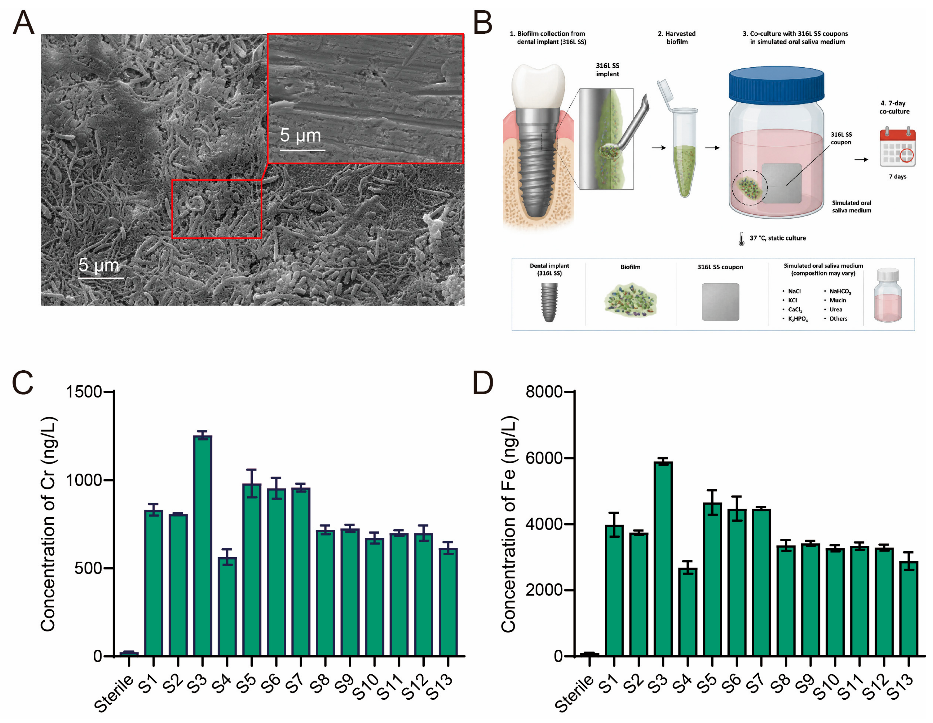
This study examined whether variation in oral biofilm communities was associated with differential corrosion of medical 316L SS and whether a defined community could reproduce selected features of a high-corrosion phenotype. The study comprised four linked components. First, a retrieved orthodontic micro-implant of medical 316L SS was examined to document the co-occurrence of surface-associated biofilm and localized corrosion features. Second, oral microbial communities from 13 donors were incubated independently with medical 316L SS coupons under standardized simulated oral conditions, and corrosion was evaluated by dissolved metal release and electrochemical measurements. Third, planktonic and coupon-associated biofilm fractions were profiled by 16S rRNA gene amplicon sequencing to identify taxa and predicted functions associated with the metal-attached state. Finally, a defined community was assembled to test how community composition was associated with biofilm development, extracellular riboflavin accumulation, and corrosion-related endpoints. Riboflavin and roseoflavin were used as chemical perturbations to assess the association between extracellular riboflavin availability and corrosion activity.
2.2. Ethics Approval, Participant Recruitment, and Oral Sample Collection
The study protocol was approved by the Ethics Committee of the China Medical University Stomatological Hospital (Shenyang, China) (approval No. 2021-17, 26 October 2021). All participants provided written informed consent before enrolment.
Oral microbial samples were obtained from 13 adult participants (age range: 20–28 years, mean age: 23.5 ± 2.2 years; 7 males and 6 females;
Table A1) recruited at the China Medical University Stomatological Hospital. Samples were collected from orthodontic micro-implant-associated plaque using sterile cotton swab and curette. To reduce major confounding effects on the oral microbiota, participants were excluded if they had received systemic antibiotics, probiotics, or antiseptic mouthwash within 14 days before sampling, or if they had predefined exclusion criteria, such as active periodontal disease, untreated caries, smoking, systemic disease, or pregnancy.
All samples were collected under standardized conditions and immediately stored at –80 °C until analysis. During transport, samples were maintained at 4 °C. Samples were handled individually throughout inoculum preparation and incubation; no donor samples were pooled for the subject-derived community experiments.
2.3. 316L SS Coupons and Pre-Treatment
Medical-grade 316L SS was used as the test material. Its nominal elemental composition (wt%) was 0.019 C, 0.43 Si, 1.18 Mn, 10.5 Ni, 16.78 Cr, 2.09 Mo, 0.032 P, 0.0006 S, and balance Fe. Coupons were machined to 10 mm × 10 mm × 5 mm. Before each experiment, coupon surfaces were sequentially ground with silicon-carbide abrasive papers from 240 to 1000 grit to standardize surface preparation. Coupons were then ultrasonically cleaned in deionized water and absolute ethanol for 15 min each, air-dried under sterile conditions, and finally sterilized by UV irradiation.
For electrochemical experiments, coupons were embedded in epoxy resin, leaving an exposed working area of 1 cm2. The exposed area was measured and used for current density normalization. Coupons were randomly assigned to experimental groups, and each coupon was used in only one independent culture–coupon experiment.
2.4. Culture Medium and Incubation Conditions
A modified artificial-saliva medium was used for subject-derived community incubations [
9], defined-community experiments, and corrosion assays unless otherwise indicated. The medium contained (mg/L) NaCl, 125.6; KCl, 963.9; KH
2PO
4, 654.5; Na
2SO
4, 336.6; NH
4Cl, 178; urea, 200; NaHCO
3, 630.8; and CaCl
2, 172. Yeast extract (4 g/L) and glucose (2 g/L) were added as sources of organic nutrients and growth factors. The pH was adjusted to 6.8 ± 0.1 with sterile phosphoric acid.
For anoxic incubations, the medium was sparged with high-purity nitrogen for 40 min before use and transferred into an anaerobic chamber to minimize oxygen exposure. Incubations were performed at 37 °C for 7 days under anaerobic conditions. The atmospheric condition was maintained using high-purity nitrogen.
2.5. Subject-Derived Oral Community Incubation and Corrosion-Phenotype Screening
For each donor, an individual oral-community inoculum was prepared using a standardized protocol involving suspension, vortexing, homogenization, dilution, or washing. Prior to inoculation, the inocula were standardized to an optical density at 600 nm (OD600) of 0.8 to ensure consistent starting biomass across all donor-derived cultures. The final inoculum volume was 1 mL per 10 mL culture volume.
Each incubation vessel contained one 316L SS coupon and 10 mL modified artificial-saliva medium. Inoculated vessels were incubated at 37 °C for 7 days under the conditions described above. Sterile medium containing a coupon served as the abiotic control in every batch. Each donor-derived community was tested in three independent culture–coupon systems per experiment, and the full experiment was repeated independently three times. In this design, the culture–coupon system was the experimental unit: the three systems within one experiment constituted technical replicates of the same donor-derived community, and the three repeated experiments, each initiated from the same frozen inoculum stock on separate occasions, constituted three independent biological replicates. For statistical analysis, the three technical replicates within each experiment were averaged first, and the reported values represent the mean ± SD of the three independent experiments (n = 3).
At the endpoint, culture supernatants were collected for quantification of dissolved Cr and Fe, and extracellular riboflavin. Planktonic and coupon-associated biofilm were recovered separately for microbial community analysis. Corrosion phenotype was assessed based on metal release, corrosion current density (icorr), and, where indicated, localized pit depth.
2.6. Recovery of Planktonic and Coupon-Associated Biofilm Fractions
At day 7, planktonic cultures were collected from each vessel prior to coupon removal. To harvest coupon-associated biomass, the coupons were gently rinsed three times with sterile PBS buffer to remove non-adherent cells. The remaining attached biofilm was recovered via vortexing and bead beating in PBS buffer for 5 min at 5 m/s. This identical recovery protocol was applied to all coupons. The recovered planktonic and biofilm fractions were either processed immediately or stored at −80 °C until DNA extraction. For each fraction, material from technical replicates was maintained separately.
2.7. DNA Extraction, 16S rRNA Gene Sequencing, and Sequence Processing
DNA was extracted from planktonic and coupon-associated biofilm samples using the DNeasy PowerSoil Kit (Qiagen, Hilden, Germany) according to the manufacturer’s instruction. The V3–V4 region of the bacterial 16S rRNA gene was amplified using primers 338F (5′–ACTCCTACGGGAGGCAGCAG–3′) and 806R (5′–GGACTACHVGGGTWTCTAAT–3′). PCR reactions were performed using ABI GeneAmp
® 9700 PCR thermocycler (ABI, Foster City, CA, USA). After PCR amplification and purification, the 16S rRNA gene was sequenced on an Illumina MiSeq PE300 platform (Illumina, San Diego, CA, USA). Raw reads were demultiplexed and quality-filtered using QIIME2 pipeline (v2023.7) [
31]. Forward and reverse reads were merged, and chimeric sequences were removed. Taxonomic classification was performed using QIIME2 classifier against the Genome Taxonomy Database (GTDB) reference database (vr2.7.2) [
32]. Taxonomic labels follow the GTDB naming convention, in which suffix codes (e.g., _A, _B, _D) distinguish genomically distinct lineages that share a genus or species name. All microbial community analyses were conducted in R (v4.0) using microeco (v2.3.0) [
33].
2.8. Functional Inference and Genome-Informed Metabolic Analysis
Predicted functional potential of 16S rRNA gene profiles was inferred using PICRUSt2 (v2.6.3) with PICRUSt2-SC database [
34]. Predicted pathway abundances were compared between planktonic and coupon-associated communities using Kruskal–Wallis.
For the community analysis, genome-scale metabolic models for OTUs were generated using CarveMe (v1.5.1) from GTDB reference database (vr2.7.2) [
35]. Pairwise metabolic interaction potential was estimated using SMETANA (v1.1.0) under medium conditions designed to approximate the modified artificial-saliva medium [
35]. Putative riboflavin biosynthesis and utilization functions were annotated using KEGG (
https://www.genome.jp/kegg/, accessed on 1 June 2024) [
36].
All PICRUSt2- and genome-model-derived results were interpreted as predictions of metabolic potential. They were used to generate hypotheses for experimental testing and were not interpreted as direct evidence of transcription, metabolite exchange, or extracellular electron transfer (EET).
2.9. Defined-Community Construction and Experimental Design
The defined community consisted of
Comamonas tsuruhatensis (laboratory stock obtained from BNCC, Biejing, China, GCA_001571325.1),
Rhodococcus erythropolis (laboratory stock obtained from BNCC, GCA_016598535.1), and
Thauera aromatica (laboratory stock obtained from Mingzhou Bio, Ningbo, China, GCA_003030465.1). The three strains were selected because they were detected at higher relative abundance in coupon-associated communities and showed high predicted metabolic complementarity in the genome-informed analysis. The taxonomic identity of each strain was confirmed by 16S rRNA gene amplification and sequencing, and GTDB-based classification (GTDB-Tk (vr2.7.2) [
32]) placed all three strains within the same GTDB genomic clusters as the corresponding taxa detected in the subject-derived communities.
Each strain was cultured separately in BHI medium at 37 °C under anaerobic conditions until reaching the mid-log phase (OD600 = 0.6). Cells were harvested by centrifugation at 5000× g for 5 min, washed 3 times with PBS buffer, and resuspended in modified artificial saliva. Cell suspensions were normalized to a density of 108 CFU/mL.
Monocultures, all three pairwise combinations, and the three-member community were inoculated at an identical total starting cell density of 10
8 cells/mL. In mixed communities, each constituent strain contributed equal cell numbers to the total inoculum. Each condition was incubated with 316L SS coupons for 7 days as described in
Section 2.4 and
Section 2.5. The subject-derived S3 community served as a reference community with a high corrosion-associated phenotype, because it showed the highest release of iron and chromium ions from 316L SS among the 13 donor-derived communities evaluated in the screening experiment.
2.10. Riboflavin and Roseoflavin Perturbation Experiments
Riboflavin and roseoflavin were obtained from Macklin (Shanghai, China) and prepared as stock solutions. Roseoflavin, a riboflavin analogue that can interfere with flavin-dependent metabolic processes [
37], was used as a chemical perturbagen to examine changes associated with extracellular riboflavin availability; its potential broader effects on microbial physiology are considered in the Discussion (
Section 4). Vehicle controls received the same final solvent concentration as compound-treated cultures. Riboflavin and roseoflavin were added at inoculation to final concentrations of 5 μg/mL and 5 mg/mL, respectively [
25,
26,
28,
38].
The following conditions were included where applicable: sterile medium, sterile medium supplemented with riboflavin, sterile medium supplemented with roseoflavin, untreated community, vehicle-treated community, community supplemented with riboflavin, and community treated with roseoflavin. Coupons were incubated for 7 days before electrochemical measurements and endpoint sampling.
2.11. Biomass, Viability, Growth, pH, and Community-Composition Controls
Biofilm structure and viability were assessed by confocal laser scanning microscopy (CLSM; LSM 900, Zeiss, Oberkochen, Germany) as described in
Section 2.14. Total biofilm biomass was quantified using crystal violet staining, and surface-associated ATP levels were measured using an ATP fluorescence detector (UPF-10ATP, UP General, Beijing, China). Culture pH was measured. These measurements were used to evaluate whether differences in corrosion rate could be explained by gross changes in biomass, cell viability, or bulk pH.
2.12. Electrochemical Measurements
Electrochemical measurements were performed at 37 °C using a three-electrode configuration. The 316L SS coupon served as the working electrode, a saturated calomel electrode (SCE) as the reference electrode, and a platinum sheet as the counter electrode. Each glass cell contained 200 mL of experimental medium and was inoculated with 2 mL of microbial suspension at standardized inoculum. The exposed surface area of the working electrode was 1 cm2.
Open-circuit potential (OCP) and potentiodynamic polarization were measured using a Gamry Reference 600 potentiostat (Gamry Instruments, Warminster, PA, USA). OCP was monitored for 0.5 h before polarization measurements. Potentiodynamic polarization was recorded after 7 days of incubation from −0.3 to 1.2 V relative to OCP at 0.333 mV/s. Corrosion current density (
icorr) was derived from Tafel extrapolation. The complete electrochemical measurement (0.5 h of OCP stabilization followed by the potentiodynamic scan) was finished within approximately 2 h, immediately after 7 days of biofilm maturation. Because this period is far shorter than the generation times of the dominant community members, meaningful changes in microbial community composition during the measurement were considered negligible, and community composition was characterized at the experimental endpoint (
Section 2.7). Three independent experiments were performed for each condition; within each experiment, measurements from three technical replicate cells were averaged before pooling across experiments (n = 3).
2.13. Metal-Release Analysis and Extracellular Riboflavin Quantification
After incubation, culture supernatants were collected by centrifugation at 10,000× g for 5 min and filtration through 0.22 μm membranes. For metal analysis, samples were acidified using a HNO3–HClO4–HF mixture (2:1:2, v/v/v) and diluted as required. Dissolved Cr and Fe concentrations were quantified by inductively coupled plasma mass spectrometry (ICP–MS; Agilent 7850, Agilent Technologies, Santa Clara, CA, USA). Calibration curves were generated from certified standards, and blank medium values were subtracted from experimental measurements.
Extracellular riboflavin concentrations were determined by high-performance liquid chromatography (HPLC; UltiMate 3000, Thermo Scientific, Waltham, MA, USA) using a C18 column (150 × 4.6 mm, 5 μm; Bonna-Agela Technologies, Tianjin, China). Samples were protected from light during collection and analysis. The mobile phase comprised methanol and 0.05 M ammonium acetate buffer (pH 6.0) at 85:15 (v/v) and was delivered isocratically at 1.0 mL/min. Riboflavin was monitored at 445 nm and quantified against an external standard curve prepared with authentic riboflavin.
2.14. Biofilm Imaging and Surface Characterization
For CLSM, coupon-associated biofilms were stained with the LIVE/DEAD BacLight bacterial viability kit (Thermo Fisher Scientific, Waltham, MA, USA) according to the manufacturer’s instructions. Samples were imaged using a Zeiss LSM 900 confocal microscope (Zeiss, Oberkochen, Germany). Three-dimensional reconstructions were generated using ZEN software (
https://www.zeiss.com/microscopy/us/products/software/zeiss-zen.html, accessed on 1 January 2025) (Zeiss), and biofilm thickness was calculated from z-stack images.
For corrosion-pit analysis, biofilms and corrosion products were removed following GB/T 4334.4-2000 [
39]. Coupons were ultrasonically cleaned in ethanol and briefly exposed to a hydrofluoric-acid/nitric-acid solution as specified by the standard. The duration of chemical cleaning was standardized across samples to minimize alteration of the underlying substrate. Pit morphology and depth were then analyzed by CLSM.
2.15. Metal-Release Assay Without an Added Organic Carbon Source
To evaluate metal dissolution associated with the oral microbial community under conditions without added organic carbon, we performed a dedicated assay without external organic carbon sources or organic electron donors in the bulk medium. Briefly, anaerobic incubation media were prepared in a simulated-saliva–based inorganic formulation and supplemented with salts and trace components as in the main corrosion assays, while omitting all organic substrates (e.g., carbohydrates, organic acids, peptides, amino acids, or any other assimilable carbon sources). Under these conditions, the 316L SS coupon represented the only intentionally supplied solid-phase material with the potential to act as an electron donor in interfacial redox processes, while the input of exogenous organic electron donors was minimized.
For each condition, pre-cleaned 316L SS coupons were incubated at 37 °C under anoxic conditions with the following groups: (i) sterile control, (ii) S3 community, (iii) S3 + riboflavin, and (iv) S3 + roseoflavin. Riboflavin and roseoflavin were added at the concentrations used in the related supplementation experiments (final concentrations stated in the main
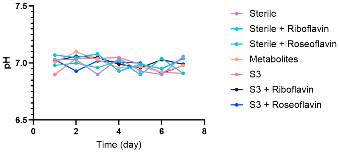
Section 2.10). Incubations were conducted for the same duration as in the corresponding metal-release experiments. After incubation, culture liquids were collected, filtered (0.22 μm), and the dissolved Cr and Fe concentrations were quantified by ICP–MS. Solution pH was measured at the endpoint toassess whether supplementation substantially altered bulk acidity.
In this assay context, increased dissolved Cr/Fe release was interpreted as evidence of enhanced metal dissolution associated with microbial presence under oligotrophic conditions (i.e., in the absence of added organic carbon), rather than as direct evidence that 316L SS served as a biological electron donor in microbial metabolism. Changes observed following riboflavin supplementation and roseoflavin treatment were interpreted as supporting an association between extracellular riboflavin availability and metal corrosion activity. All measurements were performed in three independent experiments (n = 3), each comprising triplicate technical systems that were averaged before pooling; results are reported as mean ± SD.
2.16. Statistical Analysis
Data are presented as the mean ± SD of three independent experiments (n = 3) unless otherwise stated. Normality was assessed using the Shapiro–Wilk test, and homogeneity of variances was evaluated using Levene’s test. For two-group comparisons, Student’s
t-test (normally distributed data) or the Mann–Whitney U test (non-normally distributed data) was used. For multiple-group comparisons, one-way analysis of variance (ANOVA) followed by Tukey’s HSD post hoc test was used for normally distributed data, whereas the Kruskal–Wallis test followed by Dunn’s post hoc test was used for non-normally distributed data. Differences in microbial community composition between planktonic and biofilm-associated fractions were evaluated by permutational multivariate analysis of variance (PERMANOVA; adonis2 with 999 permutations) based on Bray–curtis dissimilarity. Differentially enriched taxa were identified using LEfSe (v4.6.0) (LDA score > 2,
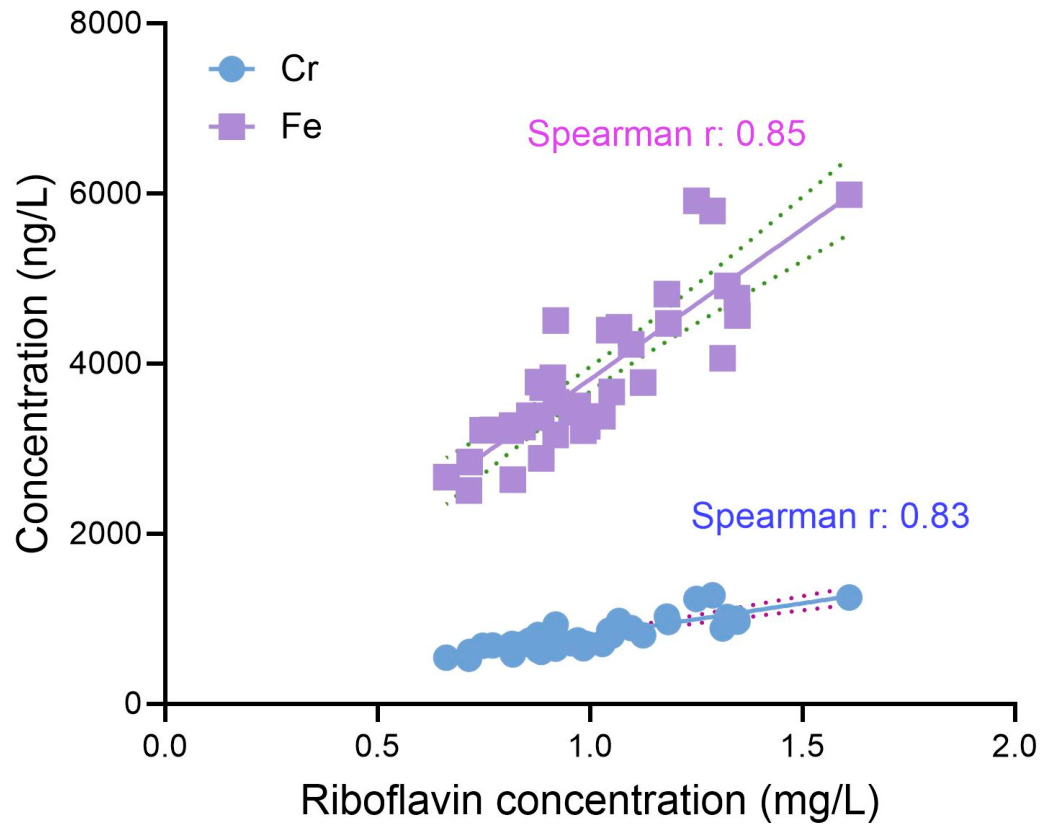
p < 0.05). Correlations between extracellular riboflavin concentrations and dissolved Cr or Fe were assessed using Spearman’s rank correlation. Statistical significance was defined as
p < 0.05. In all figures, the number of observations and the statistical tests applied are stated in the corresponding figure legends. All statistical analyses were performed in R (v4.0) using the microeco (v2.3.0) [
33] and vegan (v2.7.5) packages.
4. Discussion
Orthodontic micro-implants fabricated from 316L SS are exposed to a chemically and microbiologically heterogeneous oral environment. Although corrosion of stainless steel orthodontic devices has been documented in clinical retrieval and in vitro studies [
9,
23,
24], the extent to which oral microbial community composition and community-level metabolism shape corrosion outcomes remains incompletely understood. Here, we extend the MIC framework by demonstrating that subject-derived oral consortia markedly accelerate 316L SS corrosion under controlled ex vivo conditions. The pronounced variation in corrosion-associated outcomes among subject-derived communities further suggests that microbial composition and functional potential may contribute to inter-individual differences in corrosion behavior. Collectively, these findings motivate a community-level framework for predicting microbiota-associated corrosion resistance at oral biomaterial interfaces.
Previous work has mainly attributed intra-oral corrosion to physicochemical factors, including acidic pH, fluoride exposure, chloride-containing saliva, oxygen gradients, and mechanical disruption of the passive film [
40,
41]. MIC has more recently been recognized as a potential contributor to implant corrosion and degradation in the oral cavity [
9,
23,
24]. Subgingival microbiota have been reported to colonize 316L SS and promote localized pitting through acidic metabolites and electron-transfer-associated processes [
9,
23,
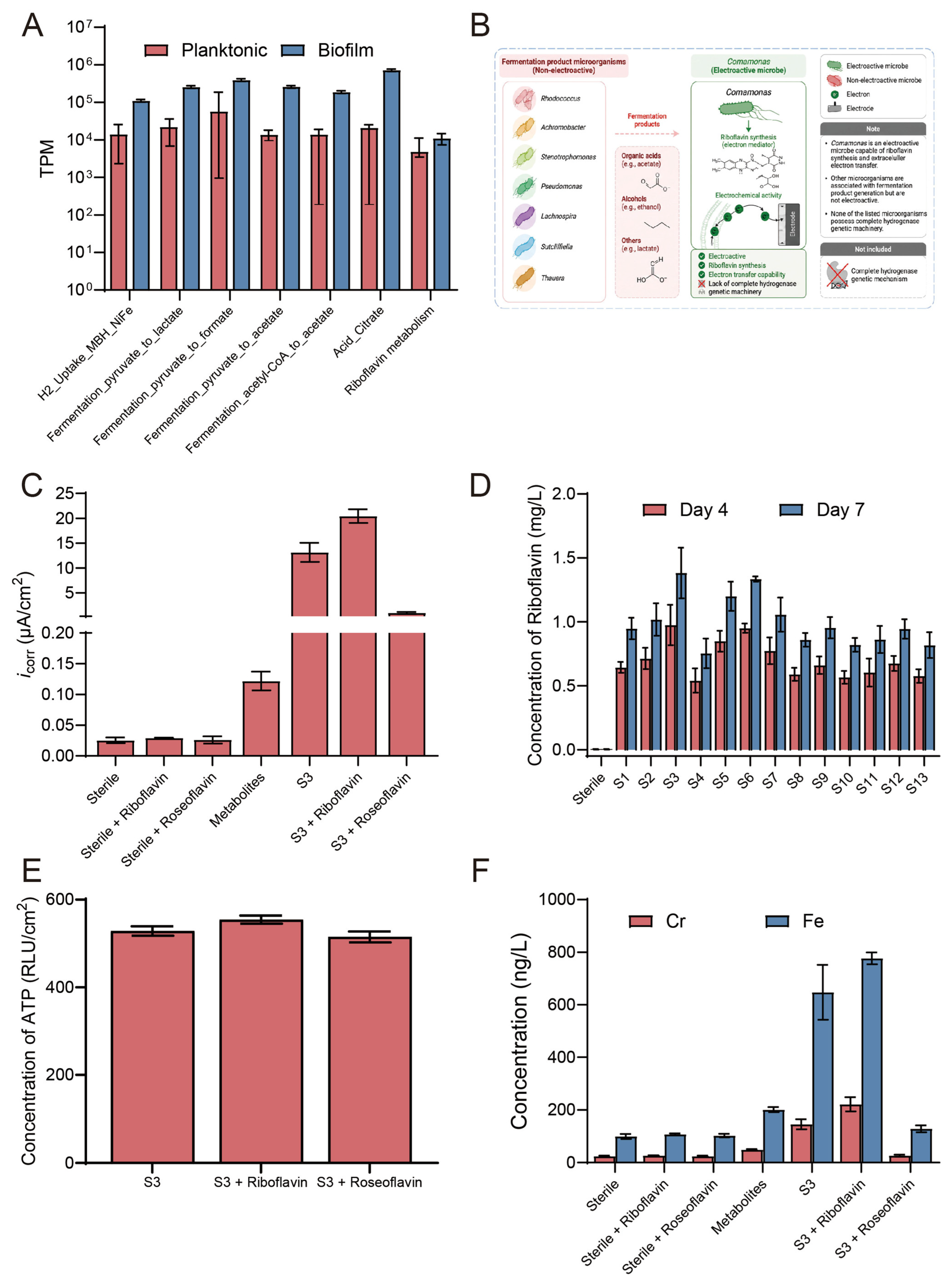
24]. Consistent with these concepts, our analyses indicate that corrosion-associated communities possess predicted functional capacities enriched for fermentation-related metabolism (e.g., pyruvate metabolism, acetyl-CoA-to-acetate conversion, citrate metabolism) and riboflavin metabolism. Importantly, culture-derived, cell-free metabolites from the high-corrosion S3 consortium increased the
icorr, but the effect was substantially weaker than that produced by the intact S3 consortium. This difference supports the view that fermentation products alone are unlikely to fully account for the high-corrosion phenotype and that community-level organization, such as biofilm development, redox gradients, extracellular polymeric substances, and interfacial redox processes that may involve electron transfer, likely contributes to the corrosion process.
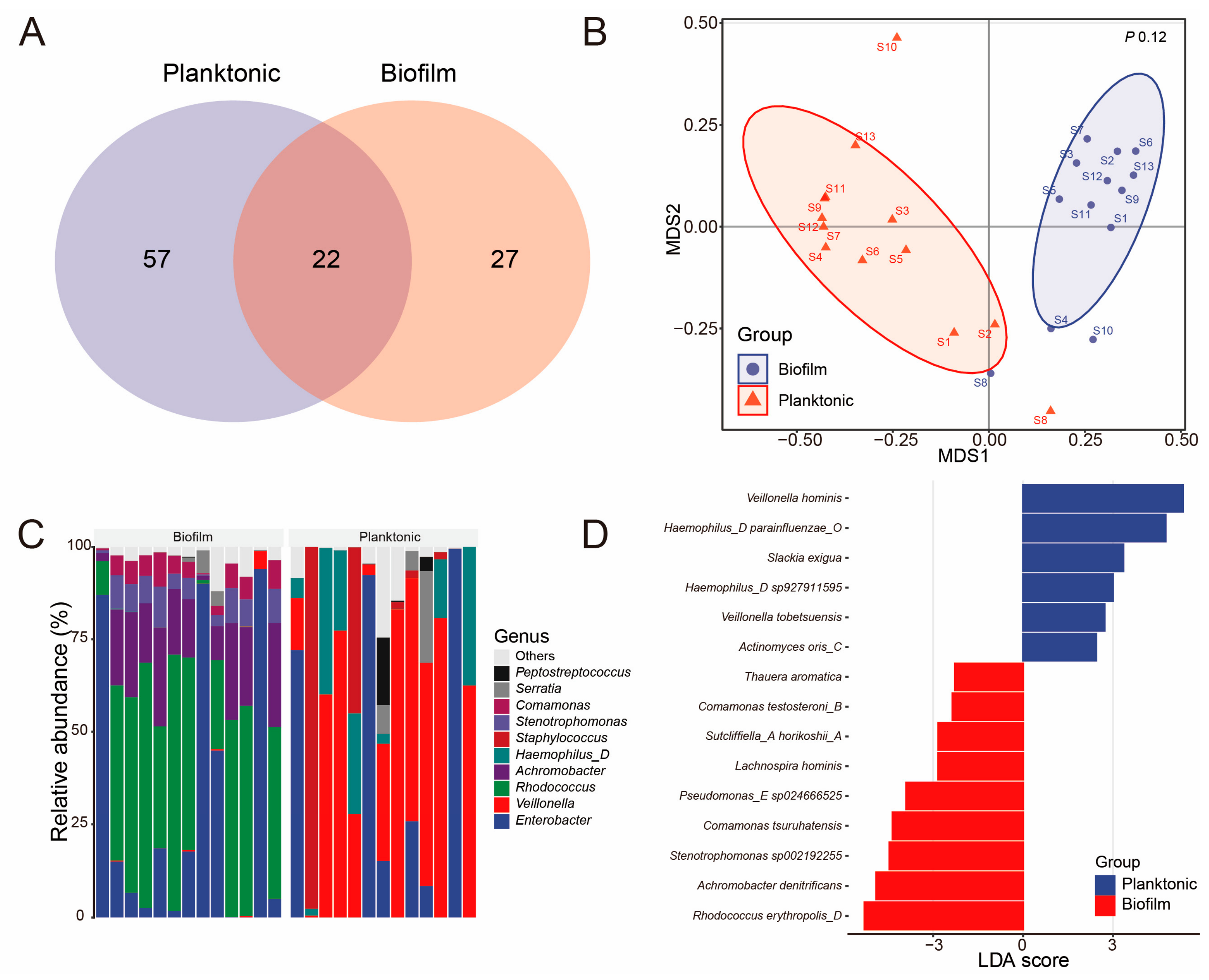
Beyond differences in microbial composition, our results suggest that the 316L SS surface may exert niche-filtering effects during interface corrosion, although the overall separation between planktonic and biofilm-associated communities was not statistically significant (PERMANOVA, p = 0.12). Although planktonic and attached-biofilm fractions contained 57 and 27 unique OTUs, respectively, with only 22 OTUs shared between fractions, ordination based on Bray–Curtis dissimilarity showed a trend toward separation. Notably, biofilm samples displayed tighter clustering in ordination space than planktonic communities, consistent with reduced variability of communities surface-associated with the metal interface relative to the heterogeneous donor-associated baseline. Genus-level profiles further supported fraction-specific enrichment: planktonic communities were dominated primarily by Veillonella and Haemophilus_D, whereas 316L SS-associated biofilms showed greater relative abundances of Enterobacter, Rhodococcus, and Comamonas, with significant enrichment of taxa including R. erythropolis_D, A. denitrificans, C. tsuruhatensis, and T. aromatica. Together, the tighter clustering and consistent enrichment patterns are suggestive of selective enrichment of functional taxa at the metal interface, although these patterns should be interpreted cautiously given the lack of statistical support for community-level separation (PERMANOVA, p = 0.12).
SRBs are widely recognized as potential contributors to MIC [
42,
43,
44,
45]. However, the abundance of SRB does not necessarily correlate with the measured corrosion rate. Their effects may depend on sulfate-reduction activity, sulfide production, localized chemical gradients within biofilms, and metabolic interactions with other microorganisms, rather than their relative abundance alone [
1]. Therefore, the low abundance or non-detection of SRB-associated taxa in this study should not be interpreted as evidence that SRB are unimportant in oral MIC. In the present dataset,
D. oralis, an SRB-associated taxon [
46], was detected only in one planktonic sample (S4; 0.12%; 1/13 participants) and was not detected in any biofilm sample. This sparse detection represents a limitation of the study. The participants were healthy adults, and samples were collected from orthodontic micro-implant-associated plaque, which may differ from more anaerobic oral niches, such as periodontal pockets, where SRB may be more readily enriched. In addition, low-abundance SRB may fall below the detection limit of V3–V4 16S rRNA gene amplicon sequencing, while ex vivo incubation may alter the composition and detectability of the original microbial communities. Future studies should employ targeted functional approaches, including dsrAB-targeted qPCR, metagenomic or metatranscriptomic sequencing, and enrichment-based assays, to determine the prevalence and activity of SRB in oral biofilms.
Our results implicate riboflavin-associated processes as a candidate factor associated with community-level corrosion behavior and a proposed interfacial redox mechanism that may involve EET. Riboflavin has been reported to act as an electron mediator in MIC systems; for example, exogenous riboflavin promoted pitting corrosion in
Desulfovibrio vulgaris biofilms [
29,
47]. Flavin-associated electron transfer has also been linked to enhanced 316L SS corrosion in other microbial settings [
25,
28], and riboflavins are established diffusible electron shuttles in electroactive bacteria such as
Shewanella [
26,
27,
48]. In oral biofilm contexts, however, riboflavin-associated contributions have been comparatively less explored at the community level.
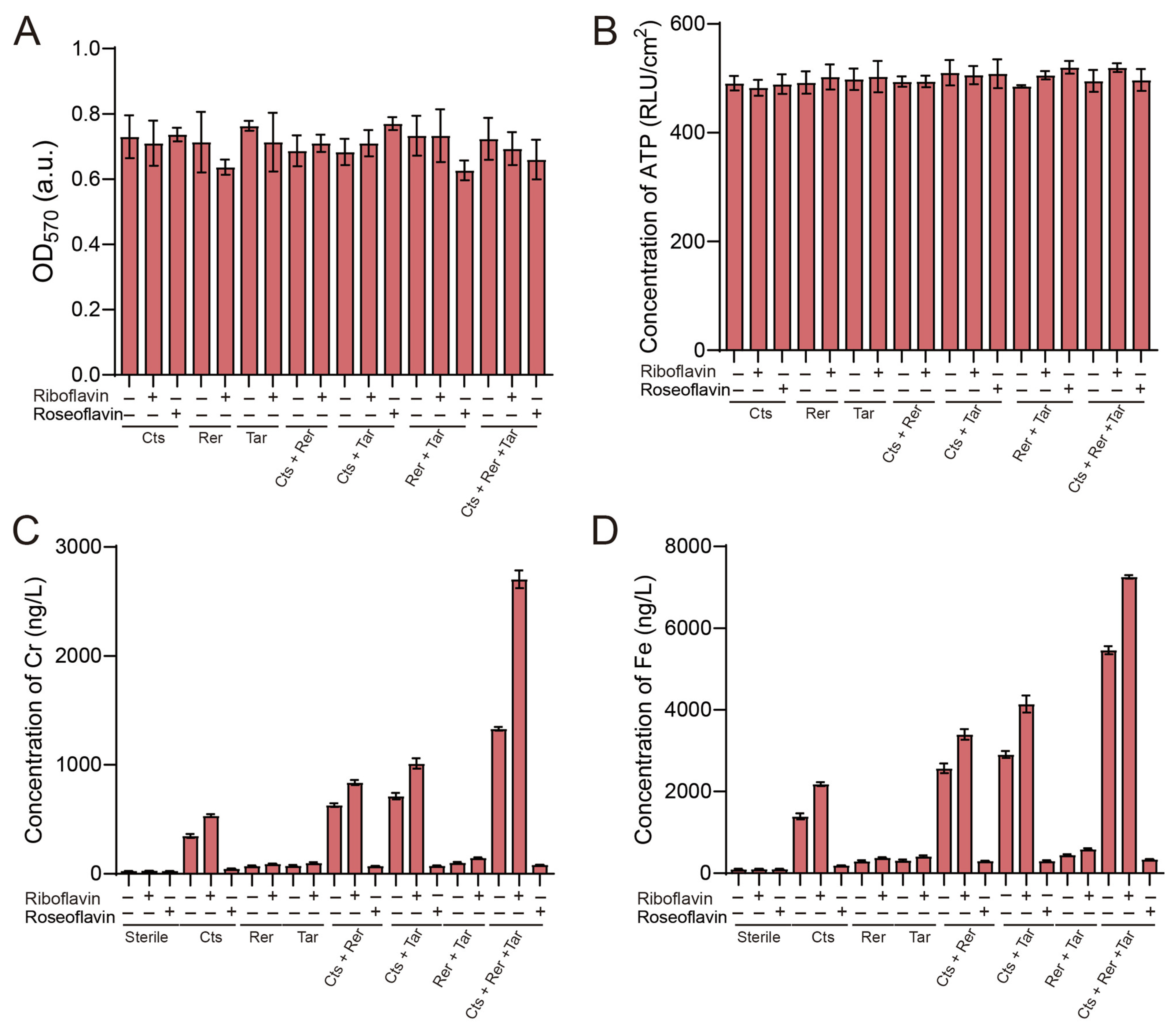
In our system, riboflavin was detected across all subject-derived consortia but remained undetectable in sterile controls, accumulated during incubation, and was highest in corrosion-active communities. Perturbation experiments provided experimental support for an association between riboflavin availability and corrosion: exogenous riboflavin increased icorr and accelerated metal corrosion and dissolution, whereas roseoflavin reduced extracellular riboflavin availability and substantially decreased icorr and dissolved Cr/Fe concentrations. Crucially, these shifts in corrosion did not coincide with gross changes in biofilm biomass or surface-associated metabolic activity (crystal violet staining and surface ATP levels showed negligible differences), indicating that riboflavin availability modulates corrosion processes rather than globally altering growth. Nevertheless, roseoflavin is a riboflavin analogue that may exert broader biological effects, including interference with flavin-dependent metabolic processes beyond the reduction in extracellular riboflavin availability, and surface-associated ATP and biomass measurements alone cannot exclude more subtle changes in microbial physiology or community composition; the roseoflavin results should therefore be interpreted with caution. Moreover, in an assay without an added organic carbon source, the S3 community increased dissolved Cr and Fe release; riboflavin further enhanced, whereas roseoflavin decreased, metal release relative to untreated S3. Because the pH remained near neutral and did not differ appreciably from controls, these findings support a riboflavin-associated contribution to corrosion in this ex vivo model, consistent with a proposed interfacial redox process. While the data support an association between riboflavin availability and corrosion activity in our assay context, the precise micro-scale pathway(s) of electron transport at the biofilm–metal interface (e.g., direct shuttling vs. alternative redox coupling) remain to be defined by direct measurements of electron transfer and riboflavin exchange.
An important mechanistic distinction should be made between the corrosion pathway examined here and the metabolite-mediated pathway reported for
Streptococcus mutans, an oral pathogen whose biofilms induce corrosion of 316L SS primarily through the production of acidic metabolites [
23,
49,
50]. In the present study, the bulk pH of the incubation medium remained near neutral and did not differ appreciably from the sterile controls throughout the 7-day experiments (
Figure A2). Under the buffered, oligotrophic conditions used here, bulk acidification is therefore unlikely to be the dominant driver of the observed corrosion. Instead, the corrosion-associated phenotype was consistently linked to extracellular riboflavin availability, and we propose that riboflavin acts as a diffusible redox mediator that can shuttle electrons between microbial cells and the steel surface [
28], thereby promoting cathodic reactions and localized metal dissolution. Because direct measurements of electron flux and riboflavin exchange at the biofilm–metal interface were not performed, this mechanism is presented as a proposal consistent with the available data, and local interfacial pH or redox conditions within the biofilm may differ from the near-neutral bulk values. This mechanistic contrast with the acid-driven
S. mutans pathway [
23] highlights the specificity of the riboflavin-associated corrosion process examined here.
Reconstructed-community experiments provide insight into the community-level basis of the corrosion phenotype. The biofilm-enriched taxa selected for reconstruction,
C. tsuruhatensis,
R. erythropolis, and
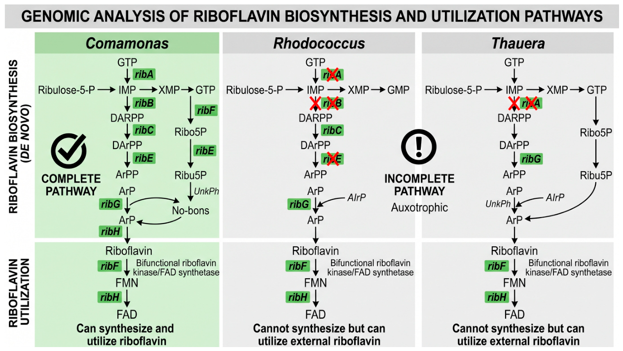
T. aromatica, showed high predicted pairwise metabolic complementarity in genome-informed analyses. Consistent with their potential involvement in electroactive biofilm phenotypes,
Comamonas species have been detected in exoelectrogenic biofilms and bioelectrochemical systems [
51],
Rhodococcus strains have been reported to exhibit flavin-associated EET [
52], and
Thauera species have been isolated from electroactive anode biofilms [
52,
53]. Consistent with a potential cooperative community effect,
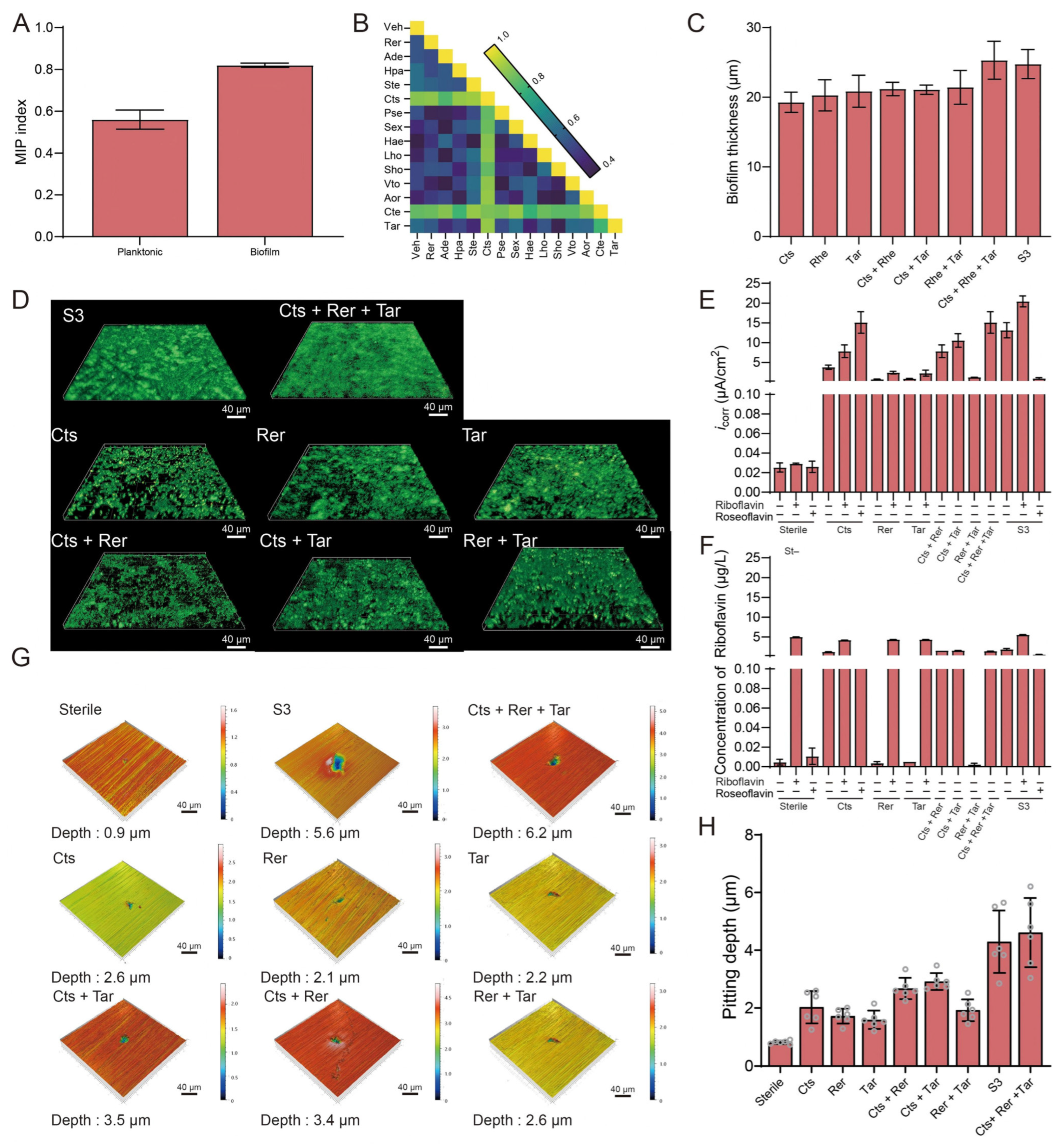
C. tsuruhatensis,
R. erythropolis, and
T. aromatica monocultures formed sparse, discontinuous surface aggregates, whereas dual-species consortia displayed moderate surface coverage and limited three-dimensional maturation. In contrast, the
C. tsuruhatensis +
R. erythropolis +
T. aromatica consortium formed a dense, continuous biofilm architecture and closely recapitulated the high-corrosion phenotype observed for the S3 consortium, including pitting severity.
Functionally, C. tsuruhatensis + R. erythropolis + T. aromatica induced markedly elevated icorr and showed the deepest localized pits among the defined communities, with activity approaching that of the S3 consortium. Notably, C. tsuruhatensis alone did not reproduce the three-strain corrosion activity, and dual-species combinations partially restored it, suggesting that the high-corrosion outcome may depends on community interactions rather than individual taxa alone. Riboflavin-perturbation within the defined consortium further supported a proposed riboflavin-linked model: riboflavin supplementation promoted higher corrosion rate of 316L SS, whereas roseoflavin treatment reduced the measured corrosion phenotypes. Extracellular riboflavin accumulation in the defined system was observed primarily in C. tsuruhatensis and increased further in the presence of R. erythropolis and T. aromatica, whereas R. erythropolis, T. aromatica, and the R. erythropolis + T. aromatica combination showed minimal riboflavin levels. Importantly, riboflavin/roseoflavin effects on corrosion were not accompanied by parallel increases in overall biofilm biomass or surface-associated ATP, reinforcing the idea that riboflavin availability modulates corrosion-linked physiology in the interspecies context rather than broadly changing biomass.
This interpretation is consistent with the broader literature on multispecies biofilms, where community-level phenotypes often cannot be predicted from monoculture behavior [
1]. Metabolic cross-feeding, spatial organization, competition, and metabolite sharing can alter biomass accumulation, local chemical gradients, and redox activity [
54,
55], and such interactions are increasingly considered important determinants of MIC dynamics [
5,
56]. In our system, the thicker and more continuous triple-species biofilm may have promoted retention of redox-active compounds and facilitated localized microenvironments that support corrosion. Nevertheless, direct measurements of riboflavin exchange kinetics and interspecies electron transfer at the interface were not performed; therefore, “riboflavin-linked interspecies interaction” should be regarded as a proposed mechanistic model supported by convergent evidence rather than directly visualized metabolite transfer.
The substantial variation in dissolved Cr and Fe release across the 13 subject-derived consortia may have clinical relevance. Corrosion products and surface degradation have been observed on retrieved orthodontic 316L SS components [
57,
58], yet clinical severity varies considerably among patients. Such variability has commonly been attributed to host and environmental factors such as oral hygiene, implantation duration, mechanical loading, saliva chemistry, and local inflammation [
59]. Our findings indicate that, even under controlled ex vivo conditions, oral microbiota composition and predicted functional capacity may contribute to variation in corrosion activity. Specifically, communities combining taxa with predicted riboflavin-biosynthesis capacity with metabolically complementary partners may be ecologically primed to assemble corrosion-promoting biofilms. This does not imply that microbiota composition alone determines clinical failure; rather, it highlights the potential interaction between microbial ecology and physicochemical conditions that regulate passive-film stability. It should be emphasized, however, that these findings were obtained in a simplified ex vivo model, and direct extrapolation to clinical conditions is limited by the differences between the experimental system and the dynamic oral environment, including salivary flow and shear forces, mechanical loading, fluctuating oxygen levels, dietary exposure, pH variations, host immune factors, and long-term exposure conditions.
Several limitations should be considered. First, functional and metabolic inferences were based primarily on 16S rRNA gene sequencing, PICRUSt2 functional predictions, and genome-based MIP. These approaches estimate metabolic potential rather than directly measuring gene expression, metabolic flux, or interspecies metabolite exchange. Metatranscriptomic, metaproteomic, and targeted metabolomic analyses would help validate the proposed processes and identify which genes and metabolic routes are active in situ. Second, the three-strain consortium serves as a tractable model but necessarily simplifies the diverse oral microbiota, excluding low-abundance, uncultured, and host-derived contributors. In addition, SRBs, which are recognized contributors to MIC in many environments and can occur in oral niches, were not detected at appreciable relative abundance in the sequencing dataset of the subject-derived communities and were not included in the defined consortium. Nevertheless, it is long established that SRB cell numbers do not correlate with measured corrosion rates; accordingly, a low SRB abundance or even non-detection does not by itself allow their potential significance in the corrosion process to be dismissed. In addition, SRB are typically low-abundance anaerobes in oral niches, and their detection by 16S rRNA gene amplicon sequencing is constrained by the sampling procedure, sample transport and storage, DNA extraction efficiency, primer specificity, and sequencing depth; our single-time-point sampling of implant-associated plaque may therefore have underestimated their presence (
Table A2). Dedicated approaches, such as targeted enrichment culture, quantitative PCR, and shotgun metagenomic sequencing, would be required to more definitively evaluate the presence and potential contribution of SRB in oral biofilm–metal systems. Their potential contribution to oral microbiota-associated corrosion therefore remains to be addressed in future studies. Third, roseoflavin, a riboflavin analogue used as a perturbation tool, may exert broader effects on flavin-dependent metabolism beyond reducing extracellular riboflavin availability, and the biomass and surface-ATP measurements performed here cannot exclude subtle changes in microbial physiology or community composition; the roseoflavin results were therefore interpreted as supporting associations rather than an established mechanism. Fourth, our ex vivo experiments were performed under static anaerobic conditions in simulated saliva, which do not fully reproduce the dynamic nature of the oral environment, including salivary flow and shear forces, mechanical loading, fluctuating oxygen levels, dietary exposure, pH variations, host immune factors, and long-term exposure conditions; the findings should therefore be regarded as evidence derived from an ex vivo experimental model rather than as direct evidence of clinical corrosion mechanisms. Fifth, the potentiodynamic polarization measurements represent endpoint snapshots obtained after 7 days of incubation rather than time-resolved corrosion monitoring; although these electrochemical results were corroborated by cumulative 7-day metal-release measurements, time-resolved techniques (e.g., electrochemical impedance spectroscopy or linear polarization resistance monitoring) would provide further insight into corrosion dynamics. Microbial abundance was not additionally monitored during the approximately 2-h electrochemical measurement window, and short-term community stability during polarization was therefore not independently verified. Finally, the cohort size (13 subjects) may not capture the full spectrum of oral microbiota diversity or corrosion phenotypes across broader populations. Sixth, the detailed multi-parameter characterization was performed for the S3 consortium selected on the basis of its highest Cr/Fe release in the primary screening; extending this characterization to additional consortia spanning the observed metal-release range (e.g., the next-highest- and lowest-release communities, S5 and S4) would further test the generalizability of the observed associations. Despite these limitations, the integration of donor-derived communities, interface-associated profiling, metabolite and electrochemical assays, and defined-community reconstruction provides an experimental ecological framework for studying biomaterial degradation as an emergent property of oral biofilm ecology.



 ,
,