Review

Human Oral-Associated Capnocytophaga Species and Their Clinical Relevance


1
Institute of Dentistry, University of Turku, Lemminkäisenkatu 2, 20520 Turku, Finland
2
Faculty of Dentistry, The University of Hong Kong, Hong Kong, China
*
Author to whom correspondence should be addressed.
Microorganisms 2026, 14(7), 1512; https://doi.org/10.3390/microorganisms14071512
Submission received: 29 May 2026 / Revised: 6 July 2026 / Accepted: 8 July 2026 / Published: 11 July 2026
(This article belongs to the Section Medical Microbiology)

Abstract

Members of the Capnocytophaga genus are common residents of the oral cavity of humans and some animals, notably dogs and cats. In humans, Capnocytophaga species have received considerably less attention than their canine compatriots due to their more nuanced roles in health and disease. Whilst they are commonly associated with oral health, many aspects regarding their respective functions, inter-species interactions, and activities within oral microbial biofilm communities remain to be established. Furthermore, there are increasing numbers of reports linking certain human oral-associated Capnocytophaga species with systemic infections, suggesting their activities as low-level pathogens. However, clinical data are currently scanty. In this narrative review, we give an overview of human oral-associated Capnocytophaga, including their taxonomy and genomic compositions, challenges in their species-level identification, their colonization patterns within the oral cavity, potential pathogenic features, and their clinical relevance in oral and non-oral infections. We discuss the similarities and differences between key aspects of human and animal oral-associated species of Capnocytophaga, including experimental methods used for their comparative functional analysis.

1. Genus Capnocytophaga

Members of the Capnocytophaga genus are known as common residents of the oral cavity of humans and some animals. In this review, our objective is to give an overview of human oral-associated Capnocytophaga, focusing on their current taxonomy and genomic compositions, challenges in their species-level identification, their colonization patterns in children and adults, as well as their clinical relevance in oral and non-oral infections.

1.1. Taxonomic Considerations

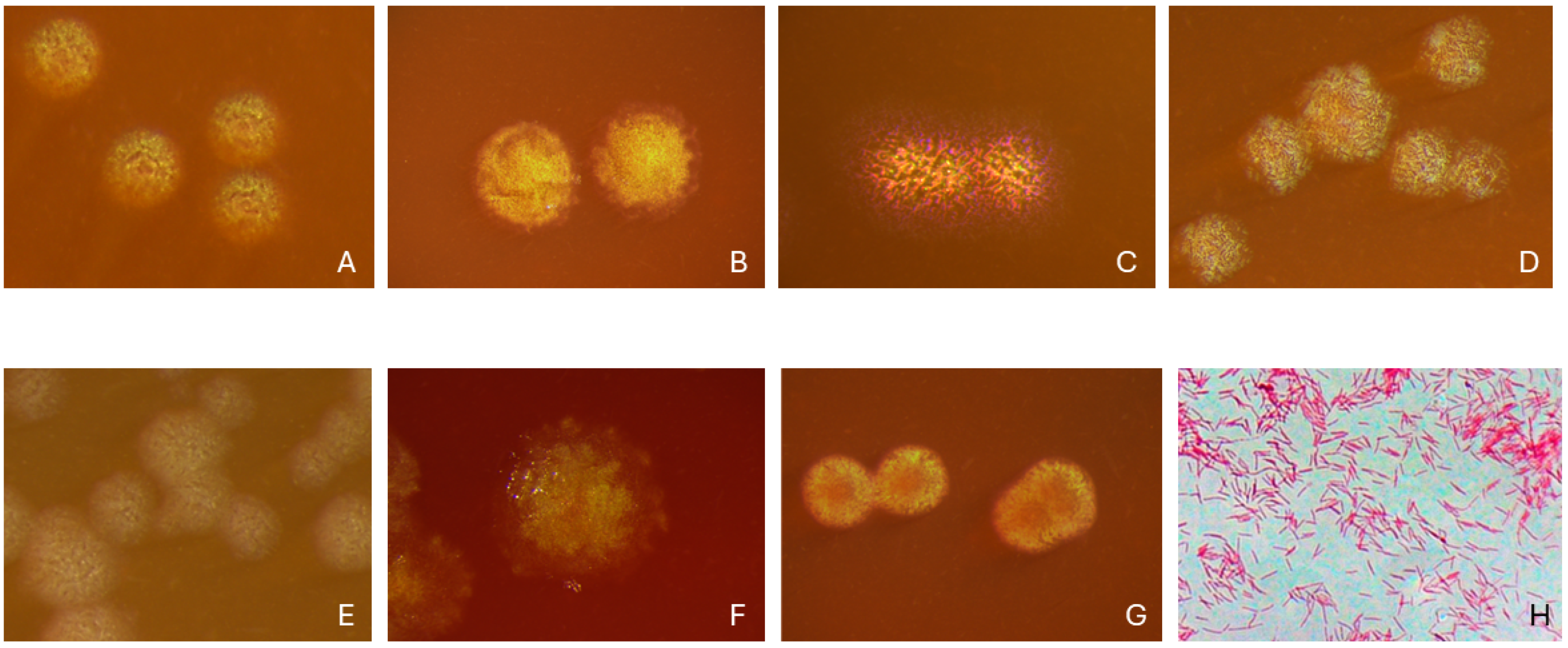
In the late 1970s, strains formerly classified as Bacteroides ochraceus and Centers for Disease Control (CDC) group DF-1 (Dysgonic fermenter 1) were re-classified into the novel genus Capnocytophaga with three novel species, including C. gingivalis, C. ochracea, and C. sputigena [1,2,3], and the novel genus name was validated in 1982. Since then, the genus has expanded, with the species being phenotypically similar; they grow under anaerobic to microaerophilic conditions, preferring an atmosphere enriched with CO2 (i.e., capnophilic), producing yellow–orange or beige, speckled colonies that typically glide on the agar surface, and the cells are thin, slender, elongated rods with a negative Gram stain reaction (Figure 1) [1,4]. The identification of human oral-associated Capnocytophaga at the genus level is feasible based on the above-mentioned basic phenotypic characteristics; however, conventional biochemical testing is not reliable for identifying the members of the genus to the species level, which requires more advanced techniques.
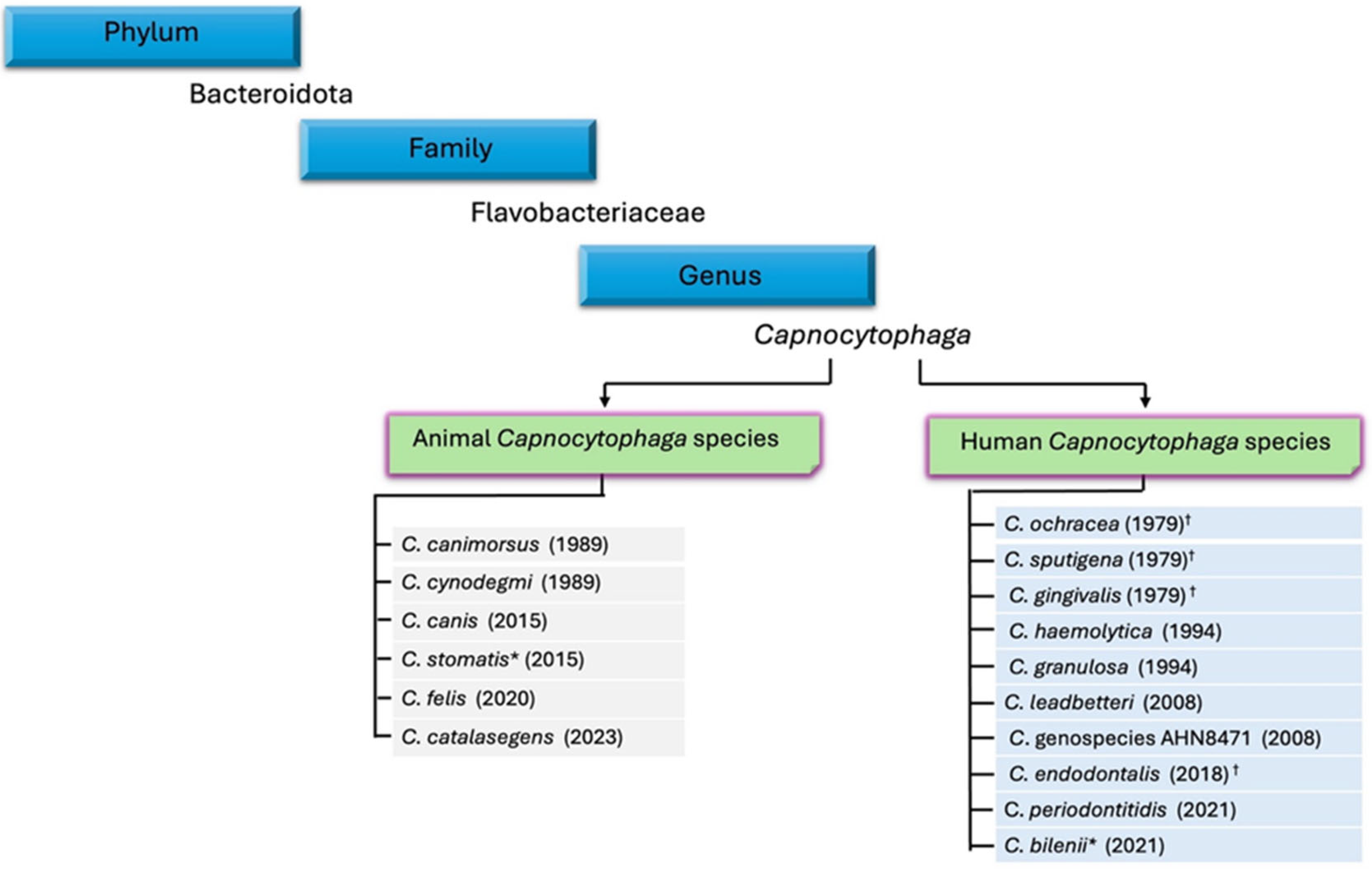
The genus Capnocytophaga currently contains eight human and five animal species that are validly published, while two ‘species candidates’ are still awaiting validation (Figure 2). The genus Capnocytophaga belongs to the family Flavobacteriaceae within the phylum Bacteroidota. C. ochracea is the type species of the genus.
While Capnocytophaga species of animal origin are well known for their role as important pathogens, especially C. canimorsus in bite infections [5,6], data on the role of human oral-associated Capnocytophaga species are rather fragmentary in the literature. However, all eight validly published Capnocytophaga species in humans are reported in the List of Recommended Names for bacteria of medical importance (LoRNs) because of the recorded risk group of the species themselves [7], and all human oral-associated Capnocytophaga species validly published before 2020 are regarded as established pathogens infecting humans [8].

1.2. Milestones in Capnocytophaga Genome Sequencing

The type strain of C. ochracea (VPI 2845T = DSM 7271T = ATCC 33596T) was the first Capnocytophaga strain to be genome sequenced in 2009 (GenBank accession: NC_013162) [9]. This revealed a single chromosome of 2,612,955 bp, encoding 2252 genes, with a G + C content of 39.6%: genomic features that are typical of Capnocytophaga taxa of both human and animal origin. The genome sequence of the ATCC 33624 type strain of C. gingivalis was also deposited in 2009 (GCA_000174755.1), but this was not accompanied by a publication.
Subsequent milestones included the elucidation of the first animal oral-associated Capnocytophaga genome sequence, that of C. canimorsus strain Cc5 [10], and the publication of 12 complete Capnocytophaga reference genomes by a team from the US CDC, including strains of C. sputigena, C. leadbetteri, C. gingivalis (all of human origin); and C. canimorsus, C. cynodegmi, and C. canis (including the CcD38 type strain), and C. stomatis (all of canine origin) [11].
The formal descriptions of the following human Capnocytophaga species were accompanied by genome sequence data for the type strains: C. endodontalis ChDC OS43T (KCTC 5562T = JCM 32133T) originally isolated from a human refractory periapical abscess (Korea) [12]; C. periodontitidis p1a2T (CGMCC 1.17337T = JCM 34126T) isolated from subgingival plaque from a deep periodontal pocket (Beijing, China) [13]. ‘C. bilenii’ Marseille-Q4570T (CSUR Q4570T) (not yet validly published) was isolated from dental plaque of a 25-year-old male with gingivitis (Marseille, France) [14].
The taxonomic standing of C. endodontalis remains controversial, and it has previously been noted that C. periodontitidis and Capnocytophaga genospecies (genomospecies) AHN8471 [15] may represent strains belonging to the same (or overlapping) species [16]. Data from our ongoing genomic analysis of human oral Capnocytophaga isolates suggest that strains originally assigned to Capnocytophaga genospecies AHN8471 (namely strains AHN8471, AHN8751, AHN9528, AHN9576, and AHN9798) encompass both the C. endodontalis and C. periodontitidis species (unpublished data). Hopefully, this taxonomic issue will be resolved in the near future through detailed comparative genomic analysis of additional strains, combined with results from a comprehensive polyphasic characterization.
In chronological order, the descriptions of the following animal Capnocytophaga species were accompanied by genome sequence data for the type strains: C. cynodegmi ATCC 49044T (CIP 103937T = DSM 19736T; from the dog oral cavity) [17]; ‘C. stomatis’ W5 (from the dog oral cavity; not yet validly published) [18]; C. canis (the first publication described three strains from this species: HP20001, HP33001, and HP40001; isolated from the dog oral cavity but not the type strain) [19]; C. felis (KC07070T = JCM 32681T= DSM 107251T, isolated from the cat oral cavity) [20]; C. catalasegens (KC07084T = JCM 32682T = DSM 107252T, isolated from the cat oral cavity) [21].
Human and animal strains of Capnocytophaga contain a single chromosome and have genome sizes ranging from ca. 2.4–3.4 Mb, with G + C contents ranging from ca. 33.5 to 44%. Strains encode ca. 2150–3150 genes. Currently, the genome of C. endodontalis ChDC OS43 is the largest (GCA_002209445.1; ca. 3.41 Mb, 3132 genes) [12], and C. canimorsus 7120 is the smallest (GCA_002302565.1; ca. 2.41 Mb, 2136 genes) [11]. Human and animal Capnocytophaga genomes generally contain three or four identical sets of the 5S, 16S, and 23S rRNA genes.

1.3. Taxonomy Beyond ‘Species’ Boundaries

The expanded Human Oral Microbiome Database (eHOMD) has been a ‘game-changing’ resource for researchers investigating human oral microbial ecology and the etiology of oral diseases [22,23]. By defining ‘species-level’ phylotypes named ‘human microbial taxon’ (HMT), formerly called ‘human oral taxon’ (HOT), within a hierarchical taxonomic and phylogenomic framework, the eHOMD enables researchers to use DNA sequence-based approaches to identify and classify bacterial taxa originating from human oral (and nasopharyngeal) niches. Focusing on Capnocytophaga, it should be noted that the animal species C. canimorsus, C. canis, C. cynodegmi, C. stomatis, C. felis, and C. catalasegens are not included in the eHOMD, even though they may be detected in serious infections originating from animal bites or wound-licking infections [5,6]. Many of these companion animal-origin taxa have been categorized within the canine oral microbiome [24] and/or feline oral microbiome [25] taxonomic frameworks.
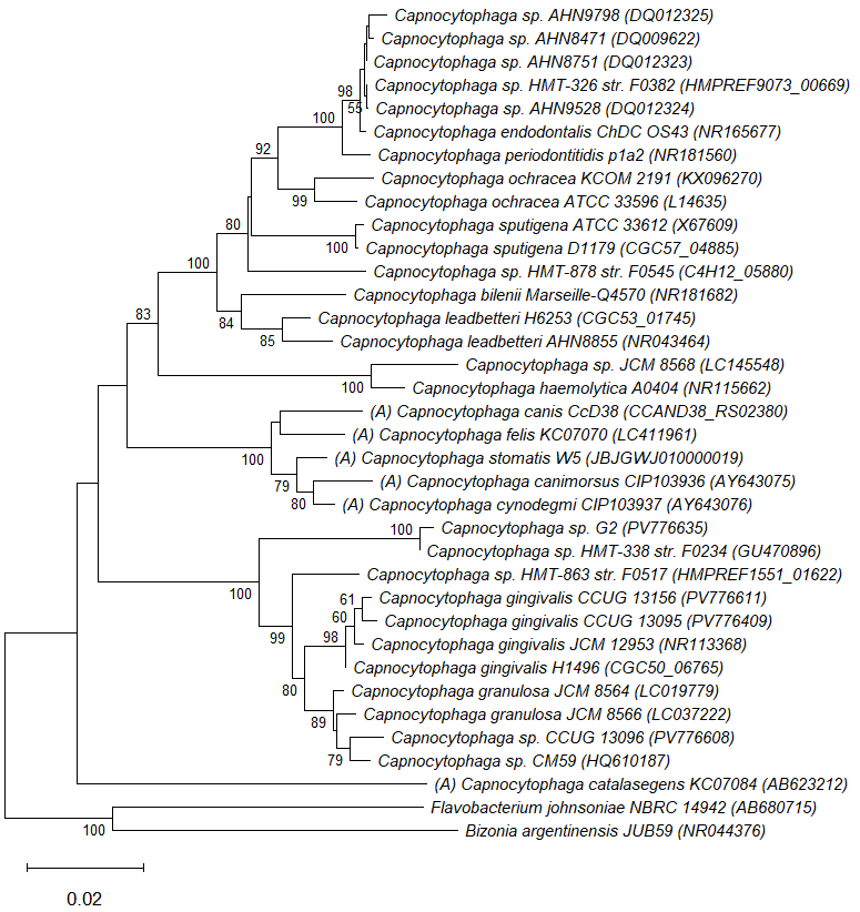
A neighbor-joining (NJ) phylogenetic tree of 16S ribosomal RNA (rRNA) sequences from representative Capnocytophaga taxa of human and animal origin is shown in Figure 3. This includes selected taxa from the HOMD (HMT-338, HMT-863, HMT-878, and HMT-326), as well as several strains that we have recently genome-sequenced, with their genomic sequence data deposited in NCBI GenBank. This includes Capnocytophaga sp. G2 (GCA_035282625.1, which corresponds to HMT-338), C. gingivalis (HMT-337) strains CCUG 13156 (GCA_035282365.1) and CCUG 13095 (GCA_035282545.1), and another CCUG reference strain, which is listed as ‘C. gingivalis’ CCUG 13096 (GCA_035282585.1), but which corresponds to HMT-471 [26], along with the genome-sequenced strain Capnocytophaga sp. CM59 (GCA_000293175.1) [27]. Both Capnocytophaga sp. CCUG 13096 and sp. CM59 are most closely related to C. granulosa (HMT-325). It should be noted that the eHOMD is a dynamic and evolving database, and there are numerous publications, as well as genome-sequenced reference strains, that classify Capnocytophaga taxa as HMT (e.g., Capnocytophaga sp. oral taxon 903 strain W10654) that are not listed in the current version of the eHOMD (v4.2φ). Somewhat confusingly, C. periodontitidis, C. endodontalis, and Capnocytophaga genospecies AHN8471 all appear to correspond to HMT-326, which is defined as being C. periodontitidis (eHOMD v4.2φ).

2. Colonization and Presence as Residents of the Oral Microbiota at Different Ages and Locations of the Mouth

The primary colonization site of Capnocytophaga is the oral cavity. In a longitudinal study on the establishment of oral anaerobes in the saliva of 44 infants during the first year of life, Capnocytophaga was categorized as a late colonizer, the detection frequency being 0% at the age of two months, 5% at the age of six months, and then increasing to 20% at the age of 12 months [29]. Earlier, another set of longitudinal data on the occurrence of gram-negative anaerobes in the oral cavity of 21 children demonstrated significant increases in the occurrence of Capnocytophaga species from 20% at the predentate stage to 100% at the dentate period (age range 24–41 months) due to the eruption of teeth and formation of gingival crevices with new oral surfaces to be colonized [30]. This culture-based observation was consistent with a recent systematic review where a total of 34 studies using 16S ribosomal RNA (rRNA) gene-targeted sequencing were included to provide comprehensive information on the oral microbiome at birth, early childhood, and adolescence [31]. It was shown that Capnocytophaga is among the genera that become dominant with a high level of abundance during the second year of life. In other words, human Capnocytophaga species are residents of the oral microbiota from early life onward, their preferred sites being dental biofilms, but they are also present on other oral surfaces. In a culture-based study of suspected periodontal pathogens in 67 children aged five to seven years, 67% of subgingival plaque, 27% of tongue surface, 36% of saliva, and 56% of tonsil samples were positive for Capnocytophaga [32].
Due to conflicting data on different Capnocytophaga species colonizing young children, 102 isolates from 12 children were selected from a culture-based study of 23 Finnish children aged two to three years [30] and were characterized using cluster analysis of multilocus enzyme electrophoresis profiles and partial 16S rRNA gene sequences by Frandsen et al. [15] to identify the genus-level isolates to the species level. Another focus was on the diversity of Capnocytophaga within children, and despite the limited number of isolates available from each child, intra-subject diversity was observed. The phylogenetic and phenotypic analyses led to the description of a novel Capnocytophaga species, C. leadbetteri, and a novel Capnocytophaga genospecies AHN8471 (=genomospecies AHN8471). Among the 102 isolates, the highest prevalence rates were observed for C. ochracea and C. sputigena, and also C. granulosa, C. leadbetteri, and the novel genospecies were present at low frequencies, whereas C. gingivalis and C. haemolytica were not detected [15].
Three PCR-based studies [33,34,35] examined the presence of Capnocytophaga at the species level in Japanese children. In dental plaque samples collected from children aged 2–12 years, C. ochracea and C. gingivalis were most prevalent, being found in 100% and 96% of the 25 healthy children and in 89% and 85% of the 85 children with gingivitis, respectively, while corresponding prevalence rates for C. sputigena were 48% and 37%, respectively [33]. In another study, the prevalence rates in subgingival plaque of 78 children aged three to nine years were 36% for C. ochracea, 50% for C. gingivalis, and 72% for C. sputigena [35]. Their findings contrasted with the absence of C. gingivalis observed in Finnish children aged 2–3 years [15]. The discrepancy may be explained by differing identification criteria and/or ethnic and/or geographical differences between the studies. Ooshima et al. [34] performed a two-year longitudinal study, where plaque and saliva samples from 119 periodontally healthy children aged 2–15 years were examined using a PCR-based approach, with C. ochracea and C. sputigena included among the 10 species targeted for study. In saliva, C. ochracea and C. sputigena were very frequently detected at all ages, but, surprisingly, were less frequent in plaque samples.
The mixed dentition stage, i.e., the period from 6 to 12 years of age, is characterized by the existence of both primary and secondary teeth. This is considered a critical transitional period for pediatric dental patients and was the focus of a recent 24-month randomized controlled trial by Zeng et al. [36] as regards the impact of sodium fluoride varnish alone or combined with oral health education, or oral health education alone, on caries increment and dental plaque microbiota. Significant changes were observed in the genus Capnocytophaga at both the 6- and 12-month assessments. After the cessation of interventions, the 24-month findings revealed that the distribution of Capnocytophaga became similar among the three intervention groups. Capnocytophaga was among the genera showing dynamic shifts in supragingival plaque composition. At the 12-month intervention, C. granulosa was less abundant in children who did not receive fluoride varnish, but the underlying reasons remain obscure [36]. A study comparing subgingival plaque findings between young African–American individuals (aged 5–21 years) with localized aggressive periodontitis (i.e., rapidly progressing periodontitis with an incisor-molar distribution) and their healthy siblings and healthy controls reported that C. granulosa was an abundant species in healthy individuals [37]. Subgingival samples from 73 generally healthy Bulgarian children, aged 10–14 years, were analyzed for the presence of nine target species, including C. gingivalis, by real-time PCR [38]. Unlike other periodontal species, C. gingivalis proved to ubiquitously colonize both periodontally healthy children and children with gingival inflammation, and was found in high quantities regardless of clinical status. One Brazilian study used PCR to determine the presence of 11 target species in gingival crevicular fluid samples collected from the first permanent molars and second deciduous molars, and two upper and two lower permanent incisors of diabetic and non-diabetic children aged 7–13 years [39]. Only C. sputigena and C. ochracea were associated with gingivitis in children with type 1 diabetes. Compared with non-diabetic children, diabetic children had significantly higher levels of C. sputigena for all tooth types and C. ochracea for permanent teeth.
In a PCR-based study [40], plaque samples were collected from 300 Indian individuals, including 100 periodontally healthy individuals, 100 with gingivitis, and 100 with periodontitis; of these, 87%, 77%, and 73%, respectively, were positive for Capnocytophaga. In the healthy group, Capnocytophaga detection was most common in young (aged 18–29 years) individuals. Prevalence rates of Capnocytophaga species varied as follows: C. ochracea 36.3%, C. granulosa 32.7%, and C. gingivalis 10%, whereas C. sputigena, C. haemolytica, C. leadbetteri, and Capnocytophaga genospecies AHN8471 (C. periodontitidis/C. endodontalis) were not found. The absence of these species, especially C. sputigena, was speculated to be due to ethnic and/or geographical differences, but also methodological aspects [41] could partly explain the wide variation between studies.
Socransky’s microbial complex theory [42] includes three Capnocytophaga species, C. gingivalis, C. ochracea, and C. sputigena, which are placed within the green complex. “Large plaques” on teeth have been shown to exhibit increased proportions of green and orange complex species [42]. In the green complex, both C. ochracea and C. gingivalis appeared at high levels on teeth with large amounts of supragingival plaque, which was collected from adult individuals [43,44]. In a study by Teles et al. [45], the focus was on revealing the ecological order of species succession during supra- and subgingival biofilm redevelopment after careful professional dental cleaning, followed by seven days of no oral hygiene. In supragingival plaque from periodontally healthy individuals, the mean proportions of C. gingivalis began to increase significantly at two days and continued to increase through four and seven days, while the proportions of C. ochracea and C. sputigena increased significantly at four days. In subgingival plaque from healthy individuals, significant increases were observed in the mean proportions of C. gingivalis at four days and at seven days of C. sputigena [45].
Li et al. [46] examined supragingival plaque samples collected before and after scaling at several time points from 40 individuals divided into three periodontal states, representing periodontal health, gingivitis, or periodontitis, with the aim of identifying similarities and differences in plaque remodeling processes. Capnocytophaga was among the few genera that had a significant correlation with the most clinical indicators, including bleeding index, plaque index, and percentage of bleeding sites on probing at different time points in the three periodontal states [46]. In a recent longitudinal one-year study, using full-length 16S rRNA and metagenomic sequencing, Zhou et al. [47] examined subgingival plaque samples collected from 30 periodontally healthy Chinese individuals, aged 18−30 years, following ultrasonic scaling (considered a perturbation to the subgingival microbiota) at 12 sequential time points. Microbial dynamics at the species and functional levels were characterized. According to their analysis, the subgingival microbiota reconstruction was mediated through coordinated changes in ecologically linked species groups, and significant positive correlations were observed among member taxa within each functional module. Within one of the modules, C. gingivalis, C. granulosa, C. sputigena, and C. leadbetteri were among the high-abundance species, with the novel species C. endodontalis (Capnocytophaga genospecies AHN8471/C. periodontitidis) also identified. Interestingly, within this late-stage module of the reconstructive process, enriched with specific functional capacities, bacterial taxa showed the highest pathogenic potential [47].
In traditional Chinese medicine, the appearance of tongue coatings has been used by clinical practitioners to differentiate ‘Cold’ and ‘Hot’ syndromes, which are considered to play roles in gastrointestinal diseases. Jiang et al. [48] investigated the microbiome composition of tongue coating samples from 19 middle-aged patients with chronic atrophic gastritis (nine diagnosed with Cold Syndrome and 10 with Hot Syndrome) and from eight young adults with no stomach discomfort, using Illumina 16S rRNA amplicon sequencing. Typical tongue coatings in Cold Syndrome patients were ‘white and greasy’, and those in Hot Syndrome patients were ‘yellow and dense’, with their respective tongue-coating microbiomes differing significantly. Capnocytophaga, C. sputigena, and C. haemolytica were detected in ‘Hot’ tongue microbiota-imbalanced networks of genus-classifiable operational taxonomic units (OTUs). It was concluded that the appearance of the tongue coating reflects the imbalanced tongue-coating microbiome in the entire human ecosystem [48].
In a more recent study, Chen et al. [49] collected swab samples from the tongue dorsum of 94 healthy Chinese individuals, which represented eight different tongue coating types with regard to color, thickness, and moisture appearance, for analysis by metagenomic sequencing, to investigate their respective microbial compositions. C. sputigena was found within the ‘yellow and thin’ tongue coating, positively correlating with Neisseria elongata. Furthermore, due to potential differences in the same tongue coating between individuals with and without disease, a comparison was performed between healthy individuals with a ‘yellow and thick and greasy’ coating and an additional, corresponding diabetic group, showing clear distinctions concerning the high abundance of Capnocytophaga in diabetic patients [49].
The impact of oral hygiene and periodontal inflammation on the prevalence of C. ochracea and C. sputigena on the tongue surface of 136 older Japanese individuals (age range 36–91 years) was investigated using real-time PCR [50]. The prevalence rates for C. sputigena and C. ochracea were 83% and 68%, respectively. It was observed that oral hygiene was poor in individuals positive for Capnocytophaga compared with those without Capnocytophaga, and the detection of both species was associated with mild periodontal inflammation.
Evidence from current experimental gingivitis studies where dental hygiene is discontinued, thus allowing natural bacterial accumulation, demonstrates a significant degree of variation in human inflammatory response to dental biofilm formation [51,52]. Among 40 periodontally and systemically healthy young adults, slow responders had a high abundance of Streptococcus and a low abundance of Capnocytophaga in supragingival plaque at baseline, whereas rapid responders had significantly higher abundances of Capnocytophaga at baseline compared with moderate and slow responders [52]. During the 14-day experimental gingivitis period, slow and moderate responders had significantly higher abundances of Capnocytophaga than rapid responders, with C. sputigena, C. granulosa, and C. leadbetteri being among the predominant bacterial species. Slow responders showed microbial resilience towards gingival inflammation, indicating their potential to return to baseline microbial conditions [52].
In a culture-based study on gram-negative anaerobic species in saliva and subgingival samples from 30 women (mean age 30 years), the majority harbored Capnocytophaga organisms, and when positive subgingivally, 64% of the saliva samples were simultaneously positive [53]. Manzoor et al. [54] used a metagenomic approach to analyze 120 saliva samples from young adults, divided into two age- and sex-matched groups, one half diagnosed as periodontally healthy/localized gingivitis (controls) and the other half as generalized gingivitis/initial periodontitis (cases). The latter group may represent a transition phase between a still reversible inflammation of the gingiva and early-stage non-reversible damage in periodontal tissues. Capnocytophaga was among the 39 ‘core’ genera shared between the groups, present in at least 90% of samples, and C. gingivalis and C. ochracea were among the ‘core’ species found in cases [54]. Acharya et al. [55] examined salivary microbiomes of 20 treated periodontitis patients and 15 periodontally healthy individuals, using a species-level resolution approach in their search for potential species able to separate individuals with well-maintained periodontal treatment from healthy individuals with similar gingival inflammation levels. C. leadbetteri was among the four species that were found to be significantly more abundant in the treated periodontitis group [55].
It is plausible that the increased pathogenicity within dental biofilms occurs due to microbial shifts with decreased or increased abundances of commensal bacteria. A recent study used a combination of high-throughput 16S rRNA gene amplicon sequencing and species-specific PCR primers targeting 10 different species of potential periodontal pathogens, including C. ochracea and C. sputigena, to investigate how various species and genera affected the colonization of ‘red complex’ species within dental plaque collected from older adults [56]. Detection rates of C. ochracea and C. sputigena were significantly higher in individuals positive for Tannerella forsythia and Porphyromonas gingivalis, respectively, than in those negative for these periodontal pathogens. It may be that individuals colonized with detectable levels of red complex species in dental biofilms—potentially supported by less pathogenic species like Capnocytophaga—are susceptible to harmful consequences for their oral and systemic health.
Analyzing vast genome-wide association study (GWAS) datasets from 455 documented tongue dorsum microbiomes and 540 documented saliva microbiomes in an East Asian cohort, He et al. [57] aimed to identify the causal relationship between the oral microbiome and five types of respiratory infections, including tonsillitis, chronic sinusitis, bronchiectasis, and pneumonia. Five genera, Prevotella, Streptococcus, Fusobacterium, Pauljensenia, and Capnocytophaga proved to play crucial but varied roles, either dampening or intensifying the progression of infection. Of these, Capnocytophaga (C. leadbetteri and C. ochracea) was found to inhibit bronchitis and pneumonia [57]. In another study focusing on the tonsillar crypt microbiota, the bacterial diversity within the crypts of the palatine tonsils was examined using 16S rRNA gene amplicon pyrosequencing in children and adults with recurrent tonsillitis, children with tonsillar hyperplasia, and healthy adults with surgery other than tonsillectomy (n = five per group) [58]. Capnocytophaga was one of the most prevalent genera with four species and non-distinguishable groups of species. Of the 33 species exceeding an average mean abundance of >0.5%, C. leadbetteri was recovered from 13/20 tonsillar samples, with two to four samples being positive in each group.
Of special note, in a recent comprehensive analysis of how spaceflight (on the SpaceX Inspiration4 mission, Dragon capsule) may affect the human microbiome [59], researchers noted a massive transient decrease in the relative metatranscriptomic activities of C. granulosa during spaceflight, the largest decrease out of all oral taxa studied. The authors proposed that this may reflect changes in microbial nutrient requirements or host dietary changes occurring during spaceflight.

3. Coaggregation and Synergistic Biofilm-Forming Activities with Different Oral Bacterial Species

Pioneering in vitro studies revealed that C. ochracea, C. sputigena, and C. gingivalis cells exhibited a variety of inter-generic cell coaggregation activities with gram-positive species that are common components of oral biofilms, including certain Streptococcus, Actinomyces, and Rothia species [60]. Broadly speaking, C. ochracea exhibited the greatest range of inter-species binding capacities [61]. Interactions were putatively mediated by high-molecular-weight, heat-sensitive ‘lectin-like’ surface proteins (also present on C. gingivalis cells), which exhibited differing degrees of inhibition with ca. 5–100 mM concentrations of sugars, e.g., N-acetylneuraminic acid, N-acetylglucosamine, L-rhamnose, and L-fucose, in a species-specific manner [62]. It was proposed that the binding of facultative anaerobes such as C. ochracea to ‘pioneer’ species in nascent biofilms may drive further oral biofilm maturation, notably via binding to the key ‘bridging’ species Fusobacterium nucleatum [63].
The coaggregation between C. ochracea and F. nucleatum cells could be inhibited by the addition of EDTA or lysine, but not by (amino-)sugars or arginine, and occurred via a heat- and proteinase K-sensitive component on F. nucleatum cells. Later studies revealed that C. ochracea formed a synergistic biofilm with F. nucleatum, and a secreted C. ochracea product (putatively the quorum sensing autoinducer AI-2) stimulated F. nucleatum biofilm formation [64]. More recently, C. ochracea was found to co-aggregate with the periodontal pathogen and oncobacterium, Parvimonas micra, and stimulate its growth in an apparently indirect manner, e.g., through a secreted product, possibly AI-2 [65].
Capnocytophaga cells are highly motile on surfaces and often exhibit a highly distinctive pattern of swirling and collective swarming behaviors [1,66,67]. They do not utilize flagellae or pili to ‘swim’ or ‘twitch’ like many bacterial species, but locomote via gliding. The complex, multi-component cell surface gliding machinery is powered by the type IX secretion system (T9SS), a rotary motor that secretes adhesins onto the outer membrane surface in a helical manner [68]. During this gliding process, other bacteria and even bacteriophage may ‘hitchhike’ on the long Capnocytophaga cells [67,69]. For example, C. gingivalis could translocate seven different non-motile species of oral bacteria, including Actinomyces sp. HMT-169, Veillonella parvula, Streptococcus sanguinis, P. micra, Porphyromonas endodontalis, Prevotella oris, and F. nucleatum [67]. This is thought to be a key process by which Capnocytophaga may drive dynamic biofilm growth, maturation, and spatial ecology.

4. Biogeography and Pangenome of Capnocytophaga Species in Human Oral Niches

4.1. Biogeography

Capnocytophaga is among the top 10 most common genera typically present in dental plaque [70]. Furthermore, pioneering RNA-sequencing studies revealed that Capnocytophaga species are highly transcriptionally active within (supragingival) dental plaque biofilms, in many cases constituting ca. 10% of all bacterial transcripts [71]. Thus, Capnocytophaga plays major roles in affecting the compositions and collective activities of oral biofilms. In their groundbreaking biogeographical analysis, Mark Welch et al. [72] used combinatorial labeling and spectral imaging fluorescence in situ hybridization (CLASI-FISH) to study the 3D architectures and taxonomic compositions (i.e., spatial ecology) of oral biofilms, and found Capnocytophaga to be the most abundant taxon present in dental plaque, being considerably more prevalent in supragingival compared with subgingival plaque. Notably, Capnocytophaga taxa were regular components of ‘hedgehog’ structures, which are commonly observed in dental biofilms, especially within a wide, putatively oxygen-depleted band in the peripheral regions, just below the surface layer [72]. Capnocytophaga taxa were also reported to be abundant in the peripheral regions of filament-rich annular (ring-like) components around ‘corncob’ structures, alongside Fusobacterium, Leptotrichia, and Streptococcus, whose central stalks were formed by Corynebacterium cells. Whilst their CLASI-FISH approach did not enable species-level identification of the Capnocytophaga taxa present, their findings reconcile well with what is known about the ecological preferences of Capnocytophaga (e.g., CO2-enriched low-oxygen niches), as well as with results from previous cell-coaggregation and synergistic growth studies involving C. ochracea (and to a lesser extent, C. sputigena and C. gingivalis) as noted above [72].

4.2. Relationships Between the Pangenome and Biogeography

Recently, Giacomini et al. [26] assembled a high-quality set of Capnocytophaga reference genomes (n = 117) from available genomic and metagenomic sequence data. From this, a human Capnocytophaga pangenome of 13,954 gene clusters was identified, with 341 core genes (gene clusters present in all genomes) and 9493 accessory genes (gene clusters present in 2–116 genomes). They revealed that the species C. sputigena, C. leadbetteri, C. granulosa, and C. gingivalis each formed two different genomic groups, based on their respective genomic compositions. By mapping specific gene-sequence reads to metagenomic data from 10 different oral niches, they were able to survey the relative abundance of Capnocytophaga species and genomic groups present within these respective niches. The two different genomic groups of C. sputigena, C. granulosa, and C. gingivalis exhibited dichotomous behaviors, with one set preferentially occupying the preferred niche of supra- and subgingival plaque, whilst the other atypical set occupied the tongue dorsum. Further (KEGG-based) predicted metabolic analysis suggested that all Capnocytophaga taxa shared many commonalities, such as carbohydrate (notably glucose) metabolism, bicarbonate (CO2) utilization, and conserved cofactor and vitamin biosynthetic capabilities. However, certain outlier groups within specific species may possess notably different metabolic adaptations, e.g., differences in oxygen utilization, nitrate/nitrite metabolism, or (aromatic) amino acid biosynthesis capabilities. The authors proposed that a single Capnocytophaga species may encompass multiple distinct genomic compositions that adapt its physiology to life in different oral niches or environments [26].
In a study by Koohi-Moghadam et al. [73], metagenomics was used to analyze biosynthetic gene clusters (BGCs) encoded by bacterial taxa present in saliva and supra- and subgingival plaques from periodontitis patients and healthy controls. Several BGCs are associated with periodontal health and disease. Notably, they identified one BGC (k141_164411) predicted to synthesize a bioactive polyketide metabolite within C. gingivalis contigs that were uniquely obtained from supra- and subgingival plaque from seven individuals with periodontitis, but were completely absent from healthy controls. This highlights the power of using genomics and metagenomics for analyzing the ‘accessory genome’ of oral bacteria to identify putative novel factors underlying health or disease [73].

5. Clinical Relevance of Capnocytophaga in the Oral Cavity

5.1. Periodontal Disease

In individuals suffering from periodontitis, an infection-driven inflammatory disease affecting tooth-supporting tissues, the taxonomic compositions and collective biological activities of subgingival microbial communities differ from those in periodontally healthy individuals [74]. Capnocytophaga levels are typically associated with periodontal health [75,76]. However, the detailed picture is more intricate and variable, and the precise roles played by individual Capnocytophaga species within diverse oral communities remain somewhat controversial. On one hand, Capnocytophaga (especially C. gingivalis and C. granulosa) is one of the genera detected at high frequencies in subgingival sites of individuals with gingival inflammation [77,78] and thus considered to have a potential pathogenic role in periodontal disease progression [79,80] and in gingivitis and periodontitis in diabetic children and adults [39,81]. Also, C. granulosa and C. haemolytica have been found in subgingival plaque of 51% and 10% of 29 periodontitis patients examined, respectively [82]. It should also be noted that the two strains of the recently described C. periodontitidis species were isolated from deep periodontal pockets [13]. A study by Nibali et al. [83] examined the periodontal microbiome in patients with aggressive periodontitis following treatment. Whilst C. granulosa (HMT-325) levels and the total amount of all Capnocytophaga taxa present within periodontal sites were positively associated with persistent disease (over ca. seven years), C. sputigena (HMT-775) levels were positively associated with a long-term successful treatment outcome [83]. In studies by Duran-Pinedo et al. [84,85], metatranscriptomics was used to analyze changes in the composition and activity of stable and progressing periodontal sites following non-surgical treatment in individuals with grade II/III periodontitis. Amongst a complex set of findings, Capnocytophaga sp. HMT-864 and HMT-338 belonged to clusters that were exclusively associated with progressing periodontal sites. However, 12 different species and HMT-phylotypes of Capnocytophaga (including C. gingivalis, C. granulosa, C. ochracea, C. sputigena, and C. leadbetteri) were associated with clusters that were exclusively associated with stable sites. Taken together, the results revealed a complex and dynamic relationship between the composition and activities of the bacterial communities and the respective physiological responses within periodontal niches, where pro-disease and pro-health periodontal microbiome communities may have many different structures.
A recent study using metagenomics aimed to investigate potential differences in subgingival microbial diversity, composition, interaction networks, and functional potential between three periodontal states, severe periodontitis (stage III) with moderate risk of progression (grade B) and high risk of progression (grade C) in comparison with periodontal health [86]. Interesting differences were seen between grades B and C, the latter having greater microbial complexity and a marked enrichment of C. granulosa and Capnocytophaga sp. CM59 (HMT-471), indicating a potential association with more rapid progression. In a complex microbial co-occurrence network in grade C periodontitis individuals, C. ochracea appeared as a hub species with key members of Socransky’s red complex but had a negative correlation with several pathogenic species. C. leadbetteri, C. sputigena, C. granulosa, and Capnocytophaga sp. CM59 (HMT-471) were among the top 50 ‘species’ in subgingival plaque, but without significant relationships to clinical indicators (in this study: bleeding on probing, pocket depth, and clinical attachment level) [86]. Furthermore, in another recent study, the abundance of Capnocytophaga exhibited a significant decreasing trend when moving from health to severe (stage III) periodontitis with increasing pocket depths of 0–3 mm, 4–5 mm, and 6–9 mm [87]. C. gingivalis, C. granulosa, and C. sputigena appeared in a co-occurrence network of species with a relative abundance of >1% in the healthy and pocket depth of 0–3 mm groups, and in the latter group, C. gingivalis and C. sputigena had a strong positive correlation with Neisseria elongata and each other. Being less abundant, C. leadbetteri and C. endodontalis (Capnocytophaga genospecies AHN8471/C. periodontitidis) appeared in the 4–5 mm group, having a weak correlation with each other [87].
In the systematic review and meta-analysis of Antezack et al. [88], the prevalence of potentially pathogenic microorganisms in subgingival plaque and/or saliva was compared between individuals with and without periodontitis. In the forest plot of the effect sizes for the association between microorganisms and periodontitis, the odds ratios (95% confidence intervals) for C. gingivalis, C. ochracea, and C. sputigena were 1.02 (0.65–1.61), 1.10 (0.74–1.63), and 1.35 (0.69–2.63), respectively. In other words, no statistically significant associations between these Capnocytophaga species and periodontitis were present [88]. Recently, Xu et al. [89], by analyzing 16S rRNA gene sequencing data, focused on identifying subgingival microbial taxa differences that may serve as potential drivers of increasing severity of periodontitis. Capnocytophaga and Paludibacter were identified as protective genera, significantly enriched in the healthy group. Interestingly, Capnocytophaga was among the genera, and, at the species level, C. leadbetteri demonstrated a decreasing correlation with periodontitis progression. In terms of microbial community re-organization, there was a gradual loss of health-associated taxa and a sequential emergence of disease-associated pathogens. In addition, microbial co-occurrence networks were less stable and resilient in advanced periodontitis and less likely to return to their original equilibrium state, compared to mild and moderate disease stages or health [89].

5.2. Periimplantitis

Similar to periodontitis, periimplantitis is a common submucosal biofilm-associated disease that affects the tissues surrounding dental implants. In the peri-implant microbiome, shifts from health to disease include an increase in diversity and a gradual depletion of commensals [90]. With the aim of clarifying bacterial interactions, community structure, and microbial stability in dysbiotic biofilms, supra- and subgingival plaque samples were collected from teeth or implants of 34 individuals with different health conditions and examined using metagenomic sequencing [91]. C. granulosa and C. sputigena were among the hub species, playing central roles in different co-occurrence networks: C. granulosa in diseased subgingival communities and C. sputigena in healthy subgingival and diseased sub- and supragingival communities. C. sputigena had several negative correlations in health, but during inflammation, the correlations weakened and were lost. In diseased communities, C. gingivalis became a concentrated node of negative correlation. Notably, with increased inflammation, subgingival bacterial networks around teeth and implants became less connected and less competitive, whereas in supragingival communities, the networks shifted in the opposite direction [91].
Recently, the submucosal microbiome around implants was examined using full-length 16S rRNA gene amplicon sequencing and metatranscriptomics to identify bacterial taxa and their activities as potential driving factors for severe tissue destruction [92]. Similar to the situation in periodontitis, Capnocytophaga was among the genera having a negative correlation with increased disease severity, and significant associations between C. gingivalis, C. sputigena, and C. leadbetteri with shallower pockets were observed. However, in another study using full-length 16S rRNA sequencing [93], higher abundances of these Capnocytophaga species were found in peri-implant pocket depths of 5–7 mm, indicating early stages of periimplantitis.

5.3. Caries and Endodontic Infections

A recent systematic review presented data on the microbial composition and functional profile of dental plaque and/or saliva samples collected from caries-free and caries-affected individuals, based on 54 studies with approximately 3500 individuals [94]. Twelve bacterial genera, among them Capnocytophaga, were frequently reported as being more abundant in caries-active individuals. In root caries, increased levels of C. granulosa have been observed on carious teeth compared to non-carious teeth [95], indicating that C. granulosa has a pathogenic role. However, this finding is hard to reconcile with its health-associated status in supragingival plaque [96].
A large variety of bacterial species have been detected within primary endodontic infections without or with a sinus tract [97]. Specific differences in the bacterial composition existed between cases with or without a sinus tract; for instance, C. sputigena and C. gingivalis were detected in significantly higher counts in the absence of a sinus tract [97]. It should also be noted that the single strain of the novel species C. endodontalis was isolated from a human refractory periapical abscess [12].
Early microbial complex species connected to endodontic-periodontal lesions in the same tooth were the focus of a systematic review presenting geographically heterogeneous data coming from only four selected articles with a total of 116 teeth [98]. Prevalence rates of two Capnocytophaga species were available from two studies: for C. granulosa, 10% in samples taken from root canals and 35% from periodontal pockets, while corresponding rates for C. sputigena varied between 4–70% and 0–30%. As such, the evidence is obscure due to the limited data available.

5.4. Mucosal Infections

In immunocompromised patients, Capnocytophaga taxa have been found in oral and gingival mucosal lesions. Oral mucositis can appear as a specific chemotherapy-induced infection with mucosal ulceration, bleeding, and severe pain. This has been linked to distinct microbiome profiles within pediatric cancer patients, where the predominant phylum is Bacteroidota, and Capnocytophaga at the genus level, and the species C. sputigena may be present with significantly increased abundances [99,100,101]. According to a case report, a biopsy taken from gingival hyperplasia of a neutropenic woman demonstrated invasion of filamentous bacteria, identified as C. sputigena, within the gingival tissue [102]. As human oral-associated Capnocytophaga spp. most commonly originate from dental biofilms; one major route of entry to the bloodstream is through inflamed periodontal tissues [103,104]. Another route of translocation of oral bacteria can occur via (micro-)aspiration to the lower respiratory tract. For instance, Tan et al. [105] collected paired subgingival plaque and tracheal aspirate samples from 53 chronic obstructive pulmonary disease (COPD) patients with severe acute exacerbations. The presence of C. sputigena at equal levels in both sample types indicates its potential contribution to this severe condition. These observations underline the importance of proper oral hygiene and preventive measures for maintaining good oral health, especially in immunocompromised individuals [105].

5.5. Nitrate Reduction and Taste Perception

C. gingivalis, C. ochracea, and C. sputigena have all been shown to be active nitrate reducers and nitrite producers in dental plaque in adults and children [106]. The conversion of nitrate to nitrite by oral bacteria may play a significant role in helping protect against diabetes and hypertension [107]. A recent clinical study investigated changes in the oral microbiome and oral metabolome relating to taste perception in response to nitrate supplementation. Uniquely, levels of C. gingivalis were significantly increased in individuals whose taste perception did not change in response to nitrate supplementation [108]. The underlying mechanisms behind this interesting correlation remain to be elucidated.

6. Species-Level Identification of Human Capnocytophaga Recoveries from Clinical Samples

Bacterial identification methods and taxonomic classifications change and evolve over time. Consequently, care and caution should be employed when interpreting results from older studies or from studies that do not accurately report the experimental methodologies used for bacterial identification. Broadly speaking, there is relatively little experimental (clinical and laboratory-based) data on human Capnocytophaga species, compared with many other species of oral bacteria. Most pertinently, we lack a holistic understanding of their pathogenic potential and antibiotic susceptibility patterns, factors that may differ dramatically between species or within specific strain lineages.
Attempts to identify Capnocytophaga organisms to the species level often remain unreliable when identification relies on conventional phenotypic and biochemical tests, including colony morphology, Gram staining, yellow/orange pigmentation, gliding motility, CO2-requiring growth, sugar fermentation, and enzymatic activities [1]. In particular, the slow, fastidious growth requirements, along with phenotypic and biochemical similarities among Capnocytophaga species, limit the accuracy of species-level identification [15]. Before the introduction of molecular methods, culture-based identification, which required laborious testing procedures, could even fail to separate bacteria similar to Capnocytophaga [109]. According to the Centers for Disease Control and Prevention [110], blood samples are usually used to identify the bacteria in culture, but identification can be difficult because automated blood culture systems may miss Capnocytophaga due to insufficient incubation time. In other words, challenges in detection and identification most likely lead to an underestimation of the true prevalence of each Capnocytophaga species as a causative agent of infections. These limitations necessitated the introduction of more advanced molecular and proteomic technologies to improve species-level identification of Capnocytophaga.
Before the adoption of ‘next-generation’ DNA sequencing technologies, approaches such as PCR-restriction fragment length polymorphism combined with Sanger sequencing of near-full-length 16S ribosomal RNA (rRNA) amplicons were a powerful and reliable approach for the species-level identification of Capnocytophaga strains or taxa present in biological samples [33,111]. However, the 16S rRNA amplicon sequencing-based approach cannot give any information about the genetic/genomic factors that may be responsible for any biologically important or atypical phenotypes, e.g., surface antigens or antibiotic resistance profiles. It is also highly dependent on a large, diverse, and clinically representative set of reference strains and 16S rRNA gene sequences. Nowadays, the use of long-read sequencing platforms for the rapid and accurate determination of complete genome sequences for clinical isolates, e.g., using the Pacific Biosciences (PacBio) or Oxford Nanopore Technologies (ONT) platforms, is highly desirable to enable accurate taxonomic identification along with the identification of putative phages, mobile genetic elements, or antibiotic resistance genes [112].
Matrix-assisted laser desorption/ionization time-of-flight mass spectrometry (MALDI-TOF MS) is a molecular identification technique introduced in the 1990s, which is particularly useful for bacterial identification based on protein spectral fingerprints [113]. Concerning Capnocytophaga, the initial focus was on animal species, especially C. canimorsus [114], but later, it was extended to human Capnocytophaga species [115,116,117,118,119]. In hospital laboratories, MALDI–TOF MS is regarded as a rapid and cost-effective diagnostic method for microbes present in clinical samples [120]. It also reliably identifies Capnocytophaga to the genus level with 100% accuracy; however, its performance at the species level is variable. As shown by Algahawi et al. [116], the accuracy and speed of species-level identification of human oral-associated Capnocytophaga are strongly influenced by the representation of strains in the MALDI-TOF MS database, where an adequate number of representative Capnocytophaga strains is needed to assure the correct identification at both the genus and species levels without customizing protocols. Strains that are underrepresented in the MALDI TOF database or are closely related require additional preparation methods, such as formic acid extraction, and the database should be enriched with these species [116]. Thus, updating and expanding the MALDI-TOF MS reference database will further improve the species-level identification accuracy of Capnocytophaga in clinical microbiology laboratories.

7. Human Oral-Associated Capnocytophaga Species in Non-Oral Infections

Clinically, the Capnocytophaga-associated disease can progress rapidly from mild, localized infection to systemic infection, sepsis, and even death [110].

7.1. Bloodstream Infections

Outside the oral cavity, most human oral-associated Capnocytophaga species have been isolated from blood. Bloodstream infections form the major infectious type with Capnocytophaga involvement, especially in individuals with weakened immune systems [121,122]. Recently, Chesdachai et al. [122] performed a retrospective review of human- and animal-associated Capnocytophaga infections, covering a 10-year period at three main hospitals of the Mayo Clinic. A total of 110 adult patients with culture-positive Capnocytophaga findings were recognized, and of them, 82 were connected to a clinically relevant infection. Among the 62 Capnocytophaga isolates identified to the species level, C. sputigena was most frequent (45%), and, in descending order, was followed by zoonosis-associated C. canimorsus (26%), C. ochracea (16%), and C. gingivalis (8%). In addition, one C. granulosa, one C. leadbetteri, and one zoonosis-associated C. cynodegmi were found. Nearly all bacteremia cases were monomicrobial, where C. sputigena was the most common finding, followed by C. canimorsus, but also C. ochracea and C. gingivalis were occasionally detected. Most non-bacteremia cases were polymicrobial in nature, with human oral-associated Capnocytophaga forming part of their infectious etiology. Notably, infections with human-associated Capnocytophaga have been proven to have a higher rate of overall six-month mortality compared to that of C. canimorsus [122]. Recently, an unusual septicemia case caused by C. ochracea after a dog bite was described [123]. According to the authors, this was the first report of bacteremia where human oral-associated Capnocytophaga (identified as C. ochracea using MALDI-TOF) was transmitted from an animal. It was speculated that transmission can occur in both directions due to close contact between humans and their pets.
In a recent review of 31 infective endocarditis cases involving Capnocytophaga species [124], only five case reports were connected to human oral-associated species; three with C. ochracea and one with C. haemolytica (identifications based on phenotypic tests). There is one further bacteremia case potentially connected to endocarditis caused by Capnocytophaga genospecies AHN8471 [125] available in the literature. As described above, the uncertain (overlapping) taxonomic positions of Capnocytophaga genospecies AHN8471, C. endodontalis, and C. periodontitidis should be well noted.
An especially frequent recovery from bloodstream infections seems to be C. sputigena. In both adult and pediatric patients with hematologic malignancies, there are several bacteremia cases involving C. sputigena [100,117,126,127,128,129,130,131,132,133]. In a catheter-related bloodstream infection caused by C. sputigena, the isolate from the patient was tested for its biofilm formation and compared with the laboratory reference strain; this clinical isolate proved to have significantly increased biofilm formation capability, which was interpreted as a pathogenic mechanism [134]. An interesting, historic sepsis case of Saint-Louis, King of France (1270 AD) was recently reported by Charlier et al. [126]; the king was assumed to suffer from scurvy and inflammatory jaw disease with the potential involvement of oral commensals. Based on careful molecular analyses of a sample from the visceral relics, C. sputigena was identified as the main pathogenic species with a uniquely high proportion (11%) of all microbial sequences. Thus, it was suggested that C. sputigena was responsible for the fatal bacteremia, leading to the king’s death [126].

7.2. Other Types of Non-Oral Infections

Sporadic case reports have been presented on infections with the involvement of human oral-associated Capnocytophaga species affecting the eye, central nervous system, and the upper and lower respiratory tracts, but also those affecting the female genital tract and the fetus during pregnancy. In addition to blood, C. sputigena seems to be the most common also in these other infectious sites outside the oral cavity. Out of 49 clinical β-lactamase-positive Capnocytophaga strains from patients treated in a Japanese university hospital, 31 identified as C. sputigena were isolated from 29 respiratory samples, one from a wound specimen, and one from blood [135]. Reports on non-oral infections vary from orbital cellulitis and adjacent subperiosteal abscess [136], sinusitis with orbital subperiosteal and intracranial abscesses [137], tonsillitis [128], community-acquired pneumonia [138], pneumonia [132], pleural empyema [139,140], and lung abscess [141,142] to soft tissue or osteo-articular infections; for instance, post-operative intra-abdominal abscess [143], primary iliopsoas abscess [144] as well as post-arthroscopic knee joint infection connected to C. sputigena have been reported [145].
In a pre-term birth case series report by Lopez et al. [146], C. sputigena was detected in two out of five cases. In case 1, the pregnancy was uncomplicated until the premature rupture of membranes occurred at 25 weeks of gestation, followed by preterm birth with low birth weight at 26 weeks of gestation. Chorioamnionitis was clinically suspected and confirmed by histological examination of the placenta. The infant was treated for respiratory distress syndrome, and C. sputigena was isolated from the tracheal aspirate and gastric fluid cultures, whereas the blood culture was negative. In case 2, an infant with low birth weight was delivered at 28 weeks of gestation. Trichomonas vaginalis was recovered in vaginal cultures of the mother. Chorioamnionitis was clinically suspected and confirmed by histological examination of the placenta. Blood culture and gastric aspirate samples revealed the presence of C. sputigena [146]. Similar cases related to chorioamnionitis and C. sputigena involvement alone [147] or together with C. leadbetteri [148] have been observed as well. Also, C. ochracea was linked with preterm birth and neonatal septicemia, as it was detected in the blood culture of a premature neonate [149]. In cases affecting the urogenital tract, insufficient oral hygiene and poor oral health status may be regarded as background factors resulting in the transfer of oral bacteria via hematogenous routes or oral sex to the female genital tract, where Capnocytophaga organisms are able to cause infection [146,147,150].
Besides C. sputigena, other traditional Capnocytophaga species, C. ochracea and C. gingivalis, are sporadic findings in a variety of non-oral infections. One explanation might be the challenges connected to their identification, as shown in a leukemia patient with pleural effusion and empyema, where anaerobic gram-negative bacilli were isolated from pleural fluid after three days of incubation, but it was not until 15 days of hospitalization that C. ochracea was identified by the reference laboratory [151]. Notably, as shown in a recent case report on C. gingivalis, bacteria with low-level pathogenicity can lead to a fatal outcome [152]. It was speculated that co-infection with COVID-19 with massive lung destruction was associated with C. gingivalis colonization of the lungs, thus leading to fulminant sepsis. Also, the recently reported first-ever case of central nervous system infection caused by C. ochracea, where a surgical operation through the oral and nasal passages was suspected to have interfered as a concomitant factor, is noteworthy [153]. In the study, a variety of methods, including metagenomics, next-generation sequencing, MALDI-TOF MS, and 16S rDNA sequencing, were used to identify the isolate from the cerebrospinal fluid of a meningioma patient. Indeed, next-generation sequencing (NGS) is a valuable method for identifying fastidious bacteria in polymicrobial infections, such as brain abscesses of odontogenic origin, where Capnocytophaga can also be found [154]. In this case, the growth from pus specimens was identified by MALDI-TOF MS as C. ochracea and another species, Arachnia propionica. The post-mortem pus sample was further analyzed using 16S deep sequencing, recognizing a wide range of oral anaerobic species, including Capnocytophaga sp. HMT-323, in this polymicrobial brain abscess [154]. In a diabetic patient with poor oral hygiene, bacteremia followed laparoscopic gastrectomy for gastric cancer, and the bacterial isolate from blood was then identified by MALDI-TOF and confirmed by 16S rRNA gene sequencing as C. ochracea [155]. This diabetic patient was estimated to have an increased risk of Capnocytophaga infection due to poor glycemic control and chronic inflammatory burden in periodontal tissues.
Infectious cases caused by Capnocytophaga are recognized in both immunocompetent and immunocompromised individuals [121,156]. A literature review collected data on 43 systemic infections associated with human oral Capnocytophaga species, published in English up to February 2016 [156]. Altogether, 25 C. ochracea, 12 C. sputigena, four C. gingivalis, one C. leadbetteri, and one C. haemolytica case were described; however, the precise species-level identification remains uncertain when based on phenotypic testing or when the methodology was not described in detail. For instance, out of the 25 publications reporting the isolation of ‘C. ochracea from non-oral infections, 12 came from studies published in the 1980s, and 6 were from the 1990s, which predates the description of several species closely related to C. ochracea. An interesting observation was that 7 of the 43 systemic infectious cases were connected to oral conditions [156].
The first septic arthritis case caused by C. gingivalis was reported in an immunocompetent young child with positive cultures from subtalar joint fluids at two separate times and identified at a reference laboratory, leading to a considerable delay in diagnosis and treatment [157]. A potential mechanism was suspected to occur via the hematogenous route from profuse gingival bleeding due to oral trauma to the subtalar joint. One C. granulosa case in an immunocompetent teenage boy, consisting of an abscess in the lumbosacral area after trauma, was reported based on extensive phenotypic tests [158]. The first report of a Bartholin’s gland abscess caused by C. ochracea was presented in an immunocompetent woman [159].
Besides the challenging diagnosis of slow-growing Capnocytophaga organisms to the species level, antimicrobial susceptibility testing can be a difficult task for clinical microbiology laboratories that may lead to delays in the initiation of effective antimicrobial therapy. As suggested by Ehrmann et al. [160], human oral-associated Capnocytophaga species form a significant β-lactamase resistance gene reservoir in the oral microbiome; the most common β-lactamase producers were C. sputigena and C. ochracea, but β-lactamase production was also detected among C. gingivalis, C. granulosa, and C. leadbetteri isolates. A few macrolide-lincosamide-streptogramin-resistant C. sputigena and one C. ochracea were observed [160]. Moreover, among the 10-year hospital-based data with 82 clinically relevant infections caused by Capnocytophaga, 30% of strains produced β-lactamases [122]. A multidrug-resistant C. ochracea strain was isolated from the blood of a boy with leukemia and later characterized as positive for a plasmid-encoded TEM-17 extended-spectrum β-lactamase [161]. In an acute exacerbation of chronic obstructive pulmonary disease, a multidrug-resistant C. gingivalis strain was found in an elderly man with urgent acute respiratory distress [156]. In a patient with severe neutropenia, C. gingivalis isolated from blood proved to produce the CSP-1 extended-spectrum-β-lactamase [162]. Taken together, this underscores the importance of susceptibility testing for selecting effective antimicrobial therapy for human oral-associated Capnocytophaga species.

8. Concluding Remarks and Future Perspectives

Dental plaque is the primary site for human oral-associated Capnocytophaga species. The majority of human Capnocytophaga species generally play protective, health-promoting roles in dental plaque and other oral biofilm niches. However, there is a paucity of mechanistic molecular evidence demonstrating exactly how they do this. We speculate that a complex network of inter-species cell-binding activities and physiological interactions between Capnocytophaga (especially C. ochracea) and other commensals helps stabilize oral biofilm structure and community composition, underpinning oral biofilm resilience and eubiosis. However, a moderate amount of evidence suggests that C. granulosa and C. gingivalis may play notable (accessory) roles in oral dysbiosis and disease.
In subgingival biofilms, Capnocytophaga is significantly enriched in health but shows a decreasing pattern from health towards disease. An interesting comparison between the subgingival microbiomes in young adults with cryptogenic ischemic stroke and stroke-free controls revealed a significantly decreased abundance of Capnocytophaga in patients [163]. Notably, in the stroke group, individuals lacking symptoms at admission and those with favorable clinical outcomes had significantly enriched populations of Capnocytophaga. To date, however, relatively little is known about this oral genus and its potential link to this and other systemic conditions. Continued progress in the determination of complete genome sequences for diverse reference strains and clinical isolates of Capnocytophaga, combined with advances in the scale and depth of high-quality clinical molecular investigations, will give us fresh insight into the relevance of this understudied group of bacteria for oral and systemic health.
The number of immunocompromised individuals will continue to increase in future years, for instance, due to the development and implementation of new immunotherapies and targeted cellular therapies, thus increasing the risk of infections caused by low-level pathogens like Capnocytophaga. Based on recent reports in the current literature, using more advanced identification techniques in clinical microbiology laboratories, the most frequent Capnocytophaga species involved in human infections seems to be C. sputigena. In contrast, C. haemolytica is one of the least frequently detected human oral-associated Capnocytophaga species. There are still many controversies regarding the clinical relevance and roles of this oral genus; thus, greater emphasis should be given to Capnocytophaga in future studies.

Author Contributions

E.K., conception, design, data interpretation, original draft preparation, writing and critically revising the manuscript; M.G., visualization, writing and critically revising the manuscript; A.A., visualization, writing and critically revising the manuscript; R.M.W., design, genome data preparation and interpretation, visualization, writing and critically revising the manuscript. All authors have read and agreed to the published version of the manuscript.

Funding

RMW acknowledges financial support from the Research Grants Council (RGC) of Hong Kong through a General Research Fund (GRF) grant # 17121820.

Institutional Review Board Statement

Not applicable.

Informed Consent Statement

Not applicable.

Data Availability Statement

No new data were created or analyzed in this study. Data sharing is not applicable to this article.

Conflicts of Interest

The authors declare no conflicts of interest.

References

  1. Leadbetter, E.R.; Holt, S.C.; Socransky, S.S. Capnocytophaga: New Genus of Gram-Negative Gliding Bacteria I. General Characteristics, Taxonomic Considerations and Significance. Arch. Microbiol. 1979, 122, 9–16. [Google Scholar] [CrossRef]
  2. Socransky, S.S.; Holt, S.C.; Leadbetter, E.R.; Tanner, A.C.R.; Savitt, E.; Hammond, B.F. Capnocytophaga: New Genus of Gram-Negative Gliding Bacteria. III. Physiological Characterization. Arch. Microbiol. 1979, 122, 29–33. [Google Scholar] [CrossRef] [PubMed]
  3. Holt, S.C.; Leadbetter, E.R.; Socransky, S.S. Capnocytophaga: New Genus of Gram-Negative Gliding Bacteria. II. Morphology and Ultrastructure. Arch. Microbiol. 1979, 122, 17–27. [Google Scholar] [CrossRef] [PubMed]
  4. Williams, B.L.; Hollis, D.; Holdeman, L.V. Synonymy of Strains of Center for Disease Control Group DF-1 with Species of Capnocytophaga. J. Clin. Microbiol. 1979, 10, 550–556. [Google Scholar] [CrossRef] [PubMed]
  5. Beauruelle, C.; Plouzeau, C.; Grillon, A.; Isnard, C.; Corvec, S.; Degand, N.; Jacquier, H.; Amara, M.; Mizrahi, A.; Diedrich, T.; et al. Capnocytophaga Zoonotic Infections: A 10-Year Retrospective Study (the French CANCAN Study). Eur. J. Clin. Microbiol. Infect. Dis. 2022, 41, 581–588. [Google Scholar] [CrossRef] [PubMed]
  6. Campagna, R.A.; Kelly, E.A.; Vugia, D.J.; Berman Watson, H.F.; Browne, C.S.; Lau, J.K.; Fritz, C.L. Clinical and Epidemiologic Review of Capnocytophaga spp. Infections Identified at a Public Health Reference Laboratory—California, 2005–2021. Zoonoses Public Health 2025, 72, 330–336. [Google Scholar] [CrossRef] [PubMed]
  7. The List of Prokaryotic Names with Standing in Nomenclature (LPSN). Available online: https://www.bacterio.net (accessed on 3 May 2026).
  8. Bartlett, A.; Padfield, D.; Lear, L.; Bendall, R.; Vos, M. A Comprehensive List of Bacterial Pathogens Infecting Humans. Microbiology 2022, 168, 001269. [Google Scholar] [CrossRef] [PubMed]
  9. Mavrommatis, K.; Gronow, S.; Saunders, E.; Land, M.; Lapidus, A.; Copeland, A.; Glavina Del Rio, T.; Nolan, M.; Lucas, S.; Chen, F.; et al. Complete Genome Sequence of Capnocytophaga ochracea Type Strain (VPI 2845T). Stand. Genom. Sci. 2009, 1, 101–109. [Google Scholar] [CrossRef] [PubMed]
  10. Manfredi, P.; Pagni, M.; Cornelis, G.R. Complete Genome Sequence of the Dog Commensal and Human Pathogen Capnocytophaga canimorsus Strain 5. J. Bacteriol. 2011, 193, 5558–5559. [Google Scholar] [CrossRef] [PubMed]
  11. Villarma, A.; Gulvik, C.A.; Rowe, L.A.; Sheth, M.; Juieng, P.; Nicholson, A.C.; Loparev, V.N.; McQuiston, J.R. Twelve Complete Reference Genomes of Clinical Isolates in the Capnocytophaga Genus. Genome Announc. 2017, 5, e01186-17. [Google Scholar] [CrossRef] [PubMed]
  12. Jo, E.; Park, S.N.; Lim, Y.K.; Paek, J.; Shin, Y.; Kim, H.; Kim, S.H.; Shin, J.H.; Chang, Y.H.; Kook, J.K. Capnocytophaga endodontalis sp. Nov., Isolated from a Human Refractory Periapical Abscess. Curr. Microbiol. 2018, 75, 420–425. [Google Scholar] [CrossRef] [PubMed]
  13. Zhang, Y.; Qiao, D.; Shi, W.; Wu, D.; Cai, M. Capnocytophaga periodontitidis sp. Nov., Isolated from Subgingival Plaque of Periodontitis Patient. Int. J. Syst. Evol. Microbiol. 2021, 71, 004979. [Google Scholar] [CrossRef] [PubMed]
  14. Antezack, A.; Boxberger, M.; La Scola, B.; Monnet-Corti, V. Isolation and Characterization of Capnocytophaga Bilenii sp. Nov., a Novel Capnocytophaga Species Detected in a Gingivitis Subject. Pathogens 2021, 10, 547. [Google Scholar] [CrossRef] [PubMed]
  15. Frandsen, E.V.G.; Poulsen, K.; Könönen, E.; Kilian, M. Diversity of Capnocytophaga Species in Children and Description of Capnocytophaga leadbetteri sp. Nov. and Capnocytophaga genospecies AHN8471. Int. J. Syst. Evol. Microbiol. 2008, 58, 324–336. [Google Scholar] [CrossRef] [PubMed]
  16. Zbinden, R.; Yagupsky, P. Fastidious or Rarely Isolated Gram-Negative Rods with a Particular Focus on Kingella kingae. Clin. Microbiol. Rev. 2025, 38, e0004825. [Google Scholar] [CrossRef] [PubMed]
  17. Manfredi, P.; Renzi, F.; Cornelis, G.R. Draft Genome Sequences of Three Capnocytophaga cynodegmi Strains Isolated from the Oral Cavity of Healthy Dogs. Genome Announc. 2015, 3, e00200-15. [Google Scholar] [CrossRef] [PubMed]
  18. Zangenah, S.; Abbasi, N.; Andersson, A.; Bergman, P. Whole Genome Sequencing Identifies a Novel Species of the Genus Capnocytophaga Isolated from Dog and Cat Bite Wounds in Humans. Sci. Rep. 2016, 6, 22919. [Google Scholar] [CrossRef] [PubMed]
  19. Suzuki, M.; Imaoka, K.; Haga, Y.; Mohri, M.; Nogami, A.; Shimojima, Y.; Irie, Y.; Sugimura, S.; Morikawa, S. Characterization of Three Strains of Capnocytophaga canis Isolated from Patients with Sepsis. Microbiol. Immunol. 2018, 62, 567–573, Erratum in Microbiol. Immunol. 2018, 62, 737. [Google Scholar] [CrossRef] [PubMed]
  20. Suzuki, M.; Umeda, K.; Kimura, M.; Imaoka, K.; Morikawa, S.; Maeda, K. Capnocytophaga felis sp. Nov. Isolated from the Feline Oral Cavity. Int. J. Syst. Evol. Microbiol. 2020, 70, 3355–3360. [Google Scholar] [CrossRef] [PubMed]
  21. Suzuki, M.; Imaoka, K.; Kimura, M.; Morikawa, S.; Maeda, K. Capnocytophaga catalasegens sp. Nov., Isolated from Feline Oral Cavities. Int. J. Syst. Evol. Microbiol. 2023, 73, 005731. [Google Scholar] [CrossRef] [PubMed]
  22. Dewhirst, F.E.; Chen, T.; Izard, J.; Paster, B.J.; Tanner, A.C.R.; Yu, W.-H.; Lakshmanan, A.; Wade, W.G. The Human Oral Microbiome. J. Bacteriol. 2010, 192, 5002–5017. [Google Scholar] [CrossRef] [PubMed]
  23. Fernández Escapa, I.; Chen, T.; Huang, Y.; Gajare, P.; Dewhirst, F.E.; Lemon, K.P. New Insights into Human Nostril Microbiome from the Expanded Human Oral Microbiome Database (eHOMD): A Resource for Species-level Identification of Microbiome Data from the Aerodigestive Tract. mSystems 2018, 3, 00187-18. [Google Scholar] [CrossRef] [PubMed]
  24. Dewhirst, F.E.; Klein, E.A.; Thompson, E.C.; Blanton, J.M.; Chen, T.; Milella, L.; Buckley, C.M.F.; Davis, I.J.; Bennett, M.-L.; Marshall-Jones, Z.V. The Canine Oral Microbiome. PLoS ONE 2012, 7, e36067, Erratum in PLoS ONE 2012, 7. https://doi.org/10.1371/annotation/c2287fc7-c976-4d78-a28f-1d4e024d568f. [Google Scholar] [CrossRef] [PubMed]
  25. Dewhirst, F.E.; Klein, E.A.; Bennett, M.-L.; Croft, J.M.; Harris, S.J.; Marshall-Jones, Z.V. The Feline Oral Microbiome: A Provisional 16S rRNA Gene Based Taxonomy with Full-Length Reference Sequences. Vet. Microbiol. 2015, 175, 294–303. [Google Scholar] [CrossRef] [PubMed]
  26. Giacomini, J.J.; Torres-Morales, J.; Dewhirst, F.E.; Borisy, G.G.; Mark Welch, J.L. Spatial Ecology of the Capnocytophaga Genus in the Human Oral Cavity. Microbiol. Spectr. 2026, 14, e0362625. [Google Scholar] [CrossRef] [PubMed]
  27. Sizova, M.V.; Hohmann, T.; Hazen, A.; Paster, B.J.; Halem, S.R.; Murphy, C.M.; Panikov, N.S.; Epstein, S.S. New Approaches for Isolation of Previously Uncultivated Oral Bacteria. Appl. Environ. Microbiol. 2012, 78, 194–203. [Google Scholar] [CrossRef] [PubMed]
  28. Kumar, S.; Stecher, G.; Suleski, M.; Sanderford, M.; Sharma, S.; Tamura, K. MEGA12: Molecular Evolutionary Genetic Analysis Version 12 for Adaptive and Green Computing. Mol. Biol. Evol. 2024, 41, msae263. [Google Scholar] [CrossRef] [PubMed]
  29. Könönen, E. Establishment of Oral Anaerobes during the First Year of Life. J. Dent. Res. 1999, 78, 1634–1639. [Google Scholar] [CrossRef] [PubMed]
  30. Könönen, E.; Asikainen, S.; Saarela, M.; Karjalainen, J.; Jousimies-Somer, H. The Oral Gram-negative Anaerobic Microflora in Young Children: Longitudinal Changes from Edentulous to Dentate Mouth. Oral Microbiol. Immunol. 1994, 9, 136–141. [Google Scholar] [CrossRef] [PubMed]
  31. D’Agostino, S.; Ferrara, E.; Valentini, G.; Stoica, S.A.; Dolci, M. Exploring Oral Microbiome in Healthy Infants and Children: A Systematic Review. Int. J. Environ. Res. Public Health 2022, 19, 11403. [Google Scholar] [CrossRef] [PubMed]
  32. Frisken, K.W.; Tagg, J.R.; Laws, A.J.; Orr, M.B. Suspected Periodontopathic Microorganisms and Their Oral Habitats in Young Children. Oral Microbiol. Immunol. 1987, 2, 60–64. [Google Scholar] [CrossRef] [PubMed]
  33. Hayashi, F.; Okada, M.; Zhong, X.; Miura, K. PCR Detection of Capnocytophaga Species in Dental Plaque Samples from Children Aged 2 to 12 Years. Microbiol. Immunol. 2001, 45, 17–22. [Google Scholar] [CrossRef] [PubMed]
  34. Ooshima, T.; Nishiyama, N.; Hou, B.; Tamura, K.; Amano, A.; Kusumoto, A.; Kimura, S. Occurrence of Periodontal Bacteria in Healthy Children: A 2-Year Longitudinal Study. Community Dent. Oral Epidemiol. 2003, 31, 417–425. [Google Scholar] [CrossRef] [PubMed]
  35. Kobayashi, N.; Ishihara, K.; Sugihara, N.; Kusumoto, M.; Yakushiji, M.; Okuda, K. Colonization Pattern of Periodontal Bacteria in Japanese Children and Their Mothers. J. Periodontal Res. 2008, 43, 156–161. [Google Scholar] [CrossRef] [PubMed]
  36. Zeng, X.; Wong, H.M.; Zhou, Q.; Liu, J.; Zhou, N. Impact of Fluoride Varnish and Oral Health Education on Caries Increment and Dental Plaque Microbiota among Children with Early Mixed Dentition: A Three-Armed 24-Month Randomized Controlled Trial. Microbiol. Spectr. 2025, 13, e0090225. [Google Scholar] [CrossRef] [PubMed]
  37. Shaddox, L.M.; Huang, H.; Lin, T.; Hou, W.; Harrison, P.L.; Aukhil, I.; Walker, C.B.; Klepac-Ceraj, V.; Paster, B.J. Microbiological Characterization in Children with Aggressive Periodontitis. J. Dent. Res. 2012, 91, 927–933. [Google Scholar] [CrossRef] [PubMed]
  38. Tankova, H.; Mitova, N. Subgingival Microbiome Profiles in Bulgarian Children Aged 10–14 with Gingival Inflammation and Healthy Periodontium: A Targeted PCR Study. Microorganisms 2025, 13, 1656. [Google Scholar] [CrossRef] [PubMed]
  39. Duque, C.; João, M.F.D.; da Cruz Galhardo Camargo, G.A.; Teixeira, G.S.; Machado, T.S.; de Souza Azevedo, R.; Mariano, F.S.; Colombo, N.H.; Vizoto, N.L.; de Oliveira Mattos-Graner, R. Microbiological, Lipid and Immunological Profiles in Children with Gingivitis and Type 1 Diabetes Mellitus. J. Appl. Oral Sci. 2017, 25, 217–226. [Google Scholar] [CrossRef] [PubMed]
  40. Idate, U.; Bhat, K.; Kotrashetti, V.; Kugaji, M.; Kumbar, V. Molecular Identification of Capnocytophaga Species from the Oral Cavity of Patients with Chronic Periodontitis and Healthy Individuals. J. Oral Maxillofac. Pathol. 2020, 24, 397. [Google Scholar] [CrossRef] [PubMed]
  41. Conrads, G.; Mutters, R.; Fischer, J.; Brauner, A.; Lütticken, R.; Lampert, F. PCR Reaction and Dot-Blot Hybridization to Monitor the Distribution of Oral Pathogens within Plaque Samples of Periodontally Healthy Individuals. J. Periodontol. 1996, 67, 994–1003. [Google Scholar] [CrossRef] [PubMed]
  42. Socransky, S.S.; Haffajee, A.D.; Cugini, M.A.; Smith, C.; Kent, R.L. Microbial Complexes in Subgingival Plaque. J. Clin. Periodontol. 1998, 25, 134–144. [Google Scholar] [CrossRef] [PubMed]
  43. Haffajee, A.D.; Teles, R.P.; Patel, M.R.; Song, X.; Veiga, N.; Socransky, S.S. Factors Affecting Human Supragingival Biofilm Composition. I. Plaque Mass. J. Periodontal Res. 2009, 44, 511–519. [Google Scholar] [CrossRef] [PubMed]
  44. Haffajee, A.D.; Teles, R.P.; Patel, M.R.; Song, X.; Yaskell, T.; Socransky, S.S. Factors Affecting Human Supragingival Biofilm Composition. II. Tooth Position. J. Periodontal Res. 2009, 44, 520–528. [Google Scholar] [CrossRef] [PubMed]
  45. Teles, F.R.; Teles, R.P.; Uzel, N.G.; Song, X.Q.; Torresyap, G.; Socransky, S.S.; Haffajee, A.D. Early Microbial Succession in Redeveloping Dental Biofilms in Periodontal Health and Disease. J. Periodontal Res. 2012, 47, 95–104. [Google Scholar] [CrossRef] [PubMed]
  46. Li, X.; Yu, C.; Zhang, B.; Shan, X.; Mao, W.; Zhang, Z.; Wang, C.; Jin, X.; Wang, J.; Zhao, H. The Recovery of the Microbial Community after Plaque Removal Depends on Periodontal Health Status. npj Biofilms Microbiomes 2023, 9, 75. [Google Scholar] [CrossRef] [PubMed]
  47. Zhou, F.; Wu, Y.; Ren, B.; Liu, Y.; Luo, K.; Li, Q.; Huang, F.; Peng, X.; Li, Y.; Su, Z.; et al. Meta-Omics Reveals Subgingival Plaque Reconstruction Dynamics. J. Oral Microbiol. 2025, 17, 2569528. [Google Scholar] [CrossRef] [PubMed]
  48. Jiang, B.; Liang, X.; Chen, Y.; Ma, T.; Liu, L.; Li, J.; Jiang, R.; Chen, T.; Zhang, X.; Li, S. Integrating Next-Generation Sequencing and Traditional Tongue Diagnosis to Determine Tongue Coating Microbiome. Sci. Rep. 2012, 2, 936, Erratum in Sci. Rep. 2012, 2, 1019. [Google Scholar] [CrossRef] [PubMed]
  49. Chen, H.; Li, Q.; Li, M.; Liu, S.; Yao, C.; Wang, Z.; Zhao, Z.; Liu, P.; Yang, F.; Li, X.; et al. Microbial Characteristics across Different Tongue Coating Types in a Healthy Population. J. Oral Microbiol. 2021, 13, 1946316. [Google Scholar] [CrossRef] [PubMed]
  50. Shigeishi, H.; Hamada, N.; Kaneyasu, Y.; Niitani, Y.; Takemoto, T.; Ohta, K. Prevalence of Oral Capnocytophaga Species and Their Association with Dental Plaque Accumulation and Periodontal Inflammation in Middle-Aged and Older People. Biomed. Rep. 2024, 20, 99. [Google Scholar] [CrossRef] [PubMed]
  51. Bamashmous, S.; Kotsakis, G.A.; Kerns, K.A.; Leroux, B.G.; Zenobia, C.; Chen, D.; Trivedi, H.M.; McLean, J.S.; Darveau, R.P. Human Variation in Gingival Inflammation. Proc. Natl. Acad. Sci. USA 2021, 118, 27. [Google Scholar] [CrossRef] [PubMed]
  52. Lundtorp-Olsen, C.; Nygaard, N.; Massarenti, L.; Constancias, F.; Damgaard, C.; Gursoy, U.K.; van Splunter, A.; Bikker, F.J.; Gursoy, M.; Markvart, M.; et al. Supragingival Microbiota, Cytokines, and Proteins in Individuals with Different Trajectories in Experimental Gingivitis. J. Oral Microbiol. 2024, 16, 2372861. [Google Scholar] [CrossRef] [PubMed]
  53. Könönen, E.; Jousimies-Somer, H.; Asikainen, S. The Most Frequently Isolated Gram-negative Anaerobes in Saliva and Subgingival Samples Taken from Young Women. Oral Microbiol. Immunol. 1994, 9, 126–128. [Google Scholar] [CrossRef] [PubMed]
  54. Manzoor, M.; Leskelä, J.; Pietiäinen, M.; Martinez-Majander, N.; Könönen, E.; Niiranen, T.; Lahti, L.; Sinisalo, J.; Putaala, J.; Pussinen, P.J.; et al. Shotgun Metagenomic Analysis of the Oral Microbiome in Gingivitis: A Nested Case-Control Study. J. Oral Microbiol. 2024, 16, 2330867. [Google Scholar] [CrossRef] [PubMed]
  55. Acharya, A.; Chen, T.; Chan, Y.; Watt, R.M.; Jin, L.; Mattheos, N. Species-Level Salivary Microbial Indicators of Well-Resolved Periodontitis: A Preliminary Investigation. Front. Cell. Infect. Microbiol. 2019, 9, 347. [Google Scholar] [CrossRef] [PubMed]
  56. Kametani, M.; Nagasawa, Y.; Usuda, M.; Kaneki, A.; Ogawa, M.; Shojima, K.; Yamazaki, H.; Tokumoto, K.; Matsuoka, D.; Suehara, K.; et al. Relationship Between the Presence of Red Complex Species and the Distribution of Other Oral Bacteria, Including Major Periodontal Pathogens in Older Japanese Individuals. Int. J. Mol. Sci. 2024, 25, 12243. [Google Scholar] [CrossRef] [PubMed]
  57. He, J.; Mao, N.; Lyu, W.; Zhou, S.; Zhang, Y.; Liu, Z.; Xu, Z. Association between Oral Microbiome and Five Types of Respiratory Infections: A Two-Sample Mendelian Randomization Study in East Asian Population. Front. Microbiol. 2024, 15, 1392473. [Google Scholar] [CrossRef] [PubMed]
  58. Jensen, A.; Fagö-Olsen, H.; Sørensen, C.H.; Kilian, M. Molecular Mapping to Species Level of the Tonsillar Crypt Microbiota Associated with Health and Recurrent Tonsillitis. PLoS ONE 2013, 8, e56418. [Google Scholar] [CrossRef] [PubMed]
  59. Tierney, B.T.; Kim, J.; Overbey, E.G.; Ryon, K.A.; Foox, J.; Sierra, M.A.; Bhattacharya, C.; Damle, N.; Najjar, D.; Park, J.; et al. Longitudinal Multi-Omics Analysis of Host Microbiome Architecture and Immune Responses during Short-Term Spaceflight. Nat. Microbiol. 2024, 9, 1661–1675. [Google Scholar] [CrossRef] [PubMed]
  60. Kolenbrander, P.E.; Andersen, R.N. Cell to Cell Interactions of Capnocytophaga and Bacteroides with Other Oral Bacteria and Their Potential Role in Development of Plaque. J. Periodontal Res. 1984, 19, 564–569. [Google Scholar] [CrossRef] [PubMed]
  61. Weiss, E.I.; London, J.; Kolenbrander, P.E.; Kagermeier, A.S.; Andersen, R.N. Characterization of Lectinlike Surface Components on Capnocytophaga ochracea ATCC 33596 That Mediate Coaggregation with Gram-Positive Oral Bacteria. Infect. Immun. 1987, 55, 1198–1202. [Google Scholar] [CrossRef] [PubMed]
  62. Kagermeier, A.S.; London, J.; Kolenbrander, P.E. Evidence for the Participation of N-acetylated Amino Sugars in the Coaggregation between Cytophaga Species Strain DR2001 and Actinomyces israelii PK16. Infect. Immun. 1984, 44, 299–305. [Google Scholar] [CrossRef] [PubMed]
  63. Kolenbrander, P.E.; Palmer, R.J., Jr.; Rickard, A.H.; Jakubovics, N.S.; Chalmers, N.I.; Diaz, P.I. Bacterial Interactions and Successions during Plaque Development. Periodontol 2000 2006, 42, 47–79. [Google Scholar] [CrossRef] [PubMed]
  64. Okuda, T.; Okuda, K.; Kokubu, E.; Kawana, T.; Saito, A.; Ishihara, K. Synergistic Effect on Biofilm Formation Between Fusobacterium nucleatum and Capnocytophaga ochracea. Anaerobe 2012, 18, 157–161. [Google Scholar] [CrossRef] [PubMed]
  65. Horiuchi, A.; Kokubu, E.; Warita, T.; Ishihara, K. Synergistic Biofilm Formation by Parvimonas micra and Fusobacterium nucleatum. Anaerobe 2020, 62, 102100. [Google Scholar] [CrossRef] [PubMed]
  66. Poirier, T.P.; Tonelli, S.J.; Holt, S.C. Ultrastructure of Gliding Bacteria: Scanning Electron Microscopy of Capnocytophaga sputigena, Capnocytophaga gingivalis, and Capnocytophaga ochracea. Infect. Immun. 1979, 26, 1146–1158. [Google Scholar] [CrossRef] [PubMed]
  67. Shrivastava, A.; Patel, V.K.; Tang, Y.; Yost, S.C.; Dewhirst, F.E.; Berg, H.C. Cargo Transport Shapes the Spatial Organization of a Microbial Community. Proc. Natl. Acad. Sci. USA 2018, 115, 8633–8638. [Google Scholar] [CrossRef] [PubMed]
  68. Kita, D.; Shibata, S.; Kikuchi, Y.; Kokubu, E.; Nakayama, K.; Saito, A.; Ishihara, K. Involvement of the Type IX Secretion System in Capnocytophaga ochracea Gliding Motility and Biofilm Formation. Appl. Environ. Microbiol. 2016, 82, 1756–1766. [Google Scholar] [CrossRef] [PubMed]
  69. Ratheesh, N.K.; Zdimal, A.M.; Calderon, C.A.; Shrivastava, A. Bacterial Swarm-Mediated Phage Transportation Disrupts a Biofilm Inherently Protected from Phage Penetration. Microbiol. Spectr. 2023, 11, e0093723. [Google Scholar] [CrossRef] [PubMed]
  70. Duran-Pinedo, A.E. Metatranscriptomic Analyses of the Oral Microbiome. Periodontol 2000 2021, 85, 28–45. [Google Scholar] [CrossRef] [PubMed]
  71. Peterson, S.N.; Meissner, T.; Su, A.I.; Snesrud, E.; Ong, A.C.; Schork, N.J.; Bretz, W.A. Functional Expression of Dental Plaque Microbiota. Front. Cell. Infect. Microbiol. 2014, 4, 108. [Google Scholar] [CrossRef] [PubMed]
  72. Mark Welch, J.L.; Rossetti, B.J.; Rieken, C.W.; Dewhirst, F.E.; Borisy, G.G. Biogeography of a Human Oral Microbiome at the Micron Scale. Proc. Natl. Acad. Sci. USA 2016, 113, E791–E800. [Google Scholar] [CrossRef] [PubMed]
  73. Koohi-Moghadam, M.; Watt, R.M.; Leung, W.K. Multi-Site Analysis of Biosynthetic Gene Clusters from the Periodontitis Oral Microbiome. J. Med. Microbiol. 2024, 73, 001898. [Google Scholar] [CrossRef] [PubMed]
  74. Könönen, E.; Gursoy, M.; Gursoy, U.K. Periodontitis: A Multifaceted Disease of Tooth-Supporting Tissues. J. Clin. Med. 2019, 8, 1135. [Google Scholar] [CrossRef] [PubMed]
  75. Segata, N.; Haake, S.K.; Mannon, P.; Lemon, K.P.; Waldron, L.; Gevers, D.; Huttenhower, C.; Izard, J. Composition of the Adult Digestive Tract Bacterial Microbiome Based on Seven Mouth Surfaces, Tonsils, Throat and Stool Samples. Genome Biol. 2012, 13, R42. [Google Scholar] [CrossRef] [PubMed]
  76. Chen, C.; Hemme, C.; Beleno, J.; Shi, Z.J.; Ning, D.; Qin, Y.; Tu, Q.; Jorgensen, M.; He, Z.; Wu, L.; et al. Oral Microbiota of Periodontal Health and Disease and Their Changes After Nonsurgical Periodontal Therapy. ISME J. 2018, 12, 1210–1224. [Google Scholar] [CrossRef] [PubMed]
  77. Iniesta, M.; Chamorro, C.; Ambrosio, N.; Marín, M.J.; Sanz, M.; Herrera, D. Subgingival Microbiome in Periodontal Health, Gingivitis and Different Stages of Periodontitis. J. Clin. Periodontol. 2023, 50, 905–920. [Google Scholar] [CrossRef] [PubMed]
  78. Park, O.J.; Yi, H.; Jeon, J.H.; Kang, S.S.; Koo, K.T.; Kum, K.Y.; Chun, J.; Yun, C.H.; Han, S.H. Pyrosequencing Analysis of Subgingival Microbiota in Distinct Periodontal Conditions. J. Dent. Res. 2015, 94, 921–927. [Google Scholar] [CrossRef] [PubMed]
  79. Kamma, J.J.; Nakou, M.; Manti, F.A. Predominant Microflora of Severe, Moderate and Minimal Periodontal Lesions in Young Adults with Rapidly Progressive Periodontitis. J. Periodontal Res. 1995, 30, 66–72. [Google Scholar] [CrossRef] [PubMed]
  80. Petsios, A.; Nakou, M.; Manti, F. Microflora in Adult Periodontitis. J. Periodontal Res. 1995, 30, 325–331. [Google Scholar] [CrossRef] [PubMed]
  81. Ciantar, M.; Gilthorpe, M.S.; Hurel, S.J.; Newman, H.N.; Wilson, M.; Spratt, D.A. Capnocytophaga spp. in Periodontitis Patients Manifesting Diabetes Mellitus. J. Periodontol. 2005, 76, 194–203. [Google Scholar] [CrossRef] [PubMed]
  82. Ciantar, M.; Spratt, D.A.; Newman, H.N.; Wilson, M. Capnocytophaga granulosa and Capnocytophaga haemolytica: Novel Species in Subgingival Plaque. J. Clin. Periodontol. 2001, 28, 701–705. [Google Scholar] [CrossRef] [PubMed]
  83. Nibali, L.; Sousa, V.; Davrandi, M.; Spratt, D.; Alyahya, Q.; Dopico, J.; Donos, N. Differences in the Periodontal Microbiome of Successfully Treated and Persistent Aggressive Periodontitis. J. Clin. Periodontol. 2020, 47, 980–990. [Google Scholar] [CrossRef] [PubMed]
  84. Duran-Pinedo, A.E.; Solbiati, J.; Teles, F.; Frias-Lopez, J. Subgingival Host-Microbiome Metatranscriptomic Changes Following Scaling and Root Planing in Grade II/III Periodontitis. J. Clin. Periodontol. 2023, 50, 316–330. [Google Scholar] [CrossRef] [PubMed]
  85. Duran-Pinedo, A.; Solbiati, J.O.; Teles, F.; Yanping, Z.; Frias-Lopez, J. Longitudinal Host-Microbiome Dynamics of Metatranscription Identify Hallmarks of Progression in Periodontitis. Microbiome 2025, 13, 119, Erratum in Microbiome 2025, 13, 147. [Google Scholar] [CrossRef] [PubMed]
  86. Zhao, Y.; Song, L.; Li, H.Y.; Liu, S.X.; Mao, F.F.; Li, X.J.; Ding, P.H. Metagenomic Insights into the Subgingival Microbiome in Periodontal Health and Different Grades of Periodontitis. J. Periodontal Res. 2025, 60, 788–798. [Google Scholar] [CrossRef] [PubMed]
  87. Wang, J.; Geng, Y.; Guo, J.; Peng, J.; Xu, H.; Zhao, B.; Huang, S.; Qin, M.; Du, W.; Tian, J. The Subgingival Microbial Composition in Health and Periodontitis with Different Probing Depths. Microorganisms 2025, 13, 930. [Google Scholar] [CrossRef] [PubMed]
  88. Antezack, A.; Etchecopar-Etchart, D.; La Scola, B.; Monnet-Corti, V. New Putative Periodontopathogens and Periodontal Health-Associated Species: A Systematic Review and Meta-analysis. J. Periodontal Res. 2023, 58, 893–906. [Google Scholar] [CrossRef] [PubMed]
  89. Xu, T.; Wei, Z.; Zeng, Q.; Feng, Q.; Zhi, M. Unveiling Potential Driver Taxa in Subgingival Plaque and Their Roles in Mediating Periodontitis Progression. J. Oral Microbiol. 2026, 18, 2609452. [Google Scholar] [CrossRef] [PubMed]
  90. Belibasakis, G.N.; Manoil, D. Microbial community-driven etiopathogenesis of peri-implantitis. J. Dent. Res. 2021, 100, 21–28. [Google Scholar] [CrossRef] [PubMed]
  91. Zhang, Y.; Li, Y.; Yang, Y.; Wang, Y.; Cao, X.; Jin, Y.; Xu, Y.; Li, S.C.; Zhou, Q. Periodontal and Peri-Implant Microbiome Dysbiosis Is Associated with Alterations in the Microbial Community Structure and Local Stability. Front. Microbiol. 2022, 12, 785191. [Google Scholar] [CrossRef] [PubMed]
  92. Joshi, A.A.; Szafrański, S.P.; Steglich, M.; Yang, I.; Behrens, W.; Schaefer-Dreyer, P.; Grischke, J.; Häussler, S.; Stiesch, M. The Submucosal Microbiome Correlates with Peri-Implantitis Severity. J. Dent. Res. 2026, 105, 313–322. [Google Scholar] [CrossRef] [PubMed]
  93. Yu, P.S.; Tu, C.C.; Wara-aswapati, N.; Wang, C.Y.; Tu, Y.K.; Hou, H.H.; Ueno, T.; Chen, I.H.; Fu, K.L.; Li, H.Y.; et al. Microbiome of Periodontitis and Peri-Implantitis before and after Therapy: Long-Read 16S RRNA Gene Amplicon Sequencing. J. Periodontal Res. 2024, 59, 657–668. [Google Scholar] [CrossRef] [PubMed]
  94. Pagnussatti, M.E.L.; de Barros Santos, H.S.; Parolo, C.C.F.; Hilgert, J.B.; Arthur, R.A. Oral Microbiota: Taxonomic Composition and Functional Profile in Caries-Free and in Caries-Affected Individuals—A Systematic Review. Arch. Oral Biol. 2024, 168, 106070. [Google Scholar] [CrossRef] [PubMed]
  95. Abram, A.M.; Szewczyk, M.M.; Park, S.G.; Sam, S.S.; Eldana, H.B.; Koria, F.J.; Ferracciolo, J.M.; Young, L.A.; Qadir, H.; Bonham, A.J.; et al. A Co-Association of Streptococcus mutans and Veillonella parvula/dispar in Root Caries Patients and In Vitro Biofilms. Infect. Immun. 2022, 90, e0035522. [Google Scholar] [CrossRef] [PubMed]
  96. Chen, L.; Qin, B.; Du, M.; Zhong, H.; Xu, Q.; Li, Y.; Zhang, P.; Fan, M. Extensive Description and Comparison of Human Supra-Gingival Microbiome in Root Caries and Health. PLoS ONE 2015, 10, e0117064. [Google Scholar] [CrossRef] [PubMed]
  97. Sassone, L.M.; Fidel, R.; Faveri, M.; Fidel, S.; Figueiredo, L.; Feres, M. Microbiological Evaluation of Primary Endodontic Infections in Teeth with and without Sinus Tract. Int. Endod. J. 2008, 41, 508–515. [Google Scholar] [CrossRef] [PubMed]
  98. Gambin, D.J.; Vitali, F.C.; Casanova, K.A.S.; DECarli, J.P.; Mazzon, R.R.; de Almeida Gomez, B.P.F.; Trentin, M.S.; Duque, T.M. Prevalence of Species of Yellow, Purple and Green Microbial Complexes in Endo-Perio Lesions: A Systematic Review. Braz. Oral Res. 2024, 38, e048. [Google Scholar] [CrossRef] [PubMed]
  99. Ye, Y.; Carlsson, G.; Agholme, M.B.; Wilson, J.A.L.; Roos, A.; Henriques-Normark, B.; Engstrand, L.; Modéer, T.; Pütsep, K. Oral Bacterial Community Dynamics in Paediatric Patients with Malignancies in Relation to Chemotherapy-Related Oral Mucositis: A Prospective Study. Clin. Microbiol. Infect. 2013, 19, E559–E567. [Google Scholar] [CrossRef] [PubMed]
  100. Immonen, E.; Paulamäki, L.; Piippo, H.; Nikkilä, A.; Aine, L.; Peltomäki, T.; Lohi, O.; Parikka, M. Oral Microbiome Diversity and Composition before and after Chemotherapy Treatment in Pediatric Oncology Patients. BMC Oral Health 2025, 25, 981. [Google Scholar] [CrossRef] [PubMed]
  101. Sánchez-Becerra, A.E.; Peña-Rodríguez, M.; Vega-Magaña, A.N.; García-Arellano, S.; Romo-Rubio, H.A.; Flores-Navarro, S.; Escobedo-Melendez, G.; Aranda-Romo, S.; Zepeda-Nuño, J.S. Oral Microbiome Dynamics in Patients with Acute Lymphoblastic Leukemia and Oral Mucositis. Microorganisms 2026, 14, 185. [Google Scholar] [CrossRef] [PubMed]
  102. Fredricks, D.N.; Schubert, M.M.; Myerson, D. Molecular Identification of an Invasive Gingival Bacterial Community. Clin. Infect. Dis. 2005, 41, e1–e4. [Google Scholar] [CrossRef] [PubMed][Green Version]
  103. Forner, L.; Larsen, T.; Kilian, M.; Holmstrup, P. Incidence of Bacteremia after Chewing, Tooth Brushing and Scaling in Individuals with Periodontal Inflammation. J. Clin. Periodontol. 2006, 33, 401–407. [Google Scholar] [CrossRef] [PubMed]
  104. Tomás, I.; Diz, P.; Tobías, A.; Scully, C.; Donos, N. Periodontal Health Status and Bacteremia from Daily Oral Activities: Systematic Review/Meta-Analysis. J. Clin. Periodontol. 2012, 39, 213–228. [Google Scholar] [CrossRef] [PubMed]
  105. Tan, L.; Wang, H.; Li, C.; Pan, Y. 16S RDNA-Based Metagenomic Analysis of Dental Plaque and Lung Bacteria in Patients with Severe Acute Exacerbations of Chronic Obstructive Pulmonary Disease. J. Periodontal Res. 2014, 49, 760–769. [Google Scholar] [CrossRef] [PubMed]
  106. Sato-Suzuki, Y.; Washio, J.; Wicaksono, D.P.; Sato, T.; Fukumoto, S.; Takahashi, N. Nitrite-Producing Oral Microbiome in Adults and Children. Sci. Rep. 2020, 10, 16652. [Google Scholar] [CrossRef] [PubMed]
  107. Rosier, B.T.; Takahashi, N.; Zaura, E.; Krom, B.P.; MartÍnez-Espinosa, R.M.; van Breda, S.G.J.; Marsh, P.D.; Mira, A. The Importance of Nitrate Reduction for Oral Health. J. Dent. Res. 2022, 101, 887–897. [Google Scholar] [CrossRef] [PubMed]
  108. Zhang, S.; Clasen, F.; Cai, H.; Do, T.; Shoaie, S.; Carpenter, G.H. Nitrate Supplementation Affects Taste by Changing the Oral Metabolome and Microbiome. npj Biofilms Microbiomes 2025, 11, 69. [Google Scholar] [CrossRef] [PubMed]
  109. De Melo Oliveira, M.G.; Abels, S.; Zbinden, R.; Bloemberg, G.V.; Zbinden, A. Accurate Identification of Fastidious Gram-Negative Rods: Integration of Both Conventional Phenotypic Methods and 16S RRNA Gene Analysis. BMC Microbiol. 2013, 13, 162. [Google Scholar] [CrossRef] [PubMed]
  110. Clinical Overview of Capnocytophaga. Available online: https://www.cdc.gov/capnocytophaga/hcp/clinical-overview/index.html (accessed on 3 May 2026).
  111. Ciantar, M.; Newman, H.N.; Wilson, M.; Spratt, D.A. Molecular Identification of Capnocytophaga spp. via 16S rRNA PCR-Restriction Fragment Length Polymorphism Analysis. J. Clin. Microbiol. 2005, 43, 894–1901. [Google Scholar] [CrossRef] [PubMed][Green Version]
  112. Peng, K.; Li, C.; Wang, Q.; Xin, X.; Wang, Z.; Li, R. The Applications and Advantages of Nanopore Sequencing in Bacterial Antimicrobial Resistance Surveillance and Research. npj Antimicrob. Resist. 2025, 3, 87. [Google Scholar] [CrossRef] [PubMed]
  113. Vargha, M.; Takáts, Z.; Konopka, A.; Nakatsu, C.H. Optimization of MALDI-TOF MS for Strain Level Differentiation of Arthrobacter Isolates. J. Microbiol. Methods 2006, 66, 399–409. [Google Scholar] [CrossRef] [PubMed]
  114. Zangenah, S.; Özenci, V.; Boräng, S.; Bergman, P. Identification of Blood and Wound Isolates of C. canimorsus and C. cynodegmi Using VITEK2 and MALDI-TOF. Eur. J. Clin. Microbiol. Infect. Dis. 2012, 31, 2631–2637. [Google Scholar] [CrossRef] [PubMed]
  115. Deng, Z.-Y.; Tong, L.-H.; Lu, Z.-T.; Liu, C.-L.; Yao, J. Capnocytophaga sputigena Bacteremia in a Nasopharyngeal Carcinoma Patient with Severe Neutropenia: A Case Report. BMC Infect. Dis. 2025, 26, 94. [Google Scholar] [CrossRef] [PubMed]
  116. Algahawi, A.; Harju, I.; Könönen, E.; Rantakokko-Jalava, K.; Gürsoy, M. Factors Affecting the Quality and Reproducibility of MALDI-TOF MS Identification for Human Capnocytophaga Species. J. Microbiol. Methods 2024, 220, 106911. [Google Scholar] [CrossRef] [PubMed]
  117. Mendes, F.R.; Bruniera, F.R.; Schmidt, J.; Cury, A.P.; Rizeck, C.; Higashino, H.; Oliveira, F.N.; Rossi, F.; Rocha, V.; Costa, S.F. Capnocytophaga sputigena Bloodstream Infection in Hematopoietic Stem Cell Transplantations: Two Case Reports and Review of the Literature. Rev. Inst. Med. Trop. Sao Paulo 2020, 62, e48. [Google Scholar] [CrossRef] [PubMed]
  118. Jolivet-Gougeon, A.; Helsens, N.; Renard, E.; Tamanai-Shacoori, Z.; Bonnaure-Mallet, M. Evaluation of Matrix-Assisted Laser Desorption Ionization-Time of Flight Mass Spectrometry for Identification of Human Oral Capnocytophaga Species. Anaerobe 2017, 48, 89–93. [Google Scholar] [CrossRef] [PubMed]
  119. Ito, S.; Hagiya, H.; Kimura, K.; Nishi, I.; Yoshida, H.; Kioka, H.; Ohtani, T.; Yamaguchi, O.; Tanabe, K.; Tomono, K.; et al. Capnocytophaga ochracea-Related Bacterium Bacteremia in a Hypertrophic Cardiomyopathy Patient without Neutropenia. Intern. Med. 2016, 55, 2731–2735. [Google Scholar] [CrossRef] [PubMed][Green Version]
  120. Biswas, S.; Rolain, J.M. Use of MALDI-TOF mass spectrometry for identification of bacteria that are difficult to culture. J. Microbiol. Methods 2013, 92, 14–24. [Google Scholar] [CrossRef] [PubMed]
  121. Bonatti, H.; Rossboth, D.W.; Nachbaur, D.; Fille, M.; Aspöck, C.; Hend, I.; Hourmont, K.; White, L.; Malnick, H.; Allerberger, F.J. A Series of Infections Due to Capnocytophaga spp. in Immunosuppressed and Immunocompetent Patients. Clin. Microbiol. Infect. 2003, 9, 380–387. [Google Scholar] [CrossRef] [PubMed]
  122. Chesdachai, S.; Tai, D.B.G.; Yetmar, Z.A.; Misra, A.; Ough, N.; Abu Saleh, O. The Characteristics of Capnocytophaga Infection: 10 Years of Experience. Open Forum Infect. Dis. 2021, 8, ofab175. [Google Scholar] [CrossRef] [PubMed]
  123. Chang, C.; Atthota, V.; Lord, M.; Bonk, M.P.; Durrani, M. Capnocytophaga ochracea Septicemia After a Dog Bite: The Case of a Usual Suspect Transmitting an Unusual Organism. Clin. Pract. Cases Emerg. Med. 2024, 8, 197–201. [Google Scholar] [CrossRef] [PubMed]
  124. Spentzouri, D.; Baliou, S.; Ioannou, P. Infective Endocarditis by Capnocytophaga Species—A Narrative Review. Medicina 2024, 60, 382. [Google Scholar] [CrossRef] [PubMed]
  125. Mills, J.M.; Lofthouse, E.; Roberts, P.; Karas, J.A. A Patient with Bacteraemia and Possible Endocarditis Caused by a Recently-Discovered Genomospecies of Capnocytophaga: Capnocytophaga genomospecies AHN8471: A Case Report. J. Med. Case Rep. 2008, 2, 369. [Google Scholar] [CrossRef] [PubMed]
  126. Charlier, P.; Augias, A.; Weil, R.; Bouchet, F.; Poupon, J.; Popescu, M.S.; Decloquement, P.; Azza, S.; Angelakis, E.; Richardin, P.; et al. Scurvy Complicated with Capnocytophaga sputigena Sepsis as a Possible Cause of Death of King Saint-Louis of France (1270 AD). Microb. Pathog. 2023, 185, 106399. [Google Scholar] [CrossRef] [PubMed]
  127. Hazırolan, G. Capnocytophaga sputigena: A Rarely Encountered Gram-Negative Bacterium in Microbiology Laboratories. J. Infect. Public Health 2021, 14, 683–684. [Google Scholar] [CrossRef] [PubMed]
  128. Heh, E.; Allen, J.C.; Raynor, M.; Hock, R.A.; Peralta, D.P. Capnocytophaga sputigena Tonsillitis in a Patient with Acute Myeloid Leukemia. Cureus 2024, 16, e56551. [Google Scholar] [CrossRef] [PubMed]
  129. Isabel, R. Capnocytophaga sputigena Bacteremia in a Neutropenic Host. IDCases 2019, 17, e00536. [Google Scholar] [CrossRef] [PubMed]
  130. Iwamoto, N.; Matsumoto, Y.; Tochitani, K.; Uchiyama, H. Capnocytophaga sputigena Sepsis in a Severe Aplastic Anemia Patient. Intern. Med. 2021, 60, 3843. [Google Scholar] [CrossRef] [PubMed]
  131. Kim, J.A.; Hong, S.K.; Kim, E.C. Capnocytophaga Sputigena Bacteremia in a Patient with Chronic Lymphocytic Leukemia. Ann. Lab. Med. 2014, 34, 325–327. [Google Scholar] [CrossRef] [PubMed]
  132. Lo, S.H.; Chang, Y.Y.; Jao, Y.T.; Wang, W.H.; Lu, P.L.; Chen, Y.H. Capnocytophaga sputigena Pneumonia and Bacteremia in a Patient with Diabetes and Gastric Cancer. J. Microbiol. Immunol. Infect. 2018, 51, 578–579. [Google Scholar] [CrossRef] [PubMed]
  133. Naruse, H.; Maeda, T.; Watanabe, N.; Tokano, M.; Koyama, S.; Tanaka, S.; Taji, Y.; Tarumoto, N.; Takahashi, N.; Mitsutake, K.; et al. Rare Capnocytophaga sputigena Bacteremia in a Patient with Acute Lymphoblastic Leukemia. J. Infect. Chemother. 2025, 31, 102771. [Google Scholar] [CrossRef] [PubMed]
  134. Fabre, S.; Malik, Y.; van De Guchte, A.; Delgado-Noguera, L.A.; Gitman, M.R.; Nowak, M.D.; Sordillo, E.M.; Hernandez, M.M.; Paniz-Mondolfi, A.E. Catheter-Related Bloodstream Infection Due to Biofilm-Producing Capnocytophaga sputigena. IDCases 2021, 25, e01231. [Google Scholar] [CrossRef] [PubMed]
  135. Matsumoto, T.; Matsumoto-Matsubara, M.; Horiuchi, K.; Arai, E.; Negishi, T.; Natori, T.; Kawakami, Y. Antibiotic Resistance and the Presence of Bla CfxA and Bla CSP Genes in β-Lactamase-Producing Clinical Capnocytophaga Isolates from a University Hospital in Japan. J. Med. Microbiol. 2021, 70, 001448. [Google Scholar] [CrossRef] [PubMed]
  136. Daigle, P.; Lee, M.H.; Flores, M.; Campisi, P.; DeAngelis, D. Capnocytophaga sputigena as a Cause of Severe Orbital Cellulitis and Subperiosteal Abscess in a Child. Can. J. Ophthalmol. 2021, 56, e90–e92. [Google Scholar] [CrossRef] [PubMed]
  137. Gandhi, K.; Pulver, C.; Foster, L.D.D.; Plemel, D.; Nashid, N.; Tay, K.; Strychowsky, J.E. Capnocytophaga sputigena Causing Complicated Sinusitis with Intracranial Involvement in a Pediatric Patient. Case Rep. Otolaryngol. 2025, 2025, 7154542. [Google Scholar] [CrossRef] [PubMed]
  138. Gosse, L.; Amrane, S.; Mailhe, M.; Dubourg, G.; Lagier, J.C. Capnocytophaga sputigena: An Unusual Cause of Community-Acquired Pneumonia. IDCases 2019, 17, e00572. [Google Scholar] [CrossRef] [PubMed]
  139. Li, A.; Tambyah, P.; Chan, D.; Leong, K.K. Capnocytophaga sputigena empyema. J. Clin. Microbiol. 2013, 51, 2772–2774. [Google Scholar] [CrossRef] [PubMed][Green Version]
  140. Joshi, T.; Albaba, I. Polymicrobial Empyema in a Patient with Lung Adenocarcinoma Containing Capnocytophaga sputigena. BMJ Case Rep. 2025, 18, e262483. [Google Scholar] [CrossRef] [PubMed]
  141. Migiyama, Y.; Anai, M.; Kashiwabara, K.; Tomita, Y.; Saeki, S.; Nakamura, K.; Okamoto, S.; Ichiyasu, H.; Fujii, K.; Kohrogi, H. Lung Abscess Following Bronchoscopy Due to Multidrug-Resistant Capnocytophaga sputigena Adjacent to Lung Cancer with High PD-L1 Expression. J. Infect. Chemother. 2018, 24, 852–855. [Google Scholar] [CrossRef] [PubMed]
  142. Thirumala, R.; Rappo, U.; Babady, N.E.; Kamboj, M.; Chawla, M. Capnocytophaga Lung Abscess in a Patient with Metastatic Neuroendocrine Tumor. J. Clin. Microbiol. 2012, 50, 204–207. [Google Scholar] [CrossRef] [PubMed][Green Version]
  143. Joglekar, R.; DeSantis, A.J.; Lynch, C.; Lorch, S. Postoperative Capnocytophaga sputigena Presenting as Diffuse Recalcitrant Intra-Abdominal Abscesses. Am. Surg. 2023, 89, 3902–3903. [Google Scholar] [CrossRef] [PubMed]
  144. Chan, J.F.W.; Wong, S.S.Y.; Leung, S.S.M.; Li, I.W.S.; To, K.K.W.; Cheng, V.C.C.; Yuen, K.Y. Capnocytophaga sputigena Primary Iliopsoas Abscess. J. Med. Microbiol. 2010, 59, 1368–1370. [Google Scholar] [CrossRef] [PubMed]
  145. Wang, Y.; Ma, Y.; Yi, J.; Li, X.; Shao, Y.; Cao, L. A Case Report of Post-Arthroscopic Knee Infection Caused by Capnocytophaga sputigena in an Anemic Patient. BMC Infect. Dis. 2026. Online ahead of print. [Google Scholar]
  146. Lopez, E.; Raymond, J.; Patkai, J.; El Ayoubi, M.; Schmitz, T.; Moriette, G.; Jarreau, P.H. Capnocytophaga Species and Preterm Birth: Case Series and Review of the Literature. Clin. Microbiol. Infect. 2010, 16, 1539–1543. [Google Scholar] [CrossRef] [PubMed]
  147. Felix, L.; Rosenberg, A.; Caraballo, K.A.; Taborga, D.P.; Hamula, C. Capnocytophaga spp. Infection Causing Chorioamnionitis: An Unusual Suspect. Anaerobe 2019, 59, 115–117. [Google Scholar] [CrossRef] [PubMed]
  148. Mekouar, H.; Voortman, G.; Bernard, P.; Hutchings, G.; Boeras, A.; Rodriguez-Villalobos, H. Capnocytophaga Species and Perinatal Infections: Case Report and Review of the Literature. Acta Clin. Belg. 2012, 67, 42–45. [Google Scholar] [CrossRef] [PubMed]
  149. Alhifany, A.A.; Almangour, T.A.; Tabb, D.E.; Levine, D.H. Premature Labor and Neonatal Septicemia Caused by Capnocytophaga ochracea. Am. J. Case Rep. 2017, 18, 674–676. [Google Scholar] [CrossRef] [PubMed]
  150. Mendz, G.L.; Kaakoush, N.O.; Quinlivan, J.A. Bacterial Aetiological Agents of Intra-Amniotic Infections and Preterm Birth in Pregnant Women. Front. Cell. Infect. Microbiol. 2013, 3, 58. [Google Scholar] [CrossRef] [PubMed]
  151. Basco, S.A.; Steele, G.M.; Henao-Martínez, A.F.; Franco-Paredes, C.; Chastain, D.B. Unexpected Etiology of a Pleural Empyema in a Patient with Chronic Lymphocytic Leukemia (CLL): Capnocytophaga ochracea. IDCases 2020, 20, e00747. [Google Scholar] [CrossRef] [PubMed]
  152. Miyazaki, D.; Kunishige, C.; Sano, S.; Urakawa, K.; Okamoto, K.; Morishita, N.; Iwata, K. A Fatal Septic Shock Caused by Capnocytophaga gingivalis Potentially Associated with COVID-19: A Case Report. Acute Med. Surg. 2024, 11, e922. [Google Scholar] [CrossRef] [PubMed]
  153. Zhang, L.; Hu, J.; Xin, Y.; Cheng, H.; Yan, Y.; Wang, Q. Capnocytophaga ochracea Detected in Cerebrospinal Fluid of a Meningioma Patient: A Case Report. BMC Infect. Dis. 2025, 25, 249. [Google Scholar] [CrossRef] [PubMed]
  154. Westerström, P.; Bivand, J.M.; Kommedal, Ø.; Ehrnström, B.; Andreassen, J.S.; Afset, J.E. Odontogenic Brain Abscess in a Hereditary Hemorrhagic Telangiectasia (HHT) Patient: Case Report with a Comprehensive Literature Review. Trop. Med. Infect. Dis. 2026, 11, 67. [Google Scholar] [CrossRef] [PubMed]
  155. Takumi, Y.; Hashimoto, T.; Tanaka, R.; Iwao, M.; Tatsuta, R.; Hiramatsu, K.; Itoh, H. Capnocytophaga ochracea Bacteremia Following Gastrectomy for Gastric Cancer in a Patient with Diabetes and Poor Oral Hygiene: A Case Report and Literature Review. Diagn. Microbiol. Infect. Dis. 2026, 116, 117430. [Google Scholar] [CrossRef] [PubMed]
  156. Ehrmann, E.; Jolivet-Gougeon, A.; Bonnaure-Mallet, M.; Fosse, T. Multidrug-Resistant Oral Capnocytophaga gingivalis Responsible for an Acute Exacerbation of Chronic Obstructive Pulmonary Disease: Case Report and Literature Review. Anaerobe 2016, 42, 50–54. [Google Scholar] [CrossRef] [PubMed]
  157. Rodgers, G.L.; Mortensen, J.E.; Goldsmith, D.P. Pyogenic Arthritis Caused by Capnocytophaga gingivalis in an Immunocompetent Three-Year-Old Male. J. Clin. Rheumatol. 2001, 7, 265–267. [Google Scholar] [CrossRef] [PubMed]
  158. Ebinger, M.; Nichterlein, T.; Schumacher, U.K.; Manncke, B.; Schmidt, D.; Böhn, I. Isolation of Capnocytophaga granulosa from an Abscess in an Immunocompetent Adolescent. Clin. Infect. Dis. 2000, 30, 606–607. [Google Scholar] [CrossRef] [PubMed][Green Version]
  159. Ligero-López, J.; Goñi, P.; López-Alonso, B.; Juan-Bañón, J.L.; Antón-Berenguer, V.; Beltrán-Rosel, A. First Report of a Bartholins’ Gland Abscess Caused by Capnocytophaga ochracea. Eur. J. Clin. Microbiol. Infect. Dis. 2025, 44, 1745–1748. [Google Scholar] [CrossRef] [PubMed]
  160. Ehrmann, E.; Handal, T.; Tamanai-Shacoori, Z.; Bonnaure-Mallet, M.; Fosse, T. High Prevalence of β-Lactam and Macrolide Resistance Genes in Human Oral Capnocytophaga Species. J. Antimicrob. Chemother. 2014, 69, 381–384. [Google Scholar] [CrossRef] [PubMed]
  161. Rosenau, A.; Cattier, B.; Gousset, N.; Harriau, P.; Philippon, A.; Quentin, R. Capnocytophaga ochracea: Characterization of a Plasmid-Encoded Extended- Spectrum TEM-17 β-Lactamase in the Phylum Flavobacter-Bacteroides. Antimicrob. Agents Chemother. 2000, 44, 760–762. [Google Scholar] [CrossRef] [PubMed][Green Version]
  162. Penven, M.; Leroyer-Gravet, H.; Cisse, A.; Lecourt, M.; Marchand, T.; Cattoir, V. Invasive CSP-1-Capnocytophaga gingivalis in a 60 Year-Old Female with Severe Neutropenia. Eur. J. Clin. Microbiol. Infect. Dis. 2025, 45, 581–585. [Google Scholar] [CrossRef] [PubMed]
  163. Manzoor, M.; Zaric, S.; Dong, A.; Leskelä, J.; Pietiäinen, M.; Könönen, E.; Ylikotila, P.; Enzinger, C.; Ropele, S.; Gattringer, T.; et al. Association of Subgingival Microbiota Composition with Risk, Severity, and Outcome of Cryptogenic Ischemic Stroke in Young Adults. J. Am. Heart Assoc. 2025, 14, e043495. [Google Scholar] [CrossRef] [PubMed]
Figure 1. Colonial Morphology of Human Oral Capnocytophaga Strains Grown on Brucella Agar for Three Days: Capnocytophaga genospecies AHN8471 (A), C. gingivalis CCUG 9715T (B), C. granulosa CCUG 32991T (C), C. haemolytica CCUG 32990T (D), C. leadbetteri AHN8855T (E), C. ochracea AHN37380 (F), and C. sputigena CCUG 9714T (G), and Gram Stain of C. haemolytica, a Gram-Negative Rod (H) (own laboratory data).
Microorganisms 14 01512 g001
Figure 2. Taxonomy and timeline of currently recognized human- and animal-associated Capnocytophaga species. The year in parentheses indicates the first description of each species; C. ochracea, C. sputigena, and C. gingivalis were validly published in the Approved Lists of Bacterial Names in 1982, and C. endodontalis was validly published in 2025. * Taxa that have not yet been officially validated.
Microorganisms 14 01512 g002
Figure 3. Neighbor-joining (NJ) phylogenetic tree of 16S rRNA gene sequences from representative Capnocytophaga strains. The respective strain names are shown to the left of the corresponding branch tips. (A) Preceding the strain name indicates that the strain is of animal origin. The accession codes or gene loci of the DNA sequences used to construct the NJ tree are shown in parentheses. Sequences were aligned using BioEdit version 7.2.0. The NJ tree was constructed using MEGA v12 [28], and bootstrap support values (above 50) from 1000 bootstrap replicates are shown at the respective branch nodes. HMT = human microbial taxon reference numbers according to the expanded Human Oral Microbiome Database (eHOMD) v4.2Φ [23]; str. = strain; sp. = species. 16S rRNA sequences from Flavobacterium johnsoniae and Bizonia argentinensis (both in the family Flavobacteriaceae) are included as outgroups.
Microorganisms 14 01512 g003
Disclaimer/Publisher’s Note: The statements, opinions and data contained in all publications are solely those of the individual author(s) and contributor(s) and not of MDPI and/or the editor(s). MDPI and/or the editor(s) disclaim responsibility for any injury to people or property resulting from any ideas, methods, instructions or products referred to in the content.