1. Introduction
The skin is a highly specialized organ, essential for maintaining homeostasis, acting as a physical, chemical, and immunological barrier [
1]. The disruption of this barrier may lead to the formation of ulcers that impair the healing process. In many cases, these lesions fail to progress properly through the stages of tissue repair, remaining in a state of persistent inflammation. Chronic wounds are characterized by delayed or incomplete healing, resulting from host-related factors, vascular alterations, and the continuous presence of microorganisms, which significantly impact quality of life and increase the risk of complications [
2,
3].
Chronic lower limb ulcers represent a major clinical and public health challenge, particularly among individuals with comorbidities. These lesions are frequently colonized by a complex microbiota where Staphylococcus aureus is identified as the most prevalent pathogen, while Pseudomonas aeruginosa is commonly recovered from deeper tissue layers, often associated with biofilm formation. Both pathogens, along with members of the Enterobacterales order, significantly complicate the healing process due to their intrinsic and acquired resistance mechanisms, which include enzymatic inactivation, target modification, and reduced drug permeability [
4]. Consequently, these infections are difficult to treat and are often associated with multidrug resistance, contributing to therapeutic failure, prolonged treatment, and increased healthcare costs. This scenario is part of a critical global trend, as antimicrobial resistance (AMR) is projected to cause 10 million deaths annually by 2050 if effective interventions are not implemented. Therefore, the molecular characterization of resistance genes in chronic wounds is essential to mitigate this burden [
5].
Expression of the
mecA gene confers resistance in
Staphylococcus aureus to penicillins and cephalosporins, giving rise to the acronym MRSA (
Methicillin-Resistant Staphylococcus aureus). Similarly, resistance to macrolides, type B streptogramins, and lincosamides defines the MLSb phenotype, encoded by the
erm gene family (
ermA and
ermC). This resistance can be inducible (iMLSb), triggered by exposure to certain antimicrobials, or constitutive (cMLSb) when the resistance is continuously expressed in the microorganism. Among Gram-negative bacteria, extended-spectrum β-lactamases (ESBLs) and class C cephalosporinases (AmpC) act as bacterial enzymes that hydrolyze β-lactam antibiotics. ESBLs confer resistance to all β-lactams except carbapenems and are often associated with the presence of the
blaCTX,
blaTEM, and
blaSHV genes. Meanwhile, AmpC enzymes display similar activity but retain susceptibility to fourth-generation cephalosporins, such as cefepime, and can be detected through the presence of the
blaCMY gene [
6,
7].
This study aims to identify the most prevalent bacterial species in chronic lower limb ulcers of patients with chronic diseases, to characterize the antimicrobial susceptibility profile of the isolates, and to detect both the phenotypic and molecular expression of genes involved in antimicrobial resistance.
3. Results
Samples were collected from 33 patients between the second half of 2022 and July 2024. The main sociodemographic characteristics, including gender, age, and educational level, are presented in
Table 1. The study population had a mean age of 63.8 years, with a predominance of male participants (64%) and individuals with incomplete education (61%). Most ulcers were of venous origin (91%), of which 58% had been chronic for at least 10 years. Sixty-seven percent of patients reported daily dressing changes, and the most common comorbidity was hypertension, affecting 55% of the patients.
A total of 116 bacterial strains were isolated from chronic lower limb ulcers in 33 patients. A higher prevalence of Gram-negative bacteria (
n = 77.6%) was observed compared to Gram-positive bacteria (
n = 39.3%). Only one patient (3%) showed a negative swab culture, while four (12%) had negative biopsy cultures.
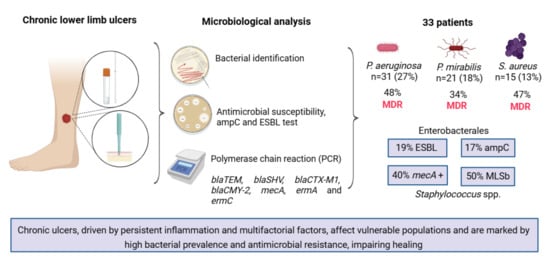
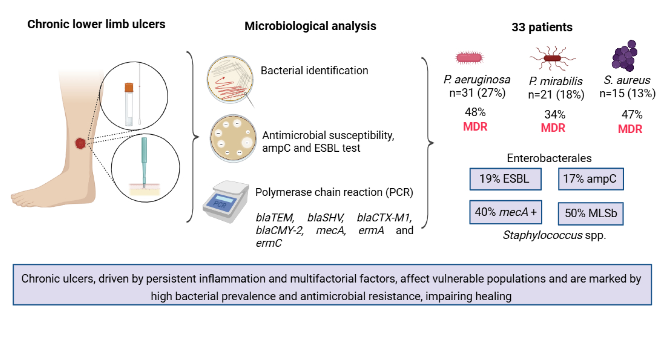
Pseudomonas aeruginosa was the most prevalent species, with 31 isolates (27%), followed by
Proteus mirabilis (
n = 21. 1%) and
Staphylococcus aureus (
n = 15.1%). Although CoNS species were analyzed separately, this group comprised a total of 15 isolates (13%), ranking among the most prevalent. Similarly, the genus
Staphylococcus spp. was the most frequent among Gram-positive bacteria (77%) and the second most prevalent overall (
n = 30), second only to the genus
Pseudomonas spp. (
n = 31). The complete list of identified microorganisms is summarized in
Table 2.
Among the resistance profiles of the isolates, Gram-negative bacteria showed 100% susceptibility to meropenem, including both non-lactose fermenters and the Enterobacterales group. Pseudomonas aeruginosa exhibited the highest antimicrobial resistance rates against penicillins (Piperacillin + Tazobactam (PIT)), cephalosporins (Cefepime (CPM), Ceftazidime (CAZ), and Cefoxitin (CFO)), monobactams (Aztreonam (ATM)), and quinolones (Ciprofloxacin (CIP) and Levofloxacin (LEV)). The Enterobacterales group also showed full susceptibility (100%) to PIT but demonstrated higher resistance rates to Amoxicillin + Clavulanic Acid (AMC), quinolones, and Sulfamethoxazole + Trimethoprim (SUT).
Staphylococcus spp. (
n = 30) exhibited 100% susceptibility to vancomycin and 97% (
n = 29) to linezolid, with the highest resistance observed to macrolides (Erythromycin (ER)) and tetracyclines (TET). Coagulase-negative staphylococci (CoNS) displayed a higher resistance profile than
S. aureus, particularly to cefoxitin, quinolones, clindamycin (CLIN), and SUT. Among other Gram-positive bacteria, such as
Enterococcus faecalis, 50% (
n = 3) showed resistance to quinolones and one isolate (17%) was resistant to vancomycin. All data are summarized in
Table 3.
Table 4 presents the multidrug resistance patterns among the main identified isolates.
P. aeruginosa showed the highest prevalence of MDR (48%) and PDR (29%) strains. Among Enterobacteriaceae,
P. mirabilis and
E. coli exhibited greater susceptibility, although a portion displayed MDR behavior (34% and 40%, respectively). Within
Staphylococcus spp.,
S. aureus demonstrated an MDR-compatible profile (47%), while PDR phenotypes were observed in
S. epidermidis (20%) and in the CoNS group (13%).
The associations between clinical-demographic variables and microbiological outcomes are summarized in
Table 5. Most clinical variables, including gender (
p = 0.394), age (
p = 1.000), diabetes mellitus (
p = 0.651), hypertension (
p = 0.202), and leprosy (
p = 0.358), showed no significant association with the detection of MDR pathogens. Similarly, the duration of the ulcer, stratified into 10 years (
p = 0.141), 11–20 years (
p = 1.000), and >20 years (
p = 0.113, did not correlate with increased resistance profiles. Notably, a significant association was found between the sampling method and the identification of MDR strains, as detailed in the comprehensive statistical analysis. Biopsy samples were significantly more likely to yield MDR isolates compared to swabs (
p = 0.008). No significant correlation was observed between the isolation of
P. aeruginosa and XDR profiles (
p = 0.115).
In the phenotypic evaluation of ESBL production, 19% (n = 8) positivity was observed among isolates belonging to the order Enterobacterales, represented by P. mirabilis (24%), P. stuartii (100%), P. rettgeri (33%), and E. coli (17%). Genotypic analysis revealed a higher prevalence of the blaTEM (36%) and blaSHV (36%) genes compared to blaCTX-M1 (27%). Although not phenotypically detected, 67% (n = 2) of K. oxytoca isolates were positive for blaTEM, and 33% (n = 1) exhibited the coexistence of all three genes.
Regarding
ampC production in
Enterobacterales, 17% (
n = 7) of isolates showed phenotypic expression, which was genotypically confirmed by the presence of the
blaCMY-2 gene in 56% (
n = 23). Phenotypically, only one
P. mirabilis isolate exhibited
ampC production, while genotypic detection was present in 43% (
n = 9). A similar pattern was observed for
E. coli, with 83% (
n = 5) molecular positivity for
blaCMY-2, despite the absence of phenotypic expression. All results are summarized in
Table 6.
The genus Staphylococcus spp. exhibited 33% of isolates with phenotypic resistance to oxacillin. Molecular detection revealed that 40% (n = 12) of microorganisms carried the gene mecA. The susceptibility of S. aureus to oxacillin remained consistent among isolates, with higher resistance observed in CoNS. In S. aureus, 20% (n = 3) showed phenotypic resistance to cefoxitin, confirmed by 33% (n = 5) mecA positive isolates, suggesting MRSA strains. Among CoNS, S. epidermidis, S. cohnii, S. lugdunensis, S. pettenkoferi, and S. haemolyticus demonstrated concomitance between the OXA-resistant phenotype and mecA positivity (40–100%).
MLSb-type resistance was detected in half (
n = 15) of the
Staphylococcus spp. isolates, with higher activity in
S. cohnii (100%),
S. argenteus (100%),
S. hominis (100%), and
S. epidermidis (80%), and a predominance of the constitutive cMLSb phenotype (30%). In
Staphylococcus spp., both
ermA and
ermC genes were detected in 37% and 40% of isolates, respectively, with a slight predominance of
ermA. The coexistence of
ermA and
ermC was observed in only two isolates (7%) from the CoNS group, according to the data in
Table 7.
Among the 33% (
n = 10) of
Staphylococcus spp. isolates exhibiting phenotypic resistance to oxacillin, 50% (
n = 5) concomitantly carried the MLSb phenotype, with a predominance (
n = 4) of the cMLSb profile (40%). Sixty-seven percent (
n = 2) of oxacillin-resistant
S. aureus (MRSA) isolates exhibited cMLSb resistance, while CoNS showed 43% (
n = 3) of the MLSb phenotype, with 29% (
n = 2) in its constitutive form. The same pattern observed phenotypically was reflected in the detection of the
mecA gene in
Staphylococcus spp. (33%), with 60% (
n = 6) of
mecA positive isolates carrying at least one gene from the
erm complex, among which the CoNS group exhibited higher positivity (67%) compared to
S. aureus (60%). All results are summarized in
Table 8.
4. Discussion
The clinical profile observed in this study reflects the complex nature of chronic wound healing, where aging, male predominance, and comorbidities such as vascular and neurological disorders act as key drivers of chronicity [
1,
2,
3]. Interestingly, the detection of MDR pathogens was not significantly associated with demographic variables such as gender (
p = 0.394) or age (
p = 1.000), nor with comorbidities including diabetes mellitus (
p = 0.651) or leprosy (
p = 0.358). These findings suggest that, in this specialized setting, MDR colonization represents a widespread risk across the patient population, regardless of individual clinical characteristics. Furthermore, the trend toward longer-standing lesions in colonized patients, despite the lack of statistical significance for
Staphylococcus spp. (
p = 0.274), reinforces the role of microbial persistence in recalcitrant ulcers.
The microbial landscape was dominated by
P. aeruginosa,
P. mirabilis, and
S. aureus, consistent with global literature [
24,
25]. In addition to this overall distribution, stratified analyses suggest that host-related factors may influence colonization patterns. For instance, 86.7% of hypertensive patients carried
P. aeruginosa (
p = 0.056), indicating that hypertension may contribute to a physiological niche favoring this species [
25].
In a study conducted by Garcia et al. [
26], which characterized bacterial colonization and microbial load in leg ulcers, an association between
P. mirabilis and infected ulcers was reported, corroborating our findings. Additionally,
P. mirabilis and coagulase-negative
Staphylococcus (CoNS) were significantly associated with female patients (
p = 0.038), suggesting a possible influence of host-related ecological factors. The statistical significance of these associations highlights patterns that warrant further investigation in larger cohorts. Moreover, although
P. aeruginosa exhibited high resistance rates, no significant correlation was observed between its isolation and XDR profiles (
p = 0.115), indicating that, in this cohort, resistance may be substantial but not maximal.
A key finding of this study relates to the sampling methodology. A significant association was identified between the sampling technique and the recovery of multidrug-resistant strains (
p = 0.008), with biopsy samples showing a higher probability of isolating these organisms compared to superficial swabs. This supports the hypothesis that resistant pathogens and biofilms are predominantly located in deeper layers of the ulcer bed, where they are less affected by topical treatments and surface cleansing. These results highlight the clinical importance of deep tissue sampling for accurate microbiological diagnosis in chronic wounds [
27].
Most antimicrobial susceptibility rates in
Staphylococcus spp. fall within the expected range for community-acquired strains. However, the indiscriminate use of antibiotics during the SARS-CoV-2 pandemic may have contributed to the emergence of resistant phenotypes, particularly affecting susceptibility to erythromycin derivatives [
28]. The 100% susceptibility to vancomycin observed across the genus is epidemiologically relevant, as this antimicrobial remains a cornerstone for the treatment of Gram-positive infections following the emergence of oxacillin resistance. In this context, the detection of the
mecA gene remains essential for monitoring β-lactam resistance and tracking MRSA dissemination.
Lincosamides may represent an alternative therapeutic option for oxacillin-resistant strains. However, phenotypic detection of MLSb-type resistance varies according to regional characteristics, prior antimicrobial exposure, and local resistance patterns. At the molecular level, the prevalence of
ermA may range from 10% to 60%, whereas
ermC is generally less frequent, reaching up to 30%. In a review by Assefa et al. [
28], 26.8% of
S. aureus mecA-positive isolates also exhibited MLSb production, primarily involving
erm and
msrA genes. These findings align with our results, in which 50% (
n = 5) of oxacillin-resistant
Staphylococcus spp. isolates exhibited the MLSb phenotype, and 60% of
mecA-positive strains carried at least one
erm gene, reinforcing the potential limitations of clindamycin therapy in MRSA infections.
The high resistance profile observed in
P. aeruginosa to piperacillin–tazobactam, cephalosporins, and monobactams may be attributed to its genomic complexity, which enables the expression of diverse regulatory mechanisms that enhance metabolic adaptability. Although this species demonstrated elevated resistance rates, this did not translate into a significant association with XDR profiles, suggesting partial rather than extreme resistance in this cohort. Intrinsic resistance mechanisms, including efflux pumps and the production of inactivating enzymes such as AmpC, likely contribute to these patterns, particularly regarding reduced susceptibility to fluoroquinolones and β-lactams [
29]. Although 77% (
n = 27) of isolates exhibited phenotypic positivity for AmpC, molecular confirmation was not performed, given the intrinsic nature of its expression in this species.
Within the order Enterobacterales, antimicrobial susceptibility patterns were consistent with those typically observed in community settings, with higher resistance rates to amoxicillin–clavulanate (42%), ciprofloxacin (45%), levofloxacin (37%), and sulfamethoxazole–trimethoprim (39%). ESBL production was phenotypically detected in 19% (n = 8) of isolates and confirmed by the presence of genes such as blaCTX-M-1. Although blaTEM-1 and blaSHV-1 are not classified as ESBLs, they encode narrow-spectrum β-lactamases and may contribute synergistically to antimicrobial resistance. Furthermore, point mutations in these genes can give rise to ESBL variants such as blaTEM-52 and blaSHV-12.
E. coli isolates showed a higher frequency of resistance genes compared to phenotypic expression, suggesting variable gene expression or regulation. Although expression analyses such as RT-PCR were not performed, this discrepancy indicates a potential latent resistance reservoir not fully captured by standard phenotypic methods. In Enterobacterales, phenotypic AmpC production was observed in 17% (
n = 7) of isolates, a lower prevalence compared to ESBLs. Unlike
P. aeruginosa, where AmpC expression is intrinsic, in Enterobacterales it may be inducible or plasmid-mediated. This dynamic regulation has important clinical implications, as exposure to β-lactam antibiotics may select for AmpC-producing mutants. Genes such as
blaCMY-2 play a key role in this process, and their plasmid-mediated dissemination facilitates spread. The absence of phenotypic expression in some genotypically positive isolates may reflect regulatory mechanisms such as induction thresholds or chromosomal derepression, potentially influencing antimicrobial response under clinical conditions [
30].
The challenges in managing infectious conditions in chronic wounds are closely linked to antimicrobial resistance, exacerbated by the widespread and often indiscriminate use of antibiotics. The detection of clinically relevant resistance genes—including
mecA,
blaTEM,
blaSHV,
blaCTX-M1,
ermA,
ermC, and
blaCMY-2—confirms the presence of multidrug resistance among bacteria isolated from chronic ulcers. However, distinguishing between colonization and true infection remains essential to guide appropriate therapy. Therefore, antimicrobial use should be guided by clinical evidence, microbiological findings, and stewardship strategies aimed at preserving the effectiveness of available treatments [
31].
Limitations of the Study
Despite the clinical relevance of our findings, this study has some limitations. First, the research was conducted at a single specialized dermatological center with a specific diagnostic demand, which may limit the generalizability of the results to general hospitals or other geographical regions. Additionally, the sample size was determined by convenience and did not involve prior power calculations, which warrants caution when generalizing the prevalence rates and statistical associations to larger populations. Furthermore, the identification of Gram-negative bacteria was based on biochemical and semi-automated methods rather than proteomic techniques like MALDI-TOF. However, we mitigated these limitations by implementing rigorous monthly quality control with ATCC reference strains and employing molecular identification (ITS-PCR and sequencing) for Gram-positive isolates to ensure taxonomic precision. Future multicenter studies with larger cohorts and a broader range of molecular resistance markers are needed to further elucidate the complex epidemiology of chronic wound infections in Southeastern Brazil.
5. Conclusions
This study demonstrates a high burden of polymicrobial colonization in chronic ulcers, with a predominance of Gram-negative bacilli, particularly P. aeruginosa and P. mirabilis, as well as species of the genus Staphylococcus. The presence of MDR and PDR phenotypes, in association with clinical and socioeconomic factors, underscores the complexity of these lesions. Notably, the sampling methodology played a decisive role in these findings, as isolates recovered via biopsy were significantly more likely to reveal MDR pathogens compared to superficial swabs (p = 0.008), highlighting the presence of resistant strains in deeper tissue layers. The detection of genetic determinants of resistance, including blaTEM, blaSHV, blaCMY-2, mecA, ermA, and ermC, reveal discrepancies between genotypic and phenotypic profiles, suggesting the presence of unexpressed resistance that may be inducible during treatment. The high prevalence of P. aeruginosa, coupled with comorbidities such as hypertension, points to its potential adaptation to vascularly compromised microenvironments. Although vancomycin and meropenem retain in vitro activity, the high frequency of mecA-positive strains and the MLSB phenotype as well as the phenotypic expression of AmpC in P. aeruginosa limit therapeutic options and complicate empirical antimicrobial selection. In this context, effective management of these lesions would benefit from a transition from empirical approaches to strategies guided by local molecular epidemiology. Continuous surveillance of resistance markers is essential to optimize wound healing, curb the dissemination of resistant pathogens, and preserve the efficacy of the antimicrobial arsenal.




 ,
,