Review

Enterococcus Species: Multifaceted Probiotic Potential and Safety Considerations

by
Ojonugwa Precious John
1,
Kayode Olayinka Afolabi
1,2,
Anayochukwu Chibuike Ngene
3,
Williams Omotola Tanimowo
1,
Mary Ayobami Adewoyin
1,
Michael Bamitale Osho
1,4 and
Rine Christopher Reuben
5,*
1
Department of Biological Sciences, Anchor University, Lagos 102213, Nigeria
2
Anchor University Centre for Global Health, Anchor University, Lagos 102213, Nigeria
3
Department of Microbiology, College of Natural Sciences, Michael Okpara University of Agriculture, Umudike 440101, Abia State, Nigeria
4
Department of Microbiology and Biotechnology, McPherson University, Seriki Sotayo 110117, Ogun State, Nigeria
5
Department of Biology, Stephen F. Austin State University, SFA Station, Nacogdoches, TX 75962, USA
*
Author to whom correspondence should be addressed.
Microorganisms 2026, 14(4), 815; https://doi.org/10.3390/microorganisms14040815
Submission received: 10 March 2026 / Revised: 29 March 2026 / Accepted: 30 March 2026 / Published: 2 April 2026
(This article belongs to the Special Issue Probiotics: Development and Application)

Abstract

Enterococcus spp. are common but not predominant commensal bacteria that inhabit the gastrointestinal tracts of humans and animals and are widely distributed in various environmental matrices and diverse food sources. Multiple strains of beneficial enterococci are increasingly utilized as protective cultures, alternatives to antibiotics, and probiotics for controlling pathogens, mitigating disease, modulating the microbiome, and supporting overall host health. They also support food fermentation and safety, enhance sensory properties, and produce bioactive compounds such as bacteriocins with strong pathogen-inhibitory activity and multifarious health benefits. Despite their advantages in health and agrifood systems, their association with healthcare-associated infections and the spread of antimicrobial resistance raises concerns about their safety. These risks underscore the need for stringent safety evaluations before their use as probiotics, alternatives to antibiotics, or protective cultures. Here, we provide a comprehensive atlas of the multifaceted probiotic and antimicrobial potential, as well as safety considerations of beneficial enterococci. This would provide a valuable resource for future research, regulatory assessments, and applications in the agrifood system and healthcare.

1. Introduction

Antimicrobial resistance (AMR) has emerged as a silent pandemic, undermining the efficacy of life-saving treatments and increasing healthcare burdens worldwide. The World Health Organization (WHO) attributes this crisis primarily to the widespread overuse of antibiotics in both clinical and agricultural settings [1]. If left unaddressed, AMR-related deaths could exceed 10 million annually within the coming decades, surpassing mortality rates from major infectious diseases [2].
As conventional antibiotics lose their effectiveness, alternative strategies, such as plant-derived compounds, bacteriophages, and probiotics, have gained increasing attention as potential countermeasures against drug-resistant infections [3,4]. Probiotics, defined as viable microbial organisms that confer significant health and nutritional advantages when consumed in sufficient quantities, are particularly promising. Among probiotic bacteria, some strains of Enterococcus spp., a subgroup of lactic acid bacteria (LAB), exhibit unique characteristics: they enhance traditional fermented foods by improving flavor, aroma, and texture, while also producing bacteriocins, bioactive peptides that exhibit potent activity against a broad spectrum of pathogens [5,6]. This functionality suggests their potential application as both natural food preservatives and alternatives to antibiotics. However, safety concerns associated with their opportunistic pathogenicity and classification among the ESKAPE (Enterococcus faecium, Staphylococcus aureus, Klebsiella pneumoniae, Acinetobacter baumannii, Pseudomonas aeruginosa, and Enterobacter species), pathogens globally known for their ability to develop resistance to multiple antibiotics, pose a significant threat in healthcare settings and are a major cause of hospital-acquired infections [7].
The safety concerns arise from the remarkable adaptability of some enterococci to hostile environments, including antibiotic-rich conditions, which increases their pathogenic potential, particularly in some E. faecalis and E. faecium strains [8,9]. The growing antibiotic resistance has positioned some enterococci as a significant contributor to the antimicrobial resistance (AMR) crisis [10]. Virulence factors such as biofilm formation, host cell adhesion, and immune evasion mechanisms further complicate their role, making them problematic pathogens in healthcare settings. These traits explain their involvement in severe bloodstream, heart valve, and urinary tract infections [11].
The Enterococcus genus exhibits strain-specific heterogeneity and environmental influences that significantly affect its pathogenicity and virulence. While clinical isolates are a major concern, several studies have identified certain strains of Enterococcus from fermented foods that carry drug resistance genes [12,13]. These environmental differences contribute to varying resistance profiles, with clinical isolates often displaying strong resistance patterns, whereas isolates from fermented foods generally exhibit weaker resistance [14,15]. Nonetheless, even antimicrobial-resistant Enterococcus strains with weak resistance can pose significant risks to immunosuppressed individuals. Such infections may lead to systemic complications and severely limit the choice of effective antibiotics, raising safety concerns about using some Enterococcus strains from food sources as probiotics in both food and healthcare settings [16]. Consequently, rigorous evaluation of the probiotic properties of beneficial enterococci, particularly regarding safety, is essential before their application in food systems. To address these concerns, regulatory authorities, including the European Food Safety Authority (EFSA), have implemented rigorous protocols for evaluating the safety of beneficial strains of Enterococcus spp. These evaluations aim to ensure that probiotic enterococci intended for use in humans or animals do not pose public health risks because of their carriage of antimicrobial and virulence determinants [16]. Enterococcus spp. have not yet achieved Generally Recognized As Safe (GRAS) or Qualified Presumption of Safety (QPS) status [10]. Therefore, the safety of beneficial (probiotic) enterococci is assessed on a strain-by-strain basis, rather than by species. This is because while some strains are considered beneficial and harmless, others are virulent and resistant to multiple antibiotics. Notwithstanding these concerns, certain commercially available probiotic strains, such as E. faecium SF68, E. lactis DSM 7134, and E. faecium 669, have exhibited substantiated therapeutic efficacy and established themselves as important contributors to the probiotic market [17,18].
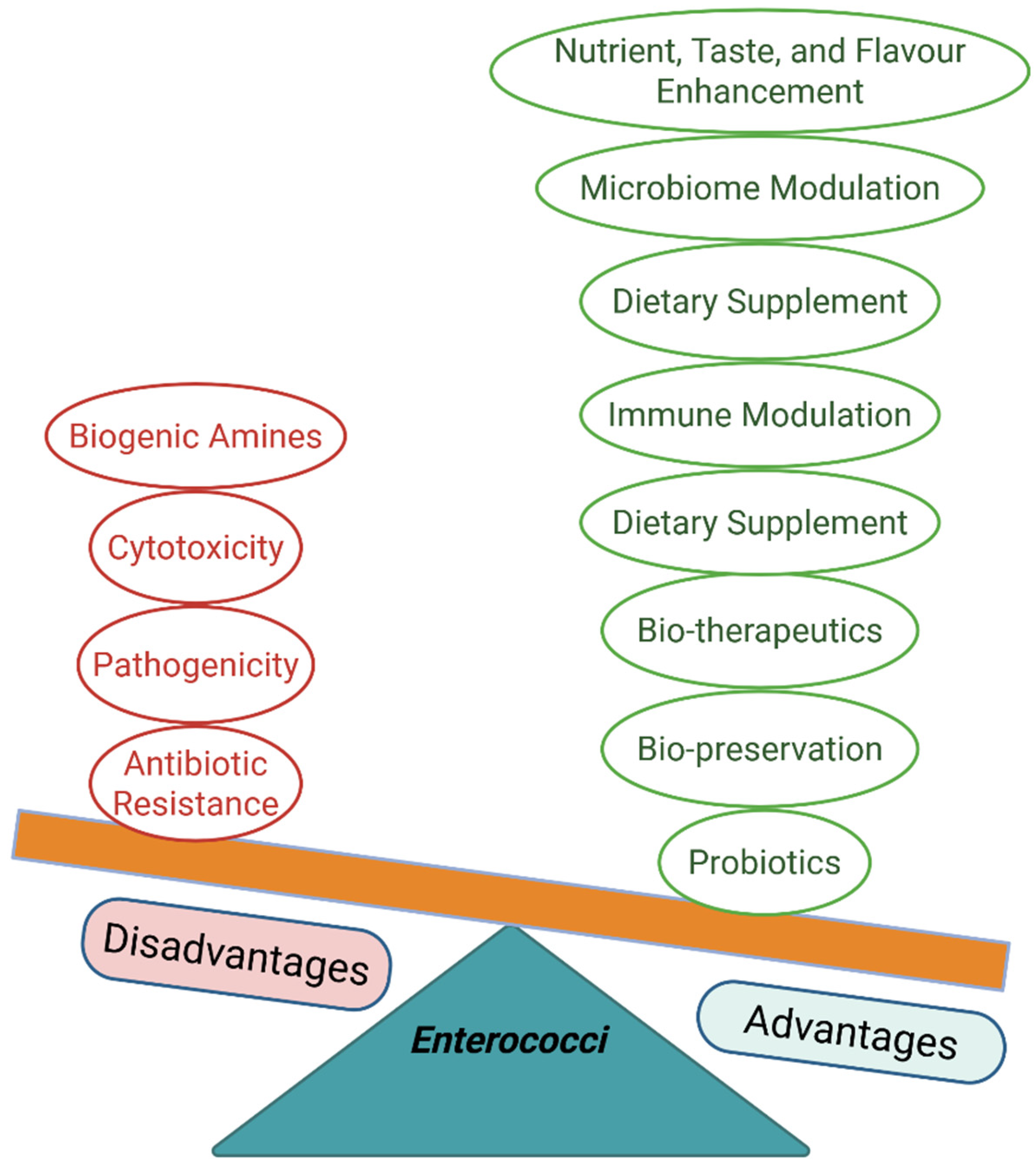
The paradoxical role of Enterococcus in food, health, and disease has sparked significant scientific debate. This review critically explores the potential of beneficial enterococci as probiotics while carefully considering their associated safety concerns. This narrative review aims to provide a comprehensive and critical analysis of the paradoxical behavior of enterococci, which could guide future applications and research, particularly in the areas of antimicrobial resistance, biotechnology, probiotic use, health, and agrifood systems.
Additionally, a comprehensive literature search was conducted across Google Scholar, Web of Science, and PubMed in April 2025 to identify and select relevant articles that highlighted multifaceted probiotic potential in agrifood and health systems. The keyword terms used included ‘Enterococcus’ OR ‘enterococci’ OR ‘probiotic Enterococcus’ OR ‘Enterococcus antimicrobial resistance’ OR ‘Enterococcus pathogenicity’ OR ‘Enterococcus virulence’ OR ‘Enterococcus nosocomial infections’ OR ‘Enterococcus fermentation’ OR ‘Enterococcus food safety’ OR ‘Enterococcus bacteriocin’. Articles published in the English language were included after an independent review of titles and abstracts by authors. Other enterococci-related publications outside the scope of this study, as well as editorials, meta-analyses, commentaries, and perspectives, were excluded.

2. Ecological Distribution of Enterococcus

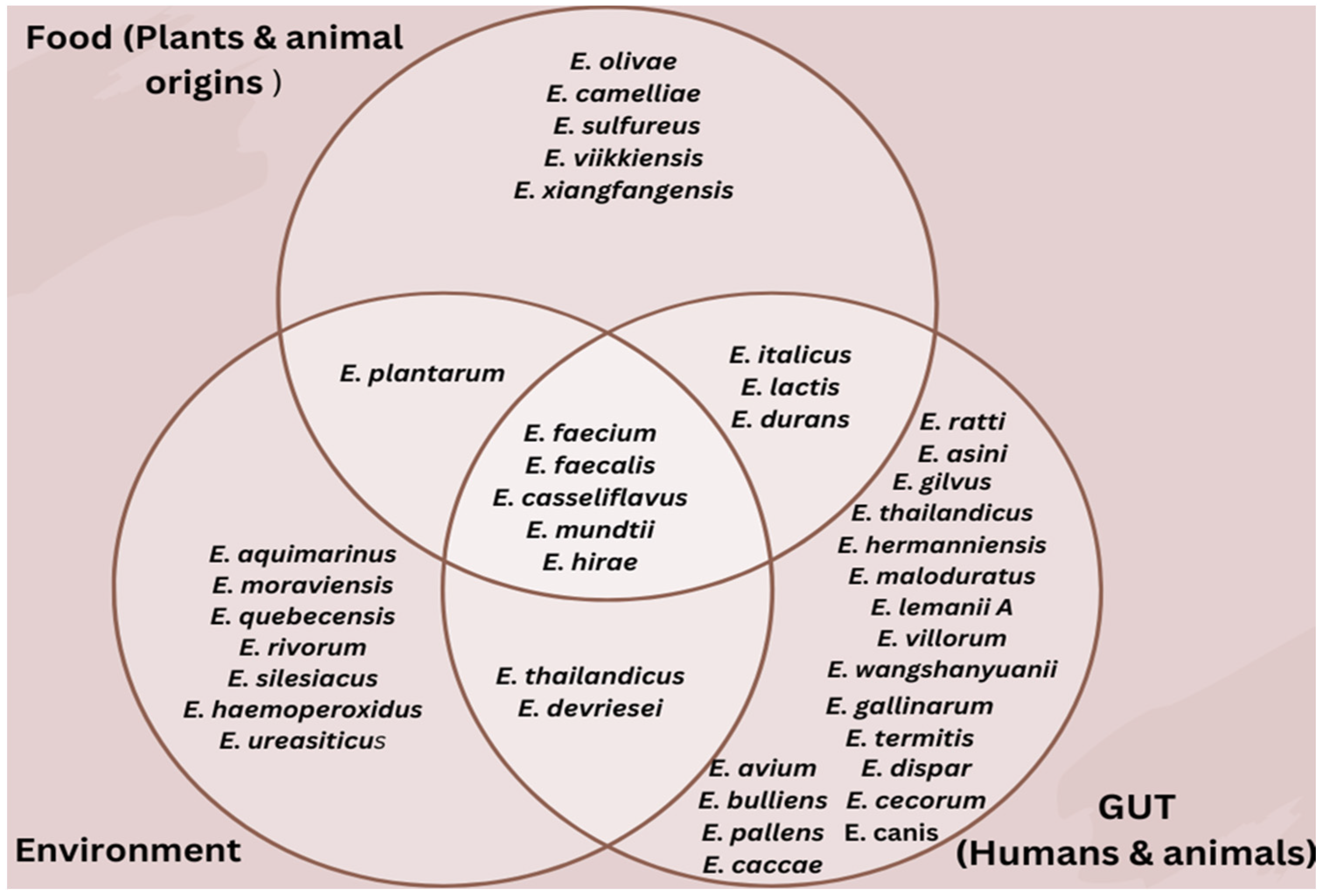
The ecological shift of Enterococcus from a historic water-based environment to a land-based environment has facilitated its adaptation and ability to thrive across multiple ecological niches [19,20,21,22,23,24,25]. This adaptability has enabled enterococci to flourish in diverse environments, ranging from natural ecosystems to human-associated settings, highlighting their remarkable ecological versatility [21,22]. However, this widespread distribution also raises concerns, as some enterococci can transition from beneficial commensals to opportunistic pathogens, particularly in clinical and food-related systems (Figure 1).

2.1. The Gut and Nasal Cavity

The intestinal tract is a multiplex network of diverse microbes that profoundly impact human and animal health. Some strains of enterococci are common but not dominant commensals of the gastrointestinal tract of animals, and they are highly adapted to the gut ecosystem, where they thrive on abundant nutrients and interact with other microorganisms and host cells [23]. Their resilience in extreme habitats, such as low pH, high bile salt concentrations, and limited oxygen, allows them to flourish in diverse hosts [24]. In the human gastrointestinal tract, E. faecalis and E. faecium constitute the predominant species, with others such as E. durans, E. hirae, and E. gallinarum present at much lower abundances. They contribute to nutrient metabolism and help maintain microbial stability [25].
In animals, the composition of enterococcal species varies widely and is influenced by factors such as diet, gut physiology, and environmental exposure. While E. faecium, E. faecalis, and E. hirae are commonly found in the gastrointestinal tracts of cattle, pigs, and sheep, other species also exhibit host specificity. In poultry, such as chickens and turkeys, E. faecium and E. durans tend to dominate [23]. Companion animals, especially dogs, often harbor E. faecalis, E. faecium, and E. canis [26]; however, wild birds and rodents are frequently associated with less-characterized species such as E. casseliflavus, E. avium, E. cecorum, and E. gallinarum. Insects, including bees, termites, and beetles, host diverse species such as E. mundtii, E. casseliflavus, and E. termites, underscoring the ecological versatility of the genus [22,23]. An insect model study using E. mundtii isolated from flour moths showed improved survival rates in beetles challenged with Bacillus thuringiensis, demonstrating probiotic effects and suggesting its potential as a probiotic or therapeutic agent [27]. Another recent study using silkworms identified E. mundtii as a beneficial intestinal commensal that colonizes and adheres to the gut, enhances carbohydrate breakdown, improves intestinal microbiome balance, protects the insect from invading pathogens, and modulates host immunity [28]. A metagenomic study on the edible insect, soybean hawkmoth, identified E. casseliflavus and E. pernyi as the dominant gut bacteria, with E. casseliflavus contributing to nutrient metabolism and overall gut health [29].
Although some enterococci inhabit the gastrointestinal tract, their colonization of the nasal cavity in humans and animals is increasingly documented [30,31,32]. Studies have shown that the most common enterococci found in nasal samples are E. faecium, E. faecalis, and E. casseliflavus [32,33,34]. Nasal colonization by some enterococci may occur in hospitalized patients, immunosuppressed individuals, and patients with chronic rhinosinusitis or nasal devices [32]. Although they rarely cause infections on their own within the nasal sites, they may contribute to biofilm formation and sinonasal infections, or act as reservoirs for the acquisition and dissemination of antimicrobial resistance genes [34].
Some enterococci play a significant role in the gut ecosystem, contributing to immunomodulation, gut health, and metabolic maintenance [35,36]. However, disturbances to the intestinal microbiome caused by indiscriminate drug use can promote the spread of drug-resistant strains by wiping out susceptible strains while surviving bacteria recolonize the gut and, through fecal discharge, are introduced into the surrounding environment, further accentuating antimicrobial resistance [25,37]. Despite these risks, numerous studies have documented the benefits of some strains of Enterococcus spp. in the gut. For example, E. casseliflavus has exhibited considerable therapeutic promise, with clinical evidence indicating improved survival outcomes in hematopoietic stem cell transplant (HSCT) recipients [38]. Nevertheless, some Enterococcus spp. can also transform into opportunistic pathogens outside the gastrointestinal region, causing conditions such as heart infections, bloodstream infections, urinary tract infections, and systemic infections [39]. Among the diverse Enterococcus species shown in Figure 1, E. faecium and E. faecalis predominate within the gut and play imperative roles in gastrointestinal health maintenance and immune modulation [35,40]. Research confirms that introducing probiotic bacteria into the gut helps maintain microbial balance, supports digestion, and strengthens immune responses [41]. Specific Enterococcus strains have been identified as possessing notable probiotic and health-promoting properties through multiple mechanisms [42,43]. A well-documented mechanism is competitive exclusion, where Enterococcus strains compete with pathogens for binding sites on the intestinal wall, thereby blocking colonization by harmful bacteria [44].
Moreover, some enterococci synthesize bacteriocins (especially enterocins), antimicrobial compounds that inhibit the establishment of detrimental bacteria in the gastrointestinal tract, thereby contributing to gut microbiota stability and overall well-being [45,46]. Additionally, some Enterococcus spp. produce fatty acid metabolites, including short-chain fatty acids (SCFAs), which are critical mediators of intestinal homeostasis. These SCFAs serve as fuel for intestinal epithelial cells, enhance intestinal barrier integrity by upregulating tight junction protein expression, attenuate pro-inflammatory cascades, and participate in immunomodulatory processes [47]. Research indicated that including probiotic E. faecalis in juvenile crayfish diets can enhance growth, strengthen the immune system, and improve survival, making it a promising supplement for aquaculture [45]. Similarly, in dogs with chronic digestive issues, E. faecalis supplementation helps balance gut bacteria, leading to improved digestion and overall health [48].
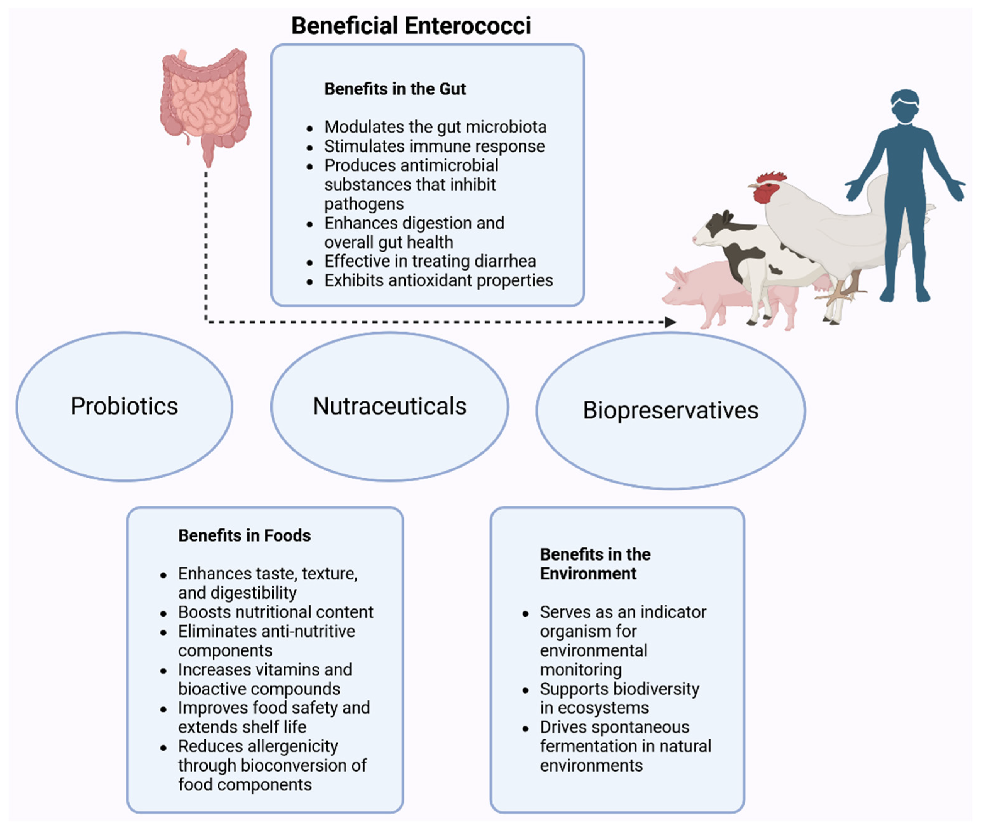
Furthermore, some strains of Enterococcus spp. also contribute significantly to the gut microbiota by metabolizing polysaccharides, particularly indigestible dietary fibers, and converting them into bioavailable nutrients. Research by Basu et al. [49] demonstrated that supplementing dairy calves with E. faecium 669 significantly enhances body weight, average daily gain, and gastrointestinal health during the late preweaning phase. Notably, treated calves showed reduced incidence of diarrhea and lower rectal temperatures, suggesting enhanced gut barrier function and immune modulation. As illustrated in Figure 2, strains such as E. faecium and E. faecalis have shown promise as probiotic supplements, with demonstrated benefits for digestive efficiency, immune response, and growth performance.

2.2. The Environment

Enterococci enter and thrive in various environmental niches, primarily due to the discharge of fecal matter from humans, animals, and sewage, as well as agricultural runoff [50]. Their remarkable adaptability allows them to be widely distributed across diverse ecological habitats, including soils, plants, and water bodies [23,50,51,52,53,54]. This ability to persist and grow in harsh environments underscores their ecological importance and functional versatility. However, these intrinsic biological properties also make Enterococcus spp. particularly effective disseminators of drug resistance in the environment [51,52].
Common Enterococcus species found in soils, including agricultural, contaminated, and natural soils, are E. hirae and E. faecium [53,54]. E. faecalis and E. faecium are frequently detected in rivers and streams, often originating from soil runoff, wastewater, or fecal contamination [55]. Additionally, E. faecalis, E. faecium, and E. casseliflavus are commonly found in plants as well as in untreated and treated wastewater [51]. Within soil environments, some Enterococcus spp. persist as minor but significant components of the microbiota, contributing to elevated bacterial counts [23].
Beyond their ecological roles (Figure 2), certain Enterococcus spp. have valuable applications in environmental remediation [56,57]. For example, Enterococcus spp. designated Cdq4–2 contribute significantly to bioremediation when used alongside plants such as Lolium perenne, enhancing plant growth and facilitating heavy metal remediation [58]. Moreover, auxin-producing Enterococcus strains have demonstrated plant growth-promoting capabilities and the potential to produce nanobiofertilizers for sustainable agriculture and food security [59].
Despite these ecological benefits, some Enterococcus spp. pose significant challenges due to their pivotal role in propagating antibiotic resistance through environmental pathways. They are well-known reservoirs of resistance, driven by both intrinsic and acquired mechanisms [60]. Soil, polar lakes, and aquatic ecosystems, especially through runoff and wastewater discharge, serve as conduits for the spread of antibiotic-resistant Enterococcus strains, thereby contributing to public health risks [52]. For example, Rehman and colleagues [61] reported that bacterial strains from drinking water and sewer overflow carried harmful virulence genes and transferable plasmids, highlighting potential health threats. This research indicates that environmental exposure to some enterococci may increase the risk of antibiotic resistance transmission to humans. Common species such as E. faecalis, E. faecium, E. hirae, and E. casseliflavus are often found in these environments, and their highly adaptable genomes enable survival under harsh conditions, making them persistent concerns [51].
Although some Enterococcus strains are known to cause infections, especially in hospital settings, these bacteria also play valuable roles in nature and industry. For instance, they are key players in traditional fermented foods, where they initiate natural fermentation processes [62]. In many developing regions, where commercial starter cultures are not always accessible, wild Enterococcus strains from the environment serve as natural fermenters. This not only enables fermentation but also improves the taste, texture, and overall quality of foods such as fermented vegetables, grains, and dairy products [63,64].
Ademola et al. reported that the genus Enterococcus is pivotal in the natural fermentation of Nigerian traditional condiments, such as Iru and Ogiri [65]. They identified Enterococcus among the naturally occurring genera in the preparation environment of these condiments, with the potential to serve as starter cultures. Their involvement influences the taste, aroma, and nutrient content of the final products [66].
In addition to their relevance in spontaneous traditional food fermentation, Enterococcus spp. serve as fundamental components of ecosystem dynamics. Research indicates that these microorganisms actively shape microbial communities by participating in biogeochemical processes, particularly the mineralization of nitrogenous compounds and carbon-rich substrates in terrestrial and aquatic systems [60]. Their metabolic activities can promote detrital decomposition and maintain critical nutrient turnover rates, making them keystone taxa that contribute to environmental sustainability.
Furthermore, Enterococcus spp. serve as critical sentinels for tracking the flow of antimicrobial resistance (AMR) genes in ecosystems. Due to their remarkable genetic adaptability and capacity for horizontal gene transfer, these bacteria function as effective biomarkers for assessing AMR dissemination, particularly in wastewater and agriculturally impacted habitats [10]. The persistent detection of AMR in such environments highlights patterns of resistance spread, underscoring the need for deeper investigation into how natural systems contribute to AMR evolution and propagation.

2.3. Foods

Food is a complex ecosystem that provides an ideal habitat for most microorganisms. Enterococci are among the predominant microbes in fermented food products [17]. While E. faecium and E. faecalis are the most common species found in foods, E. durans, E. hirae, E. lactis, E. olivae, E. italicus, E. gallinarum, and E. casseliflavus are less predominant [13,67,68]. Several of these lesser-known enterococcal species have been isolated from various fermented and raw food sources. For example, E. olivae was isolated from Spanish fermented green olives [69], E. camelliae from fermented tea leaves in Thailand [70], E. lactis from milk and dairy products [71], and E. thailandicus from fermented sausage. Additionally, species such as E. hirae, E. casseliflavus, and E. gallinarum have been reported in raw meat samples from Ghana [60].
Enterococcus spp. play significant roles in various fermented foods (Figure 2), contributing to flavor development, food safety, and quality. These bacteria dominate cheeses, where their proteolytic and lipolytic activities produce volatile compounds that increase flavor complexity [72]. Strains such as E. durans and E. hirae have been notable in traditional cheeses, enhancing microbiota diversity [73]. Similarly, in dry-cured sausages and fermented meats, E. faecium and E. faecalis improve flavor and texture through enzymatic activities [74]. However, some dairy-derived and other fermented foods containing Enterococcus spp., particularly E. faecium and E. faecalis, have been recognized as potential reservoirs of virulence and antibiotic resistance genes [75]. For instance, a survey of Turkish cheese samples revealed isolates with high resistance to lincomycin, kanamycin, low-level gentamicin, rifampin, and tetracycline, alongside several resistance genes (tetM, tetL, ermB, cat, aph(3′)-IIIa, ant(6)-Ia, aac(6′)-Ie-aph(2″)-Ia) [76]. Similar patterns have been observed in other countries. In Ethiopia, a study of fermented foods, including probiotic yogurt sold in Addis Ababa, detected Enterococcus spp. in most samples, with many isolates carrying multiple virulence genes (gelE, cylA/B/M, asa, efaA, cpd, ccf, cob) and resistance markers for vancomycin (vanC1, vanC2/C3) and erythromycin (ermB, ermC), underscoring the need for stricter microbial quality control [63]. While Enterococcus strains from cheeses were found to harbor resistance determinants for vancomycin, tetracycline, and erythromycin in Portugal [77], fermented foods-associated enterococci from Nigeria displayed multidrug resistance and carried multiple AMR genes, including parC, aac-li, ermB/ermC, and tetM [78]. Likewise, a more recent study in Greece identified Enterococcus strains from raw sheep milk exhibiting resistance to tetracyclines, macrolides, and chloramphenicol, with some phenotypic resistance not explained by detectable resistance genes [79]. Collectively, these findings highlight the widespread occurrence of virulence factors and antimicrobial resistance determinants in Enterococcus from diverse fermented food sources, raising concerns about their potential role in the transmission of resistance through the food chain.
Unlike E. faecalis and E. faecium, non-dominant Enterococcus species generally harbor a narrower range of virulence and antibiotic resistance genes [80,81]. As a result, they are increasingly being explored and thoroughly assessed for probiotic applications, particularly given E. faecium’s association with nosocomial infections and its extensive repertoire of virulence and resistance determinants. One such species, E. lactis, is closely related to E. faecium but differs in carbohydrate metabolism and 16S rRNA sequence, supporting its classification as a distinct species [82,83]. E. lactis has been reported to be relatively safe for use in the food and health sectors. For instance, its strains harbor significantly fewer acquired resistance and virulence factors compared to E. faecium [71]. However, they carry intrinsic resistance genes such as eatA (T450I), msr(C), and aac(6′)-Ii. They also express intrinsic virulence markers, including ccpA, fnm, and lysM4; however, their overall lower risk profile supports their potential as probiotics [84]. A comparative genomic study of isolates from dairy products, rice wine Koji, and human feces further confirmed the limited antibiotic resistance of E. lactis relative to E. faecium, and identified novel genetic targets for future resistance research [43].
Extending beyond food-associated contexts, Ye et al. recently identified E. lactis strain GL3 from honeybee larvae as a promising probiotic for enhancing honeybee health and controlling American foulbrood, a lethal larval disease caused by Paenibacillus larvae [85]. Strain GL3 demonstrated strong acid and bile tolerance, antioxidant activity, and potent inhibition of P. larvae [85]. Genomic analysis revealed traits for gut adaptation and the ability to produce the antibacterial compound aborycin, which could also serve as a therapeutic agent. Another species, E. italicus strain GGN10, lacked pathogenicity islands and tested negative for common resistance genes, including those for vancomycin, β-lactams, and gentamicin [86]. Similarly, E. italicus FM5 was non-hemolytic, displayed antioxidant activity, exhibited fermentation-enhancing traits, and was sensitive to a broad range of antibiotics, highlighting its safety for food applications and potential as a probiotic [81].
In fermented plant-based products like olives, sauerkraut, and kimchi, some beneficial enterococci often act as secondary microbiota, influencing fermentation dynamics without being essential to the process [87]. E. faecium and E. faecalis are prevalent in African fermented cereals, including Ogi (Nigeria), Mawe (Benin), and Kenkey (Ghana), where they contribute to acidification and microbial stability by inhibiting spoilage organisms and pathogens [66,88,89]. In legume-based condiments such as dawadawa/Iru (fermented African locust beans) and Ogiri (fermented melon seeds), E. faecium and E. durans enhance flavor through proteolysis and amino acid production, ensuring rapid fermentation and reducing spoilage risk [66]. These species are also prevalent in traditional dairy products, such as Nunu (West Africa), Kule naoto, and Mursik (East Africa), where they contribute to flavor development and inhibit pathogens through bacteriocin production [12,90]. While less prominent in alcoholic beverages, some enterococci may appear transiently in products such as palm wine, influencing early fermentation stages before being overtaken by yeasts and other bacteria [91,92]. Specific food-associated strains of E. faecium and E. faecalis are recognized for their probiotic potential, including their ability to modulate the gut microbiota, enhance immune responses, and compete with pathogens. For example, dietary supplementation with E. faecium has been shown to reduce diarrhea severity in weaned piglets, increase growth, and boost the abundance of beneficial gut microbes [93]. Similarly, E. faecalis strains have been associated with improved intestinal microbial balance and immunological homeostasis [94].
Despite their beneficial roles, some enterococci are notorious for both intrinsic and acquired antibiotic resistance, including resistance to vancomycin mediated by the vanA and vanB genes [68]. They act as reservoirs of resistance genes, which can be transferred to other foodborne spoilage and pathogenic bacteria, including Staphylococcus aureus. Additionally, some enterococci harbor virulence determinants such as aggregation substances, gelatinases, and cytolysins, making them potential opportunistic pathogens [95]. Their natural ability to transmit genetic material via mobile elements, including plasmids and transposons, facilitates the dissemination of resistance and virulence genes within microbial communities [96]. Therefore, the safety of food-associated Enterococcus strains varies significantly, with pathogenic potential largely dependent on the presence of virulence determinants. This dichotomy is exemplified by contrasting findings: while most Slovak Lump Cheese contains an E. durans strain devoid of virulence genes, indicating probiotic suitability [97], other dairy products harbor resistant, virulent E. durans strains [98]. Genomic studies corroborate this disparity, revealing that clinical isolates typically carry higher virulence gene loads than food-derived strains [99]. Such evidence highlights the importance of conducting strain-specific risk assessments prior to food applications.
Some strains of Enterococcus spp. significantly influence the organoleptic characteristics of fermented products through multiple metabolic pathways, including pH reduction via acid generation, protein degradation, fat breakdown, and citrate utilization [17]. While most Enterococcus isolates display limited acid production, E. faecalis strains demonstrate notably greater acidifying potential than E. faecium strains. Proteolytic activity appears limited in approximately 70% of strains examined, with E. faecalis consistently exhibiting the most pronounced protein-degrading capacity within the genus [100,101]. The gelE gene in Enterococcus is responsible for the degradation of casein, enhancing texture and aroma in fermented foods, especially dairy products. However, in hospital-associated strains, gelE is also linked to virulence, particularly in E. faecalis [102]. The pathogenic potential of Enterococcus depends on factors such as gene expression, environmental conditions, and strain-specific characteristics [98,103]. This highlights the importance of thorough genomic and phenotypic analyses when evaluating Enterococcus strains for food applications. In addition to protein degradation, E. faecalis demonstrates efficient citrate utilization, resulting in increased diacetyl synthesis—a crucial compound responsible for the characteristic creamy fragrance in fermented dairy products. This pronounced ability to generate flavor compounds distinguishes E. faecalis from E. faecium, which exhibits substantially reduced citrate metabolic activity [104,105].
Current shifts toward biologically derived food preservation methods have highlighted some Enterococcus spp. as potential biopreservatives. These microorganisms synthesize diverse antimicrobial metabolites, particularly enterocins and organic acids, which significantly suppress the growth of both pathogenic and spoilage organisms in food systems. A compelling example includes recent findings on E. faecium Smr18, whose enterocin ESmr18 exhibited potent anti-Salmonella activity in experimentally contaminated poultry matrices [106]. The safety profile of E. faecium Smr18 enhances its biopreservative potential, as evidenced by its antibiotic susceptibility to penicillin, ampicillin, vancomycin, and erythromycin and the absence of hemolytic activity, substantiating its suitability for food safety applications [107].

3. Multifaceted Beneficial Enterococci

3.1. Enterococcus Probiotics

The provenance of probiotics can be traced back to 2000 BC, when ancient civilizations began developing methods to preserve milk for extended periods. Through trial and error, they discovered how to produce fermented beverages by carefully observing changes in stored food [108]. These early societies noted that when fruits and grains were left in sealed containers, they transformed into wine and beer. While the mechanisms behind these transformations were not understood at the time, these practices laid the foundation for fermentation science [109].
A significant milestone in understanding fermentation occurred in 1856, when Louis Pasteur was consulted by a beetroot stillman whose production process yielded a sour, milk-like substance instead of alcohol. Pasteur analyzed the chemical composition of the problematic product and identified lactic acid as its primary byproduct. Using microscopy, he compared samples from containers that had undergone alcoholic fermentation with those producing lactic acid. His observations revealed distinct microbial populations: yeast cells were linked to alcoholic fermentation, while smaller bacterial cells were responsible for lactic acid fermentation [109]. This groundbreaking discovery not only established the distinction between alcoholic and lactic acid fermentation but also highlighted the role of microorganisms in these processes.
Building on Pasteur’s discoveries, the early 1900s experienced a surge in advances in the application of lactic acid bacteria as probiotics. A landmark development occurred in 1935, when Dr. Minoru Shirota introduced Yakult, a fermented dairy product containing Lactobacillus casei Shirota. As a product that enhances intestinal health and promotes longevity, Yakult marked the beginning of the commercial era of probiotics. Its success has spurred global interest in developing nutraceuticals to support gastrointestinal health and overall well-being [110].
The probiotic potential of some strains of Enterococcus spp. began to garner attention in the mid-20th century. Systematic studies have revealed their ability to withstand harsh gastrointestinal conditions and colonize the gut, making them robust candidates for probiotic applications. E. faecalis and E. faecium were identified by researchers as strains with significant health benefits (Table 1), laying the groundwork for their inclusion in probiotic formulations [111].
In the 1980s and 1990s, some Enterococcus strains gained prominence, particularly in Europe, where they were incorporated into commercial probiotic products [108]. These bacteria possess a range of beneficial properties, including the production of antimicrobial peptides, such as enterocin A and B, modulation of the gut microbiome, and the inhibition of harmful pathogens. Therefore, some beneficial Enterococcus strains are effective in treating various gastrointestinal diseases, such as diarrhea and irritable bowel disorder [16].
Several commercial products containing Enterococcus strains are available globally and are useful for both human and animal health. One notable product is Symbioflor 1, which contains E. faecalis. Manufactured by SymbioPharm GmbH in Germany, this probiotic is specifically used to treat recurrent upper respiratory tract infections, highlighting its therapeutic potential beyond gastrointestinal health [112]. Another product, SF68®, features E. faecium SF68® (NCIMB 10415), a strain extensively utilized for supplementation in animal feeds and as a medicine for humans. Produced by Cerbios-Pharma SA in Switzerland, SF68® has gained popularity for its effectiveness in enhancing digestive health and host immune function [113]. In the field of animal health, Cylactin®, also manufactured by Cerbios-Pharma SA, uses E. faecium as a feed supplement to improve growth and feed efficiency in livestock; this product is particularly prevalent in Switzerland and has demonstrated benefits in chicken and pig farming [114]. Similarly, Medilac-Vita, a product containing E. faecium, is widely used in China for the treatment of infantile enteritis, demonstrating its application in pediatric care [115]. Other notable Enterococcus strains include E. lactis DSM 7134, E. lactis NCIMB 10415, and E. faecium 669. These strains are integrated into various probiotic formulations to treat some health-related issues [17,18,116]. Despite their widespread use, Enterococcus probiotics are subject to stringent regulatory and safety evaluations. In the European Union, regulations such as 700/524/EEC and EG 1831/2003 permit the utilization of beneficial strains of Enterococcus spp. in animal feed [112], whereas the United States allows selected Enterococcus strains in its list of direct-fed microorganisms for livestock. However, the genus does not currently hold Generally Recognized as Safe (GRAS) status nor is it included in the Qualified Presumption of Safety (QPS) list, reflecting ongoing scrutiny of its safety profile [5]. Regardless, the probiotic potential of some enterococci is significantly supported by studies and the literature, which highlight their diverse benefits [111,117].
Probiotic qualification requires microorganisms to meet stringent physiological and genetic standards, including gastric and biliary tract survival competence, epithelial adhesion capacity, host safety assurance, antimicrobial compound production, and the absence of mobile genetic elements encoding virulence or resistance factors [112]. Some strains of Enterococcus spp. naturally satisfy these requirements through their gastrointestinal adaptation, positioning them as compelling probiotic candidates. While selected strains, such as E. faecium LCW 44 and E. durans 6HL, have broad-spectrum antibacterial efficacy against both Gram-positive and Gram-negative pathogens, only a limited number have achieved regulatory approval for commercial probiotic use [118]. Research has identified several Enterococcus strains with distinct probiotic attributes (Table 1). For example, E. durans M4-5 exhibits significant intestinal health benefits due to its potent anti-inflammatory effects on gut epithelial tissue [119]. Another noteworthy example, E. mundtii ST4SA, produces broad-spectrum antimicrobial compounds that are effective against multiple pathogenic species, further expanding the probiotic potential of this genus [120].
Emerging research highlights several Enterococcus spp. strains as producers of bioactive peptides with cardiovascular benefits. These microbial metabolites exert antihypertensive effects by selectively inhibiting angiotensin-converting enzyme (ACE), the key regulator of the renin-angiotensin system [121,122]. By blocking the conversion of angiotensin I to its potent vasoconstrictor form (angiotensin II), these peptides promote vascular relaxation and enhance renal sodium elimination, collectively reducing blood pressure [123]. With hypertension affecting nearly one-third of adults worldwide [124], further investigation into Enterococcus-derived ACE inhibitors represents a critical avenue for developing novel therapeutic interventions. Another health benefit of some beneficial enterococci is demonstrated in a stem cell transplantation study, where E. casseliflavus exhibited considerable therapeutic promise, with clinical evidence indicating improved survival outcomes [38]. Furthermore, another species, E. gallinarum, has been shown to possess significant antitumor effects, highlighting its therapeutic applications in oncology [125].
Multiple enterococci strains have demonstrated significant potential as functional supplements in animal husbandry applications. Studies indicate that these microorganisms can be effectively used in diverse agricultural roles, including livestock feed enrichment, silage fermentation enhancement, gastrointestinal microbiota modulation, and antibiotic-alternative therapies (Table 1). Research has demonstrated the multifaceted benefits of E. faecium supplementation across poultry and aquaculture systems. An earlier investigation revealed that E. faecium enhances broiler growth performance, promotes intestinal villus development, and modulates caecal microbiota composition during E. coli K88 infection [126]. Subsequent studies confirmed these findings, with Huang et al. reporting improved survival rates, enhanced immune responses, and maintenance of gut barrier function in broiler chickens challenged with E. coli O78 [127]. The probiotic effects extend to aquatic species, where 14 days of E. faecium supplementation in Tilapia feed significantly improved growth parameters and immune competence, while prolonged administration conferred protection against Aeromonas hydrophila infection [128].
Additionally, documented benefits span companion animals, aquaculture species, and other production livestock, where supplementation with some strains of Enterococcus spp. has been associated with enhanced growth performance, pathogen inhibition, immune system potentiation, improved metabolic efficiency, reduced diarrhoeal incidence, maintenance of intestinal barrier function, and anti-inflammatory effects [85,105,113,129,130,131,132].
Table 1. Applications of potential probiotic Enterococcus spp.
Probiotic OrganismComponentModel of StudyBenefitsReference
Animal Health Application
E. faeciumViable cellsBroiler chickensPromotes growth performance, improves intestinal morphology, and beneficially manipulates the cecal microflora in broilers challenged with E. coli K88.[126]
E. faecium NCIMB 10415Viable cellsBroiler chickensImproves growth performance, meat quality, and antioxidant capacity of muscle[131]
E. faciumViable cellsMiceAntagonized Salmonella Typhimurium alleviating inflammatory injury through the NF-κB/NLRP3/IL-1β signaling pathway.[133]
E. faecium R8aViable cellsGiant tiger shrimps (Penaeus monodon)It enhances immune response, combats intestinal inflammation, repairs gut barrier dysfunction, and rebalances gut flora.[134]
E. faeciumViable cellsLaying hensAntimicrobial activity against Salmonella Enteritidis, modulation of the gut microbiome, and enhances overall health and laying capacity.[135]
E. faeciumViable cellsBig-belly seahorses (Hippocampus abdominalis)Improves growth, intestinal health, and immunity. Increase in survival rate by 50% against pathogenic Edwardsiella tarda challenge[136]
E. faeciumViable cellsNile Tilapia (Oreochromis niloticus)Enhances immunity and improves intestinal flora composition.
Resistance against Francisellosis and Streptococcosis
[137]
E. faecium NCIMB11181Viable cellsBroiler chickensInhibits E. coli O78 proliferation, improves intestinal morphology, and enhances immune response[127]
E. faecium EGY_NRC1Viable cellsLactating Holstein cowsIncreased total tract digestibility of dry matter, neutral and acid detergent fiber, daily milk production, and feed efficiency.
Improved organic matter, crude protein, and nonstructural carbohydrates digestibility. Increased serum glucose and decreased serum cholesterol
[138]
E. faeciumViable cellsMilk-producing HolsteinsImproves animal growth performance and intestinal health[138]
E. faeciumHeat-killed cellsMale minksEnhances growth performance, digestibility, immune function, and intestinal development.[139]
E. duransViable cellsBroiler chickensMaintains gut epithelial health and exhibits antagonistic effects against pathogens, and improves animal weight[140]
Bio-therapeutic Application
E. faecium SF68Viable cellsMiceAdjusted the composition of the dysbiotic microbiota of High Fat Diet-fed animals, thus ameliorating clinical conditions and exerting anti-obesity effects.[141]
E. faecium KU22001Heat-killed cellsIn vitroAntioxidative and immunity-enhancing effects[132]
E. lactisViable cellsIn vitroCholesterol-lowering effects[142]
E. faecuimViable cellsIn vitroCholesterol-lowering activity[143]
E. faecalis CECT 5728Viable cellsMurineAntihypertensive activity[144]
E. faecalis EF-2001Heat-killed cellsMiceInduces human dermal papilla cell proliferation and hair regrowth[145]
E. faecalis EF-2001Heat-killed cellsMiceActivates AMPK signaling in the liver, which attenuates lipid accumulation in diet-induced obese mice[146]
E. faecalis KH2Heat-killed cellsMicePromotes re-epithelialization and granulation tissue formation during skin wound-healing[147]
Enterococcus spp.Viable cellsPatients with liver cirrhosisImproves the hepatic function of patients with liver cirrhosis. Improves haematobiochemical parameters and gut microbial composition[148]
E. faecium 96B4Viable cellsIn vitroAntilisterial activity and fatty acids biotransformation[149]
E. faecium FUA027Viable cellsIn vitroSurvival in simulated gastrointestinal environments, antimicrobial and antioxidant activity[150]
Bacterial Infection Application
E. faecium TM39Heat-killed cellsMiceProtection against Salmonella infection. Enhanced immune response in Salmonella infection[151]
E. faecium 9 N-2Viable cellsIn vitroAntibacterial activity against common foodborne pathogens, survivability in gut conditions[152]
E. faecium DC-K7 and DC-K9Viable cellsMicePromote a healthy balance of gut microbiota following antibiotic treatment.
Suppresses the growth of gut pathogens
[153]
Chemotherapeutic Application
E. hiraeViable cellsMiceAnticancer properties. Promote signaling of the innate immune sensor protein NOD2 and improved immunotherapy responses[154]
E. faecium com15, E. faecalisViable cellsMiceEnhanced cognate anticancer immune responses. Improves immune response to checkpoint inhibitor immunotherapy[155]
E. hiraeViable cellsMiceEnhances anti-tumor immune responses during cyclophosphamide (CTX) therapy[156]
Anti-fungal Application
E. mundtii CRL35 and E. faecium ST88ChViable cellsIn vitroAnti-Candida albicans activity[157]
Food Application
E. durans OP268118Viable cellsIn vitroFunctional microbial cultures for bio-preservation and fermentation of dairy matrices[158]
E. faecium UBEF-41Viable cellsArtisanal dry-fermented sausagesInhibits the growth of undesirable microorganisms and exhibits preservative effects in sausage products[159]
E. faecium (ATCC 8459)Viable cellsFermented dry-cured sausageOptimizes the sensory profile of the sausage[160]

3.2. Enterococcus Parabiotics

Parabiotics represent a novel class of nonviable microbial preparations that retain biological activity despite cellular inactivation [161]. These inactivated probiotic derivatives maintain immunomodulatory properties and can be produced through various inactivation techniques, including thermal treatment, ultraviolet irradiation, chemical exposure, and ultrasonic disruption [162]. The stability of parabiotics under processing conditions makes them particularly valuable for functional food applications, as they can deliver health benefits even after exposure to manufacturing processes that would compromise the viability of live probiotics [163]. However, unlike their viable counterparts, parabiotics lack the capacity for in situ production of biologically active compounds, such as antimicrobial peptides, during gastrointestinal transit, resulting in a more constrained spectrum of functional effects [164].
Emerging research indicates that inactivated Enterococcus strains retain significant biological activity. Fan et al. reported that heat-treated E. faecalis EF-2001 effectively attenuated adipose tissue deposition in obese rodent models [146]. Complementary findings demonstrate that thermally inactivated lactic acid bacteria consortia containing E. faecium TM39 not only prevent Salmonella establishment in murine hosts but also stimulate protective immune responses [151]. Similarly, Lee et al. found that heat-inactivated E. faecalis reduced liver fat and prevented damage in hamsters fed a high-fat diet. These findings highlight the promising potential of parabiotics for future therapeutic and functional applications [165].
Heat-killed Enterococcus strains have also shown great promise in diverse therapeutic areas, including cancer treatment, hair regrowth, biofilm inhibition, and gut health. Notably, specific E. faecium isolates (KU22001, KU22002, KU22005) derived from infant fecal microbiota exhibited measurable antitumor activity by inhibiting malignant cell growth, as assessed through tetrazolium-based viability assays [164]. Among these, strain KU22001 was particularly effective at inducing apoptosis and cell cycle arrest, suggesting its potential as a cancer and cancer-associated therapeutic agent. In addition to anticancer effects, heat-killed E. faecalis EF-2001 has demonstrated remarkable efficacy in promoting hair growth and preventing hair loss. Studies have revealed that EF-2001 enhances the proliferation of cells within hair follicles and stimulates hair regrowth in experimental models, positioning it as a promising alternative or complement to conventional treatments, such as minoxidil [145]. This underscores its potential to address hair-related disorders while minimizing the side effects often associated with existing therapies.
Furthermore, heat-killed probiotics, including some Enterococcus strains, have been shown to inhibit biofilm formation and disperse established biofilms, offering a novel approach to managing urinary tract infections (UTIs). UTIs are often complicated by biofilm-forming uropathogens and antibiotic resistance, posing significant healthcare challenges. Research has demonstrated that heat-killed probiotic bacteria, including Enterococcus, can effectively disrupt biofilms formed by pathogens such as uropathogenic Escherichia coli (UPEC), Klebsiella pneumoniae, and methicillin-resistant strains [166]. This capability not only prevents biofilm establishment but also reduces bacterial loads, suggesting a role for parabiotics in preventing recurrent UTIs.

3.3. Enterococcus Bacteriocins

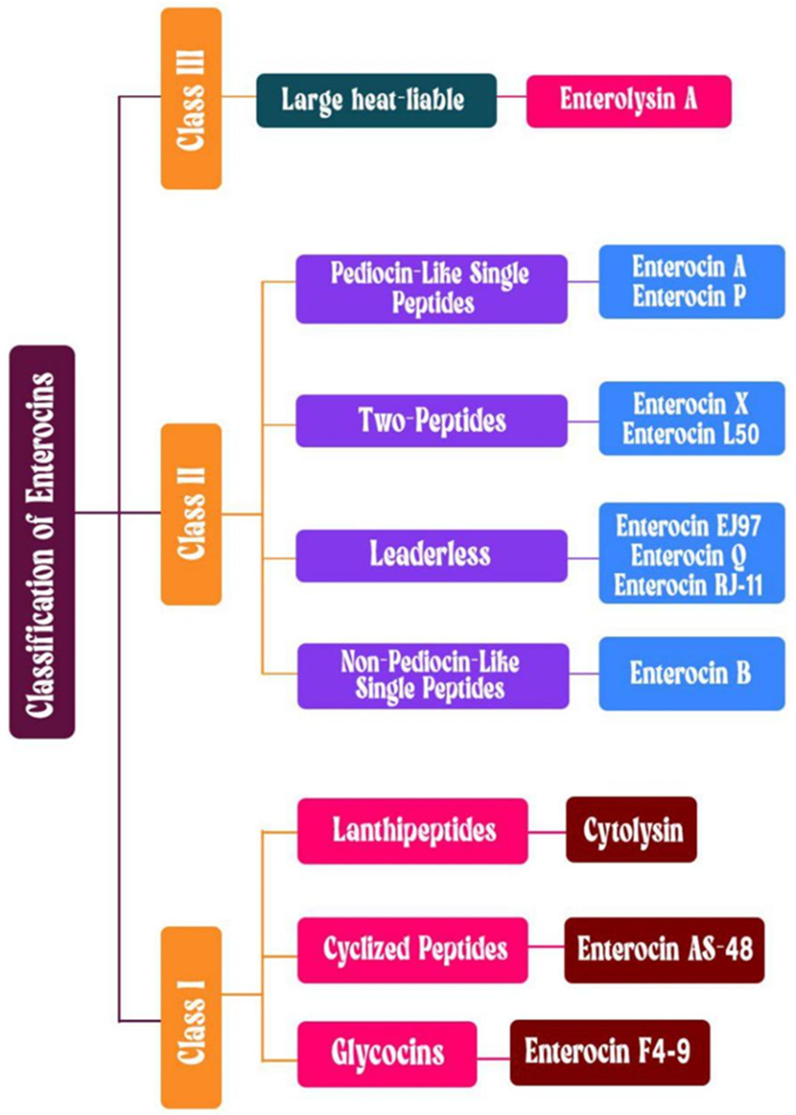
Some Enterococcus strains produce antimicrobial peptides called bacteriocins, which are secreted extracellularly and inhibit the growth of various pathogenic bacteria. The classification of bacteriocins involves multiple biochemical parameters, including structural characteristics, size distribution, specific enzymatic alterations, and distinct genetic pathways [167,168]. Bacteriocins are currently categorized into three distinct classes according to the most recent classification system (Figure 3). Class I bacteriocins are characterized by their heat-stable peptide structure, which undergoes specific modifications after translation, resulting in their distinct properties, such as enterocin AS-48, enterocin F4-9, and cytolysin. Class II includes heat-stable, unmodified peptides, which form the largest group, including enterocin A, enterocin P, enterocin DD14, enterocin K1, enterocin SEK4, enterocin L50, enterocin B, bacteriocin 32 (Bac32), and EF478. Class III comprises large, heat-labile proteins, such as enterolysin A, and Bac41 [168,169].
Cytolysin, synthesized by E. faecalis, is a hemolytic virulence factor that contributes to human disease and increased mortality [171]. The cytolysin operon contains the genes CylLL and CylLS, which code for the two distinct subunits that comprise the cytolysin molecule. These subunits are essential for the toxic activity of cytolysin, which targets mammalian cells and Gram-positive bacteria [172].
Enterocin AS-48, a circular bacteriocin also synthesized by E. faecalis, features a unique head-to-tail circular structure composed of 70 amino acids arranged into five alpha-helices. This cationic peptide disrupts bacterial membranes, resulting in cell permeabilization and subsequent cell death [46]. This compound has been shown to exhibit potent antagonism against Gram-positive pathogens. Similarly, Enterocin AS-48 exhibits enhanced activity against Gram-negative bacteria when combined with outer membrane-permeabilizing agents [173]. Enterocin AS-48 is produced during both the exponential and stationary growth phases and can be obtained in high yields when whey-based substrates are used [168].
Enterocin F4-9 contains glycosylated residues that are important for its antimicrobial activity. The enterocin F4-9 biosynthesis-related gene cluster was cloned and expressed in E. faecalis JH2-2, resulting in a peptide with improved activity against bacteria [174]. The synthesis of enterocin F4-9 involves a two-step split of the leader peptide by the EnfT protease domain and extracellular proteases. Glycosylation, mediated by enfC, is critical for both secretion and activity, whereas enfI provides immunity to producing cells; this sheds light on the minimal gene set necessary for glycocin biosynthesis and reveals a novel mechanism of glycocin production [175].
Bacteriocins 31 (bac31) and 32 (bac32) have been isolated from E. faecium strains found in dairy products, such as traditional fresh soft cheese and raw cow’s milk. Both have demonstrated activity against important foodborne pathogens, including L. monocytogenes [176]. In contrast, Bac43 was originally isolated from vancomycin-resistant Enterococcus (VRE) clinical isolates in a hospital setting and has shown a dramatic increase in prevalence, being highly associated with emerging E. faecium MLSTs in clinical cohorts over time. Despite its clinical origin, Bac43 is also active against L. monocytogenes and various Enterococcus species [177]. Additionally, a bacteriocin called Pneumolancidin, initially characterized from Streptococcus pneumoniae, has been identified through homology in patient-derived E. faecium isolates in a recent study [178]. Most bacteriocins primarily cause cell lysis by targeting the cell membrane or sugar uptake systems. Their generally narrow spectrum of activity makes them advantageous for targeted treatment or biocontrol strategies, especially in combating the continuous emergence of multidrug-resistant pathogens [179].
Some enterococcal bacteriocins exhibit broad-spectrum antagonistic activity against clinically significant pathogens, including Listeria monocytogenes, S. aureus, Escherichia coli, and Salmonella spp. (Table 2). Their bactericidal mechanisms are diverse and target-specific, including membrane permeabilization and leakage of bacterial cellular content (characteristic of pediocin-like variants), inhibition of cell wall biosynthesis via lipid II binding (exhibited by lantibiotic-class enterocins), enzymatic degradation of peptidoglycan layers (demonstrated by large bacteriolysins such as enterolysin A), and disruption of essential cellular processes, including nucleic acid metabolism and translation machinery [180,181].
Bacteriocins have significant potential as natural preservatives in fermented food systems, particularly in dairy applications (Table 2). These antimicrobial peptides show synergistic activity when combined with other lactic acid bacteria, making them effective components in multi-strain fermentation cultures [121,182]. Specific variants, including enterocins A, B, and AS-48, display potent inhibitory effects against common foodborne pathogens such as Salmonella enterica and E. coli, effectively controlling their growth in food matrices. Furthermore, enterocins can be combined with bacteriophages for enhanced pathogen control. For example, Baños et al. demonstrated the use of enterocin AS-48 in combination with phage P100 to suppress the proliferation of L. monocytogenes in fish [183]. Various studies have shown the strong antimicrobial properties of enterocins against pathogens such as Streptococcus pyogenes, S. suis, Clostridium perfringens, E. cecorum, E. faecalis, Pseudomonas aeruginosa, Campylobacter coli, Bacillus cereus, and S. aureus, highlighting their potential for combating foodborne illnesses caused by these pathogens [184,185].
Table 2. Enterococci bacteriocins and their potential applications.
BacteriocinProducing Enterococcus StrainMode of ActionModelEffectsReference
Microbiome modulation
Enterocin (Ent7420)E. faecium CCM7420 (EF2019)Stimulating the host’s immune reactionBroiler rabbitsImmune-regulating effects.
Antibacterial and anticoccidial effect. Improves serum biochemistry parameters, immunity, jejunal morphology, weight gains, feed conversion ratio, and meat quality
[186]
Enterocin A/PE. faecium P13Optimizes immune activity and gut flora compositionRabbitsSuppression of gut pathogens combined with immune-modulatory influence[169]
Bio-preservation/food safety
Enterocin AS-48E. faecium EK13 (CCM 7419)Cytoplasmic membrane perturbationCanned fruits and vegetablesInhibit Bacillus coagulans in canned fruit and vegetable foods[187]
Enterocin AS-48E. faeciumCreation of nanopores in the lipid bilayerRice-Based FoodsInhibition of Toxicogenic Bacillus cereus[173]
Enterocin F4-9E. faecalis F4-9Suppression of bacterial growthIn vitroFoodborne pathogens[175]
Enterocin AS-48E. faecalisPore formation in the cell membraneBlueberriesReduction in the viable counts of Enterobacteriaceae, Salmonella, and coliforms[188]
Enterocin-like
substances
E. faecium X2893 and X2906Not includedIn vitroAntimicrobial activity against poultry-associated Clostridium perfringens[189]
Enterocin BE. lactis 4CP3Disintegration of cellular envelopeIn vitroC. perfringens, L. monocytogenes, Propionibacterium spp., C. sporogenes, and C. tyrobutyricum[168]
Enterocin SEK4E. faecalis K-4Pore formation in cell membrane of
target organisms
In vitroC. perfringens, B. subtilis, and L. monocytogenes[168]
Enterocin LD3E. hirae LD3Cell membrane disruptionFruit juiceInhibition of Salmonella enterica subsp. enterica serovar Typhimurium ATCC 13311 in fruit juice[190]
Enterocin TJUQ1E. faecium TJUQ1Disrupts cell membrane, leading to cell lysisIn vitroAntimicrobial activity against L. monocytogenes CMCC 1595[191]
Enterocin CCM 4231E. faecium CCM 4231Formation of pores in cell membraneIn vitroS. aureus and L. monocytogenes[192]
Enterocin KT2W2GE. faecalis KT2W2GCell Membrane DepolarizationBanana peelsInhibits the growth of spoilage microorganisms isolated from spoiled banana peel.[193]
Enterocin ABPE. faecium KE82Not includedCheese production plantEnhances the inactivation of L. monocytogenes during the production of cheeses[194]
Enterocin DD14E. faecalis 14Disrupts cell membranes and cell walls, causing cytoplasmic leakage and cell death.In vitro and in vivoAntimicrobial activity against L. monocytogenes, C. perfringens, Enterococcus faecalis, and methicillin-resistant Staphylococcus aureus.[180]
Enterocin PBioengineered Pichia pastorisInteracts with negatively charged bacterial membranes, causing cell death.In vitroInhibition of Gram-positive pathogens, including L. monocytogenes, S. aureus, and Clostridium spp.[195]
Enterocin AE. faecium T136Formation of pores in the cell membraneIn vitroAnti-L. monocytogenes activity[181]
Enterocin HDX-2E. faecium HDX-2Disrupts membrane integrity, leading to cell lysis.In vitroInhibition of foodborne pathogens and spoilage bacteria.[196]
Enterocin L50E. faecium L50Membrane disintegration leading to cell deathIn vitroAnti-C. perfringens activity[190]
Bacterial infections
Enterocin A/PE. faecium EK13Immunoglobulin A (IgA) productionRabbitsImproves rabbits’ growth, immunity, and growth of rabbits, and therapeutic potential against staphylococcal infections[169]
Enterocin 416K1E. casseliflavus IM 416K1Cell membrane disruptionIn vitroAntimicrobial activity against L. monocytogenes[168]
EntK1 and EntEJ97E. faeciumDecrease in cell membrane integrityMiceTreatment of systemic vancomycin-resistant enterococci infections[197]
Enterocin LD3E. hirae LD3Cause efflux of intracellular ions and decrease in cell membrane integrityIn vitroAntimicrobial activity against Gram-negative bacteria[198]
Enterocin OS13E. faecalis OS13Formation of pores in the cell membraneIn vitroInhibitory activity against nosocomial enterococci[199]
Enterolysin AE. durans NT21Cytoplasmic membrane perturbationIn vitroAntimicrobial activity against S. aureus, M. luteus, Salmonella Typhi,
and E. faecalis
[200]
Enterocin E20CE. hirae 20CCell membrane disruptionIn vitroAntimicrobial activity against S. enterica[201]
Durancin 16AE. durans 16AMembrane perforationIn vitroInhibition of C. difficle, S. aureus, and Enterococcus (vancomycin-resistant strains)[167]
Enterocin AS-48E. faecalisDisintegration of cellular envelopeIn vitroAntimicrobial activity against Mycobacterium tuberculosis[202]
Antiviral
Enterocin CRL 35E. mundtii CRL35suppression of the late stages of replicationIn vitroInhibition of Herpes Simplex Virus-1 and Herpes Simplex Virus-2[203]
Enterocin GEn17E. duransSuppression of viral genome replicationIn vitroInhibition of poliovirus (PV-1) and herpes simplex virus 1 (HSV-1)[204]
Enterocin AAR-71E. faecalis AAR-71Suppression of viral genome replicationIn vitroAntiviral inhibitory activity and immune system stimulation[205]
Bacteriocin ST5HaE. faecium ST5HaInhibition of the key replication cycleIn vitroAnti-herpes simplex virus type 1 (HSV-1) activity[206]
EntDD14E. faecalis 14Disruption of virus assemblyIn vitroAntiviral activity against HSV[180]
Antifungal
Enterocin UNAD 046E. faecalisLoss of cell wall integrityIn vitroActivity against Aspergillus niger, Fusarium oxysporum, Pythium ultimum, and Penicillium expansum[207]
Enterocin A, B, and PE. lactis 4CP3Decreased mitochondrial membrane potentialIn vitroAntifungal activity against A. niger A79, Bacillus thuringiensis, F. equiseti F97[208]
Enterocin CHQSE. faecalis TG2Peptidoglycan disruption with concurrent oxidative stressIn vitroAntifungal activity against Candida albicans[209]
Enterocin Gr17E. faecalisLoss of cell wall integritySalmon filletsAntifungal activity, enhances sensory properties and shelf life of liquid-smoked salmon fillets[210]
Anti-cancer
Enterocin 12AE. faeciumPermeabilization of the cancer cell plasma membraneIn vitroAnticancer activity against human colon, gastric (HT-29, Caco-2, and AGS), cervical (HeLa) cancer cells[211]
Enterocin AE. faecium (por1)Triggering late-stage cell death and cell cycle arrestIn vitroActivity against gastric cancer cell lines[212]
Enterocin (LNS18)E. thailandicusCancer cell plasma membrane porationIn vitroActivity against HepG2 cancer cells[213]
Enterocin PE. faecium P13Attenuation of oncocytic membrane porosityIn vitroActivity against cancer cell lines (SW1353, HUH7, Huh-7.5, C26, B16F0)[214]
Anti-protozoan
Enterocin AS-48E. faecalis strain UGRA10Selective mitochondrial targeting and the formation of highly reactive oxygen molecules (ROS).In vitroActivity against Leishmania spp.[215]
Enterocin AS-48E. faecalis UGRA10Mitochondria depolarizationMiceEffective for the treatment of Chagas’ disease[216]
Enterocin AS-48E. faecalis UGRA10Affects and destroys cellular compartmentsIn vitroAutophagic-related Trypanosoma brucei inhibition[217]
Durancin-like
and Enterocin M
E. durans ED26E/7Formation of pores in cytoplasmic membraneIn vitro and miceAmelioration of Trichinella spiralis-associated infections[218]
Research has revealed distinct antimicrobial profiles among bacteriocin variants. The bacteriocin CRL35 exhibits potent listericidal activity as a standalone antimicrobial agent. In contrast, the engineered chimeric peptide Ent35-MccV displays broad-spectrum efficacy against resistant clinical strains, particularly E. coli isolates from healthcare settings [219,220]. Another significant antagonistic effect is observed when enterocin Durancin 61A is combined with reuterin against pathogens like Clostridium difficile, indicating its potential for treating gastrointestinal infections [167]. Emerging research also highlights the therapeutic potential of some enterocins against antibiotic-resistant pathogens. For example, durancin 61A demonstrates inhibitory activity against clinical MRSA isolates, exhibiting both standalone efficacy and synergistic effects with vancomycin therapy [167]. Complementary studies have identified enterocins DD28 and DD93 as promising candidates for treating MRSA infections, suggesting their potential application in antimicrobial strategies [221].
Contemporary research has focused on optimizing bacteriocins through chemical engineering to increase their pharmaceutical potential [129]. With this approach, Ross et al. developed novel antimicrobial peptides by structurally mimicking the α-helical domain of enterocin AS-48 [71]. Their engineered peptide library demonstrated potent antimicrobial efficacy across multiple bacterial strains, with certain variants exhibiting significant bactericidal activity at nanomolar concentrations while maintaining excellent human cell biocompatibility [222]. In light of the escalating antimicrobial resistance crisis, these findings position modified enterocins as promising candidates for next-generation therapeutics, warranting expanded investigation into their clinical applications (Table 2).

4. Safety Concerns Associated with the Use of Enterococcus spp.

4.1. Pathogenic Potential of Enterococcus spp.

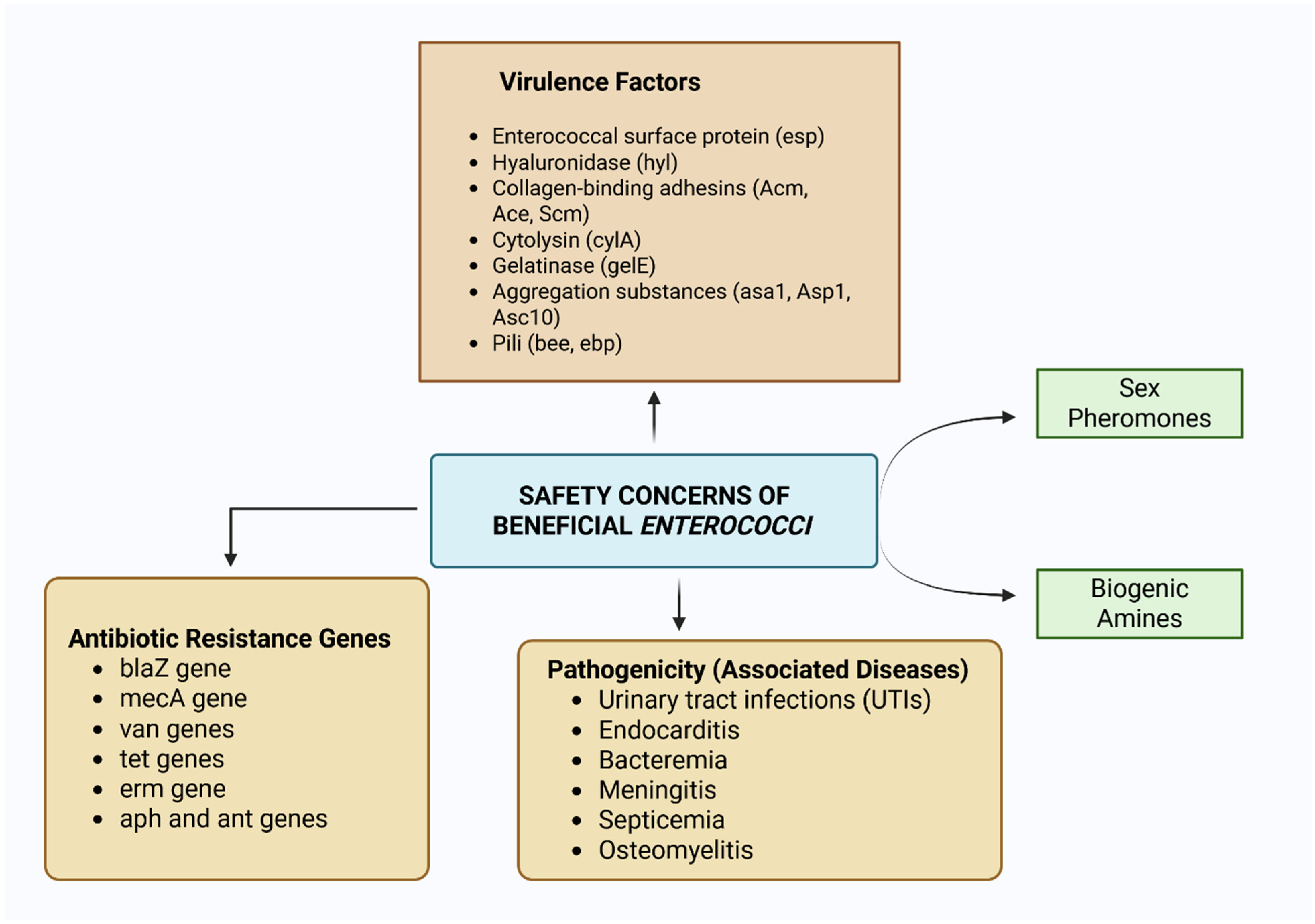
Apart from the beneficial ecological roles played by different enterococci, some strains of Enterococcus spp. are widely recognized as clinically important pathogens, raising safety concerns about their use as probiotics (Figure 4 and Figure 5). Within healthcare settings, E. faecalis and E. faecium have become predominant nosocomial threats, with distinct pathogenic profiles. While E. faecalis demonstrates greater virulence potential, E. faecium shows a heightened propensity to develop multidrug resistance, particularly against glycopeptide antibiotics such as vancomycin [223,224].
Enterococcal infections primarily manifest in vulnerable patient populations, including immunodeficient individuals, critically ill patients, and those undergoing intensive medical interventions. Extended antimicrobial therapy and impaired host defenses represent key predisposing factors for nosocomial acquisition of drug-resistant enterococcal strains. These pathogens demonstrate remarkable clinical versatility and can induce diverse pathological states, ranging from catheter-associated urinary tract infections to life-threatening systemic conditions, such as endocardial infections, bloodstream infections, neonatal sepsis, infections following surgery or burns, and meningitis [224,225].
A global monitoring report from over 40 nations pinpointed Enterococcus as a major pathogen associated with hospital-acquired diseases [226]. E. faecium was identified as the second and third most common pathogen associated with bloodstream and urinary catheters in patients from Germany [224]. Clinical surveillance at a major women’s specialty hospital in southwestern China identified E. faecalis as the secondary etiological agent in postoperative infections following gynecological surgeries, ranking only behind S. aureus in incidence frequency [227].

4.1.1. Enterococcus-Associated Urinary Tract Infections

Urinary tract infections represent a persistent global health challenge that affects populations of all ages and sexes. Although E. coli remains the predominant uropathogen, responsible for approximately four-fifths of all cases, Enterococcus spp. have gained recognition as important secondary etiological agents, collectively accounting for a substantial proportion of the remaining infections [228].
Recent epidemiological investigations have documented an increasing role of Enterococcus spp. in the pathogenesis of urinary tract infections. A 2023 meta-analysis by Noor et al. established multiple comorbidities and iatrogenic factors predisposing patients to enterococcal UTIs, notably diabetes mellitus, chronic renal impairment, malignancy, cerebrovascular accidents, and instrumentation with urinary catheters. The study further revealed that extended hospitalization (>14 days) and prior broad-spectrum antimicrobial therapy were significantly correlated with vancomycin-resistant enterococcal urinary infections [229]. Another study on a 72-year-old patient with urinary tract infection symptoms caused by Enterococcus avium and Escherichia coli showed incomplete resolution with initial antibiotic therapy (cefuroxime), as persistent haematuria was later attributed to a bleeding bladder lesion and staghorn kidney stones. Full recovery was achieved only after surgical resection of the lesion and removal of the stones, thus emphasizing that management of infections caused by rare enterococci may require both targeted antimicrobial therapy and procedural intervention [230].
The uropathogenicity of Enterococcus spp. stem largely from two key biological attributes: their capacity for biofilm development and for secreting virulence determinants. Biofilm architecture enables these microorganisms to evade host immune clearance while simultaneously enhancing antimicrobial tolerance [223]. This complex developmental process progresses through distinct phases: beginning with surface adherence and progressing to clustered cellular aggregation, followed by extracellular polymeric substance synthesis to form a mature biofilm structure, and culminating in cellular detachment for systemic spread. Urinary catheter surfaces serve as optimal substrates for Enterococcus biofilm development, often leading to catheter-associated urinary tract infections [231]. The pathogenic mechanisms of these biofilm-forming uropathogenic strains operate through several synergistic pathways: (1) generation of dense extracellular polymeric matrices that impede antimicrobial penetration, (2) horizontal acquisition of genetic resistance determinants, and (3) polymicrobial colonization with species such as E. coli that can potentiate virulence through immunomodulatory effects and synergistic pathogenicity enhancement [232].

4.1.2. Enterococcus-Associated Bloodstream Infections

Bloodstream infections caused by Enterococcus spp. (EBSIs) have become a critical healthcare challenge, presenting considerable clinical and therapeutic difficulties. While these bacteria normally colonize the intestinal tract and contribute to gut microbial balance, they can translocate across the intestinal epithelium under specific conditions, resulting in systemic infections with severe clinical consequences [233].
The pathogenesis of Enterococcus-associated bloodstream infections (EBSIs) is strongly linked to immunosuppression resulting from comorbidities, prolonged hospitalization, or invasive medical interventions. Key predisposing factors include: immunocompromised states secondary to chronic diseases (e.g., diabetes mellitus, inflammatory bowel disorders), iatrogenic factors such as indwelling catheters or prolonged intravenous access, and extended inpatient care duration, which increases exposure to nosocomial pathogens [54].
Clinical data identify E. faecalis and E. faecium as the principal etiological agents of enterococcal bloodstream infections. Although E. faecalis has a higher incidence, E. faecium infections typically present greater clinical severity. This heightened virulence profile stems largely from E. faecium’s enhanced capacity to acquire antimicrobial resistance, which frequently complicates therapeutic management and may lead to poorer patient outcomes [234]. Untreated enterococcal bloodstream infections can progress to severe systemic complications, including inflammatory endocardial disease, persistent bacteremia, central nervous system infections, bone marrow inflammation, and systemic inflammatory response syndrome. Research indicates that intestinal microbial dysbiosis and subsequent enterococcal overgrowth facilitate bacterial translocation across mucosal barriers, enabling hematogenous dissemination to distant anatomical sites and the development of these high-mortality conditions [235,236,237]. Recent genomic investigations by Chaguza et al. have elucidated the contribution of hereditary determinants to the virulence potential of E. faecalis. Their analysis revealed that the bacterial genotype accounts for 30–40% of the observed variability in clinical manifestations, significantly influencing both tissue tropism (particularly extraintestinal colonization) and epidemiological distribution patterns (with distinct strain predilection for nosocomial transmission). These findings underscore the fundamental role of microbial genomics in determining pathogenic behavior [238].
In addition to the predominant E. faecalis and E. faecium, several less common Enterococcus spp. have been implicated in bloodstream infections, including E. casseliflavus, E. durans, E. hirae, E. avium, and E. raffinosus, with most cases representing rare clinical occurrences [239,240]. Notably, E. casseliflavus is intrinsically resistant to vancomycin, necessitating alternative therapeutic approaches. For example, A clinical review of two E. casseliflavus bacteremia cases in hemodialysis patients with chronic renal failure highlighted the role of underlying comorbidities in facilitating invasive infection. Both cases did not respond to vancomycin therapy but responded rapidly to targeted daptomycin-based combination therapy (with imipenem or cefepime), underscoring the therapeutic potential of combination regimens and the need for heightened awareness of intrinsically vancomycin-resistant Enterococcus in bloodstream infections complicating chronic illness [241]. Another reported case of concomitant Enterococcus casseliflavus bacteremia and Raoultella planticola biliary colonization occurred in a 74-year-old male with pancreatic adenocarcinoma and ascending cholangitis following biliary stent placement. Blood cultures confirmed E. casseliflavus (intrinsically vancomycin-resistant via VanC), while bile cultures yielded E. casseliflavus, R. planticola, Enterobacter cloacae, and Candida albicans. The patient improved with targeted therapy including cefepime, daptomycin, metronidazole, and fluconazole, underscoring the opportunistic pathogenic potential of these rare organisms in gastrointestinal malignancy and the importance of accurate species identification to guide therapy [242]. Current evidence supports the efficacy of β-lactams (particularly ampicillin), lipopeptides (daptomycin), and carbapenem-class antibiotics against this species [243].

4.1.3. Enterococcus-Associated Central Nervous System (CNS) Infections

Enterococcal CNS infections predominantly affect vulnerable patient populations with neurological compromise, including those with recent neurosurgical interventions, structural CNS abnormalities (e.g., ventriculoperitoneal shunts, traumatic defects), and pre-existing neurological disorders. While enterococcal meningitis and ventriculitis (EMV) remain uncommon, their incidence has risen notably in pediatric inpatient settings, where invasive devices and compromised barriers facilitate microbial CNS invasion [244].
A decade-long retrospective analysis by Schnapp et al. systematically evaluated pediatric EMV cases, characterizing etiological patterns, predisposing conditions, and therapeutic challenges. The investigation revealed two predominant risk categories: iatrogenic factors, particularly cerebrospinal fluid diversion hardware (ventricular shunts and drainage systems), and developmental vulnerabilities, including premature birth. Notably, antimicrobial resistance has emerged as a critical concern, with vancomycin nonsusceptibility observed in one-fifth of the clinical strains. Rare but clinically important reports expand the pathogenic spectrum beyond E. faecalis and E. faecium. For example, the first documented case of E. casseliflavus meningitis occurred in an immunosuppressed elderly patient, where intrinsic vancomycin resistance precluded standard therapy; however, meropenem plus ampicillin–sulbactam achieved recovery [245]. Likewise, E. gallinarum, a typically poultry-associated species, caused meningitis in a 53-year-old man with alcohol abuse, who was successfully treated with high-dose ampicillin and gentamicin [246]. In pediatrics, E. gallinarum group meningitis was reported for the first time following bowel perforation from a ventriculoperitoneal shunt; its intrinsic vancomycin resistance necessitated a 6-week targeted intravenous regimen, highlighting the need for culture-guided therapy in shunt-related CNS infections [247]. A 2016 clinical investigation by Patel and colleagues demonstrated superior therapeutic outcomes with direct intraventricular vancomycin injection for enterococcal meningitis compared with conventional intravenous administration. This finding underscores the critical influence of the administration route on the treatment efficacy for central nervous system infections [248].

4.1.4. Enterococcus-Associated Animal and Zoonotic Infections

Some enterococci, especially E. faecalis and E. faecium, continue to be recognized as important pathogens in both animal health and zoonotic transmission [225,231]. In animals, these enterococcal species are associated with conditions such as UTI, mastitis, wound infections, and septicemia [225]. Companion animals, including dogs and cats, frequently harbor multidrug-resistant (MDR) strains, with studies reporting high rates of resistance to antimicrobials such as tetracycline and erythromycin [249]. The close interaction between pets and humans increases the risk of bidirectional transmission, making companion animals important reservoirs of resistant enterococcal strains [54]. Livestock farming further amplifies these concerns, as E. faecalis and E. faecium are prevalent in poultry and swine, often demonstrating less sensitivity to critical antibiotics such as vancomycin [225]. For example, E. cecorum, an emerging pathogen in poultry, is associated with systemic infections and skeletal disorders, resulting in substantial economic losses [250]. In pig farms, resistant strains not only compromise animal health but also contribute to the dissemination of AMR in farming environments [251]. Extensive use of antimicrobials in agriculture exerts selective pressure, enabling the survival and spread of resistant strains. These resistant strains can contaminate foods of animal origin, thereby increasing the risk to humans through dietary exposure [6,44]. The impact of enterococcal infections is not limited to terrestrial animals. Wildlife, particularly marine species such as sea turtles, has also been affected, with cases of septicemia and osteomyelitis caused by E. faecalis linked to environmental contamination [252]. This highlights the adaptability of enterococci, which can persist across diverse ecosystems and pose risks to multiple species.
Zoonotic infections caused by pathogenic enterococci pose significant challenges, particularly for immunocompromised individuals, where conditions such as bacteremia and endocarditis may become life-threatening [253]. For instance, a rare case of E. casseliflavus endophthalmitis following a pig-related eye injury illustrates its zoonotic potential; the organism’s intrinsic vancomycin resistance limits first-line empiric therapy, but intravenous ampicillin and dexamethasone improved symptoms, and systemic linezolid enhanced visual acuity, although intravitreal amikacin remains an important option when oral linezolid is insufficient [254]. Similarly, a recent report described a diabetic foot ulcer infection caused by E. avium in a patient with documented poultry exposure, complicated by osteomyelitis and recurrent bacterial colonization. This case required combined surgical intervention and tailored antibiotic therapy guided by microbial monitoring, underscoring the need to consider zoonotic Enterococcus species in clinical diagnoses, especially among immunocompromised or chronically ill patients [255]. The increasing prevalence of such infections highlights the urgent need for a One Health approach, fostering collaboration across human health, veterinary medicine, and environmental management to address the complex interactions that facilitate zoonotic transmission of enterococci [225].

4.1.5. Foodborne Enterococcus Infections

Enterococci serve as important microbial indicators in food safety systems, reflecting possible fecal contamination and inadequate sanitary controls during manufacturing [23]. Although they are not typical causative agents of foodborne disease, their presence in food products suggests potential co-contamination with enteric pathogens, including Salmonella, E. coli, and L. monocytogenes, which can trigger acute foodborne disease outbreaks [256]. A 2024 Kenyan surveillance study found Enterococcus contamination in 50% of sampled infant foods, indicating widespread fecal pollution in domestic food-preparation environments [257]. While epidemiological analysis failed to identify a singular contributing factor for this contamination level, the frequent detection of these indicator organisms strongly suggests inadequate hygiene protocols during food production, storage, or handling. Although most enterococci rarely cause foodborne illness, their continued presence requires the immediate adoption of improved food safety measures to prevent possible pathogen transmission [258].

4.2. Virulence Factors Associated with Enterococcus spp.

Enterococcus spp. employ diverse virulence determinants that increase their pathogenicity in their hosts. While some virulence determinants are strain-specific, others are conserved across strains within the genus. These determinants often facilitate host tissue invasion, immune evasion, and intracellular persistence, enabling colonization of multiple anatomical sites in the infected host. Key virulence mechanisms include: host cell adhesion (mediated by Asa1, efaA, and ace), biofilm formation (esp, gelE), cytolytic activity (cylA), and tissue penetration (hylA). Collectively, these factors subvert host defenses, damage cellular structures, and promote enterococci infectivity [227].
Asa1-encoded aggregation substance (AS) is a multifunctional virulence factor that mediates biofilm development and conjugative plasmid transfer, thereby enhancing the dissemination of antimicrobial resistance [101]. It also evades the immune system by binding to neutrophils independently of opsonins, impairs phagocytic clearance, and enhances macrophage adhesion, thereby promoting intracellular persistence [259]. This role in both horizontal gene transfer and host defense subversion underscores AS as a critical pathogenicity determinant in E. faecalis infections.
Enterococcal surface protein (Esp) is a critical virulence determinant that facilitates biofilm formation, host tissue adhesion, and the transfer of antibiotic resistance determinants [95]. Experimental studies in murine models have demonstrated the dual pathogenicity of Esp, which enables intestinal colonization and cardiac valve adherence, promotes endocarditis pathogenesis, and is indispensable for biofilm formation. While Esp-positive E. faecalis strains consistently form biofilms within their niches, Esp-negative variants are unable to form biofilms or establish structured communities. These findings position Esp as a master regulator of enterococcal persistence and dissemination [228,260]. Similarly, the efaA virulence gene, which exists as strain-specific variants efaAfs (E. faecalis) and efaAfm (E. faecium), is highly linked with endocarditis pathogenesis [21,261]. The EfaA surface antigen functions as a critical adhesin in E. faecalis, exhibiting host-specific expression patterns. This manganese acquisition protein demonstrates serum-inducible characteristics, showing preferential production during blood culture compared with standard media. As a component of the manganese transport system, EfaA facilitates essential nutrient uptake during bloodstream infection when manganese demand is elevated. Its expression is tightly regulated by the EfaR repressor protein, which downregulates EfaA production under manganese-replete conditions [262]. Dysfunctional EfaR regulation not only disrupts manganese homeostasis but also impairs biofilm formation and intracellular survival within phagocytic cells. This has been documented in Enterococcus-associated endocarditis, where E. faecalis adheres to the cardiac endothelium and forms antibiotic-resistant biofilms due to EfaA, which is essential for both biofilm maturation and persistent colonization of heart tissue [263]. These adaptive mechanisms significantly increase bacterial virulence, directly contributing to E. faecalis capacity to establish and maintain endocardial infections [223].
The Ace-encoded adhesin mediates E. faecalis attachment to host collagenous matrices, facilitating the colonization of biomedical implants and host tissues [259]. While both Ace and Acm proteins exhibit structural homology with microbial surface components that recognize adhesive matrix molecules (MSCRAMMs), Ace proteins demonstrate unique thermosensitive binding properties, with optimal collagen affinity at 46 °C, a temperature exceeding physiological levels [22,264]. This paradoxical behavior may reflect competitive interactions with gelatinase, a collagen-degrading enzyme produced by certain E. faecalis strains that counteract adhesion [265]. Another important virulence determinant often found in clinical E. faecalis (specifically E. faecalis subsp. liquefaciens is the GelE metalloprotease. GelE subverts innate immunity by targeting the cleavage of the complement component C3, which impairs opsonization and subsequent phagocytic clearance by neutrophils and macrophages, enabling immune evasion [266].
The cylA gene is part of a six-gene operon (cylLL, cylLS, cylM, cylB, cylA, and cylI) that coordinately regulates the biosynthesis and activation of cytolysin, a potent exotoxin capable of lysing both prokaryotic and eukaryotic cells [267]. This toxin contributes to Enterococcus pathogenicity through multiple mechanisms, including host cell lysis, which targets erythrocytes (causing anemia), destroys leukocytes and macrophages (impairing immune defenses), and induces parenchymal tissue damage in vital organs [95,268]. They are also prevalent in clinical strains, and are correlated with severe infections, promoting tissue necrosis at infection sites and amplifying inflammatory cascades through the release of DAMPs [266].

4.3. Antibiotic Resistance in Enterococcus spp.

The antimicrobial resistance (AMR) trajectory of Enterococcus spp. predate their formal taxonomic classification [269]. Initial therapeutic strategies relied on β-lactam monotherapy, which progressively lost efficacy due to the development of resistance. This prompted the adoption of synergistic β-lactam/aminoglycoside regimens, although their use was similarly compromised by emerging resistance mechanisms. The Subsequent dependence on vancomycin as a last-line agent was undermined by the first documented vancomycin-resistant Enterococcus (VRE) isolates in 1989 [8]. Alarmingly, each newly introduced anti-enterococcal agent has experienced rapid emergence of resistance, creating an escalating therapeutic crisis in the management of these infections [44,113].
Enterococcus spp. exhibit two distinct antimicrobial resistance (AMR) profiles: intrinsic (encoded by conserved chromosomal genes present across all strains) and acquired (mediated by mobile genetic elements or mutations). The innate resistance spectrum of these bacteria includes β-lactams (cephalosporins, penicillins), aminoglycosides, and macrolides [269,270]. For example, E. gallinarum and E. casseliflavus are intrinsically resistant to vancomycin and have shown high-level aminoglycoside resistance [271]. These vancomycin-resistant enterococci (VRE) have been isolated from animal feces and foods [272,273]. Acquired resistance, however, arises primarily through horizontal gene transfer via plasmids or transposons, or via mutations with high transfer potential—a process predominantly facilitated by conjugation [274]. A study on meats from Ghana detected E. hirae, E. casseliflavus, E. gallinarum, E. thailandicus, and E. lactis carrying multiple resistance genes, including those for tetracyclines (tet(M), tet(L), tet(S)), macrolides (erm(B), erm(T)), and aminoglycosides (aac(6’)-Ii, aph(3’)-III), among others. Mobile genetic elements, such as plasmids (rep1, rep2, repUS43) and transposons (Tn917, Tn6009), were found to co-occur with these genes, suggesting a potential for horizontal gene transfer [60].
The common genes that confer AMR in Enterococcus include the following:
blaZ gene: The blaZ gene, which mediates β-lactam resistance through enzymatic antibiotic inactivation, has been increasingly identified in Enterococcus spp. despite its historical association with S. aureus [98,231]. This horizontal gene transfer event suggests the intergeneric dissemination of resistance determinants. Epidemiological surveillance has detected blaZ-carrying Enterococcus strains across diverse reservoirs, including human clinical isolates, livestock-associated samples, and food production chains [275]. blaZ expression is often regulated by a coordinated system involving blaI (repressor), blaR1 (signal transducer), and blaR2 (auxiliary regulator) proteins, which collectively modulate β-lactamase production in response to antibiotic exposure [276,277]. The blaZ gene was identified in 9.6% of Enterococcus isolates from clinical samples [278] and in 48.1% of strains from fish and seafood [279]. Similarly, strains from nestling ospreys (Pandion haliaetus) and other wildlife species harbor blaZ resistance genes, highlighting the widespread prevalence of this resistance gene across diverse environments and its role in the dissemination of antibiotic resistance [280].
mecA gene: The mecA gene encodes penicillin-binding protein 2a (PBP2a), a modified transpeptidase with reduced binding affinity for β-lactam antibiotics, thereby conferring resistance to this drug class [269]. Although it is historically associated with Staphylococcus spp, the increasing detection of mecA in diverse Enterococcus strains provides compelling evidence of interspecies horizontal gene transfer, a phenomenon that significantly expands the antibiotic resistance potential of these opportunistic pathogens [269,281]. Emerging evidence indicates that mecA-positive Enterococcus strains may drive nosocomial infections through parallel pathogenic mechanisms to methicillin-resistant Staphylococcus spp. A recent clinical survey revealed methicillin resistance in 85% of the analyzed enterococcal isolates, with 95.5% testing positive for mecA carriage. Furthermore, while mecA dynamics in enterococci remain understudied, these findings suggest its potential role in propagating methicillin resistance across bacterial populations via horizontal gene transfer—a concerning prospect for antimicrobial resistance containment in healthcare environments [33].
Van genes: These genetic determinants mediate glycopeptide resistance through enzymatic alteration of peptidoglycan precursors, specifically by modifying the D-alanyl-D-alanine target site to reduce vancomycin binding affinity. The resulting structural changes in cell wall biosynthesis render glycopeptide antibiotics, such as vancomycin, therapeutically ineffective [277]. Among the various van operons, vanA and vanB are the most prevalent, with E. faecalis and E. faecium being their primary carriers [21]. VanA and vanB confer resistance by modifying the bacterial peptidoglycan, replacing the D-Ala-D-Ala terminal with D-Ala-D-Lac, which prevents vancomycin from binding and inhibiting cell wall synthesis. The VanC-mediated resistance mechanism operates through a distinct biochemical pathway, substituting the conventional D-Ala-D-Ala peptidoglycan precursor with D-Ala-D-Ser at an approximate 3:1 ratio. This genetic determinant is primarily observed in motile Enterococcus spp., including E. casseliflavus, E. gallinarum, and E. flavescens, representing an intrinsic resistance phenotype that differs fundamentally from acquired vancomycin resistance mechanisms [37]. While vancomycin resistance determinants, such as vanD, vanE, vanG, vanL, vanM, and vanN, occur sporadically in Enterococcus, surveillance of 40 commercial probiotic formulations for livestock revealed interesting findings. The vanA gene, a high-level vancomycin resistance marker, was detected in these products, with its plasmid- and transposon-borne location facilitating potential horizontal transfer to pathogenic species. This mobility underscores the risk of resistance dissemination through the use of probiotics in agriculture [282].
Studies have reported that most nasal Enterococcus isolates harbor multidrug resistance, including high-level aminoglycoside and glycopeptide resistance. Vancomycin-resistant enterococci (VRE), particularly E. faecium with VanA and VanB genotypes, are prevalent in healthcare settings and are increasingly detected in nasal samples of patients and in animal reservoirs (e.g., parrots and rabbits) [32]. Altered nasal microbiota composition, involving Enterococcus species, is associated with chronic rhinosinusitis and nasal polyps, where impaired mucosal barrier function and reduced antimicrobial peptides promote bacterial invasion and inflammation. Nasal colonization with multidrug-resistant (MDR) Enterococcus, especially VRE, is linked to increased risk of invasive infections in immunocompromised and ICU patients [34]. Surveillance data from England show a plateau in Enterococcus bloodstream infections but rising vancomycin resistance in E. faecium isolates, emphasizing ongoing clinical challenges. Furthermore, co-colonization of the nasal cavity with S. aureus facilitates the exchange of resistance genes, thereby potentiating nosocomial outbreaks [283].
Tet genes: Tetracycline resistance in Enterococcus is mediated by diverse genetic determinants encoding two primary resistance mechanisms: (1) ribosomal protection proteins (e.g., Tet(M), Tet(O), and Tet(S)) that safeguard the translation machinery, and (2) efflux pumps (e.g., Tet(K), and Tet(L)) that actively export tetracyclines from bacterial cells [269,284]. Ribosomal protection mediated by tet(M) represents the predominant tetracycline resistance mechanism in Enterococcus, although tet(L)-encoded efflux also contributes substantially to resistance profiles. Notably, the co-occurrence of tet(M) and tet(L) confers synergistic high-level resistance [285]. Both genes exhibit mobility and can be transferred interbacterially via plasmid-independent mechanisms, thereby facilitating rapid dissemination of resistance even in the absence of conjugative elements. The dissemination of tetracycline resistance genes in Enterococcus is facilitated primarily by conjugative transposons of the Tn916/Tn1545/Tn5397 family [269]. Among these mobile genetic elements, Tn916 and Tn5397 have the highest epidemiological prevalence, whereas Tn1545 occurs less frequently and is typically found only in association with other transposons of this family [285,286].
Erm genes: Macrolide resistance in Enterococcus is mediated by rRNA methylase enzymes encoded by erm genes (particularly erm(A), erm(B), and erm(C)), which dimethylate the 23S rRNA target site. This modification sterically hinders macrolide binding, conferring resistance to antibiotics such as erythromycin. Notably, these resistance determinants exhibit high mobility through transposon-mediated horizontal gene transfer. Enterococcus spp demonstrate additional macrolide resistance through mef- and msr-encoded efflux pumps [287]. E. faecalis exhibits intrinsic resistance to lincosamides (clindamycin) and streptogramins (quinupristin-dalfopristin) via the lsa efflux determinant. This species-specific resistance mechanism is chromosomally encoded in E. faecalis but absent in other enterococci, highlighting unique evolutionary adaptations within this taxon [37].
Aminoglycoside-modifying enzymes-encoding genes: Aminoglycoside resistance in Enterococcus is mediated by enzymatic modification through three primary gene families: aminoglycoside phosphotransferases (aph), aminoglycoside acetyltransferases (aac), and aminoglycoside nucleotidyltransferases (ant). These enzymes structurally alter antibiotics (e.g., gentamicin, kanamycin, and streptomycin) via phosphorylation, acetylation, or nucleotidylation, respectively. Such modifications sterically hinder drug-target interactions at the 30S ribosomal subunit, thereby conferring high-level resistance [44,288]. Enterococci possess a diverse array of aph and ant resistance determinants that specifically target different classes of aminoglycosides. The aph(3’)-III variant confers resistance to kanamycin and neomycin through phosphorylation, whereas ant(6)-Ia mediates streptomycin and tobramycin resistance via nucleotidylation. These genetic elements frequently reside on mobile platforms, particularly conjugative plasmids and transposons, enabling efficient horizontal dissemination among bacterial populations through mechanisms such as conjugation and transposition [135,289]. The selected Enterococcus spp., including E. faecium, E. durans, and E. hirae, encode aminoglycoside acetyltransferases (AACs) that confer resistance to specific aminoglycoside subclasses. Additionally, E. faecium harbors EfmM methyltransferase, a unique resistance determinant that confers high-level resistance to kanamycin and tobramycin by modifying the ribosomal target [290,291].
All Enterococcus spp. exhibit intrinsic low-level aminoglycoside resistance due to limited drug uptake across their cell wall. However, synergistic bactericidal activity occurs when aminoglycosides are combined with cell wall-targeting agents (e.g., vancomycin or ampicillin). These combinations enhance aminoglycoside penetration, overcoming the innate resistance barrier and effectively killing enterococcal cells [44,292].
Enterococcus biogenic amines (BA): Biogenic amines are nitrogenous compounds generated through the enzymatic decarboxylation of amino acids, which are naturally present in fermented foods (cheese, sausages, beer, wine). Enterococcus spp., which are prevalent in food matrices, contribute to BA accumulation and are associated with foodborne intoxication [293]. While tyramine is the most studied BA in enterococci, recent evidence confirms its capacity to synthesize additional amines (histamine, putrescine, tryptamine, phenylethylamine) via histidine/tyrosine decarboxylase activity during dairy fermentation [294]. These molecules, synthesized by BA-producing Enterococcus strains, function as neuroactive agents (e.g., norepinephrine modulation) and as virulence enhancers, exhibiting greater pathogenicity than non-producers [114,295]. A study examining quorum-sensing-mediated biogenic amine production in Sanchuan ham identified E. faecium as the primary tyramine producer. Key findings revealed that elevated temperatures and reduced sodium chloride concentrations significantly increase tyramine biosynthesis in this strain. Tyrosine serves as the exclusive precursor for tyramine generation, whereas the dipeptide alanyl-leucine functions as a quorum-sensing signal that upregulates tyrosine decarboxylase expression and modulates tyramine output [296].
Enterococcus sex pheromones: Microbial-derived peptide pheromones play pivotal roles in facilitating interbacterial communication and genetic exchange. In Enterococcus, these signaling molecules function as chemoattractants, inducing donor–recipient cell proximity to facilitate conjugative plasmid transfer [297,298]. Chromosomally encoded sex pheromone genes enable enterococcal strains to acquire exogenous genetic material through this highly efficient conjugation mechanism. Recent genomic investigations have elucidated the role of plasmid-encoded peptide mediators in facilitating horizontal gene transfer between Enterococcus strains. Tomita et al. characterized type A pheromone plasmids in vancomycin-resistant enterococci (VREs) isolated from both human and animal reservoirs. These mobile genetic elements respond to cAD1 pheromone signaling, initiating conjugative transfer of the vanA resistance determinant to recipient cells [265]. Emerging research has identified pCF10-like pheromone-responsive plasmids as key vectors in the interspecies dissemination of oxazolidinone resistance determinants between Enterococcus and Staphylococcus spp. [299,300]. This conjugative system exhibits remarkable specificity, wherein donor-derived plasmids exclusively recognize and respond to complementary pheromones secreted by recipient cells, thereby activating the expression of plasmid-encoded transfer machinery [301]. Pheromone-receptor binding in donor cells triggers AS gene upregulation, initiating plasmid transfer via aggregation substance-mediated conjugation [302,303]. In addition to genetic exchange, these signaling molecules exhibit pleiotropic effects, including immune modulation via chemotaxis of phagocytic cells, proinflammatory activity via cytokine cascade activation, and oxidative stress via superoxide production, which can cause cellular toxicity [75,266].

5. Beneficial Enterococci: Potential in the Health and Agri-Food System Versus Legislation

The regulatory framework governing the application of Enterococcus probiotics in food and healthcare has significant jurisdictional variation. National authorities typically oversee the use of probiotics through dedicated food and drug regulatory bodies. For example, in the United States, dual oversight is carried out by the Food and Drug Administration (FDA) for safety/efficacy and the Department of Agriculture (USDA) for agricultural applications. In the European Union, EFSA (European Food Safety Authority) approval is required for health claims, while in Canada, Canada’s Natural Health Products Directorate regulates probiotic supplements [304].
The European Union employs a dual regulatory framework for probiotics in food and feed applications, overseen by the European Food Safety Authority (EFSA) and the European Commission. EFSA’s mandate includes conducting scientific risk assessments, evaluating the safety and efficacy of probiotic strains for human consumption and animal feed, and implementing a streamlined safety assessment system for microorganisms used in food/feed production. Introduced in 2005, this system allows well-characterized microbial strains with established safety profiles to undergo simplified authorization processes, whereas novel strains require comprehensive evaluation [305]. The EU enforces stringent microbiological criteria for food safety through regulations such as Regulation (EC) No 2073/2005, which outlines microbiological standards for foodstuffs. While Enterococcus spp. are not directly classified as pathogens of concern, their presence is monitored as a hygiene indicator or as a marker of faecal contamination in various food products, including cheese, meat, and water. This regulation ensures that food producers maintain acceptable microbial safety levels, with Enterococcus spp. playing a key role in identifying potential contamination risks [116].
In addition to food safety, the use of Enterococcus spp. as probiotics is governed by EU guidelines to ensure public health protection. The EFSA provides comprehensive guidance on the safety assessment of bacterial strains used as food/feed additives or as probiotics. Strains intended for such purposes must undergo a QPS assessment, in which critical safety factors, including AMR, toxin production, and virulence factors, are carefully evaluated [306]. This approach ensures that only nonpathogenic, safe strains are used in food products and animal feed. Moreover, Regulation (EC) No 1831/2003 specifically regulates feed additives, including probiotic Enterococcus strains. According to this regulation, only those strains listed in the EU Register of Feed Additives that have been proven safe for animals, humans, and the environment are authorized for use. Additionally, these strains must not contribute to the dissemination of antibiotic resistance genes, aligning with broader EU efforts to curb the spread of AMR [307].
In fermented foods such as cheese, Enterococcus spp. are often considered part of the natural microbiota and are permitted within certain limits. However, it remains essential for food producers to certify that the strains used in fermentation are nonpathogenic and do not harbor transferable AMR genes. This is part of a greater effort to maintain food safety standards and reduce the risk of AMR transmission across the food chain [305]. Enterococcus spp., particularly E. faecalis and E. faecium, are also utilized as indicators of fecal contamination in water testing. European countries adhere to the EU Water Framework Directive (Directive 2000/60/EC) and Directive 98/83/EC, which regulate the quality of water intended for human consumption. These directives specify maximum allowable levels of Enterococcus spp. for various water types, including drinking and bathing water, to protect public health [116].
The European Antimicrobial Resistance Surveillance Network (EARS-Net), alongside EFSA, monitors trends in AMR in Enterococcus isolates from humans, animals, and food products. E. faecalis and E. faecium are of particular concern because of their resistance to critical drugs such as vancomycin. To address this issue, the EU has implemented measures to reduce AMR, such as restricting the use of antimicrobials in agriculture and enforcing rigorous hygiene standards in food production [306]. While these EU regulations establish a unified framework, individual countries often adopt additional, more specific guidelines or monitoring programs to address local needs and concerns. For example, Denmark and the Netherlands have emerged as leaders in AMR surveillance, implementing stringent measures to control the spread of resistant Enterococcus strains across health care settings, agriculture, and food production [116]. These country-specific initiatives complement EU-wide regulations and further mitigate the risks posed by AMR.
Regulatory agencies across Africa oversee the use of probiotics in food, feed, and health products to ensure safety and compliance. In Nigeria, the National Agency for Food and Drug Administration and Control (NAFDAC) regulates probiotics under the NAFDAC Act, Cap N1, LFN 2004, and implements measures to combat AMR, including banning antibiotics as growth promoters in livestock. To support sustainable production, NAFDAC promotes alternatives such as probiotics, prebiotics, enzymes, and organic acids. Additionally, strict regulations on the use of Enterococcus in animal feed align with European standards, ensuring safety and mitigating health risks [308].
The Qualified Presumption of Safety (QPS) system streamlines regulatory approval for microbial strains by exempting well-characterized groups from exhaustive safety evaluations, provided that no disqualifying traits are identified. For microorganisms lacking QPS status, such as certain Enterococcus spp., mandatory full safety assessments are required, including taxonomic verification, pathogenic potential analysis, intended use context, and a review of historical safety data [307,309].

6. Bioengineering of Enterococcus spp.

The bioengineering of Enterococcus spp., particularly E. faecalis and E. faecium, has attracted increasing interest because of their probiotic potential and industrial applications. These microorganisms, known for their resilience and ability to thrive in harsh environments, can be genetically modified to improve performance, benefitting healthcare, food, and agriculture. Some of the modern and emerging techniques in bioengineering for improving Enterococcus strains include the following:
CRISPR Genome Editing: Clustered Regularly Interspaced Short Palindromic Repeats (CRISPR) is a powerful tool for genome editing, allowing precise modifications to DNA in living organisms. This revolutionary technique allows for precise, targeted alterations in the Enterococcus genome. CRISPR can be used to knock out specific genes, insert new genes, or modify existing genes to enhance probiotic characteristics such as antimicrobial production, stress tolerance, or adhesion to intestinal cells [310].
This precision makes CRISPR an ideal tool for creating strains with tailored properties for applications in medicine and the food industry. For example, a study involving the multidrug-resistant E. faecium strain E745 utilized CRISPR-Cas9 to generate a deletion mutant in the lacL gene, which encodes the large subunit of E. faecium β-galactosidase [311]. Another study created E. faecium mutants in which the CRISPR-Cas12a system lacked the acpH, treA, and lacL genes. These genes are involved in phosphopantetheinylation, are responsible for trehalose-6-phosphate hydrolase activity, and encode the large subunit of β-galactosidase [312]. This method also reduced the time required for genome modification to 3 weeks, highlighting the efficiency of CRISPR.
Enterocin Engineering: Enterocins are antimicrobial peptides produced by Enterococcus and other LAB. Through bioengineering, bacterial strains can produce novel or enhanced bacteriocins [313]. For example, Pichia pastoris was genetically modified to produce a recombinant mutant capable of producing enterocin P at high concentrations [195]. In another study, E. coli strains were engineered to secrete two specific enterocins, enterocin A (EntA) and enterocin B (EntB), and the engineered E. coli strains exhibited strong antimicrobial activity against the targeted Enterococcus spp. [314].
Phage Therapy and Engineering: With increasing interest in antimicrobial resistance, engineered bacteriophages are being explored as tools to target and eliminate pathogenic Enterococcus strains [315]. Through genetic modifications, phages can be made more efficient or specific, by helping to control infections while preserving beneficial strains.

7. Limitations

Studies have continuously demonstrated the beneficial and multifaceted effects of some strains of enterococci in health agrifood systems. However, due to multiple safety concerns, the application of some beneficial enterococci strains as feed additives or probiotics has not been explicitly studied, despite their antimicrobial, health, and nutritional potential. Among others, major limitations and uncertainties include the potential dissemination of cytotoxic, multidrug-resistant, and virulence determinants through horizontal gene transfer and evolution. Also, the diversity of regulatory frameworks across countries and regions makes pre-market safety evaluations highly heterogeneous, discriminatory, and inconsistent. While we identified limitations in extrapolating from preliminary in vitro and animal data to clinical or human use of beneficial enterococci, most studies did not explicitly differentiate food-derived, commensal, environmental, and clinical enterococcal strains used in their studies.

8. Conclusions

Multiple strains of beneficial enterococci are increasingly used as protective cultures, alternatives to antibiotics, and probiotics to control pathogens, mitigate disease, modulate the microbiome, and support overall host health. They also support food fermentation and safety, enhance sensory properties, and produce bioactive compounds such as bacteriocins with strong pathogen-inhibitory activity and multifarious health benefits. Despite their advantages in health and agrifood systems, their association with healthcare-associated infections and the spread of antimicrobial resistance raises concerns about their safety. These risks underscore the need for stringent safety evaluations before their use as probiotics, alternatives to antibiotics, or protective cultures.
The genus Enterococcus demonstrates a paradoxical duality in microbial ecology, functioning both as opportunistic pathogens in healthcare settings and as beneficial probiotics in agrifood systems and health. This dichotomy is exacerbated by their frequent carriage and dissemination of AMR genes and virulence factors, posing significant public health challenges and making them both valuable and potentially hazardous across food, clinical, and environmental landscapes. The complexity of their safety profile underscores the need for thorough risk assessments and cautious, strain-by-strain rigorous evaluation and approval prior to their application in the agrifood and health sectors. Regulatory frameworks across countries and regions of the world should be consistent and nondiscriminatory and must evolve alongside advances in biotechnology to ensure only safe and effective strains are utilized. Functional probiotic properties are strain-specific. Therefore, the future of enterococcal probiotics in clinical use is delicate and requires balancing the exploitation of their multifaceted therapeutic and antimicrobial potential across multiple infectious and non-infectious diseases with the successful management of associated risks. Future research should focus on the high-resolution genomic and functional characterization of emerging and less-studied Enterococcus species, the development of precision tools such as CRISPR-Cas systems for targeted removal of undesirable traits, and the exploration of synthetic biology approaches to engineer safer, customized probiotic strains. Such efforts will be essential in harnessing the full potential of Enterococcus spp. while enhancing food security and safeguarding public health.

Author Contributions

O.P.J. conducted the literature review and prepared the initial manuscript draft. A.C.N., W.O.T., M.A.A., K.O.A. and M.B.O. were responsible for the visual presentation of the data (tables/figures) and manuscript revisions. R.C.R. provided study conceptualization, secured funding, and critically edited the manuscript. All authors reviewed and approved the final version prior to submission. All authors have read and agreed to the published version of the manuscript.

Funding

This work was carried out with the aid of a grant in the UNESCO-TWAS program “Seed Grant for African Principal Investigator” financed by the German Federal Ministry of Education and Research (BMBF).

Institutional Review Board Statement

Not applicable.

Informed Consent Statement

Not applicable.

Data Availability Statement

No new data were created or analyzed in this study.

Conflicts of Interest

The authors declare no conflicts of interest.

References

  1. World Health Organization. Antimicrobial resistance. In WHO Newsroom Fact Sheet 2023; World Health Organization: Geneva, Switzerland, 2023; Available online: https://www.who.int/news-room/fact-sheets/detail/antimicrobial-resistance (accessed on 13 September 2025).
  2. Walsh, T.R.; Gales, A.C.; Laxminarayan, R.; Dodd, P.C. Antimicrobial Resistance: Addressing a Global Threat to Humanity. PLoS Med. 2023, 20, e1004264. [Google Scholar] [CrossRef] [PubMed]
  3. Vinayamohan, P.G.; Viju, L.S.; Joseph, D.; Venkitanarayanan, K. Fermented Foods as a Potential Vehicle of Antimicrobial-Resistant Bacteria and Genes. Fermentation 2023, 9, 688. [Google Scholar] [CrossRef]
  4. Elshobary, M.E.; Badawy, N.K.; Ashraf, Y.; Zatioun, A.A.; Masriya, H.H.; Ammar, M.M.; Mohamed, N.A.; Mourad, S.; Assy, A.M. Combating Antibiotic Resistance: Mechanisms, Multidrug-Resistant Pathogens, and Novel Therapeutic Approaches: An Updated Review. Pharmaceuticals 2025, 18, 402. [Google Scholar] [CrossRef] [PubMed]
  5. Im, E.J.; Lee, H.H.; Kim, M.; Kim, M.K. Evaluation of Enterococcal Probiotic Usage and Review of Potential Health Benefits, Safety, and Risk of Antibiotic-Resistant Strain Emergence. Antibiotics 2023, 12, 1327. [Google Scholar] [CrossRef]
  6. Odey, T.O.; Tanimowo, W.O.; Afolabi, K.O.; Jahid, I.K.; Reuben, R.C. Antimicrobial Use and Resistance in Food Animal Production: Food Safety and Associated Concerns in Sub-Saharan Africa. Int. Microbiol. 2024, 27, 1–23. [Google Scholar] [CrossRef]
  7. Jiang, Y.; Wu, J.; Tian, L.; Liu, Y.; Zhao, F.; He, Z.; Mao, Y.; Jia, J.; Guan, T. The Therapeutic Effects of Fermented Milk with Lactic Acid Bacteria from Traditional Daqu on Hypertensive Mice. J. Dairy Sci. 2024, 107, 742–758. [Google Scholar] [CrossRef]
  8. García-Solache, M.; Rice, L.B. The Enterococcus: A Model of Adaptability to Its Environment. Clin. Microbiol. Rev. 2019, 32, e00058-18. [Google Scholar] [CrossRef]
  9. Hussain, A.; Abid Ali, S. The Emergence and Prevalence of Antibiotic Resistance in Genus Enterococcus and Their Implications on Probiotics. In Antimicrobial Resistance—New Insights and Works; IntechOpen: Rijeka, Croatia, 2025. [Google Scholar] [CrossRef]
  10. Cho, S.; Hiott, L.M.; Read, Q.D.; Damashek, J.; Westrich, J.; Edwards, M.; Seim, R.F.; Glinski, D.A.; Bateman McDonald, J.M.; Ottesen, E.A.; et al. Distribution of Antibiotic Resistance in a Mixed-Use Watershed and the Impact of Wastewater Treatment Plants on Antibiotic Resistance in Surface Water. Antibiotics 2023, 12, 1586. [Google Scholar] [CrossRef]
  11. Wei, Y.; Palacios Araya, D.; Palmer, K.L. Enterococcus faecium: Evolution, Adaptation, Pathogenesis and Emerging Therapeutics. Nat. Rev. Microbiol. 2024, 22, 705–721. [Google Scholar] [CrossRef]
  12. Obioha, P.I.; Anyogu, A.; Awamaria, B.; Ghoddusi, H.B.; Ouoba, L.I.I. Antimicrobial Resistance of Lactic Acid Bacteria from Nono, a Naturally Fermented Milk Product. Antibiotics 2023, 12, 843. [Google Scholar] [CrossRef]
  13. Tadesse, B.T.; Svetličić, E.; Zhao, S.; Berhane, N.; Jers, C.; Solem, C.; Mijaković, I. Bad to the Bone?—Genomic Analysis of Enterococcus Isolates from Diverse Environments Reveals That Most Are Safe and Display Potential as Food Fermentation Microorganisms. Microbiol. Res. 2024, 283, 127702. [Google Scholar] [CrossRef]
  14. Boehm, A.B.; Sassoubre, L.M. Enterococci as Indicators of Environmental Fecal Contamination. In Enterococci: From Commensals to Leading Causes of Drug Resistant Infection; Gilmore, M.S., Clewell, D.B., Ike, Y., Shankar, N., Eds.; Massachusetts Eye and Ear Infirmary: Boston, MA, USA, 2014. [Google Scholar]
  15. AlSheikh, H.M.; Sultan, I.; Kumar, V.; Rather, I.A.; Al-Sheikh, H.; Tasleem Jan, A.; Haq, Q.M.R. Plant-Based Phytochemicals as Possible Alternatives to Antibiotics in Combating Bacterial Drug Resistance. Antibiotics 2020, 9, 480. [Google Scholar] [CrossRef] [PubMed]
  16. Xiao, Y.; Hassani, M.; Moghaddam, M.B.; Fazilat, A.; Ojarudi, M.; Valilo, M. Contribution of the Tumor Microenvironment (TME) to Tumor Apoptosis, Angiogenesis, Metastasis, and Drug Resistance. Med. Oncol. 2025, 42, 108. [Google Scholar] [CrossRef] [PubMed]
  17. Graham, K.; Stack, H.; Rea, R. Safety, Beneficial and Technological Properties of Enterococci for Use in Functional Food Applications—A Review. Crit. Rev. Food Sci. Nutr. 2020, 60, 3836–3861. [Google Scholar] [CrossRef] [PubMed]
  18. Cappellozza, B.I.; Segura, A.; Milora, N.; Galschioet, C.; Schjelde, M.; Copani, G. Stability of Bacillus and Enterococcus faecium 669 Probiotic Strains When Added to Different Feed Matrices Used in Dairy Production. Animals 2023, 13, 2350. [Google Scholar] [CrossRef]
  19. Lebreton, F.; Manson, A.L.; Saavedra, T.J.; Straub, T.J.; Earl, A.M.; Gilmore, M.S. Tracing the Enterococci from Paleozoic Origins to the Hospital. Cell 2017, 169, 849–861. [Google Scholar] [CrossRef]
  20. Schwartzman, J.A.; Lebreton, F.; Salamzade, R.; Martin, M.J.; Schaufler, K.; Urhan, A.; Abeel, T.; Camargo, I.L.; Sgardioli, B.F.; Prichula, J.; et al. Global Diversity of Enterococci and Description of 18 Novel Species. bioRxiv 2023. preprint. [Google Scholar] [CrossRef]
  21. Monteiro Marques, J.; Coelho, M.; Santana, A.R.; Pinto, D.; Semedo-Lemsaddek, T. Dissemination of Enterococcal Genetic Lineages: A One Health Perspective. Antibiotics 2023, 12, 1140. [Google Scholar] [CrossRef]
  22. Macena Leite, V.L.; Faria, A.R.; Guerra, C.F.; Souza, S.D.; Freitas, A.D.; Morais, J.M.; Carreira Merquior, V.L.; Planet, P.J.; Teixeira, L.M. Hidden Diversity in Enterococcus faecalis Revealed by CRISPR2 Screening: Eco-Evolutionary Insights into a Novel Subspecies. bioRxiv 2025. preprint. [Google Scholar] [CrossRef]
  23. Tamai, S.; Suzuki, Y. Diversity of Fecal Indicator Enterococci Among Different Hosts: Importance to Water Contamination Source Tracking. Microorganisms 2023, 11, 2981. [Google Scholar] [CrossRef]
  24. Schwartzman, J.A.; Lebreton, F.; Salamzade, R.; Shea, T.; Martin, M.J.; Schaufler, K.; Urhan, A.; Abeel, T.; Camargo, I.L.; Sgardioli, B.F.; et al. Global Diversity of Enterococci and Description of 18 Previously Unknown Species. Proc. Natl. Acad. Sci. USA 2024, 121, e2310852121. [Google Scholar] [CrossRef] [PubMed]
  25. Almeida-Santos, A.C.; Novais, C.; Peixe, L.; Freitas, A.R. Enterococcus spp. as a Producer and Target of Bacteriocins: A Double-Edged Sword in the Antimicrobial Resistance Crisis Context. Antibiotics 2021, 10, 1215. [Google Scholar] [CrossRef] [PubMed]
  26. Ben Said, L.; Dziri, R.; Sassi, N.; Lozano, C.; Ben Slama, K.; Ouzari, I.; Torres, C.; Klibi, N. Species Distribution, Antibiotic Resistance and Virulence Traits in Canine and Feline Enterococci in Tunisia. Acta Vet. Hung. 2017, 65, 173–184. [Google Scholar] [CrossRef] [PubMed]
  27. Grau, T.; Vilcinskas, A.; Joop, G. Probiotic Enterococcus mundtii Isolate Protects the Model Insect Tribolium castaneum against Bacillus thuringiensis. Front. Microbiol. 2017, 8, 1261. [Google Scholar] [CrossRef]
  28. Li, G.; Wu, M.; Xiao, Y.; Tong, Y.; Li, S.; Qian, H.; Zhao, T. Multi-Omics Reveals the Ecological and Biological Functions of Enterococcus mundtii in the Intestine of Lepidopteran Insects. Comp. Biochem. Physiol. D Genom. Proteom. 2024, 52, 101309. [Google Scholar] [CrossRef]
  29. Qian, L.; Wang, Y.; Deng, P.; Zhang, J.; Qin, Y.; Li, Z.; Liao, H.; Chen, F. Enterococcus casseliflavus Regulates Amino Acid Metabolism in Edible Insect Clanis bilineata tsingtauica: A Functional Metagenomics Study. Front. Microbiol. 2024, 15, 1343265. [Google Scholar] [CrossRef]
  30. Yameen, M.A.; Iram, S.; Mannan, A.; Khan, S.A.; Akhtar, N. Nasal and Perirectal Colonization of Vancomycin-Sensitive and Resistant Enterococci in Patients of Paediatrics ICU (PICU) of Tertiary Health Care Facilities. BMC Infect. Dis. 2013, 13, 156. [Google Scholar] [CrossRef]
  31. Lopes, J.; De Lencastre, H.; Conceição, T. Genomic Analysis of Enterococcus faecium from Non-Clinical Settings: Antimicrobial Resistance, Virulence, and Clonal Population in Livestock and the Urban Environment. Front. Microbiol. 2024, 15, 1466990. [Google Scholar] [CrossRef]
  32. Samir, A.; Mosallam, T.; Aboul-Ella, H.; Ali, A.; Samir, O.; Hegab, M.; Erian, M.; Youssef, F.; Zaher, H. Zoonotic Relevance of Multidrug-Resistant Bacteria in Parrots with Respiratory Illness. Vet. Res. Commun. 2025, 49, 194. [Google Scholar] [CrossRef]
  33. González-Azcona, C.; Jiménez-Ruiz, S.; Santos, N.; Del Campo-Fernández, I.; Rojas-Tigasi, K.; Álvarez-Gómez, T.; Marañón-Clemente, I.; Eguizábal, P.; Abdullahi, I.N.; Alonso, C.A.; et al. Culturomics Profiling of Nasal Cavities of European Wild Rabbits on the Iberian Peninsula: Antimicrobial Resistance and Detection of Microorganisms of Public Health Interest. Pathogens 2025, 14, 317. [Google Scholar] [CrossRef]
  34. Gan, W.; Xiang, Y.; Wei, B.; Liu, S.; Liu, F. The Inflammatory Microenvironment of Nasal Polyps and Its Relationship with the Nasal Microbiome in Patients with Chronic Rhinosinusitis. Asian J. Surg. 2024, 47, 124–133. [Google Scholar] [CrossRef]
  35. Zhong, Y.; Wang, S.; Di, H.; Deng, Z.; Liu, J.; Wang, H. Gut Health Benefit and Application of Postbiotics in Animal Production. J. Anim. Sci. Biotechnol. 2022, 13, 38. [Google Scholar] [CrossRef] [PubMed]
  36. Wang, X.; Huang, Y.; Yang, Z.; Yang, Y.; Wei, F.; Yan, M.; Li, F.; Wang, C. Effect of Probiotics Combined with Immune Checkpoint Suppressors and Chemotherapeutic Agents on Digestive System Function, Intestinal Immunity and Prognosis in Patients with Metastatic Colorectal Carcinoma: A Quasi-Experimental Study. BMC Gastroenterol. 2025, 25, 38. [Google Scholar] [CrossRef] [PubMed]
  37. Urban-Chmiel, R.; Marek, A.; Stępień-Pyśniak, D.; Wieczorek, K.; Dec, M.; Nowaczek, A.; Osek, J. Antibiotic Resistance in Bacteria—A Review. Antibiotics 2022, 11, 1079. [Google Scholar] [CrossRef] [PubMed]
  38. Rashidi, A.; Ebadi, M.; Shields-Cutler, R.; DeFor, T.E.; Knights, D.; Weisdorf, D.J. Pre-Engraftment Gut Colonization with Enterococcus casseliflavus Improves Survival after Allogeneic Hematopoietic Cell Transplantation. Blood 2018, 132, 813. [Google Scholar] [CrossRef]
  39. Krawczyk, B.; Wityk, P.; Gałęcka, M.; Michalik, M. The Many Faces of Enterococcus spp.—Commensal, Probiotic and Opportunistic Pathogen. Microorganisms 2021, 9, 1900. [Google Scholar] [CrossRef]
  40. Atanasova, K.; Knödler, L.L.; Reindl, W.; Ebert, M.P.; Thomann, A.K. Role of the Gut Microbiome in Psychological Symptoms Associated with Inflammatory Bowel Diseases. Semin. Immunopathol. 2025, 47, 12. [Google Scholar] [CrossRef]
  41. Latif, A.; Shehzad, A.; Niazi, S.; Zahid, A.; Ashraf, W.; Iqbal, M.W.; Rehman, A.; Riaz, T.; Aadil, R.M.; Khan, I.M.; et al. Probiotics: Mechanism of Action, Health Benefits and Their Application in Food Industries. Front. Microbiol. 2023, 14, 1216674, Correction in Front. Microbiol. 2024, 15, 1378225. [Google Scholar] [CrossRef]
  42. Farman, M.; Yasir, M.; Al-Hindi, R.R.; Farraj, S.A.; Jiman-Fatani, A.A.; Alawi, M.; Azhar, E.I. Genomic Analysis of Multidrug-Resistant Clinical Enterococcus faecalis Isolates for Antimicrobial Resistance Genes and Virulence Factors from the Western Region of Saudi Arabia. Antimicrob. Resist. Infect. Control 2019, 8, 55. [Google Scholar] [CrossRef]
  43. Lu, J.; Shen, T.; Zhang, Y.; Ma, X.; Xu, S.; Awad, S.; Du, M.; Zhong, Z. Safety Assessment of Enterococcus lactis Based on Comparative Genomics and Phenotypic Analysis. Front. Microbiol. 2023, 14, 1196558. [Google Scholar] [CrossRef]
  44. Hota, S.; Patil, S.R.; Mane, P.M.; Patil, S.; Mane, P.N. Enterococcus: Understanding Their Resistance Mechanisms, Therapeutic Challenges, and Emerging Threats. Cureus 2025, 17, 2. [Google Scholar] [CrossRef] [PubMed]
  45. Safari, O.; Paolucci, M. Effect of In Vitro Selected Synbiotics (Galactooligosaccharide and Mannanoligosaccharide with or without Enterococcus faecalis) on Growth Performance, Immune Responses and Intestinal Microbiota of Juvenile Narrow-Clawed Crayfish, Astacus leptodactylus leptodactylus Eschscholtz, 1823. Aquac. Nutr. 2018, 24, 247–259. [Google Scholar] [CrossRef]
  46. Merzoug, M.; Bendida, K.; Aireche, M.; Zater, Z.Y.; Brakna, C.N.; Hammadi, A.I.; Saidi, Y.; Todorov, S.D.; Saidi, D. Isolation and Characterization of Enterocin-Producing Enterococcus faecium Strains from Algerian Traditional Food “Dried Figs Marinated in Olive Oil”: Functional and Safety Evaluations. Foods 2025, 14, 766. [Google Scholar] [CrossRef] [PubMed]
  47. Bui, T.N.; Paul, A.; Guleria, S.; O’Sullivan, J.M.; Toldi, G. Short-Chain Fatty Acids—A Key Link Between the Gut Microbiome and T-Lymphocytes in Neonates? Pediatr. Res. 2025, 1–9. [Google Scholar] [CrossRef]
  48. Pilla, R.; Guard, B.C.; Steiner, J.M.; Gaschen, F.P.; Olson, E.; Werling, D.; Allenspach, K.; Salavati Schmitz, S.; Suchodolski, J.S. Administration of a Synbiotic Containing Enterococcus faecium Does Not Significantly Alter Fecal Microbiota Richness or Diversity in Dogs with and without Food-Responsive Chronic Enteropathy. Front. Vet. Sci. 2019, 6, 277. [Google Scholar] [CrossRef]
  49. Basu, B.; Prajapati, B.G.; Mukherjee, S.; Roy, T.K.; Roy, A.; Hossain, C.M.; Prajapati, J.B.; Patel, J. Therapeutic Drug Monitoring (TDM) and Toxicological Studies in Alternative Biological Matrices. In Recent Advances in Therapeutic Drug Monitoring and Clinical Toxicology; Dasgupta, A., Krasowski, M.D., Eds.; Springer International Publishing: Cham, Switzerland, 2022; pp. 95–116. [Google Scholar] [CrossRef]
  50. Zaheer, R.; Cook, S.R.; Barbieri, R.; Goji, N.; Cameron, A.; Petkau, A.; Polo, R.O.; Tymensen, L.; Stamm, C.; Song, J.; et al. Surveillance of Enterococcus spp. Reveals Distinct Species and Antimicrobial Resistance Diversity Across a One-Health Continuum. Sci. Rep. 2020, 10, 3937, Erratum in Sci. Rep. 2020, 10, 13401. [Google Scholar] [CrossRef]
  51. Basode, V.K.; Abdulhaq, A.; Alamoudi, M.U.; Tohari, H.M.; Quhal, W.A.; Madkhali, A.M.; Hobani, Y.H.; Hershan, A.A. Prevalence of a Carbapenem-Resistance Gene (KPC), Vancomycin-Resistance Genes (vanA/B) and a Methicillin-Resistance Gene (mecA) in Hospital and Municipal Sewage in a Southwestern Province of Saudi Arabia. BMC Res. Notes 2018, 11, 30. [Google Scholar] [CrossRef]
  52. Ibekwe, A.M.; Obayiuwana, A.C.; Murinda, S.E. Enterococcus Species and Their Antimicrobial Resistance in an Urban Watershed Affected by Different Anthropogenic Sources. Water 2023, 16, 116. [Google Scholar] [CrossRef]
  53. Song, Q.; Li, R.; Song, X.; Clausen, M.P.; Orlien, V.; Giacalone, D. The Effect of High-Pressure Processing on Sensory Quality and Consumer Acceptability of Fruit Juices and Smoothies: A Review. Food Res. Int. 2022, 157, 111250. [Google Scholar] [CrossRef]
  54. Bhatti, J.M.; Raza, S.A.; Alam, A.F.; Khan, Y.N.; Mala, A.; Batool, I.; Sameeullah, F.N. Antibiotic Choices Among Healthcare Professionals for Enterococcal Bacteremia with Patterns of Resistance and Risk Factors of Mortality in Settings of Poor Antibiotic Stewardship Programs—A Five-Year Retrospective Cohort Study. BMC Infect. Dis. 2023, 23, 514. [Google Scholar] [CrossRef]
  55. Kalinowska, A.; Jankowska, K.; Fudala-Książek, S.; Pierpaoli, M.; Łuczkiewicz, A. The Microbial Community, Its Biochemical Potential, and the Antimicrobial Resistance of Enterococcus spp. in Arctic Lakes Under Natural and Anthropogenic Impact (West Spitsbergen). Sci. Total Environ. 2021, 763, 142998. [Google Scholar] [CrossRef] [PubMed]
  56. Ceballos-Escalera, A.; Pous, N.; Balaguer, M.D.; Puig, S. Nitrate Electro-Bioremediation and Water Disinfection for Rural Areas. Chemosphere 2024, 352, 141370. [Google Scholar] [CrossRef] [PubMed]
  57. Lejri, R.; Ellafi, A.; Tebar, J.V.; Chaieb, M.; Mekki, A.; Džunková, M.; Younes, S.B. Phenotypic Characterization for Bioremediation Suitability of Isolates from Southern Tunisian Tannery Effluent. Microbiol. Res. 2024, 285, 127771. [Google Scholar] [CrossRef] [PubMed]
  58. Song, L.; Zhou, J.; Xu, X.; Na, M.; Xu, S.; Huang, Y.; Zhang, J.; Li, X.; Zheng, X. Inoculation of Cadmium-Tolerant Bacteria to Regulate Microbial Activity and Key Bacterial Populations in Cadmium-Contaminated Soils During Bioremediation. Ecotoxicol. Environ. Saf. 2024, 271, 115957. [Google Scholar] [CrossRef]
  59. Batool, S.; Safdar, M.; Naseem, S.; Sami, A.; Saleem, R.S.; Larrainzar, E.; Shahid, I. A Novel Enterococcus-Based Nanofertilizer Promotes Seedling Growth and Vigor in Wheat (Triticum aestivum L.). Plants 2024, 13, 2875. [Google Scholar] [CrossRef]
  60. Amuasi, G.R.; Dsani, E.; Owusu-Nyantakyi, C.; Owusu, F.A.; Mohktar, Q.; Nilsson, P.; Adu, B.; Hendriksen, R.S.; Egyir, B. Enterococcus Species: Insights into Antimicrobial Resistance and Whole-Genome Features of Isolates Recovered from Livestock and Raw Meat in Ghana. Front. Microbiol. 2023, 14, 1254896. [Google Scholar] [CrossRef]
  61. Rehman, M.; Hasan, K.A.; Bin-Asif, H.; Akram, S.; Zahid, S.; Mirani, Z.A.; Ali, S.A. Differentiating Enterococcus Lineages in Combined Sewer Overflow and Potable Water: Combating Hospital-Acquired High-Level β-Lactam Resistance. Environ. Chall. 2021, 4, 100094. [Google Scholar] [CrossRef]
  62. Malongane, F.; Berejena, T. Exploring the Microbiome Present in Fermented Indigenous African Foods and Their Potential Impact on Human Health. J. Agric. Food Res. 2024, 16, 101101. [Google Scholar] [CrossRef]
  63. Mariam, S.H. A Sampling Survey of Enterococci within Pasteurized, Fermented Dairy Products and Their Virulence and Antibiotic Resistance Properties. PLoS ONE 2021, 16, e0254390. [Google Scholar] [CrossRef]
  64. Olanbiwoninu, A.; Greppi, A.; Awotundun, T.; Adebayo, E.A.; Spano, G.; Mora, D.; Russo, P. Microbial-Based Biofortification to Mitigate African Micronutrient Deficiency: A Focus on Plant-Based Fermentation as a Source of B-Group Vitamins. Food Biosci. 2023, 55, 102996. [Google Scholar] [CrossRef]
  65. Ademola, O.M.; Adeyemi, T.E.; Ezeokoli, O.T.; Ayeni, K.I.; Obadina, A.O.; Somorin, Y.M.; Omemu, A.M.; Adeleke, R.A.; Nwangburuka, C.C.; Oluwafemi, F.; et al. Phylogenetic Analyses of Bacteria Associated with the Processing of Iru and Ogiri Condiments. Lett. Appl. Microbiol. 2018, 67, 354–362. [Google Scholar] [CrossRef]
  66. Zannou, O.; Chabi, I.B.; Kpoclou, Y.E.; Kayodé, A.P.; Galanakis, C.M.; Selli, S. Traditional Fermented Foods of Benin Republic: Microbiological Safety and Health Benefits. Discov. Food 2023, 3, 3. [Google Scholar] [CrossRef]
  67. M’hir, S.; Minervini, F.; Di Cagno, R.; Chammem, N.; Hamdi, M. Technological, Functional and Safety Aspects of Enterococci in Fermented Vegetable Products: A Mini-Review. Ann. Microbiol. 2012, 62, 469–481. [Google Scholar] [CrossRef]
  68. Cebeci, T. Species Prevalence, Virulence Genes, and Antibiotic Resistance of Enterococci from Food-Producing Animals at a Slaughterhouse in Turkey. Sci. Rep. 2024, 14, 13191. [Google Scholar] [CrossRef]
  69. Lucena-Padrós, H.; González, J.M.; Caballero-Guerrero, B.; Ruiz-Barba, J.L.; Maldonado-Barragán, A. Enterococcus olivae sp. nov., Isolated from Spanish-Style Green-Olive Fermentations. Int. J. Syst. Evol. Microbiol. 2014, 64, 2534–2539. [Google Scholar] [CrossRef]
  70. Sukontasing, S.; Tanasupawat, S.; Moonmangmee, S.; Lee, J.S.; Suzuki, K.I. Enterococcus camelliae sp. nov., Isolated from Fermented Tea Leaves in Thailand. Int. J. Syst. Evol. Microbiol. 2007, 57, 2151–2154. [Google Scholar] [CrossRef]
  71. Ross, T.A.; Janice, J.; Arredondo-Alonso, S.; Löhr, I.H.; Holsbø, E.; Corander, J.; Pöntinen, A.K.; Kampffmeyer, M.; Hegstad, K. Enterococcus lactis Is Ecologically and Genetically Distinct from the Major Opportunistic Pathogen Enterococcus faecium. Microb. Genom. 2025, 11, 001420. [Google Scholar] [CrossRef]
  72. Sonei, A.; Dovom, M.R.; Yavarmanesh, M. Evaluation of Probiotic, Safety, and Technological Properties of Bacteriocinogenic Enterococcus faecium and Enterococcus faecalis Strains Isolated from Lighvan and Koozeh Cheeses. Int. Dairy J. 2024, 148, 105807. [Google Scholar] [CrossRef]
  73. Kouhi, F.; Mirzaei, H.; Nami, Y.; Khandaghi, J.; Javadi, A. Potential Probiotic and Safety Characterisation of Enterococcus Bacteria Isolated from Indigenous Fermented Motal Cheese. Int. Dairy J. 2022, 126, 105247. [Google Scholar] [CrossRef]
  74. Mao, J.; Wang, X.; Chen, H.; Zhao, Z.; Liu, D.; Zhang, Y.; Nie, X. The Contribution of Microorganisms to the Quality and Flavor Formation of Chinese Traditional Fermented Meat and Fish Products. Foods 2024, 13, 608. [Google Scholar] [CrossRef]
  75. Bhardwaj, A.; Malik, R.K.; Chauhan, P. Functional and Safety Aspects of Enterococci in Dairy Foods. Indian J. Microbiol. 2008, 48, 317–325. [Google Scholar] [CrossRef] [PubMed]
  76. Kürekci, C.; Önen, S.P.; Yipel, M.; Aslantaş, Ö.; Gündoğdu, A. Characterisation of Phenotypic and Genotypic Antibiotic Resistance Profile of Enterococci from Cheeses in Turkey. Korean J. Food Sci. Anim. Resour. 2016, 36, 352. [Google Scholar] [CrossRef] [PubMed]
  77. Salamandane, A.; Cahango, G.; Muetanene, B.A.; Malfeito-Ferreira, M.; Brito, L. Multidrug Resistance in Enterococci Isolated from Cheese and Capable of Producing Benzalkonium Chloride-Resistant Biofilms. Biology 2023, 12, 1353. [Google Scholar] [CrossRef] [PubMed]
  78. Duche, R.T.; Singh, A.; Wandhare, A.G.; Sangwan, V.; Sihag, M.K.; Nwagu, T.N.; Panwar, H.; Ezeogu, L.I. Antibiotic Resistance in Potential Probiotic Lactic Acid Bacteria of Fermented Foods and Human Origin from Nigeria. BMC Microbiol. 2023, 23, 142. [Google Scholar] [CrossRef]
  79. Glykeria-Myrto, A.; Theodora, S.; Vasileios, T.; Loulouda, B.; Marios, M. Comparative Genomic Analysis and Antimicrobial Resistance Profile of Enterococcus Strains Isolated from Raw Sheep Milk. Vet. Sci. 2025, 12, 685. [Google Scholar] [CrossRef]
  80. Tytarenko, N.; Tesliuk, N.; Merlich, A.; Haertle, T.; Ivanytsia, V. Impact of Enterococcus italicus ONU547 on the Growth and Acclimatization of Micropropagated Rubus fruticosus L. and Paulownia tomentosa Steud. Plants to Ex Vitro Conditions. BioTechnologia 2023, 104, 301. [Google Scholar] [CrossRef]
  81. Huo, Y.; Zhang, D.; Wang, X.; Xu, G.; Dai, M.; Zhang, S. Biofunctional Attributes and Storage Study of Milk Fermented by Enterococcus italicus. Int. J. Food Microbiol. 2024, 423, 110844. [Google Scholar] [CrossRef]
  82. Moradi, M.; Mardani, K.; Tajik, H. Characterization and Application of Postbiotics of Lactobacillus spp. on Listeria monocytogenes In Vitro and in Food Models. LWT—Food Sci. Technol. 2019, 111, 457–464. [Google Scholar] [CrossRef]
  83. Morandi, S.; Cremonesi, P.; Povolo, M.; Brasca, M. Enterococcus lactis sp. nov., from Italian Raw Milk Cheeses. Int. J. Syst. Evol. Microbiol. 2012, 62, 1992–1996. [Google Scholar] [CrossRef]
  84. Fu, X.; Lyu, L.; Wang, Y.; Zhang, Y.; Guo, X.; Chen, Q.; Liu, C. Safety Assessment and Probiotic Characteristics of Enterococcus lactis JDM1. Microb. Pathog. 2022, 163, 105380. [Google Scholar] [CrossRef]
  85. Ye, M.; Jiang, Y.; Han, Q.; Li, X.; Meng, C.; Ji, C.; Ji, F.; Zhou, B. Probiotic Potential of Enterococcus lactis GL3 Strain Isolated from Honeybee (Apis mellifera L.) Larvae: Insights into Its Antimicrobial Activity against Paenibacillus larvae. Vet. Sci. 2025, 12, 165. [Google Scholar] [CrossRef] [PubMed]
  86. Gaaloul, N.; Ben Braiek, O.; Berjeaud, J.-M.; Arthur, T.I.; Cavera, V.L.; Chikindas, M.L.; Hani, K.; Ghrairi, T. Evaluation of Antimicrobial Activity and Safety Aspect of Enterococcus italicus GGN10 Strain Isolated from Tunisian Bovine Raw Milk. J. Food Saf. 2014, 34, 300–311. [Google Scholar] [CrossRef]
  87. Mefleh, M.; Darwish, A. Fermentation: An Old and New Tool for Improved Alternative Proteins and Plant-Based Foods. In Handbook of Plant-Based Food and Drinks Design; Academic Press: London, UK, 2024; pp. 155–166. [Google Scholar] [CrossRef]
  88. Madilo, F.K.; Parry-Hanson Kunadu, A.; Tano-Debrah, K.; Saalia, F.K.; Kolanisi, U. Diversity of Production Techniques and Microbiology of African Cereal-Based Traditional Fermented Beverages. J. Food Qual. 2024, 2024, 1241614. [Google Scholar] [CrossRef]
  89. Gebre, T.S.; Emire, S.A.; Aloo, S.O.; Chelliah, R.; Vijayalakshmi, S.; Oh, D.-H. Unveiling the Potential of African Fermented Cereal-Based Beverages: Probiotics, Functional Drinks, Health Benefits and Bioactive Components. Food Res. Int. 2024, 191, 114656. [Google Scholar] [CrossRef]
  90. Khayeka-Wandabwa, C.; Choge, J.K.; Linnemann, A.R.; Schoustra, S. Linking Fermented Foods to Microbial Composition and Valorisation: Blueprint for Kenya. Food Rev. Int. 2024, 40, 3424–3444. [Google Scholar] [CrossRef]
  91. Oluwole, O.; Kosoko, S.; Familola, O.; Ibironke, O.; Cheikyoussef, A.; Raheem, D.; Saraiva, A.; Raposo, A. Fermented Traditional Wine from Palm Trees: Microbial, Nutritional Attributes and Health Impacts. Front. Food Sci. Technol. 2023, 3, 1225762. [Google Scholar] [CrossRef]
  92. Ambe, N.F.; Bobga, T.P.; Tatsinkou, L.L.; Taiwe, G.S.; Fossi, B.T. Ameliorative Effects of Probiotic Limosilactobacillus fermentum and Enterococcus lactis Isolated from Cameroonian Traditionally Processed Milk and Palm Wine Against Chronic Constriction Injury-Induced Neuropathic Pain in Mice. J. Ethnopharmacol. 2024, 334, 118560. [Google Scholar] [CrossRef]
  93. Sureshkumar, S.; Park, J.H.; Kim, I.H. Effects of Enterococcus faecium SLB 130 Probiotic on the Performance of Weaning Pigs. Vet. Med. 2022, 67, 562. [Google Scholar] [CrossRef]
  94. Matsumoto, H.; Miyagawa, M.; Yin, Y.; Oosumi, T. Effects of Organic Acid, Enterococcus faecalis Strain EC-12 and Sugar Cane Extract in Feed Against Enterotoxigenic Escherichia coli-Induced Diarrhea in Pigs. AMB Express 2021, 11, 68. [Google Scholar] [CrossRef]
  95. Chajęcka-Wierzchowska, W.; Zadernowska, A.; García-Solache, M. Ready-to-Eat Dairy Products as a Source of Multidrug-Resistant Enterococcus Strains: Phenotypic and Genotypic Characteristics. J. Dairy Sci. 2020, 103, 4068–4077. [Google Scholar] [CrossRef]
  96. Acero-Pimentel, D.; Romero-Sánchez, D.I.; Fuentes-Curiel, S.N.; Quirasco, M. Study of an Enterococcus faecium Strain Isolated from an Artisanal Mexican Cheese: Whole-Genome Sequencing, Comparative Genomics, and Bacteriocin Expression. Antonie Van Leeuwenhoek 2024, 117, 40. [Google Scholar] [CrossRef]
  97. Lauková, A.; Tomáška, M.; Kmeť, V.; Strompfová, V.; Pogány Simonová, M.; Dvorožňáková, E. Slovak Local Ewe’s Milk Lump Cheese, a Source of Beneficial Enterococcus durans Strain. Foods 2021, 10, 3091. [Google Scholar] [CrossRef]
  98. Abouzaid, A.Z.; Al-Azeem, A.; Mohamed, W.; Mohamed, H. Molecular Detection of Virulence and Resistance Genes of Enterococcus Species Isolated from Milk and Milk Products. SVU-Int. J. Vet. Sci. 2022, 5, 1–7. [Google Scholar] [CrossRef]
  99. Oh, S.E.; Heo, S.; Lee, G.; Park, H.J.; Jeong, D.W. Novel Starter Strain Enterococcus faecium DMEA09 from Traditional Korean Fermented Meju. Foods 2023, 12, 3008. [Google Scholar] [CrossRef]
  100. Anagnostopoulos, D.A.; Bozoudi, D.; Tsaltas, D. Enterococci Isolated from Cypriot Green Table Olives as a New Source of Technological and Probiotic Properties. Fermentation 2018, 4, 48. [Google Scholar] [CrossRef]
  101. Dapkevicius, M.D.; Sgardioli, B.; Câmara, S.P.; Poeta, P.; Malcata, F.X. Current Trends of Enterococci in Dairy Products: A Comprehensive Review of Their Multiple Roles. Foods 2021, 10, 821. [Google Scholar] [CrossRef] [PubMed]
  102. Bíscola, V.; Tulini, F.L.; Choiset, Y.; Rabesona, H.; Ivanova, I.; Chobert, J.M.; Todorov, S.D.; Haertlé, T.; Franco, B.D.G.M. Proteolytic Activity of Enterococcus faecalis VB63F for Reduction of Allergenicity of Bovine Milk Proteins. J. Dairy Sci. 2016, 99, 5144–5154. [Google Scholar] [CrossRef] [PubMed]
  103. Iweriebor, B.C.; Obi, L.C.; Okoh, A.I. Virulence and Antimicrobial Resistance Factors of Enterococcus spp. Isolated from Fecal Samples from Piggery Farms in Eastern Cape, South Africa. BMC Microbiol. 2015, 15, 136. [Google Scholar] [CrossRef]
  104. Jaouani, I.; Abbassi, M.S.; Ribeiro, S.C.; Khemiri, M.; Mansouri, R.; Messadi, L.; Silva, C.C. Safety and Technological Properties of Bacteriocinogenic Enterococci Isolates from Tunisia. J. Appl. Microbiol. 2015, 119, 1089–1100. [Google Scholar] [CrossRef]
  105. Tian, H.; Shi, Y.; Zhang, Y.; Yu, H.; Mu, H.; Chen, C. Screening of Aroma-Producing Lactic Acid Bacteria and Their Application in Improving the Aromatic Profile of Yogurt. J. Food Biochem. 2019, 43, e12837, Erratum in J. Food Biochem. 2020, 44, e13294. [Google Scholar] [CrossRef]
  106. Albayrak, Ç.B.; Duran, M. Isolation and Characterization of Aroma-Producing Lactic Acid Bacteria from Artisanal White Cheese for Multifunctional Properties. LWT 2021, 150, 112053. [Google Scholar] [CrossRef]
  107. Alemayehu, T.; Hailemariam, M. Prevalence of Vancomycin-Resistant Enterococcus in Africa in a One Health Approach: A Systematic Review and Meta-Analysis. Sci. Rep. 2020, 10, 20542. [Google Scholar] [CrossRef] [PubMed]
  108. Ozen, M.; Dinleyici, E.C. The History of Probiotics: The Untold Story. Benef. Microbes 2015, 6, 159–166. [Google Scholar] [CrossRef] [PubMed]
  109. Gasbarrini, G.; Bonvicini, F.; Gramenzi, A. Probiotics History. J. Clin. Gastroenterol. 2016, 50, S116–S119. [Google Scholar] [CrossRef]
  110. Nagata, S.; Chiba, Y.; Wang, C.; Yamashiro, Y. The Effects of the Lactobacillus casei Strain on Obesity in Children: A Pilot Study. Benef. Microbes 2017, 8, 535–544. [Google Scholar] [CrossRef]
  111. Franz, C.M.A.P.; Huch, M.; Abriouel, H.; Holzapfel, W.; Gálvez, A. Enterococci as Probiotics and Their Implications in Food Safety. Int. J. Food Microbiol. 2011, 151, 125–140. [Google Scholar] [CrossRef]
  112. Zommiti, M.; Cambronel, M.; Maillot, O.; Barreau, M.; Sebei, K.; Feuilloley, M.; Ferchichi, M.; Connil, N. Evaluation of Probiotic Properties and Safety of Enterococcus faecium Isolated from Artisanal Tunisian Meat “Dried Ossban”. Front. Microbiol. 2018, 9, 1685. [Google Scholar] [CrossRef]
  113. Hanchi, H.; Mottawea, W.; Sebei, K.; Hammami, R. The Genus Enterococcus: Between Probiotic Potential and Safety Concerns—An Update. Front. Microbiol. 2018, 9, 1791. [Google Scholar] [CrossRef]
  114. Ferchichi, M.; Sebei, K.; Boukerb, A.M.; Karray-Bouraoui, N.; Chevalier, S.; Feuilloley, M.G.; Connil, N.; Zommiti, M. Enterococcus spp.: Is It a Bad Choice for a Good Use—A Conundrum to Solve? Microorganisms 2021, 9, 2222. [Google Scholar] [CrossRef]
  115. Deng, F.; Chen, Y.; Zhou, X.; Xiao, H.; Sun, T.; Deng, Y.; Wen, J. New Insights into the Virulence Traits and Antibiotic Resistance of Enterococci Isolated from Diverse Probiotic Products. Microorganisms 2021, 9, 726. [Google Scholar] [CrossRef]
  116. EFSA Panel on Additives and Products or Substances Used in Animal Feed (FEEDAP). Guidance on the Safety Assessment of Enterococcus faecium in Animal Nutrition. EFSA J. 2012, 10, 2682. [CrossRef]
  117. Rahmani, M.; Saffari, F.; Domann, E.; Zimmermann, K.; Langroudi, L.; Mansouri, S. Enterococci as Intestinal Microbiota: Investigation of Characteristics and Probiotic Potential in Isolates from Adults and Breast-Fed Infants. Probiotics Antimicrob. Proteins 2022, 14, 1139–1150. [Google Scholar] [CrossRef] [PubMed]
  118. Vimont, A.; Fernandez, B.; Hammami, R.; Ababsa, A.; Daba, H.; Fliss, I. Bacteriocin-Producing Enterococcus faecium LCW 44: A High Potential Probiotic Candidate from Raw Camel Milk. Front. Microbiol. 2017, 8, 865. [Google Scholar] [CrossRef] [PubMed]
  119. Avram-Hananel, L.; Stock, J.; Parlesak, A.; Bode, C.; Schwartz, B. Enterococcus durans Strain M4–5 Isolated from Human Colonic Flora Attenuates Intestinal Inflammation. Dis. Colon Rectum 2010, 53, 1676–1686. [Google Scholar] [CrossRef]
  120. Van Zyl, W.F.; Deane, S.M.; Dicks, L.M.T. Enterococcus mundtii ST4SA and Lactobacillus plantarum 423 Exclude Listeria monocytogenes from the GIT, as Shown by Bioluminescent Studies in Mice. Benef. Microbes 2016, 7, 227–236. [Google Scholar] [CrossRef]
  121. Graham, K.; Rea, R.; Simpson, P.; Stack, H. Enterococcus faecalis Milk Fermentates Display Antioxidant Properties and Inhibitory Activity Towards Key Enzymes Linked to Hypertension and Hyperglycaemia. J. Funct. Foods 2019, 58, 292–300. [Google Scholar] [CrossRef]
  122. Das, S.; Hati, S. Food-Derived ACE Inhibitory Peptides: Science to Application. In Nutritional and Functional Foods for Boosting Digestive, Metabolic, and Immune Health; Bagchi, D., Ohia, S.E., Eds.; Academic Press: London, UK, 2022; pp. 39–54. [Google Scholar] [CrossRef]
  123. Herman, L.L.; Padala, S.A.; Ahmed, I.; Bashir, K. Angiotensin-Converting Enzyme Inhibitors (ACEI). In StatPearls; StatPearls Publishing: Treasure Island, FL, USA, 2023. Available online: https://www.ncbi.nlm.nih.gov/books/NBK431051/ (accessed on 23 December 2025).
  124. Zhou, B.; Carrillo-Larco, R.M.; Danaei, G.; Riley, L.M.; Paciorek, C.J.; Stevens, G.A.; Gregg, E.W.; Bennett, J.E.; Solomon, B.; Singleton, R.K.; et al. Worldwide Trends in Hypertension Prevalence and Progress in Treatment and Control from 1990 to 2019: A Pooled Analysis of 1201 Population-Representative Studies with 104 Million Participants. Lancet 2021, 398, 957–980, Erratum in Lancet 2022, 399, 520. [Google Scholar] [CrossRef]
  125. Lauté-Caly, D.L.; Raftis, E.J.; Cowie, P.; Hennessy, E.; Holt, A.; Panzica, D.A.; Sparre, C.; Minter, B.; Stroobach, E.; Mulder, I.E. The Flagellin of Candidate Live Biotherapeutic Enterococcus gallinarum MRx0518 Is a Potent Immunostimulant. Sci. Rep. 2019, 9, 801. [Google Scholar] [CrossRef]
  126. Cao, G.T.; Zeng, X.F.; Chen, A.G.; Zhou, L.; Zhang, L.; Xiao, Y.P.; Yang, C.M. Effects of a Probiotic, Enterococcus faecium, on Growth Performance, Intestinal Morphology, Immune Response, and Cecal Microflora in Broiler Chickens Challenged with Escherichia coli K88. Poult. Sci. 2013, 92, 2949–2955. [Google Scholar] [CrossRef]
  127. Huang, L.; Luo, L.; Zhang, Y.; Wang, Z.; Xia, Z. Effects of the Dietary Probiotic, Enterococcus faecium NCIMB11181, on the Intestinal Barrier and Systemic Immune Status in Escherichia coli O78-Challenged Broiler Chickens. Probiotics Antimicrob. Proteins 2019, 11, 946–956. [Google Scholar] [CrossRef]
  128. Tachibana, L.; Telli, G.S.; de Carla Dias, D.; Gonçalves, G.S.; Ishikawa, C.M.; Cavalcante, R.B.; Natori, M.M.; Hamed, S.B.; Ranzani-Paiva, M.J. Effect of Feeding Strategy of Probiotic Enterococcus faecium on Growth Performance, Hematologic and Biochemical Parameters, and Non-Specific Immune Response of Nile Tilapia. Aquac. Rep. 2020, 16, 100277. [Google Scholar] [CrossRef]
  129. Rowe, S.M.; Spring, D.R. The Role of Chemical Synthesis in Developing RiPP Antibiotics. Chem. Soc. Rev. 2021, 50, 4245–4258. [Google Scholar] [CrossRef] [PubMed]
  130. Bai, W.Q.; Li, H.; Meng, K.; Zi, Z.F.; Li, J.; Zhao, L.J. Isolation and identification of 1 strain of sheep pathogenic Enterococcus faecium. Chin. Anim. Husb. Vet. Med. 2021, 48, 3889–3895. [Google Scholar]
  131. Yu, L.; Peng, Z.; Dong, L.; Wang, H.; Shi, S. Enterococcus faecium NCIMB 10415 supplementation improves the meat quality and antioxidant capacity of muscle of broilers. J. Anim. Physiol. Anim. Nutr. 2019, 103, 1099–1106. [Google Scholar] [CrossRef]
  132. Ha, J.S.; Kim, J.W.; Lee, N.K.; Paik, H.D. Antioxidative and immunity-enhancing effects of heat-killed probiotic Enterococcus faecium KU22001 without toxin or antibiotic resistance. Microb. Pathog. 2024, 195, 106875. [Google Scholar] [CrossRef]
  133. Chen, D.; Liang, X.; Lei, J.; Shen, F.; Yang, F.; Tang, C. Enterococcus faecium inhibits NF-κB/NLRP3/IL-1β signaling pathway and antagonizes Salmonella-mediated inflammatory response. Future Microbiol. 2024, 19, 131–140. [Google Scholar] [CrossRef]
  134. Sun, X.; Fang, Z.; Yu, H.; Zhao, H.; Wang, Y.; Zhou, F.; Zhao, L.; Sun, J.; Tian, Y. Effects of Enterococcus faecium (R8a) on nonspecific immune gene expression, immunity and intestinal flora of giant tiger shrimp (Penaeus monodon). Sci. Rep. 2024, 14, 1823. [Google Scholar] [CrossRef]
  135. Huang, S.; Rong, X.; Liu, M.; Liang, Z.; Geng, Y.; Wang, X.; Zhang, J.; Ji, C.; Zhao, L.; Ma, Q. Intestinal mucosal immunity-mediated modulation of the gut microbiome by oral delivery of Enterococcus faecium against Salmonella enteritidis pathogenesis in a laying hen model. Front. Immunol. 2022, 13, 853954. [Google Scholar] [CrossRef]
  136. Tian, Y.; Wang, F.; Su, X.; Zhang, L.; Ma, Z.; Gao, L.; Yan, H.; Xue, Y.; Lv, C.; Zhang, X.; et al. Supplementation with Enterococcus faecium enhances growth performance, intestinal health and immunity of big-belly seahorses (Hippocampus abdominalis) during diet conversion. Aquac. Rep. 2023, 28, 101466. [Google Scholar] [CrossRef]
  137. Suphoronski, S.A.; de Souza, F.P.; Chideroli, R.T.; Mantovani Favero, L.; Ferrari, N.A.; Ziemniczak, H.M.; Gonçalves, D.D.; Lopera Barrero, N.M.; Pereira, U.D. Effect of Enterococcus faecium as a water and/or feed additive on the gut microbiota, hematologic and immunological parameters, and resistance against Francisellosis and Streptococcosis in Nile tilapia (Oreochromis niloticus). Front. Microbiol. 2021, 12, 743957. [Google Scholar] [CrossRef]
  138. Azzaz, H.H.; Kholif, A.E.; Murad, H.A.; Vargas-Bello-Pérez, E. A newly developed strain of Enterococcus faecium isolated from fresh dairy products to be used as a probiotic in lactating Holstein cows. Front. Vet. Sci. 2022, 9, 989606. [Google Scholar] [CrossRef]
  139. Cao, L.; Sun, F.; Ren, Q.; Jiang, Z.; Chen, J.; Li, Y.; Wang, L. Effects of dietary supplementation of Enterococcus faecium postbiotics on growth performance and intestinal health of growing male mink. Front. Vet. Sci. 2024, 11, 1409127. [Google Scholar] [CrossRef]
  140. Rashid, H.; Zaidi, A.; Anwar, M.A.; Tariq, M. A synbiotic made of an autochthonous Enterococcus durans strain and microbial polysaccharides improves broiler chicken health. J. Agric. Food Res. 2023, 14, 100812. [Google Scholar] [CrossRef]
  141. Panattoni, A.; Calvigioni, M.; Benvenuti, L.; D’Antongiovanni, V.; Pellegrini, C.; Di Salvo, C.; Mazzantini, D.; Celandroni, F.; Fornai, M.; Antonioli, L.; et al. The administration of Enterococcus faecium SF68 counteracts compositional shifts in the gut microbiota of diet-induced obese mice. Front. Microbiol. 2022, 13, 1054097. [Google Scholar] [CrossRef] [PubMed]
  142. Albano, C.; Morandi, S.; Silvetti, T.; Casiraghi, M.C.; Manini, F.; Brasca, M. Lactic acid bacteria with cholesterol-lowering properties for dairy applications: In vitro and in situ activity. J. Dairy Sci. 2018, 101, 10807–10818. [Google Scholar] [CrossRef] [PubMed]
  143. Singhal, N.; Maurya, A.K.; Mohanty, S.; Kumar, M.; Virdi, J.S. Evaluation of bile salt hydrolases, cholesterol-lowering capabilities, and probiotic potential of Enterococcus faecium isolated from rhizosphere. Front. Microbiol. 2019, 10, 1567. [Google Scholar] [CrossRef]
  144. Miguel, M.; Muguerza, B.; Sánchez, E.; Delgado, M.A.; Recio, I.; Ramos, M.; Aleixandre, M.A. Changes in arterial blood pressure in hypertensive rats caused by long-term intake of milk fermented by Enterococcus faecalis CECT 5728. Br. J. Nutr. 2005, 94, 36–43. [Google Scholar] [CrossRef]
  145. Baek, Y.H.; Lee, J.H.; Chang, S.J.; Chae, Y.; Lee, M.H.; Kim, S.H.; Han, K.I.; Kim, T.J. Heat-Killed Enterococcus faecalis EF-2001 Induces Human Dermal Papilla Cell Proliferation and Hair Regrowth in C57BL/6 Mice. Int. J. Mol. Sci. 2022, 23, 5413. [Google Scholar] [CrossRef]
  146. Fan, M.; Choi, Y.J.; Wedamulla, N.E.; Tang, Y.; Han, K.I.; Hwang, J.Y.; Kim, E.K. Heat-Killed Enterococcus faecalis EF-2001 Attenuates Lipid Accumulation in Diet-Induced Obese (DIO) Mice by Activating AMPK Signaling in the Liver. Foods 2022, 11, 575. [Google Scholar] [CrossRef]
  147. Tanno, H.; Kanno, E.; Kurosaka, S.; Oikawa, Y.; Watanabe, T.; Sato, K.; Kasamatsu, J.; Miyasaka, T.; Ishi, S.; Shoji, M.; et al. Topical administration of heat-killed Enterococcus faecalis strain KH2 promotes re-epithelialization and granulation tissue formation during skin wound-healing. Biomedicines 2021, 9, 1520. [Google Scholar] [CrossRef]
  148. Wu, Z.; Zhou, H.; Liu, D.; Deng, F. Alterations in the gut microbiota and the efficacy of adjuvant probiotic therapy in liver cirrhosis. Front. Cell. Infect. Microbiol. 2023, 13, 1218552. [Google Scholar] [CrossRef] [PubMed]
  149. Abedini, R.; Zaghari, G.; Jabbari, L.; Salekdeh, G.H.; Hashemi, M. A potential probiotic Enterococcus faecium isolated from camel rumen: Fatty acids biotransformation, antilisteria activity and safety assessment. Int. Dairy J. 2023, 145, 105706. [Google Scholar] [CrossRef]
  150. Xia, M.; Mu, S.; Fang, Y.; Zhang, X.; Yang, G.; Hou, X.; He, F.; Zhao, Y.; Huang, Y.; Zhang, W.; et al. Genetic and Probiotic Characteristics of Urolithin A Producing Enterococcus faecium FUA027. Foods 2023, 12, 1021. [Google Scholar] [CrossRef] [PubMed]
  151. Chen, C.Y.; Tsen, H.Y.; Lin, C.L.; Lin, C.K.; Chuang, L.T.; Chen, C.S.; Chiang, Y.C. Enhancement of the Immune Response Against Salmonella Infection in Mice by Heat-Killed Multispecies Combinations of Lactic Acid Bacteria. J. Med. Microbiol. 2013, 62, 1657–1664. [Google Scholar] [CrossRef]
  152. Dikbaş, N.; Orman, Y.C.; Alım, Ş.; Uçar, S.; Tülek, A. Evaluating Enterococcus faecium 9 N-2 as a probiotic candidate from traditional village white cheese. Food Sci. Nutr. 2024, 12, 1847–1856. [Google Scholar] [CrossRef]
  153. Yao, X.; Nie, W.; Chen, X.; Zhang, J.; Wei, J.; Qiu, Y.; Liu, K.; Shao, D.; Liu, H.; Ma, Z.; et al. Two Enterococcus faecium isolates demonstrated modulating effects on the dysbiosis of mice gut microbiota induced by antibiotic treatment. Int. J. Mol. Sci. 2024, 25, 5405. [Google Scholar] [CrossRef]
  154. Goubet, A.G.; Wheeler, R.; Fluckiger, A.; Qu, B.; Lemaître, F.; Iribarren, K.; Mondragón, L.; Tidjani Alou, M.; Pizzato, E.; Durand, S.; et al. Multifaceted modes of action of the anticancer probiotic Enterococcus hirae. Cell Death Differ. 2021, 28, 2276–2295. [Google Scholar] [CrossRef]
  155. Griffin, M.E.; Espinosa, J.; Becker, J.L.; Luo, J.D.; Carroll, T.S.; Jha, J.K.; Fanger, G.R.; Hang, H.C. Enterococcus peptidoglycan remodeling promotes checkpoint inhibitor cancer immunotherapy. Science 2021, 373, 1040–1046. [Google Scholar] [CrossRef]
  156. Daillère, R.; Vétizou, M.; Waldschmitt, N.; Yamazaki, T.; Isnard, C.; Poirier-Colame, V.; Duong, C.P.; Flament, C.; Lepage, P.; Roberti, M.P.; et al. Enterococcus hirae and Barnesiella intestinihominis facilitate cyclophosphamide-induced therapeutic immunomodulatory effects. Immunity 2016, 45, 931–943. [Google Scholar] [CrossRef]
  157. Todorov, S.D.; Weeks, R.; Popov, I.; Franco, B.D.; Chikindas, M.L. In vitro anti-Candida albicans mode of action of Enterococcus mundtii and Enterococcus faecium. Microorganisms 2023, 11, 602. [Google Scholar] [CrossRef]
  158. Nasr, N.M.; Abd-Alhalim, L.R. Characterization and identification of Lactobacillus rhamnosus and Enterococcus durans as probiotic potential isolated from selected dairy products in Egypt. J. Umm Al-Qura Univ. Appl. Sci. 2024, 10, 168–177. [Google Scholar] [CrossRef]
  159. Cenci-Goga, B.T.; Karama, M.; Sechi, P.; Iulietto, M.F.; Novelli, S.; Selvaggini, R.; Barbera, S. Effect of a novel starter culture and specific ripening conditions on microbiological characteristics of nitrate-free dry-cured pork sausages. Ital. J. Anim. Sci. 2016, 15, 358–374. [Google Scholar] [CrossRef]
  160. de Carvalho, C.C.; Santos, V.A.; Gomes, R.G.; Hoffmann, F.L. Fermented sausage production using E. faecium as starter culture: Physicochemical and microbiological profile, sensorial acceptance and cellular viability. Acta Sci. Technol. 2017, 39, 395. [Google Scholar] [CrossRef]
  161. Akter, S.; Park, J.H.; Jung, H.K. Potential Health-Promoting Benefits of Paraprobiotics, Inactivated Probiotic Cells. J. Microbiol. Biotechnol. 2020, 30, 477. [Google Scholar] [CrossRef]
  162. Nataraj, B.H.; Ali, S.A.; Behare, P.V.; Yadav, H. Postbiotics–Parabiotics: The New Horizons in Microbial Biotherapy and Functional Foods. Microb. Cell Fact. 2020, 19, 168. [Google Scholar] [CrossRef]
  163. Park, E.; Ha, J.; Lim, S.; Kim, G.; Yoon, Y. Development of Postbiotics by Whey Bioconversion with Enterococcus faecalis M157 KACC81148BP and Lactococcus lactis CAU2013 KACC81152BP for Treating Periodontal Disease and Improving Gut Health. J. Dairy Sci. 2021, 104, 12321–12331. [Google Scholar] [CrossRef]
  164. Ha, J.S.; Lee, N.K.; Paik, H.D. Heat-Killed Enterococcus faecium KU22001 Having Effective Anti-Cancer Effects on HeLa Cell Lines at a Lower Temperature. J. Microbiol. Biotechnol. 2024, 34, 902. [Google Scholar] [CrossRef]
  165. Lee, J.H.; Woo, K.J.; Hong, J.; Han, K.I.; Kim, H.S.; Kim, T.J. Heat-Killed Enterococcus faecalis Inhibits FL83B Hepatic Lipid Accumulation and High-Fat Diet-Induced Fatty Liver Damage in Rats by Activating Lipolysis Through Regulation of the AMPK Signaling Pathway. Int. J. Mol. Sci. 2023, 24, 4486. [Google Scholar] [CrossRef]
  166. Chen, T. Unveiling the Significance of Inducible Nitric Oxide Synthase: Its Impact on Cancer Progression and Clinical Implications. Cancer Lett. 2024, 592, 216931. [Google Scholar] [CrossRef]
  167. Hanchi, H.; Hammami, R.; Gingras, H.; Kourda, R.; Bergeron, M.G.; Ben Hamida, J.; Ouellette, M.; Fliss, I. Inhibition of MRSA and of Clostridium difficile by Durancin 61A: Synergy with Bacteriocins and Antibiotics. Future Microbiol. 2017, 12, 205–212. [Google Scholar] [CrossRef]
  168. Wu, Y.; Pang, X.; Wu, Y.; Liu, X.; Zhang, X. Enterocins: Classification, Synthesis, Antibacterial Mechanisms and Food Applications. Molecules 2022, 27, 2258. [Google Scholar] [CrossRef] [PubMed]
  169. Pogány Simonová, M.; Chrastinová, Ľ.; Ščerbová, J.; Focková, V.; Plachá, I.; Tokarčíková, K.; Žitňan, R.; Lauková, A. The Effect of Enterocin A/P Dipeptide on Growth Performance, Glutathione Peroxidase Activity, IgA Secretion and Jejunal Morphology in Rabbits After Experimental Methicillin-Resistant Staphylococcus epidermidis P3Tr2a Infection. Vet. Res. Commun. 2024, 48, 507–517, Erratum in Vet. Res. Commun. 2024, 48, 519. [Google Scholar] [CrossRef] [PubMed]
  170. Reuben, R.C.; Torres, C. Bacteriocins: Potentials and prospects in health and agrifood systems. Arch. Microbiol. 2024, 206, 233. [Google Scholar] [CrossRef] [PubMed]
  171. Cabré, N.; Yang, Y.; Wang, Y.; Schnabl, B. Development of a Quantitative PCR Method for Detecting Enterococcus faecalis Cytolysin in Human Stool Samples. Methods Protoc. 2023, 6, 107. [Google Scholar] [CrossRef]
  172. Ali, L.; Mustafa, M.; Xiao, Z.R.; Islam, W.; Ara, U.; Ajmal, M.; Khan, K.A.; Ghramh, H.A.; Yu, D. Responses of Enterococcus faecalis Resistance and Cytolysin Up-Regulation to Nutrients in Constructed Mesocosms. J. King Saud Univ. Sci. 2022, 34, 101680. [Google Scholar] [CrossRef]
  173. Grande, M.J.; Lucas, R.; Abriouel, H.; Valdivia, E.; Omar, N.B.; Maqueda, M.; Martínez-Bueno, M.; Martínez-Cañamero, M.; Gálvez, A. Inhibition of Toxicogenic Bacillus cereus in Rice-Based Foods by Enterocin AS-48. Int. J. Food Microbiol. 2006, 106, 185–194. [Google Scholar] [CrossRef]
  174. Ahlawat, S.; Shukla, B.N.; Singh, V.; Sharma, Y.; Choudhary, P.; Rao, A. Glycocins: The Sugar-Peppered Antimicrobials. Biotechnol. Adv. 2024, 75, 108415. [Google Scholar] [CrossRef]
  175. Maky, M.A.; Ishibashi, N.; Nakayama, J.; Zendo, T. Characterization of the Biosynthetic Gene Cluster of Enterocin F4-9, a Glycosylated Bacteriocin. Microorganisms 2021, 9, 2276. [Google Scholar] [CrossRef]
  176. Popović, N.; Veljović, K.; Radojević, D.; Brdarić, E.; Stevanović, D.; Živković, M.; Kojić, M. Insight into the Probiogenomic Potential of Enterococcus faecium BGPAS1-3 and Application of a Potent Thermostable Bacteriocin. Foods 2024, 13, 2637. [Google Scholar] [CrossRef]
  177. Tedim, A.P.; Almeida-Santos, A.C.; Lanza, V.F.; Novais, C.; Coque, T.M.; Freitas, A.R.; Peixe, L.; from the ESCMID Study Group on Food- and Water-Borne Infections (EFWISG). Bacteriocin Distribution Patterns in Enterococcus faecium and Enterococcus lactis: Bioinformatic Analysis Using a Tailored Genomics Framework. Appl. Environ. Microbiol. 2024, 90, e01376-24. [Google Scholar] [CrossRef]
  178. Garretto, A.; Dawid, S.; Woods, R. Increasing Prevalence of Bacteriocin Carriage in a 6-Year Hospital Cohort of Enterococcus faecium. J. Bacteriol. 2024, 206, e00294-24. [Google Scholar] [CrossRef] [PubMed]
  179. Popović, N.; Stevanović, D.; Radojević, D.; Veljović, K.; Đokić, J.; Golić, N.; Terzić-Vidojević, A. Insight into the Postbiotic Potential of the Autochthonous Bacteriocin-Producing Enterococcus faecium BGZLM1-5 in the Reduction of the Abundance of Listeria monocytogenes ATCC19111 in a Milk Model. Microorganisms 2023, 11, 2844. [Google Scholar] [CrossRef] [PubMed]
  180. Ladjouzi, R.; Dussert, E.; Teiar, R.; Belguesmia, Y.; Drider, D. A Review on Enterocin DD14, the Leaderless Two-Peptide Bacteriocin with Multiple Biological Functions and Unusual Transport Pathway. Antibiotics 2023, 12, 1188. [Google Scholar] [CrossRef] [PubMed]
  181. Landete, J.M.; Montiel, R.; Rodríguez-Mínguez, E.; Arqués, J.L. Enterocins Produced by Enterococci Isolated from Breast-Fed Infants: Antilisterial Potential. Children 2024, 11, 261. [Google Scholar] [CrossRef]
  182. Kim, D.H.; Kim, S.A.; Jo, N.G.; Bae, J.H.; Nguyen, M.T.; Jo, Y.M.; Han, N.S. Phenotypic and Genomic Analyses of Bacteriocin-Producing Probiotic Enterococcus faecium EFEL8600 Isolated from Korean Soy-Meju. Front. Microbiol. 2023, 14, 1237442. [Google Scholar] [CrossRef]
  183. Baños, A.; García-López, J.D.; Núñez, C.; Martínez-Bueno, M.; Maqueda, M.; Valdivia, E. Biocontrol of Listeria monocytogenes in Fish by Enterocin AS-48 and Listeria Lytic Bacteriophage P100. LWT 2016, 66, 672–677. [Google Scholar] [CrossRef]
  184. Chakraborty, S.; Dutta, H. Use of Nature-Derived Antimicrobial Substances as Safe Disinfectants and Preservatives in Food Processing Industries: A Review. J. Food Process. Preserv. 2022, 46, e15999. [Google Scholar] [CrossRef]
  185. Aljohani, A.B.; Al-Hejin, A.M.; Shori, A.B. Bacteriocins as Promising Antimicrobial Peptides: Definition, Classification, and Their Potential Applications in Cheeses. Food Sci. Technol. 2023, 43, e118021. [Google Scholar] [CrossRef]
  186. Pogány Simonová, M.; Chrastinová, Ľ.; Lauková, A. Autochthonous strain Enterococcus faecium EF2019 (CCM7420), its bacteriocin and their beneficial effects in broiler rabbits—A review. Animals 2020, 10, 1188. [Google Scholar] [CrossRef]
  187. Lucas, R.; Grande, M.J.; Abriouel, H.; Maqueda, M.; Omar, N.B.; Valdivia, E.; Martínez-Cañamero, M.; Gálvez, A. Application of the broad-spectrum bacteriocin enterocin AS-48 to inhibit Bacillus coagulans in canned fruit and vegetable foods. Food Chem. Toxicol. 2006, 44, 1774–1781. [Google Scholar] [CrossRef]
  188. López, J.R.; GrandeBurgos, M.J.; De Filippis, F.; Pulido, R.P.; Ercolini, D.; Gálvez, A.; Lucas, R. Determination of the effect of the bacteriocin enterocin AS-48 on the microbial loads and bacterial diversity of blueberries. Heliyon 2023, 9, e15967. [Google Scholar] [CrossRef] [PubMed]
  189. García-Vela, S.; Ben Said, L.; Soltani, S.; Guerbaa, R.; Fernández-Fernández, R.; Ben Yahia, H.; Ben Slama, K.; Torres, C.; Fliss, I. Targeting enterococci with antimicrobial activity against Clostridium perfringens from poultry. Antibiotics 2023, 12, 231. [Google Scholar] [CrossRef] [PubMed]
  190. Sheoran, P.; Yadav, M.K.; Kumari, I.; Tiwari, S.K. Enterocin LD3 from Enterococcus hirae LD3 inhibits the growth of Salmonella enterica subsp. enterica serovar Typhimurium ATCC 13311 in fruit juice. Probiotics Antimicrob. Proteins 2024, 16, 1205–1213. [Google Scholar] [CrossRef] [PubMed]
  191. Qiao, X.; Du, R.; Wang, Y.U.; Han, Y.E.; Zhou, Z. Purification, characterization and mode of action of enterocin, a novel bacteriocin produced by Enterococcus faecium TJUQ1. Int. J. Biol. Macromol. 2020, 144, 151–159. [Google Scholar] [CrossRef]
  192. Lauková, A.; Kandričáková, A.; Bino, E. Susceptibility to enterocins and lantibiotic bacteriocins of biofilm-forming enterococci isolated from Slovak fermented meat products available on the market. Int. J. Environ. Res. Public Health 2020, 17, 9586. [Google Scholar] [CrossRef]
  193. Issouffou, C.; Suwansri, S.; Salaipeth, L.; Domig, K.J.; Hwanhlem, N. Synergistic effect of essential oils and enterocin KT2W2G on the growth of spoilage microorganisms isolated from spoiled banana peel. Food Control 2018, 89, 260–269. [Google Scholar] [CrossRef]
  194. Sameli, N.; Skandamis, P.N.; Samelis, J. Application of Enterococcus faecium KE82, an enterocin ABP–producing strain, as an adjunct culture enhances inactivation of Listeria monocytogenes during traditional Protected Designation of Origin Galotyri processing. J. Food Prot. 2021, 84, 87–98. [Google Scholar] [CrossRef]
  195. Tanhaeian, A.; Majidzadeh Heravi, R.; Pakbaten, B. Production of enterocin P by a bioengineered Pichia pastoris to control pathogen bacteria. J. Epigenet. 2024, 5, 1. [Google Scholar] [CrossRef]
  196. Du, R.; Ping, W.; Ge, J. Purification, characterization and mechanism of action of enterocin HDX-2, a novel class IIa bacteriocin produced by Enterococcus faecium HDX-2. LWT 2022, 153, 112451. [Google Scholar] [CrossRef]
  197. Reinseth, I.; Diep, D.B.; Kjos, M.; Tønnesen, H.H.; Carlsen, H. Exploring the feasibility of bacteriocins EntK1 and EntEJ97s in treatment of systemic vancomycin-resistant enterococci infections in mice. J. Appl. Microbiol. 2024, 135, lxae054. [Google Scholar] [CrossRef]
  198. Sheoran, P.; Tiwari, S.K. Enterocin LD3 from Enterococcus hirae LD3 causing efflux of intracellular ions and UV-absorbing materials in Gram-negative bacteria. J. Appl. Microbiol. 2019, 126, 1059–1069. [Google Scholar] [CrossRef] [PubMed]
  199. El-Gendy, A.O.; Brede, D.A.; Essam, T.M.; Amin, M.A.; Ahmed, S.H.; Holo, H.; Nes, I.F.; Shamikh, Y.I. Purification and characterization of bacteriocin-like inhibitory substances from food-isolated Enterococcus faecalis OS13 with activity against nosocomial enterococci. Sci. Rep. 2021, 11, 3795. [Google Scholar] [CrossRef] [PubMed]
  200. Tarek, N.; El-Gendy, A.O.; Khairalla, A.S.; Abdel-Fattah, M.; Tawfik, E.; Azmy, A.F. Genomic analysis of Enterococcus durans NT21, a putative bacteriocin-producing isolate. Mol. Biol. Res. Commun. 2022, 11, 143. [Google Scholar] [CrossRef] [PubMed]
  201. Sharma, P.; Rashid, M.; Kaur, S. Novel enterocin E20c purified from Enterococcus hirae 20c synergised with β-lactams and ciprofloxacin against Salmonella enterica. Microb. Cell Fact. 2020, 19, 98. [Google Scholar] [CrossRef]
  202. Aguilar-Perez, C.; Gracia, B.; Rodrigues, L.; Vitoria, A.; Cebrian, R.; Deboosere, N.; Song, O.R.; Brodin, P.; Maqueda, M.; Ainsa, J.A. Synergy between circular bacteriocin AS-48 and ethambutol against Mycobacterium tuberculosis. Antimicrob. Agents Chemother. 2018, 62, e0128-18. [Google Scholar] [CrossRef]
  203. Wachsman, M.B.; Castilla, V.; de Ruiz Holgado, A.P.; de Torres, R.A.; Sesma, F.; Coto, C.E. Enterocin CRL35 inhibits late stages of HSV-1 and HSV-2 replication in vitro. Antivir. Res. 2003, 58, 17–24. [Google Scholar] [CrossRef]
  204. Cavicchioli, V.Q.; de Carvalho, O.V.; de Paiva, J.C.; Todorov, S.D.; Júnior, A.S.; Nero, L.A. Inhibition of herpes simplex virus 1 (HSV-1) and poliovirus (PV-1) by bacteriocins from Lactococcus lactis subsp. lactis and Enterococcus durans strains isolated from goat milk. Int. J. Antimicrob. Agents 2018, 51, 33–37. [Google Scholar] [CrossRef]
  205. Al Kassaa, I.; Hober, D.; Hamze, M.; Chihib, N.E.; Drider, D. Antiviral potential of lactic acid bacteria and their bacteriocins. Probiotics Antimicrob. Proteins 2014, 6, 177–185. [Google Scholar] [CrossRef]
  206. Todorov, S.D.; Wachsman, M.; Tomé, E.; Dousset, X.; Destro, M.T.; Dicks, L.M.; de Melo Franco, B.D.; Vaz-Velho, M.; Drider, D. Characterisation of an antiviral pediocin-like bacteriocin produced by Enterococcus faecium. Food Microbiol. 2010, 27, 869–879. [Google Scholar] [CrossRef]
  207. David, O.M.; Onifade, O.E. Effect of partially purified enterocins from Enterococcus faecalis strains on the growth of some phytopathogenic fungi. Ruhuna J. Sci. 2018, 9, 160. [Google Scholar] [CrossRef]
  208. Braïek, O.B.; Cremonesi, P.; Morandi, S.; Smaoui, S.; Hani, K.; Ghrairi, T. Safety characterisation and inhibition of fungi and bacteria by a novel multiple enterocin-producing Enterococcus lactis 4CP3 strain. Microb. Pathog. 2018, 118, 32–38. [Google Scholar] [CrossRef] [PubMed]
  209. Wang, Q.; Pan, L.; Han, Y.; Zhou, Z. Antimicrobial mechanisms of enterocin CHQS against Candida albicans. Curr. Microbiol. 2022, 79, 191. [Google Scholar] [CrossRef] [PubMed]
  210. Duan, J.; Nie, R.; Du, J.; Sun, H.; Liu, G. Effect of nanoemulsion containing enterocin GR17 and cinnamaldehyde on microbiological, physicochemical and sensory properties and shelf life of liquid-smoked salmon fillets. Foods 2022, 12, 78. [Google Scholar] [CrossRef] [PubMed]
  211. Ankaiah, D.; Esakkiraj, P.; Perumal, V.; Ayyanna, R.; Venkatesan, A. Probiotic characterization of Enterococcus faecium POR1: Cloning, overexpression of Enterocin-A and evaluation of antibacterial, anti-cancer properties. J. Funct. Foods 2017, 38, 280–292. [Google Scholar] [CrossRef]
  212. Jalalvand, N.; Esmaeili, D.; Bashi, M.M.; Raiszadeh, M.; Naeimi, S. Evaluation of physicochemical activity of anticancer fusion proteins; enterocin A-R type pyocin-lactocin-ligand against gastric cancer cell line by real-time RT PCR technique. Int. J. Pept. Res. Ther. 2021, 27, 1167–1175. [Google Scholar] [CrossRef]
  213. Al-Madboly, L.A.; El-Deeb, N.M.; Kabbash, A.; Nael, M.A.; Kenawy, A.M.; Ragab, A.E. Purification, characterization, identification, and anticancer activity of a circular bacteriocin from Enterococcus thailandicus. Front. Bioeng. Biotechnol. 2020, 8, 450. [Google Scholar] [CrossRef]
  214. Pirkhezranian, Z.; Tanhaeian, A.; Mirzaii, M.; Sekhavati, M.H. Expression of Enterocin-P in HEK platform: Evaluation of its cytotoxic effects on cancer cell lines and its potency to interact with cell-surface glycosaminoglycan by molecular modeling. Int. J. Pept. Res. Ther. 2020, 26, 1503–1512. [Google Scholar] [CrossRef]
  215. Abengózar, M.Á.; Cebrián, R.; Saugar, J.M.; Gárate, T.; Valdivia, E.; Martínez-Bueno, M.; Maqueda, M.; Rivas, L. Enterocin AS-48 as evidence for the use of bacteriocins as new leishmanicidal agents. Antimicrob. Agents Chemother. 2017, 61, e0128-17. [Google Scholar] [CrossRef]
  216. Martín-Escolano, R.; Cebrián, R.; Maqueda, M.; Romero, D.; Rosales, M.J.; Sánchez-Moreno, M.; Marín, C. Assessing the effectiveness of AS-48 in experimental mice models of Chagas’ disease. J. Antimicrob. Chemother. 2020, 75, 1537–1545. [Google Scholar] [CrossRef]
  217. Martínez-García, M.; Bart, J.M.; Campos-Salinas, J.; Valdivia, E.; Martínez-Bueno, M.; González-Rey, E.; Navarro, M.; Maqueda, M.; Cebrián, R.; Pérez-Victoria, J.M. Autophagic-related cell death of Trypanosoma brucei induced by bacteriocin AS-48. Int. J. Parasitol. Drugs Drug Resist. 2018, 8, 203–212. [Google Scholar] [CrossRef]
  218. Petrová, M.; Hurníková, Z.; Lauková, A.; Dvorožňáková, E. Antiparasitic activity of Enterocin M and Durancin-like from beneficial enterococci in mice experimentally infected with Trichinella spiralis. Microorganisms 2024, 12, 923. [Google Scholar] [CrossRef] [PubMed]
  219. Acuña, L.; Picariello, G.; Sesma, F.; Morero, R.D.; Bellomio, A. A New Hybrid Bacteriocin, Ent35–MccV, Displays Antimicrobial Activity Against Pathogenic Gram-Positive and Gram-Negative Bacteria. FEBS Open Bio 2012, 2, 12–19. [Google Scholar] [CrossRef] [PubMed]
  220. Salvucci, E.; Saavedra, L.; Hebert, E.M.; Haro, C.; Sesma, F. Enterocin CRL35 Inhibits Listeria monocytogenes in a Murine Model. Foodborne Pathog. Dis. 2012, 9, 68–74. [Google Scholar] [CrossRef] [PubMed]
  221. Al Atya, A.K.; Belguesmia, Y.; Chataigne, G.; Ravallec, R.; Vachée, A.; Szunerits, S.; Boukherroub, R.; Drider, D. Anti-MRSA Activities of Enterocins DD28 and DD93 and Evidence on Their Role in the Inhibition of Biofilm Formation. Front. Microbiol. 2016, 7, 817, Correction in Front. Microbiol. 2025, 16, 1646639. [Google Scholar] [CrossRef]
  222. Varghese, S.; Sanders, J.; Monogue, M.; Yen, C. 245. Comparing Enterococcus faecalis and Enterococcus faecium Bloodstream Infections’ Severity and Mortality. Open Forum Infect. Dis. 2023, 10, ofad500.318. [Google Scholar] [CrossRef]
  223. Fiore, E.; Van Tyne, D.; Gilmore, M.S. Pathogenicity of Enterococci. Microbiol. Spectr. 2019, 7, 4. [Google Scholar] [CrossRef]
  224. Gastmeier, P.; Kola, A.; Schwab, F.; Behnke, M.; Geffers, C. Etiology of Nosocomial Infections in Intensive Care Patients in German Hospitals: An Analysis of Trends Between 2008 and 2022. Int. J. Med. Microbiol. 2024, 314, 151594. [Google Scholar] [CrossRef]
  225. Eichel, V.M.; Last, K.; Brühwasser, C.; Von Baum, H.; Dettenkofer, M.; Götting, T.; Grundmann, H.; Güldenhöven, H.; Liese, J.; Martin, M.; et al. Epidemiology and Outcomes of Vancomycin-Resistant Enterococcus Infections: A Systematic Review and Meta-Analysis. J. Hosp. Infect. 2023, 141, 119–128. [Google Scholar] [CrossRef]
  226. McAuley, C.M.; Britz, M.L.; Gobius, K.S.; Craven, H.M. Prevalence, Seasonality, and Growth of Enterococci in Raw and Pasteurized Milk in Victoria, Australia. J. Dairy Sci. 2015, 98, 8348–8358. [Google Scholar] [CrossRef]
  227. Wang, X.; Yang, Y.; Huycke, M.M. Risks Associated with Enterococci as Probiotics. Food Res. Int. 2020, 129, 108788. [Google Scholar] [CrossRef]
  228. Codelia-Anjum, A.; Lerner, L.B.; Elterman, D.; Zorn, K.C.; Bhojani, N.; Chughtai, B. Enterococcal Urinary Tract Infections: A Review of the Pathogenicity, Epidemiology, and Treatment. Antibiotics 2023, 12, 778. [Google Scholar] [CrossRef] [PubMed]
  229. Noor, N.M.; Hamat, R.; Ibrahim, R. Antibiotic Susceptibility Patterns, Risk Factors, and Outcomes of Urinary Tract Infections Caused by Enterococcus Species in Hospital Kuala Lumpur from January 2020 Until December 2020. Int. J. Infect. Dis. 2023, 130, S118–S119. [Google Scholar] [CrossRef]
  230. Abo-Zed, A.; Hegazy, S.; Phan, T. Detection of Enterococcus avium in a Case of Urinary Tract Infection and Haematuria. Access Microbiol. 2022, 4, 000349. [Google Scholar] [CrossRef] [PubMed]
  231. Gaston, J.R.; Andersen, M.J.; Johnson, A.O.; Bair, K.L.; Sullivan, C.M.; Guterman, L.B.; White, A.N.; Brauer, A.L.; Learman, B.S.; Flores-Mireles, A.L.; et al. Enterococcus faecalis Polymicrobial Interactions Facilitate Biofilm Formation, Antibiotic Recalcitrance, and Persistent Colonization of the Catheterized Urinary Tract. Pathogens 2020, 9, 835. [Google Scholar] [CrossRef]
  232. Xu, W.; Fang, Y.; Zhu, K. Enterococci Facilitate Polymicrobial Infections. Trends Microbiol. 2024, 32, 162–177. [Google Scholar] [CrossRef]
  233. Eid, R.; Zahar, J.R.; Ait Ali, C.; Mizrahi, A.; Ibrahim, R.; Banh, E.; Halouani, H.; Jauréguy, F.; Pilmis, B.; Saliba, R. Bloodstream Infections: Comparison of Diagnostic Methods and Therapeutic Consequences Between a Hospital in a Resource-Limited Setting and Two French Hospitals. Microorganisms 2023, 11, 2136. [Google Scholar] [CrossRef]
  234. Hernández, R.B.; Juliá, M.B. Enterococcus spp. and Streptococcus spp. Bloodstream Infections: Epidemiology and Therapeutic Approach. Rev. Esp. Quimioter. 2023, 36, 2. [Google Scholar] [CrossRef]
  235. Wells, C.L.; Jechorek, R.P.; Erlandsen, S.L. Evidence for the Translocation of Enterococcus faecalis Across the Mouse Intestinal Tract. J. Infect. Dis. 1990, 162, 82–90. [Google Scholar] [CrossRef]
  236. Ubeda, C.; Taur, Y.; Jenq, R.R.; Equinda, M.J.; Son, T.; Samstein, M.; Viale, A.; Socci, N.D.; Van Den Brink, M.R.M.; Kamboj, M.; et al. Vancomycin-Resistant Enterococcus Domination of Intestinal Microbiota Is Enabled by Antibiotic Treatment in Mice and Precedes Bloodstream Invasion in Humans. J. Clin. Investig. 2010, 120, 4332–4341. [Google Scholar] [CrossRef]
  237. Archambaud, C.; Derré-Bobillot, A.; Lapaque, N.; Rigottier-Gois, L.; Serror, P. Intestinal Translocation of Enterococci Requires a Threshold Level of Enterococcal Overgrowth in the Lumen. Sci. Rep. 2019, 9, 8926. [Google Scholar] [CrossRef]
  238. Chaguza, C.; Pöntinen, A.K.; Top, J.; Arredondo-Alonso, S.; Freitas, A.R.; Novais, C.; Torres, C.; Bentley, S.D.; Peixe, L.; Coque, T.M.; et al. The Population-Level Impact of Enterococcus faecalis Genetics on Intestinal Colonization and Extraintestinal Infection. Microbiol. Spectr. 2023, 11, e00201-23. [Google Scholar] [CrossRef] [PubMed]
  239. Motie, I.; Burns, K.; Thompson, R.; Friar, E.; Bermingham, I.; Ranasinghe, U.; Wiese-Rometsch, W. Acinetobacter radioresistens and Enterococcus casseliflavus Co-Infection with Endocarditis, Bacteremia, and Pneumonia. IDCases 2022, 30, e01622. [Google Scholar] [CrossRef] [PubMed]
  240. Piccinini, D.; Bernasconi, E.; Di Benedetto, C.; Martinetti Lucchini, G.; Bongiovanni, M. Enterococcus hirae Infections in Clinical Practice. Infect. Dis. 2023, 55, 71–73. [Google Scholar] [CrossRef] [PubMed]
  241. Bilman, F.B.; Çiçek, B. Enterococcus casseliflavus Bacteremia: A Report of Two Cases and Review of the Literature. Haseki Tıp Bülteni 2015, 53, 175–178. [Google Scholar] [CrossRef]
  242. Prasad, V.; Barouni, B.; Khiatah, B.; Saeed, M. A Rare Case of Raoultella planticola and Enterococcus casseliflavus Coinfection. Case Rep. Infect. Dis. 2022, 2022, 3377331. [Google Scholar] [CrossRef]
  243. Okumura, N.; Watanabe, T.; Teranishi, S.; Suzuki, D.; Hashimoto, T.; Takahashi, K.; Hara, T. Successful Treatment of Aortic Valve Endocarditis Caused by Enterococcus casseliflavus: A Case Report. BMC Infect. Dis. 2021, 21, 447. [Google Scholar] [CrossRef]
  244. Çay, Ü.; Alabaz, D.; Özgür Gündeşlioğlu, Ö.; Kibar, F.; Çetin, C.; Oktay, K. Experience with Enterococcal Meningitis/Ventriculitis in Children. Pediatr. Int. 2023, 65, e15398. [Google Scholar] [CrossRef]
  245. Iaria, C.; Stassi, G.; Costa, G.B.; Di Leo, R.; Toscano, A.; Cascio, A. Enterococcal Meningitis Caused by Enterococcus casseliflavus: First Case Report. BMC Infect. Dis. 2005, 5, 3. [Google Scholar] [CrossRef]
  246. Antonello, V.S.; Zenkner, F.D.; França, J.; Santos, B.R. Enterococcus gallinarum Meningitis in an Immunocompetent Host: A Case Report. Rev. Inst. Med. Trop. Sao Paulo 2010, 52, 111–112. [Google Scholar] [CrossRef]
  247. Şahin, M.H.; Temtek, U. Enterococcus gallinarum Group Meningitis After Transanal Migration of the Ventriculoperitoneal Shunt: A Pediatric Case Report. Childs Nerv. Syst. 2023, 39, 1093–1096. [Google Scholar] [CrossRef]
  248. Haddad, N.; Carr, M.; Balian, S.; Lannin, J.; Kim, Y.; Toth, C.; Jarvis, J. The Blood–Brain Barrier and Pharmacokinetic/Pharmacodynamic Optimization of Antibiotics for the Treatment of Central Nervous System Infections in Adults. Antibiotics 2022, 11, 1843. [Google Scholar] [CrossRef] [PubMed]
  249. Moon, B.Y.; Ali, M.S.; Choi, J.H.; Heo, Y.E.; Lee, Y.H.; Kang, H.S.; Kim, T.S.; Yoon, S.S.; Moon, D.C.; Lim, S.K. Antimicrobial Resistance Profiles of Enterococcus faecium and Enterococcus faecalis Isolated from Healthy Dogs and Cats in South Korea. Microorganisms 2023, 11, 2991. [Google Scholar] [CrossRef] [PubMed]
  250. Kodana, M.; Imai, K.; Takayama, T.; Suzuki, M.; Kawamura, T.; Omachi, R.; Asano, H.; Maeda, T. Acute Cholangitis Caused by Coinfection with Zoonotic Pathogens of Gallibacterium anatis and Enterococcus cecorum: A Case Report. Int. J. Infect. Dis. 2025, 150, 107292. [Google Scholar] [CrossRef] [PubMed]
  251. Chechet, O.N.; Gorbatyuk, O.I.; Rublenko, I.O.; Kuryata, N.V.; Buchkovska, G.A.; Musiets, I.V.; Shchur, N.V.; Shalimova, L.O.; Ordynska, D.O.; Balanchuk, L.V.; et al. Zoonotic and Commensal Bacteria from Pigs with Acquired Antimicrobial Resistance. Regul. Mech. Biosyst. 2023, 14, 624–629. [Google Scholar] [CrossRef]
  252. Ebani, V.V. Bacterial Infections in Sea Turtles. Vet. Sci. 2023, 10, 333. [Google Scholar] [CrossRef]
  253. Prescott, J.F.; Sykes, J.E.; Daniels, J.B. Streptococcal and Enterococcal Infections. In Greene’s Infectious Diseases of the Dog and Cat; Greene, C.E., Ed.; Elsevier (WB Saunders): St. Louis, MO, USA, 2021; pp. 597–610. [Google Scholar] [CrossRef]
  254. Bao, Q.D.; Liu, T.X.; Xie, M.; Tian, X. Exogenous Endophthalmitis Caused by Enterococcus casseliflavus: A Case Report. World J. Clin. Cases 2019, 7, 3904. [Google Scholar] [CrossRef]
  255. Jin, Y.; Zhu, T.; Cai, X.; Fu, Z.; Pan, Q.; Tu, H.; Wang, S.; Li, Y. Identification and Treatment of Enterococcus avium-Induced Diabetic Foot Ulcer: A Case Report and Microbiome Analysis. Front. Med. 2024, 11, 1502337. [Google Scholar] [CrossRef]
  256. USDA Food Safety and Inspection Service. National Antimicrobial Resistance Monitoring System (NARMS). U.S. Department of Agriculture: Washington, DC, USA, 2023. Available online: http://www.fsis.usda.gov/science-data/national-antimicrobial-resistance-monitoring-system-narms (accessed on 10 April 2024).
  257. Gutema, F.D.; Cumming, O.; Mumma, J.; Simiyu, S.; Attitwa, E.; Okoth, B.; Denge, J.; Sewell, D.; Baker, K.K. Enterococcus Contamination of Infant Foods and Implications for Exposure to Foodborne Pathogens in Peri-Urban Neighbourhoods of Kisumu, Kenya. Epidemiol. Infect. 2024, 152, e23. [Google Scholar] [CrossRef]
  258. Wambui, J.; Tasara, T.; Njage, P.M.; Stephan, R. Species Distribution and Antimicrobial Profiles of Enterococcus spp. Isolates from Kenyan Small and Medium Enterprise Slaughterhouses. J. Food Prot. 2018, 81, 1445–1449. [Google Scholar] [CrossRef]
  259. Yang, F.; Zhang, S.; Shang, X.; Wang, X.; Yan, Z.; Li, H.; Li, J. Antimicrobial Resistance and Virulence Genes of Enterococcus faecalis Isolated from Subclinical Bovine Mastitis Cases in China. J. Dairy Sci. 2019, 102, 140–144. [Google Scholar] [CrossRef]
  260. Zou, J.; Shankar, N. The Opportunistic Pathogen Enterococcus faecalis Resists Phagosome Acidification and Autophagy to Promote Intracellular Survival in Macrophages. Cell. Microbiol. 2016, 18, 831–843. [Google Scholar] [CrossRef]
  261. Preethee, T.; Kandaswamy, D.; Hannah, R. Molecular Identification of an Enterococcus faecalis Endocarditis Antigen (efaA) in Root Canals of Therapy-Resistant Endodontic Infections. J. Conserv. Dent. Endod. 2012, 15, 319–322. [Google Scholar] [CrossRef] [PubMed]
  262. Brunson, D.N.; Colomer-Winter, C.; Lam, L.N.; Lemos, J.A. Identification of Multiple Iron Uptake Mechanisms in Enterococcus faecalis and Their Relationship to Virulence. Infect. Immun. 2023, 91, e00496-22. [Google Scholar] [CrossRef] [PubMed]
  263. Kang, X.; Wei, Y.; Fan, X.; Luo, S.; Luo, X.; Zhao, S.; Wang, G. Analysis of Virulence Genes, Drug Resistance Detection, and Pathogenicity in Enterococcus from Farm Animals. Microb. Pathog. 2022, 171, 105745. [Google Scholar] [CrossRef] [PubMed]
  264. Nallapareddy, S.R.; Qin, X.; Weinstock, G.M.; Hök, M.; Murray, B.E. Enterococcus faecalis Adhesin, Ace, Mediates Attachment to Extracellular Matrix Proteins Collagen Type IV and Laminin as Well as Collagen Type I. Infect. Immun. 2000, 68, 5218–5224. [Google Scholar] [CrossRef]
  265. Tomita, H.; Lu, J.J.; Ike, Y. High Incidence of Multiple-Drug-Resistant Pheromone-Responsive Plasmids and Transmission of VanA-Type Vancomycin-Resistant Enterococcus faecalis Between Livestock and Humans in Taiwan. Antibiotics 2023, 12, 1668. [Google Scholar] [CrossRef]
  266. Elashiry, M.M.; Bergeron, B.E.; Tay, F.R. Enterococcus faecalis in Secondary Apical Periodontitis: Mechanisms of Bacterial Survival and Disease Persistence. Microb. Pathog. 2023, 183, 106337. [Google Scholar] [CrossRef]
  267. Singh, N.V.; Singh, K.V.; Dinh, A.Q.; Arias, C.A.; Shropshire, W.C.; Hanson, B.M.; Murray, B.E. Colocalization of Linezolid Resistance (cfr) and Virulence Factors Cytolysin and Hemolysin (cln and hln) on a Plasmid in Enterococcus faecalis. Antimicrob. Agents Chemother. 2023, 67, e00259-23. [Google Scholar] [CrossRef]
  268. Ankoud, H.H.; Hadi, Z.M.; Abd, H.S. Pathogenicity, Antibiotic Resistance, and Metabolites in Enterococci. J. Curr. Med. Res. Opin. 2023, 6, 1801–1812. [Google Scholar]
  269. Torres, C.; Alonso, C.A.; Ruiz-Ripa, L.; León-Sampedro, R.; Del Campo, R.; Coque, T.M. Antimicrobial Resistance in Enterococcus spp. of Animal Origin. In Antimicrobial Resistance in Bacteria from Livestock and Companion Animals; Schwarz, S., Cavaco, L.M., Shen, J., Eds.; ASM Press: Washington, DC, USA, 2018; pp. 185–227. [Google Scholar] [CrossRef]
  270. Rogers, R.; Rice, L.B. State-of-the-Art Review: Persistent Enterococcal Bacteremia. Clin. Infect. Dis. 2024, 78, e1–e11. [Google Scholar] [CrossRef]
  271. Abamecha, A.; Wondafrash, B.; Abdissa, A. Antimicrobial Resistance Profile of Enterococcus Species Isolated from Intestinal Tracts of Hospitalized Patients in Jimma, Ethiopia. BMC Res. Notes 2015, 8, 213. [Google Scholar] [CrossRef] [PubMed]
  272. Wist, V.; Morach, M.; Schneeberger, M.; Cernela, N.; Stevens, M.J.; Zurfluh, K.; Stephan, R.; Nüesch-Inderbinen, M. Phenotypic and Genotypic Traits of Vancomycin-Resistant Enterococci from Healthy Food-Producing Animals. Microorganisms 2020, 8, 261, Erratum in Microorganisms 2021, 9, 847. [Google Scholar] [CrossRef] [PubMed]
  273. Gundog, D.A.; Onmaz, N.E.; Gungor, C.; Koskeroglu, K.; Ozkaya, Y.; Karadal, F. Enterococcus faecalis and E. faecium in Dairy Production Line: Antibiotic Resistance Profile and Virulence Characteristics. Int. Dairy J. 2025, 165, 106209. [Google Scholar] [CrossRef]
  274. Osei Sekyere, J.; Mensah, E. Molecular Epidemiology and Mechanisms of Antibiotic Resistance in Enterococcus spp., Staphylococcus spp., and Streptococcus spp. in Africa: A Systematic Review from a One Health Perspective. Ann. N. Y. Acad. Sci. 2020, 1465, 29–58. [Google Scholar] [CrossRef]
  275. Younis, W.; Mohamed, A.A.; Abd Al-Azeem, M.W.; Mohamed, H. Isolation and Identification of Enterococcus faecium from Meat Products with Special Reference to Some Virulence and Antibiotic Resistance Factors. SVU Int. J. Vet. Sci. 2022, 5, 1–6. [Google Scholar] [CrossRef]
  276. Van Melckebeke, H.; Vreuls, C.; Gans, P.; Filée, P.; Llabres, G.; Joris, B.; Simorre, J.P. Solution Structural Study of BlaI: Implications for the Repression of Genes Involved in β-Lactam Antibiotic Resistance. J. Mol. Biol. 2003, 333, 711–720. [Google Scholar] [CrossRef]
  277. Shaku, M.; Ealand, C.; Matlhabe, O.; Lala, R.; Kana, B.D. Peptidoglycan Biosynthesis and Remodeling Revisited. Adv. Appl. Microbiol. 2020, 112, 67–103. [Google Scholar] [CrossRef]
  278. Alipour, M.; Rajabi, M.; Khalili, R.; Torkamanzadeh, P. Prevalence of Antibiotic Resistance Genes among Enterococcus Strains Isolated from Clinical Specimens. Gene Rep. 2021, 23, 101092. [Google Scholar] [CrossRef]
  279. Abdel-Raheem, S.M.; Khodier, S.M.; Almathen, F.; Hanafy, A.S.; Abbas, S.M.; Al-Shami, S.A.; Al-Sultan, S.I.; Alfifi, A.; El-Tarabili, R.M. Dissemination, Virulence Characteristics, and Antibiotic Resistance Determinants of Emerging Linezolid- and Vancomycin-Resistant Enterococcus spp. in Fish and Crustaceans. Int. J. Food Microbiol. 2024, 418, 110711. [Google Scholar] [CrossRef]
  280. Loncaric, I.; Szostak, M.P.; Cabal-Rosel, A.; Grünzweil, O.M.; Riegelnegg, A.; Misic, D.; Müller, E.; Feßler, A.T.; Braun, S.D.; Schwarz, S.; et al. Molecular Characterization, Virulence, and Antimicrobial and Biocidal Susceptibility of Selected Bacteria Isolated from the Cloaca of Nestling Ospreys (Pandion haliaetus) from Mono Lake, California, USA. PLoS ONE 2024, 19, e0311306. [Google Scholar] [CrossRef]
  281. Resende, J.A.; Fontes, C.O.; Ferreira-Machado, A.B.; Nascimento, T.C.; Silva, V.L.; Diniz, C.G. Antimicrobial-Resistance Genetic Markers in Potentially Pathogenic Gram-Positive Cocci Isolated from Brazilian Soft Cheese. J. Food Sci. 2018, 83, 377–385. [Google Scholar] [CrossRef] [PubMed]
  282. Berreta, A.; Baumgardner, R.M.; Kopper, J.J. Evaluation of Commercial Veterinary Probiotics Containing Enterococci for Transferrable Vancomycin Resistance Genes. BMC Res. Notes 2020, 13, 275. [Google Scholar] [CrossRef] [PubMed]
  283. UK Health Security Agency. Laboratory Surveillance of Enterococcus spp. Bacteraemia (England): 2023; UK Government: London, UK, 2023. Available online: https://www.gov.uk/government/publications/enterococcus-spp-bacteraemia-voluntary-surveillance-2023/laboratory-surveillance-of-enterococcus-spp-bacteraemia-england-2023 (accessed on 17 September 2025).
  284. Adegoke, A.A.; Madu, C.E.; Reddy, P.; Fatunla, O.K.; Stenström, T.A.; Okoh, A.I. Virulence and Antibiotic Resistance Genes in Enterococcus from Wastewater for Reuse and Their Health Impact. Microorganisms 2025, 13, 1045. [Google Scholar] [CrossRef]
  285. Mussa, E.A.; Alsalahi, A.; Aljaberi, M.A.; Jasni, A.S.; Desa, M.N.; Al-Mahdi, A.Y.; Hamat, R.A. Acquired Tetracycline Resistance Genes by Transposons and Virulence Factors in Enterococci Recovered from Overland and Aquatic Animals: A Systematic Review. Rev. Aquac. 2022, 14, 399–413. [Google Scholar] [CrossRef]
  286. Ramos, S.; Igrejas, G.; Capelo-Martinez, J.-L.; Poeta, P. Antibiotic resistance and mechanisms implicated in fecal enterococci recovered from pigs, cattle and sheep in a Portuguese slaughterhouse. Ann. Microbiol. 2012, 62, 1485–1494. [Google Scholar] [CrossRef]
  287. Ahmadpoor, N.; Ahmadrajabi, R.; Esfahani, S.; Hojabri, Z.; Moshafi, M.H.; Saffari, F. High-level resistance to erythromycin and tetracycline and dissemination of resistance determinants among clinical enterococci in Iran. Med. Princ. Pract. 2021, 30, 272–276. [Google Scholar] [CrossRef]
  288. Moussa, A.A.; Md Nordin, A.F.; Hamat, R.A.; Jasni, A.S. High level aminoglycoside resistance and distribution of the resistance genes in Enterococcus faecalis and Enterococcus faecium from a teaching hospital in Malaysia. Infect. Drug Resist. 2019, 12, 3269–3274. [Google Scholar] [CrossRef]
  289. del Campo, R.; Galán, J.C.; Tenorio, C.; Ruiz-Garbajosa, P.; Zarazaga, M.; Torres, C.; Baquero, F. New aac(6′)-I genes in Enterococcus hirae and Enterococcus durans: Effect on β-lactam/aminoglycoside synergy. J. Antimicrob. Chemother. 2005, 55, 1053–1055. [Google Scholar] [CrossRef]
  290. Galimand, M.; Schmitt, E.; Panvert, M.; Desmolaize, B.; Douthwaite, S.; Mechulam, Y.; Courvalin, P. Intrinsic resistance to aminoglycosides in Enterococcus faecium is conferred by the 16S rRNA m5C1404-specific methyltransferase EfmM. RNA 2011, 17, 251–262. [Google Scholar] [CrossRef]
  291. Lombardi, A.; Ripabelli, G.; Sammarco, M.L.; Tamburro, M. Enterococcus faecium as an emerging pathogen: Molecular epidemiology and antimicrobial resistance in clinical strains. Pathogens 2025, 14, 483. [Google Scholar] [CrossRef]
  292. Shete, V.; Grover, N.; Kumar, M. Analysis of aminoglycoside modifying enzyme genes responsible for high-level aminoglycoside resistance among enterococcal isolates. J. Pathog. 2017, 2017, 3256952. [Google Scholar] [CrossRef] [PubMed]
  293. Aspri, M.; O’Connor, P.M.; Field, D.; Cotter, P.D.; Ross, P.; Hill, C.; Papademas, P. Application of bacteriocin-producing Enterococcus faecium isolated from donkey milk in the bio-control of Listeria monocytogenes in fresh whey cheese. Int. Dairy J. 2017, 73, 1–9. [Google Scholar] [CrossRef]
  294. Gardini, F.; Martuscelli, M.; Caruso, M.C.; Galgano, F.; Crudele, M.A.; Favati, F.; Guerzoni, M.E.; Suzzi, G. Effects of pH, temperature and NaCl concentration on the growth kinetics, proteolytic activity and biogenic amine production of Enterococcus faecalis. Int. J. Food Microbiol. 2001, 64, 105–117. [Google Scholar] [CrossRef] [PubMed]
  295. Özogul, F.; Hamed, I. The importance of lactic acid bacteria for the prevention of bacterial growth and their biogenic amines formation: A review. Crit. Rev. Food Sci. Nutr. 2018, 58, 1660–1670. [Google Scholar] [CrossRef]
  296. Zhang, Y.; Shan, B.; Gong, J.; Hu, Y. Mechanism of biogenic amine synthesis of Enterococcus faecium isolated from Sanchun ham. Food Sci. Nutr. 2022, 10, 2036–2049. [Google Scholar] [CrossRef]
  297. Breuer, R.J.; Hirt, H.; Dunny, G.M. Mechanistic features of the enterococcal pCF10 sex pheromone response and the biology of Enterococcus faecalis in its natural habitat. J. Bacteriol. 2018, 200, e00128-18. [Google Scholar] [CrossRef]
  298. Yang, X.; Niu, Y.; Yang, Y.; Zhou, H.; Li, J.; Fu, X.; Shen, Z.; Wang, J.; Qiu, Z. Pheromone effect of estradiol regulates the conjugative transfer of pCF10 carrying antibiotic resistance genes. J. Hazard. Mater. 2023, 451, 131087. [Google Scholar] [CrossRef]
  299. Tang, B.; Zou, C.; Schwarz, S.; Xu, C.; Hao, W.; Yan, X.M.; Huang, Y.; Ni, J.; Yang, H.; Du, X.D.; et al. Linezolid-resistant Enterococcus faecalis of chicken origin harbored chromosome-borne optrA and plasmid-borne cfr, cfr(D), and poxtA2 genes. Microbiol. Spectr. 2023, 11, e02741-22. [Google Scholar] [CrossRef]
  300. Ratna, T.A.; Sharon, B.M.; Barros Velin, C.A.; Palmer, K. Factors affecting CRISPR-Cas defense against antibiotic resistance plasmids harbored by Enterococcus faecalis laboratory model strains and clinical isolates. Microbiology 2025, 171, 001601. [Google Scholar] [CrossRef]
  301. Cinthi, M.; Coccitto, S.N.; Simoni, S.; Gherardi, G.; Palamara, A.T.; Di Lodovico, S.; Di Giulio, M.; Du, X.D.; Vignaroli, C.; Brenciani, A.; et al. The optrA, cfr(D) and vanA genes are co-located on linear plasmids in linezolid- and vancomycin-resistant enterococcal clinical isolates in Italy. J. Antimicrob. Chemother. 2025, 80, 1362–1370. [Google Scholar] [CrossRef]
  302. Clewell, D.B.; Francia, M.V.; Flannagan, S.E.; An, F.Y. Enterococcal plasmid transfer: Sex pheromones, transfer origins, relaxases, and the Staphylococcus aureus issue. Plasmid 2002, 48, 193–201. [Google Scholar] [CrossRef]
  303. Dunny, G.M.; Berntsson, R.P. Enterococcal sex pheromones: Evolutionary pathways to complex, two-signal systems. J. Bacteriol. 2016, 198, 1556–1562. [Google Scholar] [CrossRef]
  304. Bamgbose, T.; Alberdi, P.; Abdullahi, I.O.; Inabo, H.I.; Bello, M.; Sinha, S.; Anvikar, A.R.; Mateos-Hernandez, L.; Torres-Maravilla, E.; Bermúdez-Humarán, L.G.; et al. Functional characterization of α-Gal producing lactic acid bacteria with potential probiotic properties. Sci. Rep. 2022, 12, 7484. [Google Scholar] [CrossRef] [PubMed]
  305. European Food Safety Authority (EFSA). Opinion of the Scientific Committee on a request from EFSA related to a generic approach to the safety assessment by EFSA of microorganisms used in food/feed and the production of food/feed additives. EFSA J. 2005, 3, 226. [Google Scholar] [CrossRef]
  306. EFSA Panel on Additives and Products or Substances used in Animal Feed (FEEDAP); Bampidis, V.; Azimonti, G.; Bastos, M.D.; Christensen, H.; Durjava, M.; Dusemund, B.; Kouba, M.; López-Alonso, M.; López Puente, S.; et al. Efficacy of the feed additives consisting of Enterococcus faecium ATCC 53519 and E. faecium ATCC 55593 for all animal species (FEFANA asbl). EFSA J. 2023, 21, e08166. [Google Scholar] [CrossRef] [PubMed]
  307. EFSA Panel on Additives and Products or Substances used in Animal Feed (FEEDAP); Bampidis, V.; Azimonti, G.; Bastos, M.D.; Christensen, H.; Durjava, M.; Dusemund, B.; Kouba, M.; López-Alonso, M.; López Puente, S.; et al. Safety and efficacy of the feed additive consisting of Enterococcus lactis NCIMB 10415 (Cylactin®) for all poultry for fattening or reared for laying/breeding and ornamental birds, all Suidae, calves and other ruminant species for fattening or rearing (DSM Nutritional Products Ltd.). EFSA J. 2023, 21, e08347. [Google Scholar] [CrossRef]
  308. Adminito. Public Alert No: 001/2019—Warning Against the Purchase and Consumption of the Following Unregistered Products. NAFDAC 2019. Available online: https://nafdac.gov.ng/public-alert-no-001-2019-warning-against-the-purchase-and-consumption-of-the-following-unregistered-products/ (accessed on 17 September 2025).
  309. EFSA Panel on Biological Hazards (BIOHAZ). Scientific opinion on the maintenance of the list of QPS microorganisms intentionally added to food or feed (2009 update). EFSA J. 2009, 7, 1431. [Google Scholar] [CrossRef]
  310. Echavarria Galindo, M.; Lai, Y. CRISPR-based genetic tools for the study of host-microbe interactions. Infect. Immun. 2025, 93, e00510-24. [Google Scholar] [CrossRef]
  311. de Maat, V.; Stege, P.B.; Dedden, M.; Hamer, M.; van Pijkeren, J.P.; Willems, R.J.; van Schaik, W. CRISPR-Cas9-mediated genome editing in vancomycin-resistant Enterococcus faecium. FEMS Microbiol. Lett. 2019, 366, fnz256. [Google Scholar] [CrossRef]
  312. Chua, M.J.; Collins, J. Rapid, efficient, and cost-effective gene editing of Enterococcus faecium with CRISPR-Cas12a. Microbiol. Spectr. 2022, 10, e02427-21. [Google Scholar] [CrossRef]
  313. Liu, W.X.; Wang, J.J.; Xiao, X.K.; Chen, C.R.; Lu, X.; Zhang, X.Y.; Lin, L.B.; Wang, F. Antimicrobial effects and metabolomics analysis of cell-free supernatant produced by Pediococcus acidilactici LWX 401 isolated from Yunnan traditional pickles. LWT 2024, 191, 115626. [Google Scholar] [CrossRef]
  314. Rutter, J.W.; Dekker, L.; Clare, C.; Slendebroek, Z.F.; Owen, K.A.; McDonald, J.A.; Nair, S.P.; Fedorec, A.J.; Barnes, C.P. A bacteriocin expression platform for targeting pathogenic bacterial species. Nat. Commun. 2024, 15, 6332. [Google Scholar] [CrossRef]
  315. Ribes-Martínez, L.; Muñoz-Egea, M.C.; Yuste, J.; Esteban, J.; García-Quintanilla, M. Bacteriophage therapy as a promising alternative for antibiotic-resistant Enterococcus faecium: Advances and challenges. Antibiotics 2024, 13, 1120. [Google Scholar] [CrossRef]
Figure 1. Distribution of enterococci across multiple niches.
Microorganisms 14 00815 g001
Figure 2. Benefits of Enterococcus spp. in the gastrointestinal tract of animals and humans, foods, and the environment.
Microorganisms 14 00815 g002
Figure 3. Classification of bacteriocins produced by enterococci (adapted and modified [170]).
Microorganisms 14 00815 g003
Figure 4. Major safety concerns associated with enterococci.
Microorganisms 14 00815 g004
Figure 5. Highlights of the advantages and disadvantages of Enterococcus on a scale of impact.
Microorganisms 14 00815 g005
Disclaimer/Publisher’s Note: The statements, opinions and data contained in all publications are solely those of the individual author(s) and contributor(s) and not of MDPI and/or the editor(s). MDPI and/or the editor(s) disclaim responsibility for any injury to people or property resulting from any ideas, methods, instructions or products referred to in the content.