1. Introduction
Klebsiella pneumoniae (
K. pneumoniae) is one of the most prevalent opportunistic Gram-negative pathogens. It is responsible for diverse healthcare and community-associated infections, such as liver abscesses, sepsis, pneumonia, urinary tract infections, and meningitis [
1]. Notably, hypervirulent
K. pneumoniae (hvKp) strains are generally characterized by a hypermucoviscous phenotype and increased pathogenic potential; thus, the mucoid phenotype of a strain is often used as a rapid indicator to distinguish the virulence level of
K. pneumoniae [
2,
3]. However, some
K. pneumoniae strains exhibiting a hypermucoviscous phenotype display virulence comparable to that of non-string-forming, low-virulence classical
K. pneumoniae (cKp) strains [
4,
5]. With the spread of virulence genes and the improper use of antimicrobial agents, the global incidence of multidrug-resistant
K. pneumoniae (MDR-Kp) is increasing significantly, representing a major risk to public health [
6]. Meanwhile, the development of new antimicrobial agents has been slow, and the update of existing drugs cannot keep pace with the emergence and spread of resistant bacteria. Overall, gaining further insight into the virulence mechanisms of
K. pneumoniae and identifying potential virulence-associated drug targets are of great significance.
The capsule polysaccharide is a major virulence factor of
K. pneumoniae and plays a crucial role in resisting phagocytosis and complement-mediated killing [
7]. In
K. pneumoniae, capsular polysaccharide synthesis relies on the Wzx/Wzy-dependent pathway, in which genes such as
galF,
wzc,
wzi, and
wcaJ are essential [
8]. Beyond capsule synthesis, anchoring of polysaccharide chains is also critical to ensure the capsule functions properly. Previous studies have shown that GalA residues are often located at the reducing end or within specific structural units of capsular polysaccharides, facilitating their firm attachment to the bacterial surface [
9]. Deletion of the GalA-associated gene
wabG in certain
K. pneumoniae strains has been reported to disrupt polysaccharide anchoring, leading to capsule loss and reduced virulence [
9]. Therefore, investigating
K. pneumoniae virulence regulation from the perspectives of capsule synthesis and anchoring is of great importance.
H-NS (histone-like nucleoid-structuring protein) is a highly conserved DNA-binding protein found in a variety of Gram-negative bacteria [
10]. As a global transcriptional regulator and key component of the bacterial nucleoid, H-NS forms nucleoprotein complexes that bind and bridge DNA, restricting RNA polymerase progression and silencing target genes in response to environmental cues [
11,
12]. In enteric bacteria like
Escherichia coli (
E. coli), H-NS has been shown to negatively regulate capsule biosynthesis, reflecting its broader role in controlling virulence-associated traits [
13,
14]. Similarly, in a clinical
K. pneumoniae isolate (capsular serotype K39 and non-hypermucoid isolates), disruption of H-NS led to increased capsule production and enhanced colony mucoidy compared with the wild-type strain, supporting the notion that H-NS suppresses capsule expression [
10]. Despite these observations, the precise regulatory mechanisms and the broader functional impact of H-NS in
K. pneumoniae remain incompletely understood, highlighting the need for further investigation into how this global regulator shapes bacterial physiology and virulence.
In our previous study, we observed that K. pneumoniae strains lacking the hns gene formed colonies with a more pronounced mucoid phenotype, suggesting that H-NS may influence capsule expression or the attachment of capsular polysaccharide chains to the bacterial cell. This study aims to elucidate the role of H-NS in capsule synthesis, anchoring, and virulence in K. pneumoniae.
2. Materials and Methods
2.1. Bacterial Strains and Growth Conditions
FK6741 was originally isolated from the First Affiliated Hospital of Wenzhou Medical University, Wenzhou, China. Bacterial strains were preserved in Luria–Bertani (LB) medium with 30% glycerol (Solarbio, G8190, Beijing, China) at −80 °C. The strains were inoculated onto Columbia blood agar plates (Oxoid Ltd., Basingstoke, UK) for routine cultivation, and then, a single colony was transferred to LB broth for overnight culture at 37 °C with shaking at 180 rpm. During gene knockout or complementation procedures, strains harboring the temperature-sensitive pCasKP plasmid were incubated at 30 °C. Antibiotics were supplemented to the medium at the following concentrations: hygromycin (Solarbio, IH0160, 100 μg/mL, Beijing, China), spectinomycin (Solarbio, S8290, 50 μg/mL, Beijing, China), and chloramphenicol (Solarbio, C8050, 100 μg/mL, Beijing, China). All strains and plasmids used in this study are shown in
Table 1.
2.2. Construction of hns Gene Deletion and Complementation Strains
The
hns gene of FK6741 was deleted using the CRISPR-Cas9 system, as described in previous studies [
15]. In brief, electrocompetent FK6741 cells were prepared using 10% (
v/
v) ice-cold glycerol. The pCasKP plasmid, encoding the Cas9 protein, was introduced into these competent cells via electroporation, and transformants were selected on LB agar containing hygromycin. The 20 bp spacer sequence, located upstream of the sgRNA, guides Cas9 to the target gene. The pSGKP plasmid carries the sgRNA. When the spacer sequence correctly base-pairs with the target site upstream of the protospacer adjacent motif (PAM), Cas9 is activated to generate a double-strand break. To construct the sgRNA-expressing plasmid, the pSGKP vector was linearized with BsaI (NEB, Ipswich, MA, USA), and the spacer sequence, PCR-amplified from the FK6741 genome, was ligated into the vector using T4 DNA ligase to generate the pSGKP spacer. Correct insertion of the spacer was confirmed using PCR. The recombinant plasmid was initially amplified in
E. coli DH5α, with positive clones selected on spectinomycin-containing LB agar. For the homologous recombination template, 500 bp regions flanking the
hns gene were amplified. The pSGKP-spacer plasmid was linearized with NdeI (NEB, Ipswich, MA, USA), and the amplified flanking sequences were cloned using the ClonExpress II One-Step Cloning Kit (Vazyme, Nanjing, China), yielding pSGKP-spacer template. After amplification in
E. coli DH5α, the plasmid was introduced into FK6741 cells already carrying pCasKP. Transformants were selected on LB agar containing both hygromycin and spectinomycin. Plasmid curing was then performed to complete the
hns deletion. All positive colonies were validated using PCR, and the PCR products were purified using the Takara MiniBEST DNA Fragment Purification Kit Ver.4.0 (TaKaRA, Kusatsu, Japan). The primers are listed in
Table 2 and were designed using NCBI Primer-BLAST (
https://www.ncbi.nlm.nih.gov/tools/primer-blast/index.cgi?LINK_LOC=BlastHome, accessed on 7 March 2025) and Vazyme CeTool (
https://crm.vazyme.com/cetool/simple.html, accessed on 7 March 2025).
For complementation of the
hns gene, the full-length
hns was amplified from FK6741 genomic DNA. The pACYC184 vector was linearized with XmnI (NEB, Ipswich, MA, USA), and the amplified
hns fragment was inserted using the ClonExpress II One-Step Cloning Kit to generate pACYC184-
hns. This plasmid was then introduced into electrocompetent FK6741 Δ
hns cells, with transformants selected on LB agar containing chloramphenicol. Positive clones were confirmed using PCR, and products were purified as described above. Primer information is provided in
Table 2.
2.3. String Test
The string test was performed as previously described [
17] with some modifications. Briefly, FK6741, FK6741 Δ
hns, and FK6741 Δ
hns + p
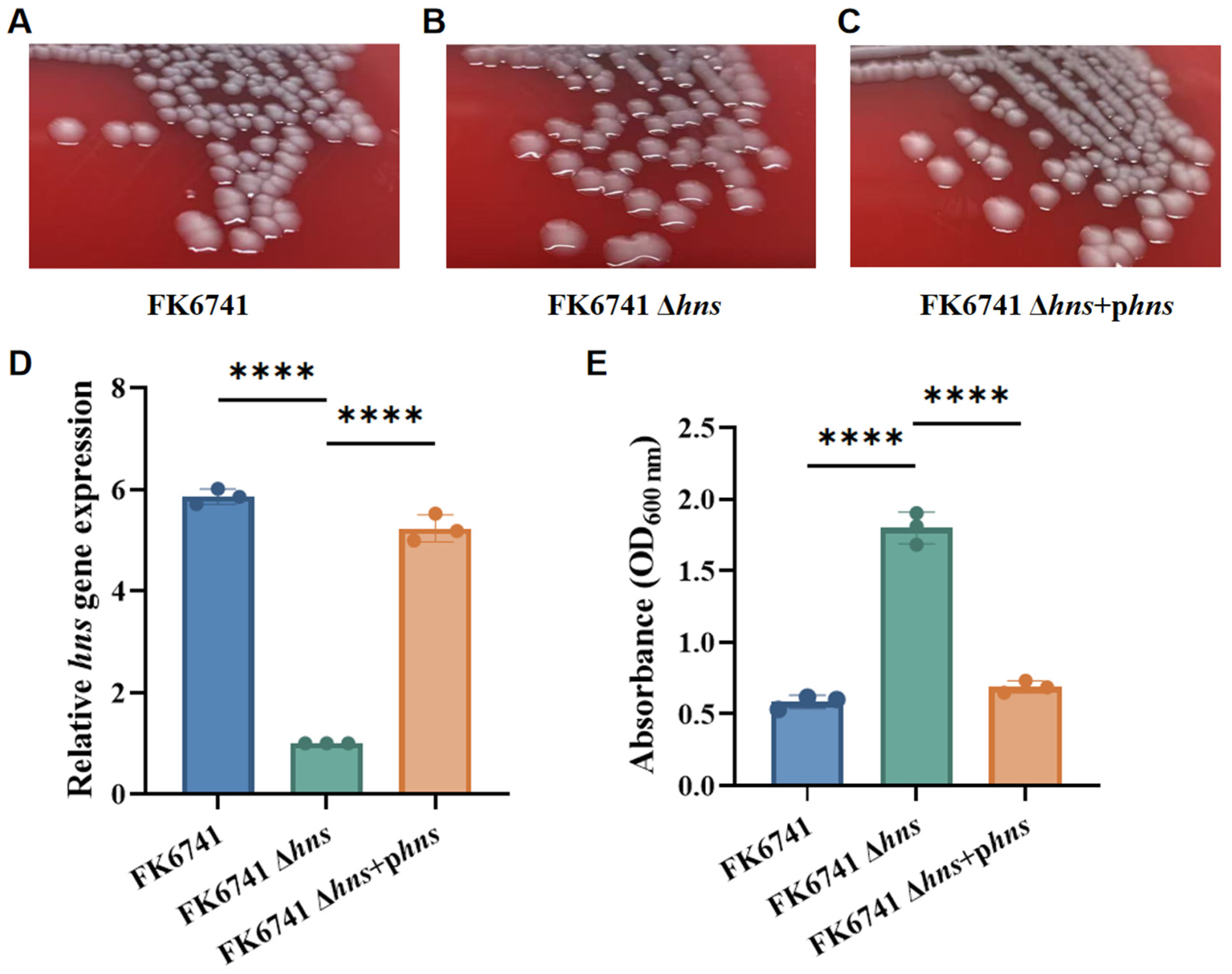
hns were inoculated onto Columbia blood agar plates. A standard inoculation loop was then used to gently lift each colony and assess the formation of mucoid strings. Colonies that produced a viscous filament of ≥5 mm were considered positive for the string test.
2.4. Mucoviscosity Assay
FK6741, FK6741 Δhns, and FK6741 Δhns + phns strains were inoculated into LB broth and incubated for 6 h at 37 °C and 180 rpm. To more accurately compare capsule production among the strains, a low-speed centrifugation assay was performed. Bacterial cultures in LB broth were aliquoted (1 mL each) and centrifuged at 2000× g for 5 min. After centrifugation, 1 mL of the supernatant was collected, and its optical density at 600 nm (OD600) was measured with a microplate reader (Multiskan FC, Thermo Fisher Scientific, Waltham, MA, USA) to estimate relative capsular content. It should be noted that, due to differences in growth rates among strains or variations caused by environmental conditions, the bacterial concentration in the same culture volume may differ between groups at a given time point. Therefore, cell numbers in each group were quantified with a Neubauer counting chamber under a microscope, and cultures were subsequently diluted with LB broth. As a result, 1 mL of the overnight culture contained equal bacterial numbers across all groups. This experiment was independently repeated at least three times.
2.5. Growth Kinetics Assay
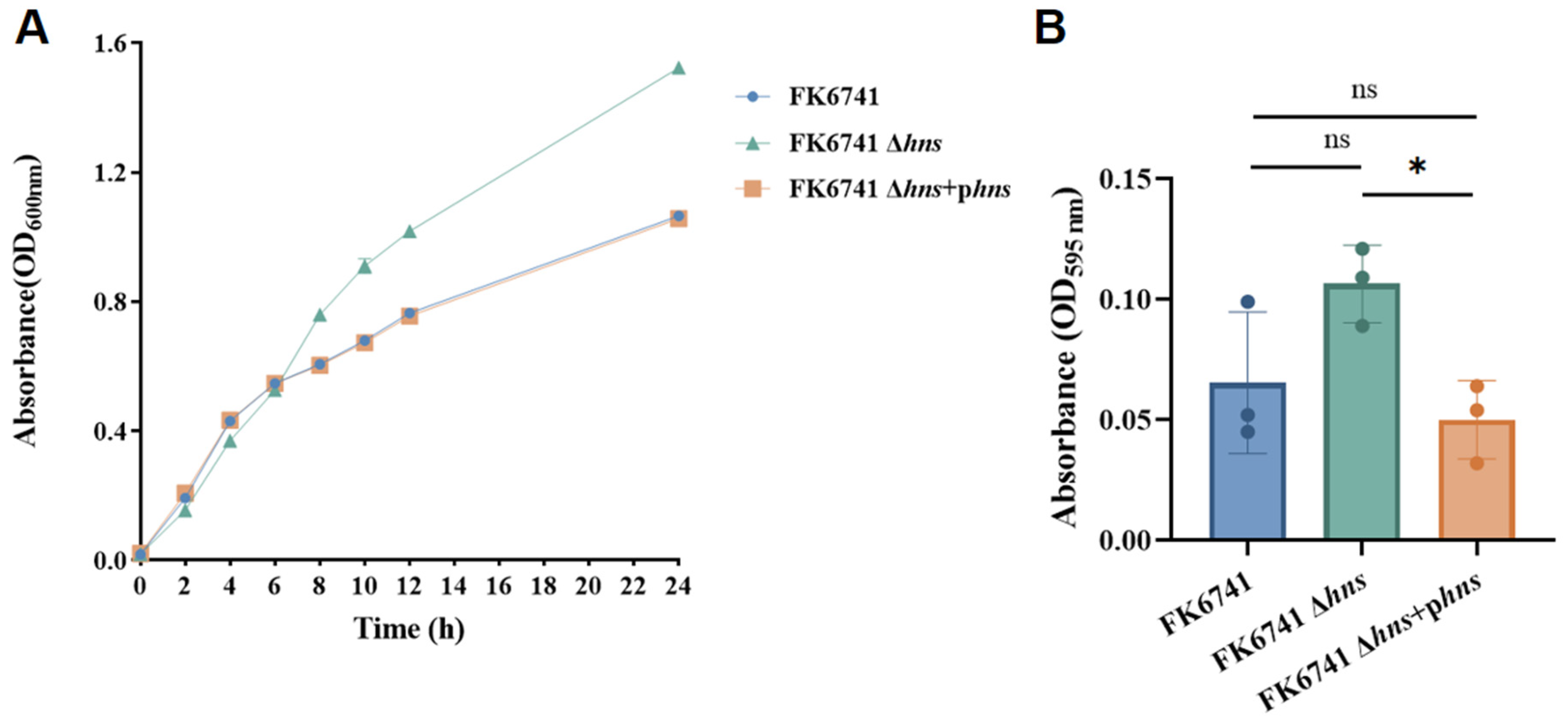
In this experiment, the growth kinetics of FK6741, FK6741 Δhns, and FK6741 Δhns + phns strains were evaluated in LB broth. Three 50 mL centrifuge tubes were prepared, each containing 20 mL of LB broth. The three strains were first streaked onto Columbia blood agar plates and incubated for 16 h. Then, colonies were inoculated into centrifuge tubes containing LB broth. After incubation, 200 μL of each overnight culture was transferred into prepared centrifuge tubes containing 20 mL LB broth and mixed thoroughly. An initial OD600 measurement (0 h) was taken by sampling 200 μL from each culture. Subsequently, all cultures were incubated at 37 °C with shaking at 180 rpm, and OD600 values were recorded at 2, 4, 6, 8, 10, 12, and 24 h to plot the growth curves. OD600 values were read by a microplate reader (Multiskan FC, Thermo Fisher Scientific, Waltham, MA, USA). This experiment was independently repeated at least three times.
2.6. Biofilm Formation Assay
The biofilm formation assay was conducted in 96-well plates following a previously established method [
18]. Briefly, overnight cultures of each strain grown on LB agar plates were adjusted to a 0.5 McFarland standard using 0.9% NaCl. The bacterial suspensions were then diluted 1:100 in LB broth. A total of 100 μL of each diluted suspension was added to wells of a 96-well microtiter plate, and an additional 100 μL of LB broth was added to each well. Each group was tested in triplicate wells and incubated at 37 °C for 48 h. After incubation, the culture medium was discarded, and the wells were gently washed with phosphate-buffered saline (PBS, Solarbio, P1020, Beijing, China) to remove unbound cells. The adherent biofilm was then stained with 200 μL of 1% crystal violet solution (Solarbio, Beijing, China) for 15 min. The dye was subsequently solubilized using a 200 μL solution containing 95% ethanol, and 5% acetic acid was transferred to a fresh 96-well plate. The absorbance at 595 nm (OD595) was measured using a microplate reader (Multiskan FC, Thermo Fisher Scientific, Waltham, MA, USA). Wells containing sterile LB broth only were used as negative controls. Biofilm-forming ability was compared across groups based on OD595 readings. This experiment was independently repeated at least three times.
2.7. Quantification of Capsular Polysaccharides and Free Polysaccharide Chains
The extraction and quantification of polysaccharide chains were performed as described previously [
19] with some modifications. Single colonies were inoculated into LB broth and incubated at 37 °C with shaking for 6 h. To remove exopolysaccharides (EPS) and other extracellular materials that could affect capsule measurement, 500 μL of each culture was centrifuged at 7200×
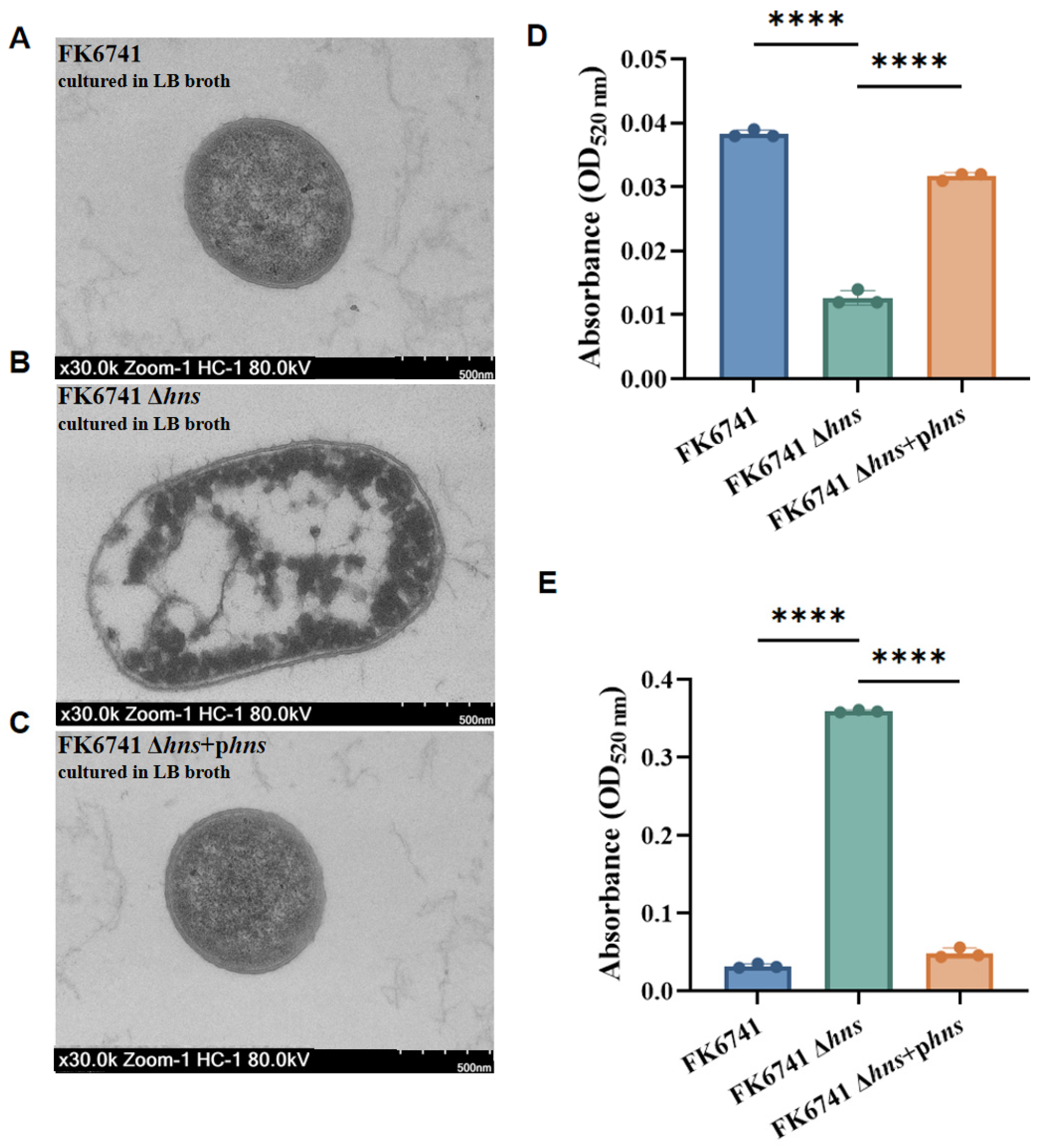
g for 5 min, and the resulting bacterial pellet was washed twice with 500 μL of 0.9% NaCl. Bacterial concentrations were standardized using turbidity measurements. Next, 500 μL of the resuspended cells was mixed with 100 μL of 1% Zwittergent 3–14 detergent (MedChemExpress, HY-W099581, Monmouth Junction, NJ, USA) and incubated at 50 °C for 20 min. After a second centrifugation, 300 μL of the supernatant was combined with 1.2 mL of absolute ethanol (JINSHANHUAXUE, AR5LAR5L, Shanghai, China) and incubated at 4 °C for 20 min. The mixture was centrifuged at maximum speed for 5 min, and the pellet was air-dried and resuspended in 200 μL of distilled water. Then, 1.2 mL of 12.5 mM sodium tetraborate in sulfuric acid was added, and the samples were boiled at 100 °C for 5 min, followed by cooling on ice for 10 min. Finally, 30 μL of 0.15% 3-phenylphenol (MACKLIN, P816935, Shanghai, China) was added into 0.5% NaOH, and after 5 min at room temperature, the absorbance at 520 nm (OD520) was measured using a microplate reader to determine relative capsule levels. All experiments were independently repeated at least three times.
Simultaneously, the supernatant obtained after initial centrifugation was mixed with four volumes of anhydrous ethanol and incubated overnight at 4 °C. The ethanol-insoluble free long-chain polysaccharides were then collected by centrifugation at 20,000 rpm for 20 min, and their quantification was performed using the same method as described above for capsular polysaccharides. Each experiment was independently repeated at least three times.
2.8. Transmission Electron Microscopy (TEM)
TEM was performed to observe the capsule formation of FK6741, FK6741 Δhns, and FK6741 Δhns + phns. The samples were fixed for at least 2 h at room temperature in 3% glutaraldehyde, followed by pre-embedding in 1% agarose and fixation with 1% osmium tetroxide prepared in 0.1 M PBS (Solarbio, P1020, pH = 7.4, Beijing, China) for 2 h in the dark at room temperature. Post-fixation was carried out using 1% osmium tetroxide. After this, the samples were dehydrated through a graded ethanol series, incubated with propylene oxide, and infiltrated overnight in a 1:1 mixture of propylene oxide and low-viscosity epoxy resin. On the following day, the samples were embedded in epoxy resin and polymerized. Ultrathin sections (60–80 nm) were cut using a Leica UC7 ultramicrotome (Leica Microsystems, Wetzlar, Germany), transferred onto copper grids, stained with lead citrate, and imaged using an HT-7800 transmission electron microscope (HITACHI, Tokyo, Japan) by Scientific Compass (Hangzhou, China).
2.9. Construction of Mouse Infection Model
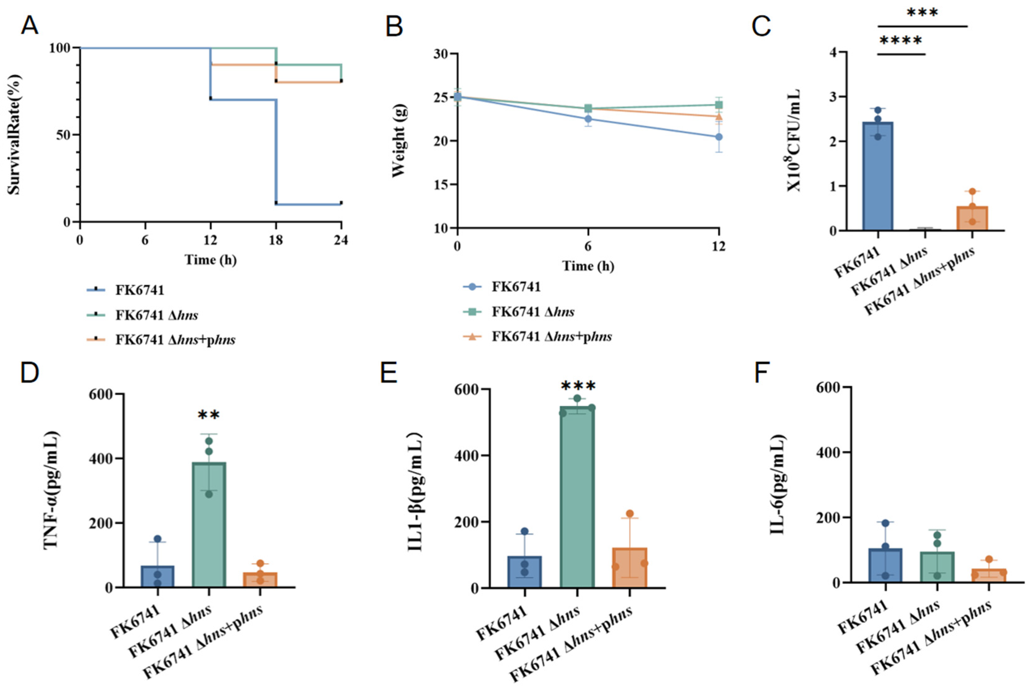
To further investigate the differences between strains, we established a mouse model to examine intraperitoneal infection. Three experimental groups were compared: mice infected with K. pneumoniae FK6741 (wild type), the FK6741 Δhns mutant strain, and the FK6741 Δhns + phns strain. The wild-type strain served as the control group. A total of 60 male ICR mice (Vital River, Jiaxing, China), aged 4–5 weeks and weighing 22–25 g, were randomly divided into three groups. The experimental unit used was a single mouse. No specific inclusion or exclusion criteria were predefined prior to the experiment. The bacterial concentrations after 6 h of culture in LB broth were determined using the plate colony counting method. A total of 200 µL (containing 3 × 10⁷ CFU) of the prepared bacterial suspension was injected into the mice. The survival rates and body weights of mice were recorded for 24 h at 6 h intervals. After 24 h, all mice were anesthetized with 2.0–5.0% isoflurane gas for approximately 5 min to achieve adequate anesthesia, as indicated by loss of consciousness and lack of response to noxious stimuli. Then, peritoneal lavage was performed on each group of mice using PBS, and the lavage fluid was used for plate colony counting. Meanwhile, blood was collected from the orbital sinus of each mouse, and the levels of TNF-α, IL-1β, and IL-6 in the serum were determined using ELISA kits (RayBio® Sandwich-based ELISA Kits, RayBiotech, Norcross, GA, USA) based on the double-antibody sandwich method. The absorbance was measured using an ELX-800 microplate reader (Bio-Tek, Winooski, VT, USA).
2.10. Quantitative Real-Time PCR
Total RNA from bacterial cells was isolated using the Trizol method [
20], which is well-suited for Gram-negative bacteria. In brief, the Trizol reagent, containing phenol and guanidinium thiocyanate, was added to lyse the cells and inactivate RNases. Chloroform was subsequently added, vortexed vigorously and centrifuged to separate the phases. The upper aqueous layer containing RNA was carefully collected, and an equal volume of isopropanol was used to precipitate the RNA. After incubation and centrifugation, the RNA pellet was washed with 75% ethanol, air-dried at room temperature, and dissolved in DEPC-treated water to obtain total RNA. RNA concentration and purity were evaluated using a NanoDrop 2000/2000c spectrophotometer (Thermo Scientific, Waltham, MA, USA); only samples with an A260/A280 ratio of 1.8–2.0 were used for downstream experiments. Reverse transcription was performed using the PrimeScript RT Reagent Kit (Takara, Kusatsu, Japan) according to the manufacturer’s instructions. Quantitative real-time PCR (qRT-PCR) was performed using the Takara fluorescence quantitative kit on a QuantStudio 5 system (Thermo Fisher Scientific, Waltham, MA, USA), following established protocols [
10]. Melting curve analysis was performed after each run to confirm specific amplification. The 16S rRNA gene served as the internal reference, and relative expression of target genes was calculated using the 2
−ΔCt method [
21]. All primers were designed with Primer3Plus (
https://www.primer3plus.com/ (accessed on 7 March 2025)) and are listed in
Table 2.
2.11. Transcriptomic Sequencing
Transcriptomic profiling of the bacterial strain was performed by Novogene (Beijing, China) using RNA-Seq. Ribosomal RNA was removed from total RNA, before being fragmented and reverse-transcribed to first-strand cDNA with random hexamers. During second-strand synthesis, dTTP replaced dUTP to maintain strand specificity. Directional libraries were prepared (end repair, A-tailing, adapter ligation, size selection, amplification, and purification) and checked for quality and concentration using Qubit, real-time PCR, and a bioanalyzer. Libraries were pooled and sequenced on an Illumina platform. Raw FASTQ reads were processed with fastp to remove adapters, low-quality reads, and poly-N sequences. Clean reads were mapped to the reference genome using Bowtie2 (v2.5.4). Rockhopper was used to identify novel genes, operons, TSS/TTS, and antisense transcripts. Upstream 700 bp sequences were extracted to perform promoter prediction using TDNN. UTRs, Shine–Dalgarno sequences, terminators, and novel intergenic transcripts were predicted using RBSfinder (v1.0), TransTermH (v2.0.9), and Blastx (
https://blast.ncbi.nlm.nih.gov/Blast.cgi?PROGRAM=blastx&PAGE_TYPE=BlastSearch&LINK_LOC=blasthome, accessed on 11 June 2025). Candidate sRNAs were analyzed for secondary structure and target genes using RNAfold (v2.0) and IntaRNA (v3.4.1). Gene expression was quantified with FeatureCounts (v2.0.6), and FPKM values were calculated. Differential expression analysis was performed using DESeq2 (v1.42.0) for replicates and using edgeR (v4.0.16) for non-replicates, with Benjamini–Hochberg correction (padj ≤ 0.05 or ≤0.005, |log2fold change| thresholds applied). GO and KEGG enrichment analyses were conducted using clusterProfiler (v4.8.1), with adjusted
p values < 0.05 considered significant.
2.12. Statistical Analysis
The results are expressed as the mean ± standard deviation of three replicates. Statistical analysis was performed using one-way ANOVA. For all analyses, the following connotations are used: ns: not statistically significant; *: p < 0.05; **: p < 0.01; ***: p < 0.001; and ****: p < 0.0001. Statistical analysis was performed with Prism 8 (GraphPad Software LLC, San Diego, CA, USA).
4. Discussion
In this study, we investigated the regulatory role of H-NS in controlling the mucoviscous phenotype, bacterial fitness, and virulence traits of
K. pneumoniae, and further analyzed molecular mechanisms. We first observed that deletion of
hns resulted in markedly increased colony viscosity compared to the parental strain. It is generally believed that an increased production of capsules contributes to a mucoviscous colony phenotype [
24]. However, our findings suggest a more complex scenario. Although FK6741 Δ
hns exhibited a greater number of mucoviscous colonies, quantitative capsule analysis revealed a lower capsule level compared to the wild-type strain but a higher number of free polysaccharides. In our previous studies on phage depolymerases, transmission electron microscopy (TEM) images of a hypermucoviscous, hypercapsulated K2-type hvKp strain revealed a densely anchored, wheat spike-like capsular layer on the bacterial outer membrane [
25]. By contrast, the wild-type FK6741 strain used in this study did not exhibit such a capsular structure. These findings suggest that the mucoid phenotype does not fully correspond to the amount of capsule synthesized but may be related to the localization or anchoring status of the capsule. Consistently, the capsule was barely visible under transmission electron microscopy (TEM) in the Δ
hns mutant. Moreover, FK6741 Δ
hns exhibited attenuated virulence in the mouse infection model.
Based on previous studies [
9,
22,
23], these results led us to hypothesize that the hypermucoviscous phenotype in the
hns mutant is not caused by thickening of the capsule layer on the bacterial surface, but rather by impaired anchoring of the capsular polysaccharides. Downregulation of H-NS may affect the expression of proteins or lipids responsible for tethering the capsule to the cell envelope, leading to the secretion of unanchored polysaccharide chains.
To test this hypothesis, we conducted molecular biology analyses, starting with an investigation into the regulatory role of H-NS during capsule formation in
K. pneumoniae. Previous studies have shown that
K. pneumoniae utilizes a Wzx/Wzy-dependent pathway for capsule assembly and export [
8]. The process begins with the biosynthesis of nucleotide sugar precursors specific to each K-antigen type [
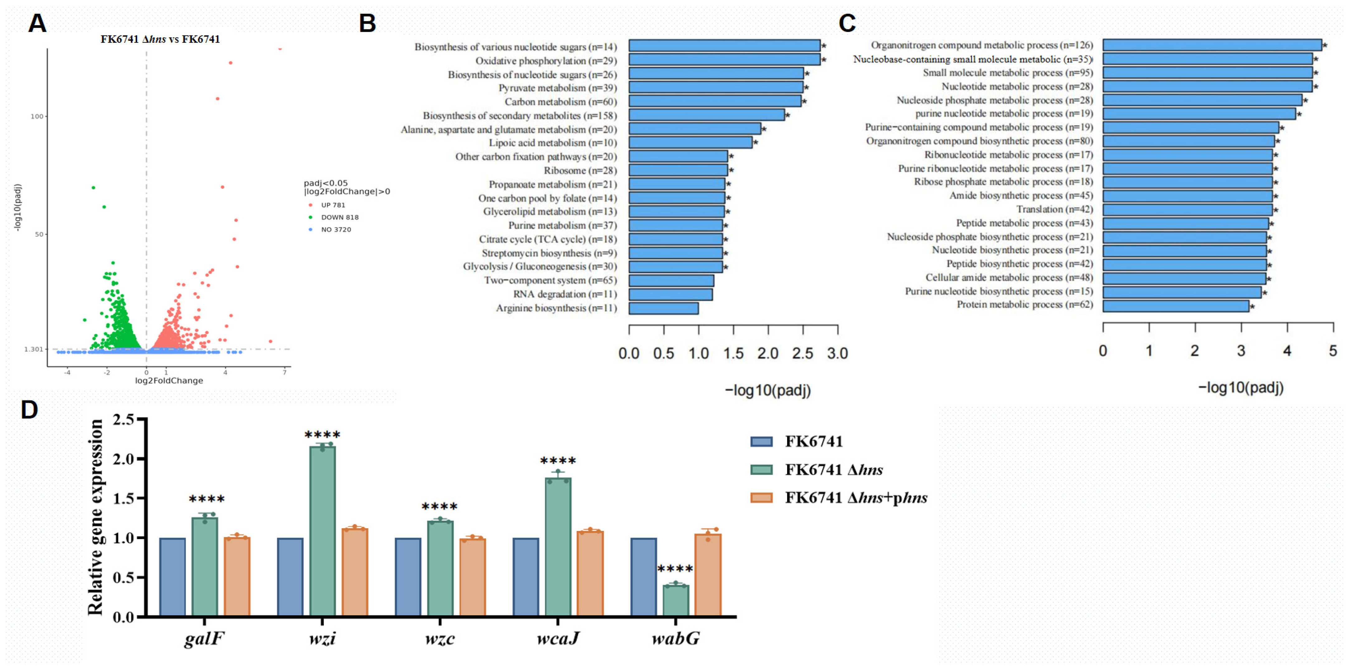
26], which are synthesized by glucose metabolism, including UDP-glucose, UDP-galactose and more. Therefore, in our analysis, we focused primarily on genes associated with the Wzx/Wzy-dependent pathway and metabolic pathways related to sugar precursors for capsule biosynthesis. We found that H-NS inherently suppresses carbohydrate metabolism, inhibits the formation of precursors required for capsule synthesis, and represses the expression of genes associated with capsule formation. Further analysis of our results enabled us to identify genes potentially involved in anchoring capsular polysaccharide chains in
K. pneumoniae, among which the glycosyltransferase family 2 gene
wabG, related to GalA residues, was significantly downregulated. Therefore, we propose that H-NS, as a global regulator, exerts multiple effects: on the one hand, deletion of
hns may relieve its inhibitory effect on capsule synthesis in
K. pneumoniae; on the other hand, it leads to abnormal anchoring of capsular polysaccharide chains. These unanchored polysaccharides are easily lost from the bacterial surface into the surrounding environment, resulting in the absence of visible capsule layers under electron microscopy. Moreover, these polysaccharide chains lose their original biological functions, such as anti-phagocytic and anti-complement activities, ultimately leading to altered bacterial virulence.
Notably, in the mouse intraperitoneal infection model, the survival curves of mice infected with the
hns deletion strain and the complemented strain were unexpectedly similar (
Figure 4A). A plausible explanation for this observation is the instability of the complementation plasmid in vivo. In animal infection models, antibiotic selection pressure cannot be maintained for prolonged periods, which significantly compromises plasmid retention [
27]. Moreover, plasmid carriage generally imposes a metabolic burden on bacteria; under the competitive conditions encountered within the host, bacterial cells that lose the plasmid may gain a growth or survival advantage [
28]. In addition, because the complemented
hns gene is carried on an extrachromosomal plasmid rather than being integrated into the chromosome, the plasmid is prone to segregation loss during bacterial replication. Consequently, the complementation plasmid may be progressively lost during infection, resulting in a gradual reversion of the complemented strain toward a phenotype resembling that of the
hns deletion mutant. Similar plasmid instability in in vivo infection models has been reported previously and is recognized as a common limitation of plasmid-based complementation strategies. Therefore, the comparable survival outcomes observed for the deletion and complemented strains in vivo are likely attributable to plasmid loss rather than a true restoration of virulence by
hns complementation.
In this study, we also assessed the impact of H-NS on bacterial fitness. We first observed that a loss of H-NS may inhibit bacterial growth during the early stages of cultivation, a phenomenon previously reported [
10]. As a key regulator of bacterial physiology, disruption of the H-NS function can adversely affect growth. At later stages, however, the OD values of the H-NS-deficient strain exceeded those of the wild-type and complemented strains. This can be explained by the hypermucoviscous phenotype observed in the H-NS mutant: the large-scale production of abnormally anchored polysaccharide chains causes the culture medium to become highly viscous, leading to artificially elevated OD readings. Similarly, these excessively secreted free polysaccharide chains, functioning like extracellular polysaccharides, surround the bacterial cells and, to some extent, promote the formation of biofilms.
Overall, our study indicates that capsule formation in K. pneumoniae depends not only on the amount of capsule synthesized but also on the proper anchoring and functional integrity of capsular polysaccharides. From a clinical perspective, this creates potential for an intervention strategy by targeting H-NS or the genes involved in capsule anchoring. By disrupting the H-NS regulatory network or capsule polysaccharide anchoring, bacterial virulence may attenuate without directly killing the bacteria, thereby reducing the severity of infection. Therefore, interventions targeting H-NS-mediated regulatory mechanisms may offer novel preventive and therapeutic strategies for high-risk patients with severe K. pneumoniae infections, providing a theoretical basis for the development of new agents to treat K. pneumoniae infections. This study also deepens understanding of the physiological regulatory functions of H-NS and contributes to research elucidating the mechanisms underlying the virulence of K. pneumoniae.




 ,
,