Article

The Effect of Variations in Temperature and Contact Time of Zingerone, [6]-Gingerol and Shogaol as Disinfectants on Staphylococcus aureus, Escherichia coli, Pseudomonas aeruginosa and Candida albicans


1
Department of Biomedical Engineering, Near East University, Mersin 10, Nicosia 99138, Turkey
2
Department of Medical and Clinical Microbiology, Near East University, Mersin 10, Nicosia 99138, Turkey
*
Author to whom correspondence should be addressed.
Microorganisms 2026, 14(3), 539; https://doi.org/10.3390/microorganisms14030539
Submission received: 13 January 2026 / Revised: 12 February 2026 / Accepted: 20 February 2026 / Published: 26 February 2026

Abstract

Rising microbial resistance to synthetic disinfectants has intensified the search for chemically synthesized natural alternatives, such as ginger-derived bioactive compounds. Several bioactive compounds, including Zingerone, Gingerols, and Shogaols, have been shown to possess antimicrobial activities. However, the antimicrobial efficacy of these compounds as disinfectants at varying temperatures and contact times is poorly understood. Therefore, understanding the temperature- and time-dependent effects of contact is crucial for optimizing the potential application of these compounds in various antimicrobial strategies. In this study, the antimicrobial activities of three chemicals, 10% [6]-Gingerol, Zingerone, and Shogaols, were evaluated against Staphylococcus aureus, Escherichia coli, Pseudomonas aeruginosa, and Candida albicans. Furthermore, the variations in temperatures (5 °C, 25 °C, and 37 °C) and contact time (1, 5, and 60 min) were assessed based on CFU counts, log10 reductions, percent kill, and decimal reduction time. The findings of this study indicated that 10% Zingerone completely inactivated all tested organisms in 60 min at all temperatures. Although 10% [6]-Gingerol and Shogaol exhibited temperature- and time-dependent effects, they failed to completely inactivate the bacteria and fungi after 60 min. Furthermore, both temperature and contact time were shown to influence the efficacy of the tested disinfectants, providing a significant time- and temperature-dependent reduction in viable cells across all tested organisms.

1. Introduction

The COVID-19 pandemic significantly increased the use of biocides, highlighting their role in infection control but also exacerbating concerns over antimicrobial resistance (AMR) and environmental toxicity associated with synthetic disinfectants, such as quaternary ammonium compounds (QACs) and alcohols [1,2,3,4,5,6]. While effective, these agents face limitations, including the potential to drive AMR, reduced efficacy in the presence of organic matter, and safety issues [7,8,9,10,11,12,13]. Plant-based alternatives offer eco-friendly profiles but often suffer from variable composition, standardization challenges, and lower potency [14,15,16,17].
Ginger (Zingiber officinale Roscoe) is a prominent source of bioactive compounds with demonstrated antimicrobial properties. Key constituents include Zingerone, Gingerols (e.g., [6]-Gingerol), and Shogaols, which are formed from Gingerols via dehydration [18,19]. These compounds exhibit a range of pharmacological activities, including antimicrobial effects against various bacteria and fungi [20,21,22,23,24,25,26,27]. For instance, ginger extracts and specific compounds like [6]-Gingerol have shown activity against pathogens such as Staphylococcus aureus, Escherichia coli, Pseudomonas aeruginosa, and Candida albicans [23,24,27].
Shogaols, Zingerone, and Gingerols are some of the disinfectants reported in the literature. Zingerone, chemically known as vanillylacetone, is a naturally occurring chemical compound derived from ginger (Zingiber officinale). The plant-derived compound is associated with several pharmacological properties, including antimicrobial, antioxidant, anti-inflammatory, antidiabetic, anxiolytic, and appetite stimulant effects [28]. Zingerone antimicrobial activity involves its ability to inhibit the growth of certain microbes. Thus, the compound has great potential as a remedy for various infectious diseases. However, further studies are required to fully understand its mechanism of action and pharmacological potency [28,29].
Like Zingerone, Gingerol is another plant-based compound derived from Zingiber officinale. Gingerols contain several phenolic compounds, including [6]-Gingerol, [8]-Gingerol, and [10]-Gingerol. The compound is associated with the spicy flavor of Zingiber officinale and several pharmacological activities, including antimicrobial, antioxidant, anti-cancer, and anti-inflammatory effects [20,30,31].
Shogaols are another class of chemical compounds derived from Zingiber officinale, produced by dehydrating Gingerols during processing or storage. Shogaols differ from Gingerols based on the replacement of hydroxyl groups (Gingerols) with ketone groups. There are several forms of Shogaols, including 4-, 6- (regarded as the most potent), 8-, 10-, and 12- Shogaols. These compounds are associated with part of ginger’s pungent flavor and numerous medicinal and biological properties, including antimicrobial, antioxidant, and anti-inflammatory effects [32,33,34].
Chemically synthesized versions of natural compounds, such as those derived from ginger, offer potential advantages including consistent quality, scalable supply, and the ability to study structure/activity relationships. However, empirical data are lacking on their performance under varied environmental conditions. Moreover, the practical application of disinfectants is critically influenced by operational factors such as contact time and temperature. Deviations from recommended contact times or use at non-standard temperatures can drastically reduce efficacy and promote resistance [35,36,37,38]. Therefore, there is a pressing need for effective disinfectants that maintain activity across a range of realistic use conditions. To address this gap, this study aimed to carry out the following:
  • Investigate the efficacy of three synthetic ginger-derived compounds (Zingerone, [6]-Gingerol, and Shogaol) as alternative disinfectants.
  • Evaluate their activity against representative Gram-positive (S. aureus) and Gram-negative (E. coli, P. aeruginosa) bacteria, and the fungus C. albicans.
  • Systematically assess the impact of temperature (5 °C, 25 °C, 37 °C) and contact time (1, 5, 60 min) on antimicrobial efficacy.
  • Generate empirical data to inform potential real-world applications in healthcare, the food industry, or environmental settings.

2. Materials and Methods

2.1. Materials

Chemical reagents: Zingerone (100 mg: Cat. No.: HY-14621.CAS No.: 122-48-5. Molecular Formula: C11H14O3. Molecular Weight: 194.23), [6]-Gingerol (100 mg: Cat. No.: HY-14615. CAS No.: 23513-14-6. Molecular Formula: C17H26O4. Molecular Weight: 294.39), and Shogaol (50 mg: Cat. No.: HY-14616. CAS No.: 555-66-8. Molecular Formula: C17H24O3. Molecular Weight: 276.37) were purchased from MedChemTronica European branch of MCE (MedChemExpress) MedChemTronica Sollentuna, Sweden. We also purchased Tween (80: Cat. No.: HY-Y1891. CAS No.: 9005-65-6), dimethyl sulfoxide (DMSO: Cat. No.: HY-Y0320. CAS No.: 67-68-5 Molecular Formula: C2H6OS. Molecular Weight: 78.13), and Phosphate-Buffered Saline (PBS Buffer (1×): Cat. No.: HY-K3005) from the same company. Growth medium: Mueller–Hinton agar MHA and Sabouraud Dextrose Agar (SDA). Microorganism strains: Staphylococcus aureus ATCC25923, Escherichia coli ATCC25922, Pseudomonas aeruginosa ATCC27853, and Candida albicans ATCC10231. The step-by-step preparation of disinfectants is presented in the Supplementary File.

2.2. Preparation of Chemical Disinfectants

2.2.1. Preparation of Dimethyl Sulfoxide (DMSO) Solution

To prepare a 0.5% DMSO (dimethyl sulfoxide) solution, used to dissolve Zingerone, [6]-Gingerol, and Shogaol, 50 μL of pure DMSO was transferred using a sterile micropipette into a tube [33].
Then, 9.95 mL of the selected diluent PBS was added to a sterile conical 20 mL tube. The solution was mixed gently but thoroughly using vortex mixer vortexing. The container was labeled with 0.5% DMSO, 10 mL, and the date.

2.2.2. Preparation of Tween Solution

Next, 160 μL of pure Tween 80 was transferred using a sterile pipette into a 20 mL sterile container, and 19.84 mL of sterile PBS was subsequently added into the container. The solution was mixed using vortexing, and the container was labeled with “0.8% Tween-80” and the date.

2.2.3. Preparation of 10% Zingerone, 10% [6]-Gingerol, and 10% Shogaol

Using a sterile pipette, 1 mL and 0.5 mL of 0.5% DMSO were transferred into three 1.5 mL Eppendorf tubes; each separate tube was mixed with 100 mg of Zingerone, 100 mg of [6]-Gingerol, and 50 mg Shogaol using vortexing. The containers were subsequently labeled with 10% Zingerone, 10% [6]-Gingerol, and 10% Shogaol.

2.2.4. Preparation of Bacterial and Fungal Suspension

To prepare the bacterial suspension, colonies were transferred using a sterile loop into a sterile tube containing 10 mL of PBS. Based on McFarland standards:
0.5 McFarland ≈ 1.5 × 10⁸ CFU/mL
The starting suspension (assumed) was 1.5 × 10⁸ CFU/mL, and the target suspension was 5.7 × 10⁵ CFU/mL [33].
According to the formula
C1 × V1 = C2 × V,
A total of 4981 μL PBS is mixed with 19 μL of stock concentration, leading to a target suspension concentration of 5.7 × 10⁵.
To prepare the fungal suspension, colonies were transferred using a sterile loop into a sterile tube containing 10 mL of PBS. Based on McFarland standards:
1 McFarland ≈ 1 × 10⁷ CFU/mL
The starting suspension (assumed) was 1 × 10⁷ CFU/mL, and the target suspension was 1 × 10⁶ CFU/mL [30,31].
Using the same formula used for bacteria (i.e., the one mentioned above), 9 mL PBS is mixed with 1 mL of stock concentration, leading to a target suspension concentration of 1 × 10⁶.

2.3. Time–Kill Assay with Neutralization

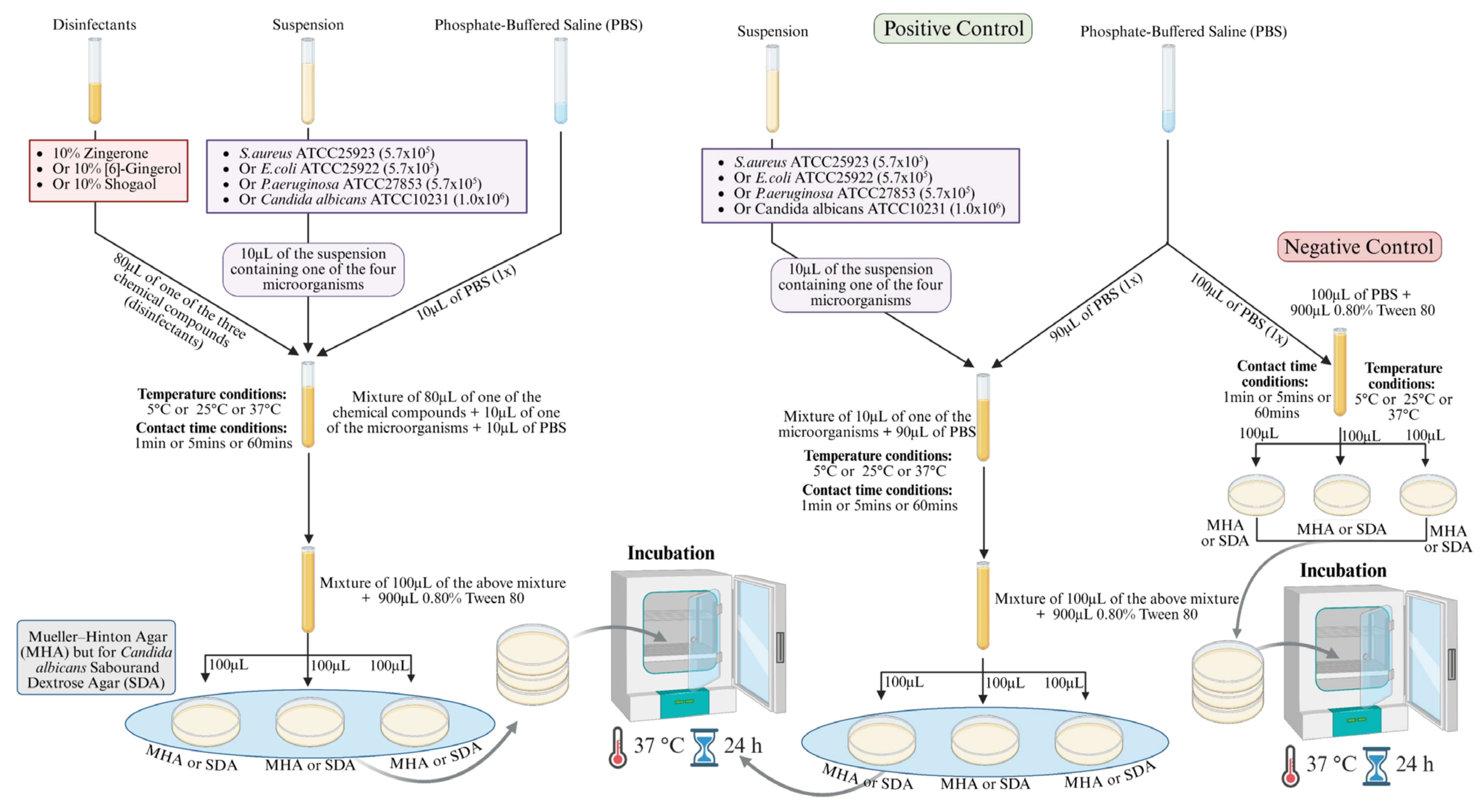
The time–kill assay was performed following the standard methodology [39] with modifications. Figure 1 summarizes the 5 steps involved in testing the efficacy of the disinfectants (10% Zingerone, 10% [6]-Gingerol, and 10% Shogaol) against selected microbes under various temperatures and contact times. The protocol for the reaction mixture involved transferring 80 μL of 10% Zingerone or 10% [6]-Gingerol or 10% Shogaol into separate sterile tubes, followed by the addition of 10 μL of S. aureus ATCC25923 or E. coli ATCC25922 or P. aeruginosa ATCC27853 or C. albicans ATCC10231, and 10 μL of PBS (1×). The same protocol was repeated for 3 temperatures and 3 contact times. The reaction mixture was incubated at three different temperatures—5 °C, 25 °C, and 37 °C—and at three different contact times—1 min, 5 min, and 60 min. After the specified contact time at varied temperatures, 100 μL was transferred into a new tube containing 900 μL of 0.8% Tween 80, serving as a neutralizer. Subsequently, 100 μL (from the 1000 μL) is plated on Mueller–Hinton agar (MHA) for bacterial growth or Sabouraud Dextrose Agar (SDA) for fungal growth and incubated at 37 °C for 24 h. The viable count of each microorganism cell after exposure to Zingerone, [6]-Gingerol, and Shogaol was determined by colony-forming units (CFU/mL). The results were calculated by comparing the CFU/mL values of each microorganism before and after exposure to the tested compounds, and the reduction was calculated accordingly.

Controls

  • Positive control (without disinfectant)
First, 10 μL of the microbial suspension is mixed with 90 μL of PBS in a sterile tube. The reaction is mixed using vortexing and incubated at one of the three different temperatures, 5 °C, 25 °C, or 37 °C, and at one of the three different contact times, 1 min, 5 min, or 60 min. Subsequently, 100 μL of the mixture is transferred into a sterile tube containing 900 μL of 0.8% Tween 80. Finally, 100 μL (from the 1000 μL) is plated on MHA for bacterial growth or SDA for fungal growth and incubated at 37 °C for 24 h.
  • Negative control (without microbes)
First, 80 μL of 10% Zingerone, 10% [6]-Gingerol, or 10% Shogaol is transferred into a separate sterile tube and mixed with 20 μL of PBS. The reaction is mixed using vortexing and incubated at one of the three different temperatures, 5 °C, 25 °C, or 37 °C, and at one of the three different contact times, 1 min, 5 min, or 60 min. Subsequently, 100 μL is transferred into a sterile tube containing 900 μL of 0.8% Tween 80. Finally, 100 μL (from the 1000 μL) is plated on MHA for bacterial growth or SDA for fungal growth and incubated at 37 °C for 24 h.

2.4. Statistical Analysis

A one-way analysis of variance (ANOVA) was conducted separately for each temperature to determine whether there were statistically significant differences in the mean D-values among the four tested microorganisms. Prior to ANOVA, the assumptions of normality and homogeneity of variances were evaluated using the Shapiro–Wilk test and Levene’s test, respectively. Data were considered normally distributed and variances homogeneous if p > 0.05. While the ANOVA results indicated significant differences (p < 0.05), post hoc pairwise comparisons were performed using Tukey’s Honestly Significant Difference (HSD) test to identify specific group differences. The results were expressed as mean ± standard deviation (SD), and a significance level of 0.05 was used throughout.

3. Results

3.1. Evaluation of Antibacterial and Antifungal Activity of 10% Zingerone

The results of the antimicrobial efficacy of 10% Zingerone were evaluated against four representative microorganisms, including E. coli, P. aeruginosa, S. aureus, and C. albicans, at three different temperatures (5 °C, 25 °C, and 37 °C) and across various contact times (0, 1, 5, and 60 min) are summarized in Table 1. Overall, the results demonstrated that 10% Zingerone is capable of achieving complete microbial inactivation within 60 min under different temperature conditions. The rate of microbial kill was enhanced at higher temperatures and varied slightly between organisms, with Gram-negative bacteria (E. coli) showing slightly faster reductions than S. aureus, while C. albicans exhibited consistent susceptibility.

3.1.1. Percent Kill After Exposure to 10% Zingerone

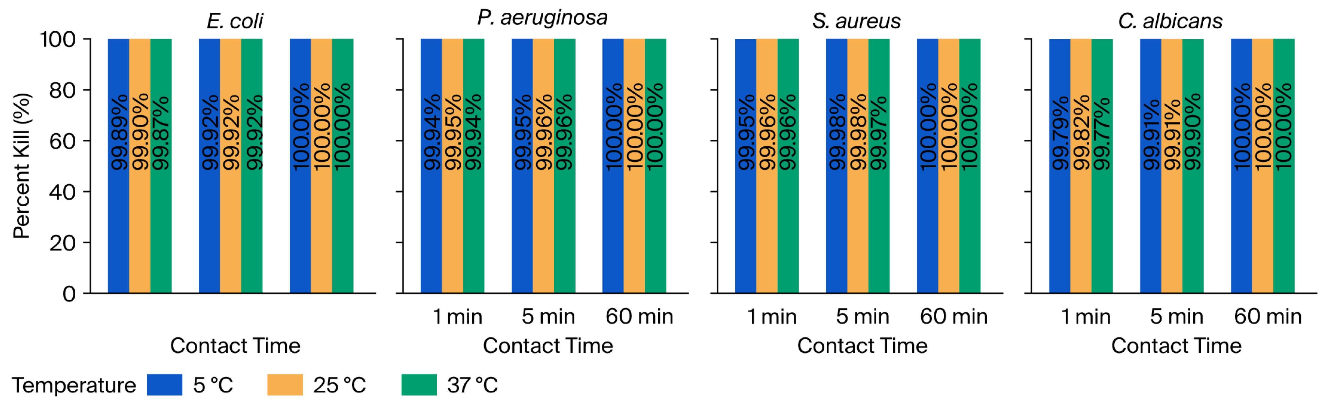
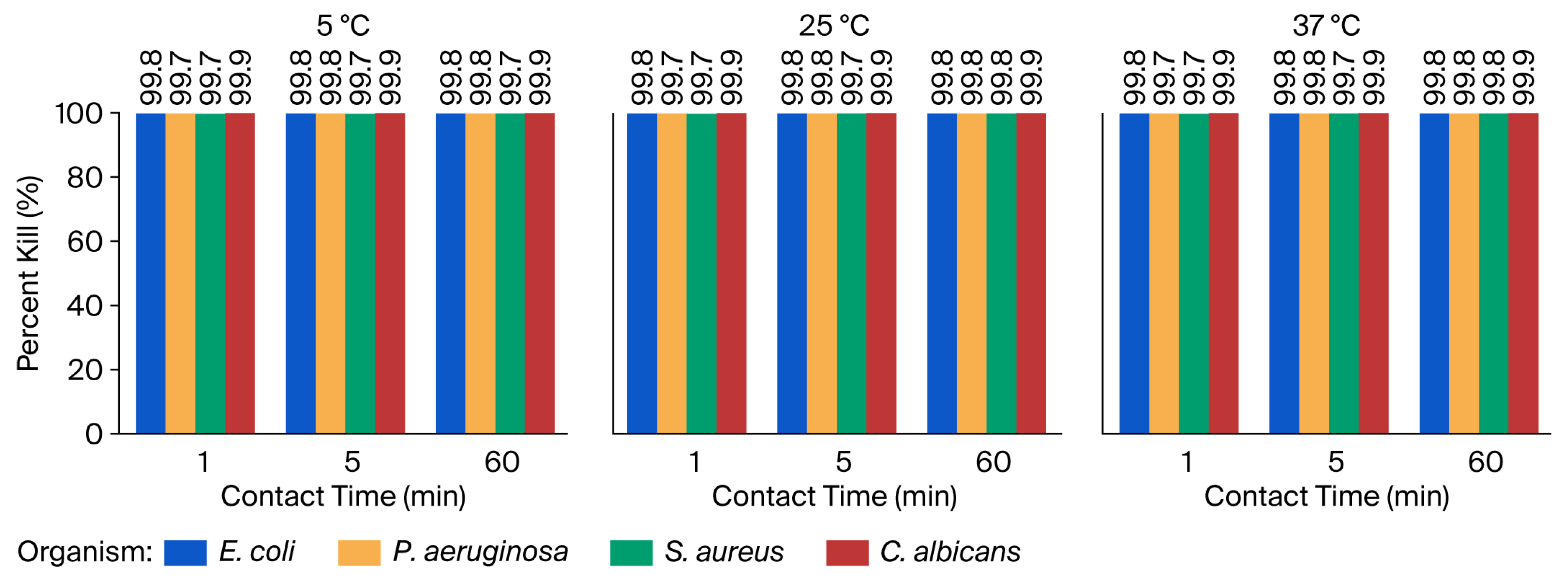
The antimicrobial efficacy of 10% Zingerone was also evaluated by determining the percent kill of E. coli, P. aeruginosa, S. aureus, and C. albicans following exposure at various temperatures and contact times. All organisms exhibited a rapid reduction in viability within the first minute of exposure, with further enhancement over time, culminating in complete inactivation (100%) by 60 min across all temperature conditions, as shown in Figure 2. Overall, the results demonstrated that 10% Zingerone exerts a broad-spectrum and time-dependent antimicrobial effect. Figure 2 clearly illustrates this pattern, with temperature-coded bars representing kill percentages for each organism over the contact time course. The vertical display of exact percent values within each bar further emphasizes the consistent and complete reduction achieved after 60 min.

3.1.2. Decimal Reduction Times (D-Values) After Exposure to 10% Zingerone

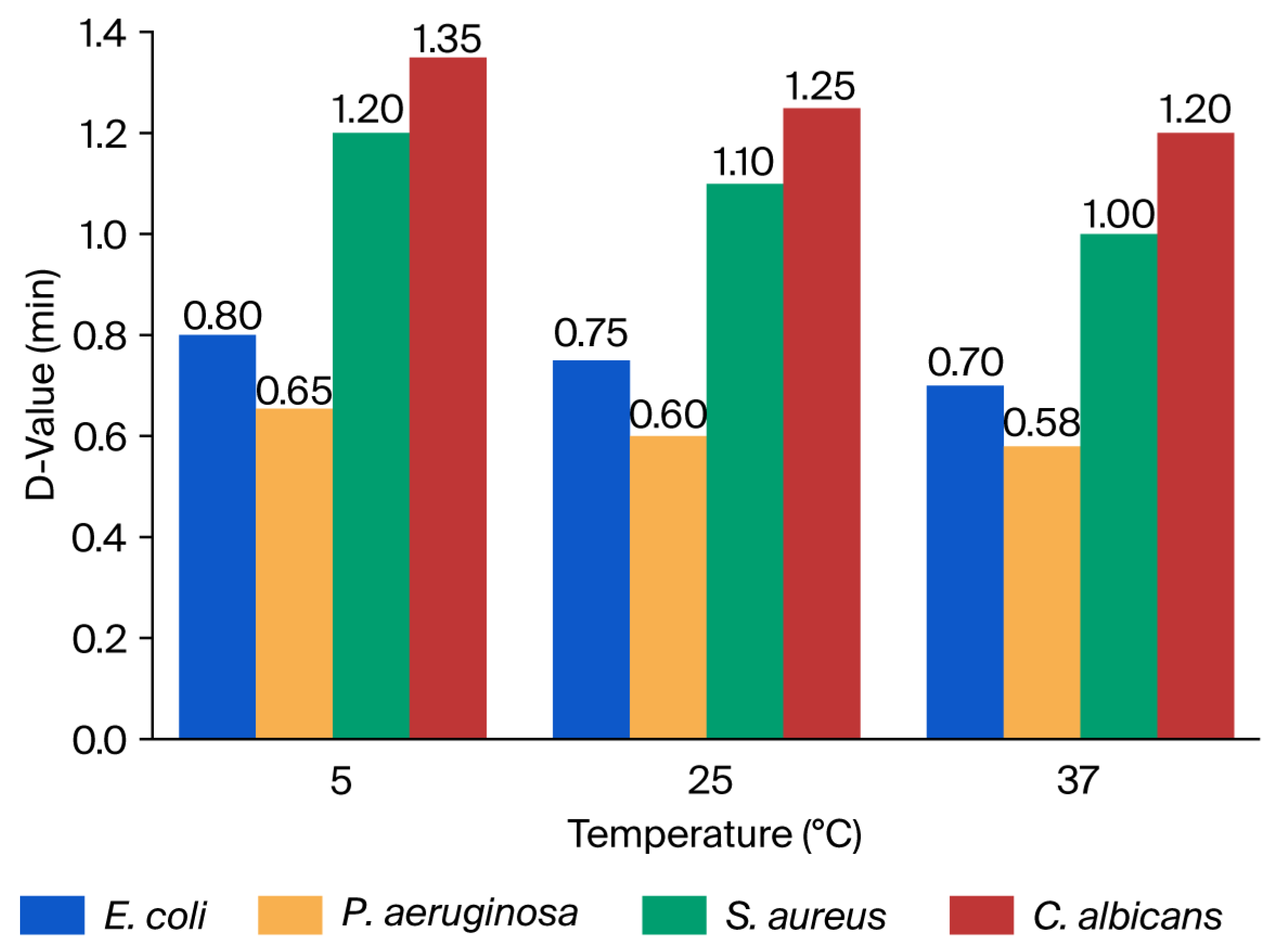
The D-values indicate the time required to achieve a 1-log10 (90%) reduction in viable cell counts. The value represents the time required to achieve a 90% reduction in viable microbial count. Lower D-values indicate faster antimicrobial action. The D-values of E. coli, P. aeruginosa, S. aureus, and C. albicans following exposure to 10% Zingerone were evaluated across the three temperatures. The results revealed that P. aeruginosa exhibited the lowest D-values across all temperatures, ranging from 0.58 min at 37 °C to 0.65 min at 5 °C. E. coli followed closely, with D-values ranging from 0.70 to 0.80 min. These findings indicate that Gram-negative bacteria are more susceptible to rapid inactivation by Zingerone under the tested conditions. In contrast, S. aureus and C. albicans demonstrated significantly higher D-values, ranging from 1.00 to 1.35 min. The longest D-value was observed in C. albicans at 5 °C (1.35 min), suggesting greater thermal stability or resistance to the compound at lower temperatures. S. aureus showed intermediate D-values, between 1.00 and 1.20 min, across all conditions.
A slight temperature-dependent reduction in D-values was observed for all tested organisms, particularly among Gram-negative species. For example, the D-value for E. coli decreased from 0.80 min at 5 °C to 0.70 min at 37 °C. This trend suggests enhanced antimicrobial activity of Zingerone at elevated temperatures, possibly due to an increased diffusion rate, better solubility, or greater membrane penetration. The D-values of the tested microbes using Zingerone are presented in Figure 3. Log10 reduction in microorganisms after exposure to 10% Zingerone at different temperatures and contact times is presented in Figure S1 of the Supplementary File. A comparison of D-values, minutes, and 1 min kill percentages of Zingerone against the tested microbes is presented in Figure S2 of the Supplementary File.

3.2. Evaluation of Antibacterial and Antifungal Activity of 10% [6]-Gingerol

The antimicrobial activity of 10% [6]-Gingerol was assessed against four microbial strains. Each organism was exposed to 10% [6]-Gingerol at three different temperatures and sampled at four contact times. The results, which are summarized in Table 2, indicate that 10% [6]-Gingerol exerts a rapid and strong antimicrobial effect across a broad range of microbial types. The effect was slightly enhanced at higher temperatures, supporting the temperature-dependent enhancement of its action. These results highlight [6]-Gingerol’s potential as a broad-spectrum antimicrobial agent effective in both bacterial and fungal applications.

3.2.1. Percent Kill After Exposure to 10% [6]-Gingerol

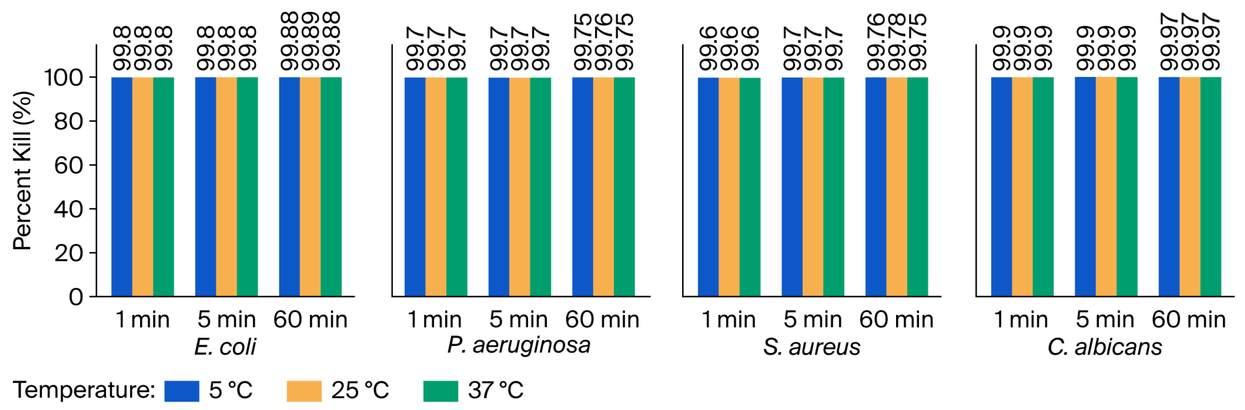
As shown in Figure 4, the antimicrobial activity of 10% [6]-Gingerol was evaluated against four test microbes at three different incubation temperatures and contact times. Overall, the figure shows a clustered bar chart illustrating the percent kill of each microorganism at different contact times across the three tested temperatures. C. albicans consistently exhibited the highest susceptibility to [6]-Gingerol, especially at 25 °C and 37 °C, suggesting a greater sensitivity of fungal and bacterial strains.

3.2.2. Decimal Reduction Times (D-Values) After Exposure to 10% [6]-Gingerol

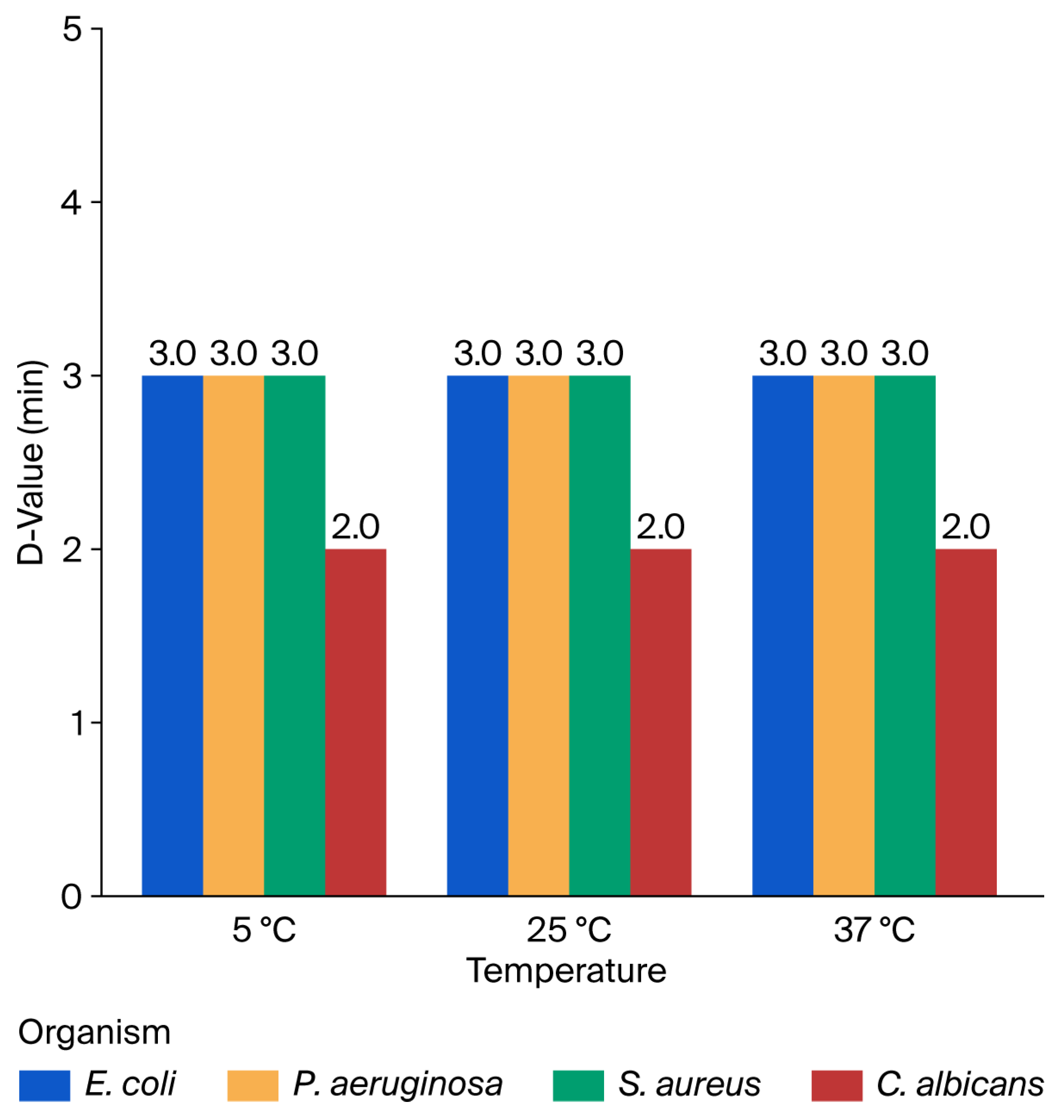
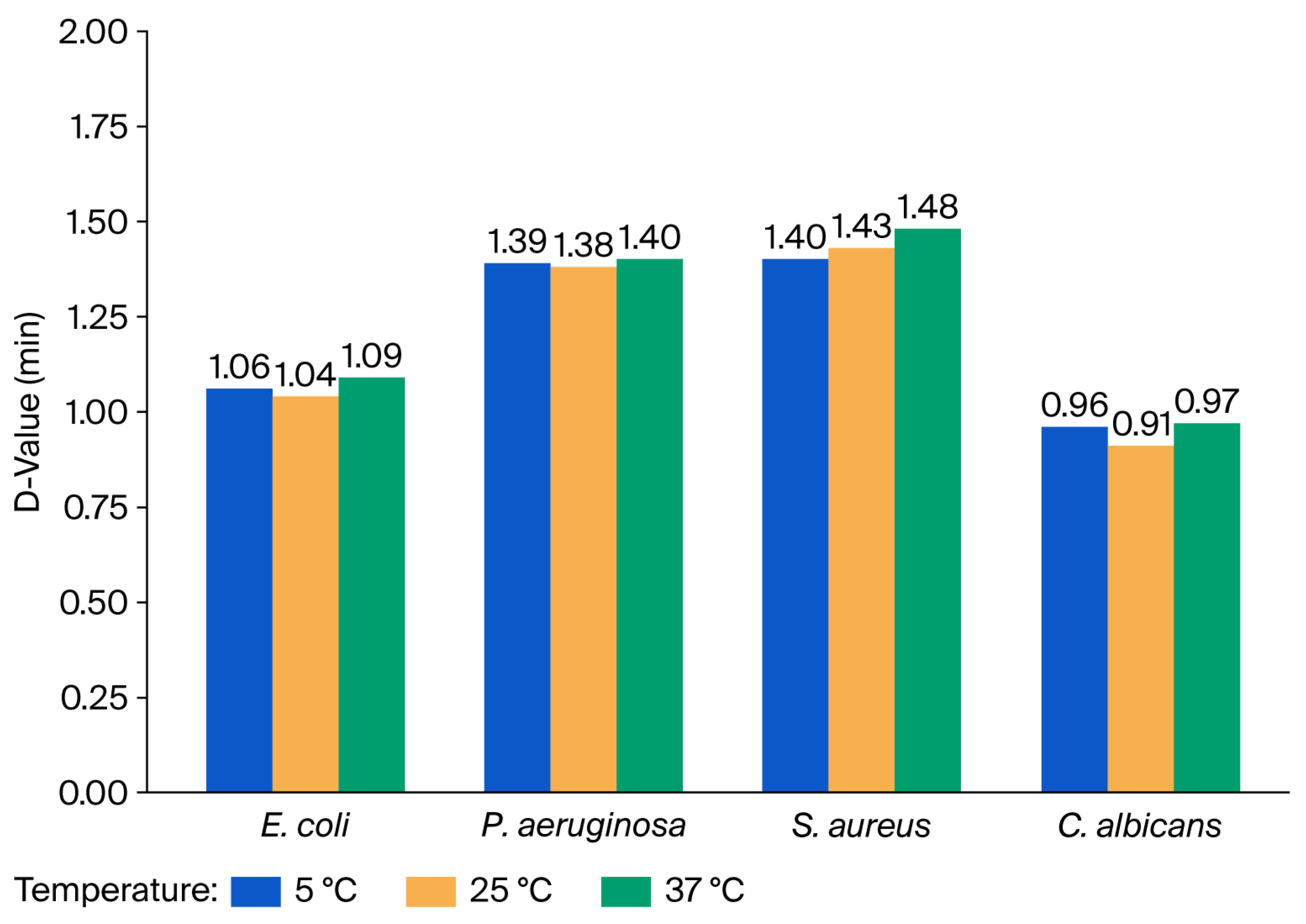
The decimal reduction time (D-value) was determined for the test microbes following treatment with 10% [6]-Gingerol at three different temperatures (5 °C, 25 °C, and 37 °C), as shown in Figure 5. The D-values for E. coli, P. aeruginosa, and S. aureus were consistent across all three tested temperatures, with values of 3.0 min. This uniformity indicates that temperature had a minimal influence on the early killing kinetics of [6]-Gingerol against these bacterial strains. In contrast, C. albicans showed a consistently lower D-value of 2.0 min, demonstrating a more rapid decline in viable fungal cells during the same exposure period. This suggests a higher susceptibility of C. albicans to [6]-Gingerol compared to the bacterial strains tested. Log10 reduction in microorganisms after exposure to 10% [6]-Gingerol at different temperatures and contact times is presented in Figure S3 of the Supplementary File. A comparison of D-values, minutes, and 1 min kill percentages of [6]-Gingerol against the tested microbes is presented in Figure S4 of the Supplementary File.

3.3. Evaluation of Antibacterial and Antifungal Activity of 10% Shogaol

Table 3 summarizes the antimicrobial activity of 10% Shogaol against four representative microorganisms across three incubation temperatures and over four contact times. Overall, the results indicate that 10% Shogaol demonstrated significant antimicrobial effects, especially against E. coli and C. albicans, with slightly less pronounced activity against P. aeruginosa and S. aureus. These findings support the potential application of Shogaol as an antimicrobial agent effective across multiple pathogenic species and a wide temperature range.

3.3.1. Percent Kill After Exposure to 10% Shogaol

Figure 6 presents the percent kill of E. coli, P. aeruginosa, S. aureus, and C. albicans after treatment with 10% Shogaol. The results demonstrate rapid microbial reductions within the first minute, with enhanced killing at longer contact times and consistent antimicrobial activity irrespective of temperature. The results revealed that E. coli and C. albicans are highly susceptible to 10% Shogaol, with percent kills exceeding 99.7% within the first minute of exposure across all temperatures. This rapid and potent antimicrobial effect was sustained for 60 min, where the percent kill approached or exceeded 99.9%. P. aeruginosa and S. aureus showed slightly lower susceptibility, with percent kills ranging from approximately 99.5% to 99.8% after 1 min of exposure, suggesting a moderate level of resistance compared to E. coli and C. albicans. However, longer contact times consistently improved microbial kill, culminating in reductions exceeding 99.7% at 60 min. Importantly, the antimicrobial efficacy of 10% Shogaol was consistent across the range of temperatures studied, indicating its potential applicability in diverse environmental or clinical settings.

3.3.2. Decimal Reduction Times (D-Values) After Exposure to 10% Shogaol

To assess the antimicrobial efficacy of 10% Shogaol, D-values were calculated for four clinically significant microorganisms. The results demonstrated that D-values varied across both microbial species and temperatures. Overall, the results suggest that 10% Shogaol is effective across a range of microorganisms and temperatures, with the most pronounced effects observed against C. albicans and E. coli. The antimicrobial efficacy appeared optimal at 25 °C, particularly for fungal pathogens, though the differences across temperatures were generally subtle. The comparative resistance of P. aeruginosa highlights the need for more potent strategies or a combination of disinfectants when targeting this organism. Figure 7 summarizes the D-values of each microorganism at all three temperatures. Log10 reduction in microorganisms after exposure to 10% Shogaol at different temperatures and contact times is presented in Figure S5 of the Supplementary File. A comparison of D-values, minutes, and 1 min kill percentages of Shogaol against the test microbes is presented in Figure S6 of the Supplementary File.

3.4. Statistical Analysis

ANOVA was performed to compare the D-values and 1 min kill percentages of four representative microorganisms after exposure to 10% Zingerone, [6]-Gingerol, and Shogaol across various temperatures. The results indicated that, after exposure to 10% Zingerone, the D-values varied significantly among the microorganisms at each temperature, with P. aeruginosa generally showing the lowest D-values (highest susceptibility), and S. aureus and C. albicans demonstrating higher D-values. The 1 min kill percentages were consistently high (>99.7%) across all organisms and temperatures, reflecting rapid microbial inactivation.
After exposure to 10% [6]-Gingerol, the results indicated a highly significant difference between groups (F(3, 8) = 1.01 × 1031, p < 0.0001), indicating that the microbial response to [6]-Gingerol varied significantly by species. Tukey’s HSD test confirmed that C. albicans exhibited significantly lower D-values than all bacterial strains (p < 0.001), highlighting its greater sensitivity to 10% [6]-Gingerol. In contrast, no significant differences were found among E. coli, P. aeruginosa, and S. aureus, indicating a consistent bactericidal effect of the compound across these organisms.
The results revealed a statistically significant difference among the tested organisms (F(3,8) = 118.83, p < 0.0001) after exposure to 10% Shogaol, indicating that microbial susceptibility to Shogaol varied significantly based on species. To further examine these differences, Tukey’s Honestly Significant Difference (HSD) post hoc test was applied. The test confirmed that C. albicans exhibited significantly higher 1 min kill percentages than all bacterial species (p < 0.01), reflecting its high sensitivity to Shogaol. In contrast, P. aeruginosa demonstrated the lowest kill rates, with statistically significant differences from C. albicans, E. coli, and S. aureus (p < 0.05). Although E. coli and S. aureus showed similar mean kill percentages, their values were still statistically distinct from C. albicans and P. aeruginosa.

4. Discussion

The chemical structure of ginger-derived compounds plays a vital role in their antimicrobial potency. The key differences between these compounds lie in the functional groups on their side chain, which directly influence their reactivity, hydrophobicity, and ability to interact with microbial cellular components. For example, Zingerone, a hydrogenated metabolite, has a saturated side chain, while Gingerol, the primary pungent compound in fresh ginger, possesses a β-hydroxy keto group, and Shogaol (which forms as a result of the dehydration of Gingerol) possesses an α, β-unsaturated carbonyl moiety. Thus, these basic structural variations determined their distinct mechanisms and efficacy [40,41,42].
The mode of action of these compounds also varies due to differences in their chemical structure. The mechanism of action of Zingerone relies on its ability to integrate into membranes due to its hydrophobicity, causing fluidity changes and mild disruption [43]. The mechanism of action of Gingerol relies more on its phenolic hydroxyl group, which can act as a pro-oxidant, thereby generating reactive oxygen species that cause oxidative stress in microbial cells. Unlike Shogaol, Gingerol lacks a strong Michael acceptor system; however, it can also disrupt membrane integrity, albeit less effectively compared with Shogaol due to its higher polarity from the β-hydroxy group [42]. The α, β-unsaturated carbonyl group in Shogaol acts as a Michael acceptor, making it a highly reactive electrophile. This chemical structure readily forms covalent bonds with nucleophilic thiol (-SH) groups in cysteine residues of microbial proteins and enzymes. This non-specific, irreversible binding leads to simultaneous disruption of multiple essential cellular functions (i.e., energy metabolism, cell wall biosynthesis, and enzyme activity) [44].
The evaluation of the antimicrobial efficacy of Zingerone has shown that it is effective against all the test microbes, with percent kill values of over 99% at 5 °C, 25 °C, and 37 °C, after 1 min and 5 min, and 100% after 60 min (as shown in Table 1 and Figure 2).
A comparative analysis between the test microbes indicated that Zingerone is more effective against Gram-negative bacteria (E. coli and P. aeruginosa) compared with S. aureus (Gram-positive bacterium). On the contrary, [26] reported the susceptibility of natural ginger extract (standardized to gingerol content) against Gram-positive bacteria compared with Gram-negative bacteria. The study opted for disk diffusion, MIC, and biofilm inhibition to evaluate the antimicrobial activity of natural ginger extracts against S. aureus, Bacillus cereus, E. coli, P. aeruginosa, Salmonella Typhi, and C. albicans. The results also indicated that Zingerone is more effective against bacteria than fungi.
Nonetheless, the assessment of the antimicrobial efficacy of [6]-Gingerol has shown that it exerts a rapid and strong antimicrobial effect across all the tested microbes, with percent kill values of over 99.6% after 1 min. However, unlike in Zingerone, the total kill was not recorded after prolonged exposure (60 min), with a percent kill of over 99.7% (as shown in Table 2 and Figure 4). The effect of 6-Gingerol on E. coli, P. aeruginosa, S. aureus, and C. albicans at 250 C after 1, 5, and 60 min is presented in Figure S7 of the Supplementary File. The comparative analysis between the test microbes indicated that [6]-Gingerol is more effective against C. albicans (fungi) than bacteria. Moreover, a comparison between Gram-positive and Gram-negative bacteria has shown that [6]-Gingerol is more effective against Gram-negative bacteria (E. coli and P. aeruginosa) compared with S. aureus (Gram-positive bacterium). On the contrary, the study reported by [20] indicated that S. aureus (Gram-positive) is more susceptible than Gram-negative bacteria, with the highest inhibition at 30 mg/mL (20 mm). The study assesses antimicrobial activity (based on disk diffusion–inhibition zone) of naturally derived gingerol (0.1–0.3% w/v, 37 °C, and after 24 h) against vaginitis-associated microbes (S. aureus, E. coli, Klebsiella spp., C. albicans, C. tropicalis, C. krusei) in comparison with antibiotics. Similarly, the study reported by [27] revealed that commercially sourced [6]-Gingerol synergies with antibiotics showed stronger activity against Gram-positive bacteria than Gram-negative bacteria. The study tested the efficacy of [6]-Gingerol (37 °C, time–kill up to 24 h) against S. aureus (including MRSA), E. coli, and P. aeruginosa.
Consequently, the evaluation of the antimicrobial efficacy of Shogaol has shown that it is effective against the test microbes, with a percent kill of over 95% within the first minute of exposure. A similar trend with [6]-Gingerol was also recorded for prolonged exposure (as shown in Table 3 and Figure 6). A comparative analysis between the tested microbes indicated that Shogaol is more effective against E. coli and C. albicans, with percent kills exceeding 99.7% within the first minute of exposure. This rapid and potent antimicrobial effect was sustained for 60 min, where the percent kill approached or exceeded 99.9%. The potent antifungal and antibiofilm activity of 6-Shogaol against Candida spp., especially C. auris, is also reported by [45]. The study evaluated the antifungal activity of 6-Shogaol (37 °C, time–kill up to 24 h) against C. auris, C. albicans, and C. tropicalis using broth dilution, biofilm inhibition, and time–kill.
Based on the evaluation of the impact of temperature (5 °C, 25 °C, 37 °C) and contact time (1, 5, 60 min) on antimicrobial efficacy, the result has shown that the rate of antimicrobial efficacy of Zingerone was enhanced at higher temperatures and varied slightly between organisms, with E. coli (Gram-negative bacteria) exhibiting slightly faster reductions than S. aureus (Gram-positive bacteria) and C. albicans (fungi), showing consistent susceptibility. In the case of [6]-Gingerol (as shown in Table 2), the antimicrobial efficacy increased with the increase in temperature, while in the case of Shogaol (as shown in Table 3), temperature variations had minimal impact on the antimicrobial activity. Based on contact time, Zingerone is more effective, reaching 100% kill after 60 min compared with [6]-Gingerol and Shogaol.

5. Conclusions

The emergence and re-emergence of pathogenic microbes continue to impose a burden on the global healthcare sector, and despite numerous antimicrobial agents, the surge in microbial resistance to synthetic disinfectants continues to pose a threat, necessitating intensified research on chemically synthesized natural bioactive compounds. In this study, we investigate how temperature modulates the antimicrobial efficacy of Zingerone, [6]-Gingerol, and Shogaols against S aureus, E. coli, P. aeruginosa, and C. albicans, with findings indicating that 10% Zingerone demonstrated effective and rapid antimicrobial action against both bacterial and fungal species, [6]-Gingerol demonstrated rapid and strong antimicrobial activity with temperature-independent early-phase efficacy, and Shogaol demonstrated strong activity under all temperature conditions with optimal performance at 25 °C. Future studies will attempt to evaluate the efficacy of the three disinfectants on other Gram-positive and Gram-negative bacteria, such as Salmonella enteritidis, Salmonella typhimurium, Bacillus cereus, Listeria monocytogenes, and Enterococcus faecalis, as well as other fungi, including Candida auris, Aspergillus flavus, and Cryptococcus neoformans.

6. Patents

The patent numbers obtained for this work are listed below:
Zingerone Application No. 2025/009718;
[6]-Gingerol Application No. 2025/009721;
Shogaol Application No. 2025/009723.

Supplementary Materials

The following supporting information can be downloaded at: https://www.mdpi.com/article/10.3390/microorganisms14030539/s1. Figure S1: Log10 Reduction in microorganisms after exposure to 10% Zingerone at different temperatures and contact times. Figure S2: Comparison of decimal reduction times (D-values, minutes) and 1 min kill percentages of E. coli, P. aeruginosa, S. aureus, and C. albicans following exposure to 10% Zingerone at 5 °C, 25 °C, and 37 °C. Figure S3. Log10 reduction in microorganisms after exposure to 10% [6]-Gingerol at different temperatures and contact times. Figure S4: Comparison of decimal reduction times (D-values, minutes) and 1 min kill percentages of E. coli, P. aeruginosa, S. aureus, and C. albicans following exposure to 10% [6]-Gingerol at 5 °C, 25 °C, and 37 °C. Figure S5. Log10 reduction in microorganisms after exposure to 10% Shogaol at different temperatures and contact times. Figure S6: Comparison of decimal reduction times (D-values, minutes) and 1 min kill percentages of E. coli, P. aeruginosa, S. aureus, and C. albicans following exposure to 10% Shogaol at 5 °C, 25 °C, and 37 °C. Figure S7. The effect of 6-Gingerol on E. coli, P. aeruginosa, S. aureus, and C. albicans at 25 °C after 1, 5, and 60 min.

Author Contributions

Conceptualization, F.A.A.H. and K.S.; methodology, F.A.A.H. and A.U.I.; validation, K.S. and S.A.; writing—original draft preparation, F.A.A.H. and A.U.I.; writing—review and editing, F.A.A.H. and A.U.I.; supervision, K.S. and S.A. All authors have read and agreed to the published version of the manuscript.

Funding

This research received no external funding.

Institutional Review Board Statement

Not applicable.

Informed Consent Statement

Not applicable.

Data Availability Statement

The original contributions presented in this study are included in the article.

Conflicts of Interest

The authors declare no conflicts of interest.

Abbreviations

The following abbreviations are used in this manuscript:
AMRAntimicrobial Resistance
ANOVAAnalysis of Variance
CFUsColony-Forming Units
DMSODimethyl Sulfoxide
HSDHonestly Significant Difference
MHAMueller–Hinton Agar
PBSPhosphate-Buffered Saline
QACsQuaternary Ammonium Compounds
SDASabouraud Dextrose Agar
SGDDsSynthetic Ginger-Derived Disinfectants

References

  1. Choi, H.; Chatterjee, P.; Lichtfouse, E.; Martel, J.A.; Hwang, M.; Jinadatha, C.; Sharma, V.K. Classical and alternative disinfection strategies to control the COVID-19 virus in healthcare facilities: A review. Environ. Chem. Lett. 2021, 3, 1945–1951. [Google Scholar] [CrossRef]
  2. Marteinson, S.C.; Lawrence, M.J.; Taranu, Z.E.; Kosziwka, K.; Taylor, J.J.; Green, A.; Winegardner, A.K.; Rytwinski, T.; Reid, J.L.; Dubetz, C.; et al. Increased use of sanitizers and disinfectants during the COVID-19 pandemic: Identification of antimicrobial chemicals and considerations for aquatic environmental contamination. Environ. Rev. 2022, 31, 76–94. [Google Scholar] [CrossRef]
  3. Dewey, H.M.; Jones, J.M.; Keating, M.R.; Budhathoki-Uprety, J. Increased use of disinfectants during the COVID-19 pandemic and its potential impacts on health and safety. ACS Chem. Health Saf. 2021, 29, 27–38. [Google Scholar] [CrossRef]
  4. Ukuhor, H.O. The interrelationships between antimicrobial resistance, COVID-19, past, and future pandemics. J. Infect. Public Health 2021, 14, 53–60. [Google Scholar] [CrossRef] [PubMed]
  5. Lu, J.; Guo, J. Disinfection spreads antimicrobial resistance. Science 2021, 371, 474. [Google Scholar] [CrossRef] [PubMed]
  6. McDonnell, G.; Burke, P. Disinfection: Is it time to reconsider Spaulding? J. Hosp. Infect. 2011, 78, 163–170. [Google Scholar] [CrossRef]
  7. Wand, M.E.; Bock, L.J.; Bonney, L.C.; Sutton, J.M. Mechanisms of increased resistance to chlorhexidine and cross-resistance to colistin following exposure of Klebsiella pneumoniae clinical isolates to chlorhexidine. Antimicrob. Agents Chemother. 2017, 61, e01162-16. [Google Scholar] [CrossRef]
  8. Ledwoch, K.; Dancer, S.J.; Otter, J.A.; Kerr, K.; Roposte, D.; Rushton, L.; Weiser, R.; Mahenthiralingam, E.; Muir, D.D.; Maillard, J.Y. Beware biofilm! Dry biofilms containing bacterial pathogens on multiple healthcare surfaces; a multi-centre study. J. Hosp. Infect. 2018, 100, e47–e56. [Google Scholar] [CrossRef]
  9. Qureshi, K.A.; Imtiaz, M.; Parvez, A.; Rai, P.K.; Jaremko, M.; Emwas, A.H.; Bholay, A.D.; Fatmi, M.Q. In vitro and in silico approaches for the evaluation of antimicrobial activity, time-kill kinetics, and anti-biofilm potential of thymoquinone (2-Methyl-5-propan-2-ylcyclohexa-2,5-diene-1,4-dione) against selected human pathogens. Antibiotics 2022, 11, 79. [Google Scholar] [CrossRef] [PubMed]
  10. Qureshi, K.A.; Parvez, A.; Ismatullah, H.; Almahasheer, H.; Al Rugaie, O. Exploring the antimicrobial and antibiofilm potency of four essential oils against selected human pathogens using in vitro and in silico approaches. PLoS ONE 2025, 20, e0315663. [Google Scholar] [CrossRef] [PubMed]
  11. Van Dijk, H.F.; Verbrugh, H.A. Resisting disinfectants. Commun. Med. 2022, 2, 6. [Google Scholar] [CrossRef]
  12. Hart, A.; Warren, J.; Wilkinson, H.; Schmidt, W. Environmental surveillance of antimicrobial resistance (AMR), perspectives from a national environmental regulator in 2023. Eurosurveillance 2023, 28, 2200367. [Google Scholar] [CrossRef] [PubMed]
  13. Wu, R.A.; Feng, J.; Yue, M.; Liu, D.; Ding, T. Overuse of food-grade disinfectants threatens a global spread of antimicrobial-resistant bacteria. Crit. Rev. Food Sci. Nutr. 2024, 64, 6870–6879. [Google Scholar] [CrossRef]
  14. Nazzaro, F.; Fratianni, F.; Coppola, R.; De Feo, V. Essential oils and antifungal activity. Pharmaceuticals 2017, 10, 86. [Google Scholar] [CrossRef] [PubMed]
  15. Gutiérrez-del-Río, I.; López-Ibáñez, S.; Magadán-Corpas, P.; Fernández-Calleja, L.; Pérez-Valero, Á.; Tuñón-Granda, M.; Miguélez, E.M.; Villar, C.J.; Lombó, F. Terpenoids and polyphenols as natural antioxidant agents in food preservation. Antioxidants 2021, 10, 1264. [Google Scholar] [CrossRef]
  16. Adeeyo, A.O.; Ojelade, B.S.; Alabi, M.A.; Makungo, R. Disinfectants of plant origin: Emerging application, standardization and meta-analysis. Desalin. Water Treat. 2023, 313, 125–129. [Google Scholar] [CrossRef]
  17. Gebel, J.; Exner, M.; French, G.; Chartier, Y.; Christiansen, B.; Gemein, S.; Goroncy-Bermes, P.; Hartemann, P.; Heudorf, U.; Kramer, A.; et al. The role of surface disinfection in infection prevention. GMS Hyg. Infect. Control 2013, 8, Doc10. [Google Scholar] [CrossRef]
  18. Sharifi-Rad, M.; Varoni, E.M.; Salehi, B.; Sharifi-Rad, J.; Matthews, K.R.; Ayatollahi, S.A.; Kobarfard, F.; Ibrahim, S.A.; Mnayer, D.; Zakaria, Z.A.; et al. Plants of the genus Zingiber as a source of bioactive phytochemicals: From tradition to pharmacy. Molecules 2017, 22, 2145. [Google Scholar] [CrossRef]
  19. Kiyama, R. Nutritional implications of ginger: Chemistry, biological activities and signaling pathways. J. Nutr. Biochem. 2020, 86, 108486. [Google Scholar] [CrossRef]
  20. Rashed, H.H.; Al-Hussaini, I.M.; Al-Marzoqi, A.H. Evaluation of the inhibitory effect of Gingerol on bacteria and fungi isolated from the vagina. Med. J. Babylon 2024, 21, 85–93. [Google Scholar] [CrossRef]
  21. Park, M.; Bae, J.; Lee, D.S. Antibacterial activity of [10]-gingerol and [12]-gingerol isolated from ginger rhizome against periodontal bacteria. Phytother. Res. 2008, 22, 1446–1449. [Google Scholar] [CrossRef]
  22. Ghasemzadeh, A.; Jaafar, H.Z.; Baghdadi, A.; Tayebi-Meigooni, A. Formation of 6-, 8- and 10-shogaol in ginger through application of different drying methods: Altered antioxidant and antimicrobial activity. Molecules 2018, 23, 1646. [Google Scholar] [CrossRef]
  23. Wang, X.; Shen, Y.; Thakur, K.; Han, J.; Zhang, J.G.; Hu, F.; Wei, Z.J. Antibacterial activity and mechanism of ginger essential oil against Escherichia coli and Staphylococcus aureus. Molecules 2020, 25, 3955. [Google Scholar] [CrossRef]
  24. Dos Santos, E.A.; Tadielo, L.E.; Schmiedt, J.A.; Possebon, F.S.; Pereira, M.O.; Pereira, J.G.; dos Santos Bersot, L. Effect of ginger essential oil and 6-gingerol on a multispecies biofilm of Listeria monocytogenes, Salmonella Typhimurium, and Pseudomonas aeruginosa. Braz. J. Microbiol. 2023, 54, 3041–3049. [Google Scholar] [CrossRef]
  25. Zhang, C.; Xie, Y.; Qiu, W.; Mei, J.; Xie, J. Antibacterial and antibiofilm efficacy and mechanism of ginger (Zingiber officinale) essential oil against Shewanella putrefaciens. Plants 2023, 12, 1720. [Google Scholar] [CrossRef]
  26. Akbari, S.; Didar, Z.; Vazifedoost, M.; Hajirostamloo, B.; Mohtashami, M. Antibiofilm activity of ginger (Zingiber officinale) extracts in vitro and food model. J. Food Process. Preserv. 2023, 2023, 5134332. [Google Scholar] [CrossRef]
  27. Elfaky, M.A.; Okairy, H.M.; Abdallah, H.M.; Koshak, A.E.; Mohamed, G.A.; Ibrahim, S.R.; Alzain, A.A.; Hegazy, W.A.; Khafagy, E.S.; Seleem, N.M. Assessing the antibacterial potential of 6-gingerol: Combined experimental and computational approaches. Saudi Pharm. J. 2024, 32, 102041. [Google Scholar] [CrossRef] [PubMed]
  28. Ahmad, B.; Rehman, M.U.; Amin, I.; Arif, A.; Rasool, S.; Bhat, S.A.; Afzal, I.; Hussain, I.; Bilal, S.; Mir, M.U. A review on pharmacological properties of zingerone (4-(4-Hydroxy-3-methoxyphenyl)-2-butanone). Sci. World J. 2015, 2015, 816364. [Google Scholar] [CrossRef] [PubMed]
  29. Kharga, K.; Dhar, I.; Kashyap, S.; Sengupta, S.; Kumar, D.; Kumar, L. Zingerone inhibits biofilm formation and enhances antibiotic efficacy against Salmonella biofilm. World J. Microbiol. Biotechnol. 2023, 39, 268, Erratum in World J. Microbiol. Biotechnol. 2023, 39, 285. https://doi.org/10.1007/s11274-023-03726-w. [Google Scholar] [CrossRef] [PubMed]
  30. Beristain-Bauza, S.D.; Hernández-Carranza, P.; Cid-Pérez, T.S.; Ávila-Sosa, R.; Ruiz-López, I.I.; Ochoa-Velasco, C.E. Antimicrobial activity of ginger (Zingiber officinale) and its application in food products. Food Rev. Int. 2019, 35, 407–426. [Google Scholar] [CrossRef]
  31. Yahyazadeh, R.; Baradaran Rahimi, V.; Yahyazadeh, A.; Mohajeri, S.A.; Askari, V.R. Promising effects of gingerol against toxins: A review article. BioFactors 2021, 47, 885–913. [Google Scholar] [CrossRef] [PubMed]
  32. Yocum, G.T.; Hwang, J.J.; Mikami, M.; Danielsson, J.; Kuforiji, A.S.; Emala, C.W. Ginger and its bioactive component 6-shogaol mitigate lung inflammation in a murine asthma model. Am. J. Physiol. Lung Cell. Mol. Physiol. 2020, 318, L296–L303. [Google Scholar] [CrossRef] [PubMed]
  33. Attallah, K.A.; El-Dessouki, A.M.; Abd-Elmawla, M.A.; Ghaiad, H.R.; Abo-Elghiet, F.; Mustafa, A.M.; El-Shiekh, R.A.; Elosaily, H. The therapeutic potential of naturally occurring 6-shogaol: An updated comprehensive review. Inflammopharmacology 2025, 1–24. [Google Scholar] [CrossRef]
  34. Bischoff-Kont, I.; Fürst, R. Benefits of ginger and its constituent 6-shogaol in inhibiting inflammatory processes. Pharmaceuticals 2021, 14, 571. [Google Scholar] [CrossRef]
  35. U.S. Environmental Protection Agency (EPA). Guidance for Efficacy Evaluation of Products with Sporicidal Claims; EPA Office of Pesticide Programs: Washington, DC, USA, 2018. Available online: https://nepis.epa.gov/Exe/ZyPURL.cgi?Dockey=P100Y0B8.txt (accessed on 25 September 2025).
  36. Boyce, J.M. Alcohols as surface disinfectants in healthcare settings. Infect. Control Hosp. Epidemiol. 2018, 39, 323–328. [Google Scholar] [CrossRef] [PubMed]
  37. Møretrø, T.; Fanebust, H.; Fagerlund, A.; Langsrud, S. Whole room disinfection with hydrogen peroxide mist to control Listeria monocytogenes in food industry related environments. Int. J. Food Microbiol. 2019, 292, 118–125. [Google Scholar] [CrossRef]
  38. Rutala, W.A.; Weber, D.J. Surface disinfection: Treatment time (wipes and sprays) versus contact time (liquids). Infect. Control Hosp. Epidemiol. 2018, 39, 329–331. [Google Scholar] [CrossRef]
  39. Belley, A.; Neesham-Grenon, E.; Arhin, F.F.; McKay, G.A.; Parr, T.R., Jr.; Moeck, G. Assessment by time-kill methodology of the synergistic effects of oritavancin in combination with other antimicrobial agents against Staphylococcus aureus. Antimicrob. Agents Chemother. 2008, 52, 3820–3822. [Google Scholar] [CrossRef]
  40. Verma, R.; Bisen, P.S. Ginger-a potential source of therapeutic and pharmaceutical compounds. J. Food Bioact. 2022, 18, 67–76. [Google Scholar] [CrossRef]
  41. Darekar, S.U.; Nagrale, S.N.; Babar, V.B.; Pondkule, A. Review on ginger: Chemical constituents and biological effects. J. Pharmacogn. Phytochem. 2023, 12, 267–271. [Google Scholar] [CrossRef]
  42. Wang, S.; Zhang, C.; Yang, G.; Yang, Y. Biological properties of 6-gingerol: A brief review. Nat. Prod. Commun. 2014, 9, 1934578X1400900736. [Google Scholar] [CrossRef]
  43. Kumar, L.; Chhibber, S.; Kumar, R.; Kumar, M.; Harjai, K. Zingerone silences quorum sensing and attenuates virulence of Pseudomonas aeruginosa. Fitoterapia 2015, 102, 84–95. [Google Scholar] [CrossRef]
  44. Dugasani, S.; Pichika, M.R.; Nadarajah, V.D.; Balijepalli, M.K.; Tandra, S.; Korlakunta, J.N. Comparative antioxidant and anti-inflammatory effects of [6]-gingerol, [8]-gingerol, [10]-gingerol and [6]-shogaol. J. Ethnopharmacol. 2010, 127, 515–520. [Google Scholar] [CrossRef] [PubMed]
  45. Kim, H.R.; Eom, Y.B. Antifungal and anti-biofilm effects of 6-shogaol against Candida auris. J. Appl. Microbiol. 2021, 130, 1142–1153. [Google Scholar] [CrossRef] [PubMed]
Figure 1. Schematic overview of the time–kill assay procedure to evaluate the antimicrobial effect of chemical disinfectants on bacteria and fungi.
Microorganisms 14 00539 g001
Figure 2. Percent kill of E. coli, P. aeruginosa, S. aureus, and C. albicans after exposure to 10% Zingerone at 5 °C, 25 °C, and 37 °C and different contact times.
Microorganisms 14 00539 g002
Figure 3. Decimal reduction times (D-values, minutes) of E. coli, P. aeruginosa, S. aureus, and C. albicans following exposure to 10% Zingerone at 5 °C, 25 °C, and 37 °C.
Microorganisms 14 00539 g003
Figure 4. Percent kill of E. coli, P. aeruginosa, S. aureus, and C. albicans after exposure to 10% [6]-Gingerol at 5 °C, 25 °C, and 37 °C and different contact times.
Microorganisms 14 00539 g004
Figure 5. D-values (minutes) of E. coli, P. aeruginosa, S. aureus, and C. albicans following exposure to 10% [6]-Gingerol for 0–5 min at 5 °C, 25 °C, and 37 °C.
Microorganisms 14 00539 g005
Figure 6. Percent kill of microorganisms following exposure to 10% Shogaol at 5 °C, 25 °C, and 37 °C over contact times of 1, 5, and 60 min.
Microorganisms 14 00539 g006
Figure 7. Decimal reduction times (D-values) and one-minute kill percentages of E. coli, P. aeruginosa, S. aureus, and C. albicans following exposure to 10% Shogaol at 5 °C, 25 °C, and 37 °C.
Microorganisms 14 00539 g007
Table 1. Microbial counts (CFU/mL) after exposure to 10% Zingerone at contact times of 0, 1, 5, and 60 min.
Microbes/Mean ± SDTemperature (°C)0 min1 min5 min60 min
E. coli55.7 × 1053.4 × 1022.7 × 1020
255.7 × 1053.0 × 1022.5 × 1020
375.7 × 1053.2 × 1022.4 × 1020
Mean ± SD 5.70 ± 0.00 × 1053.20 ± 0.20 × 1022.53 ± 0.15 × 10²⁰
P. aeruginosa55.7 × 1052.6 × 1021.3 × 1020
255.7 × 1052.0 × 1021.0 × 1020
375.7 × 1052.4 × 1021.6 × 1020
Mean ± SD 5.70 ± 0.00 × 1052.33 ± 0.31 × 1021.30 ± 0.30 × 10²⁰
S. aureus55.7 × 1051.2 × 1035.3 × 1020
255.7 × 1051.0 × 1035.0 × 1020
375.7 × 1051.3 × 1035.5 × 1020
Mean ± SD 5.70 ± 0.00 × 1051.17 ± 0.15 × 1035.27 ± 0.25 × 10²⁰
C. albicans51 × 1061.1 × 1037.8 × 1020
251 × 1061.0 × 1037.5 × 1020
371 × 1061.3 × 1037.7 × 1020
Mean ± SD 1.00 ± 0.00 × 1061.13 ± 0.15 × 1037.67 ± 0.15 × 10²
Table 2. Microbial counts (CFU/mL) after exposure to 10% [6]-Gingerol at contact times of 0, 1, 5, and 60 min.
Microbes/Mean ± SDTemperature (°C)0 min1 min5 min60 min
E. coli55.7 × 1051.2 × 1031.1 × 1039.5 × 102
255.7 × 1051.1 × 1031.0 × 1039.0 × 102
375.7 × 1051.4 × 1031.1 × 1039.4 × 102
Mean ± SD 5.70 ± 0.00 × 1051.23 ± 0.15 × 1031.07 ± 0.06 × 1039.30 ± 0.25 × 10²
P. aeruginosa55.7 × 1051.5 × 1031.3 × 1039.8 × 102
255.7 × 1051.4 × 1031.2 × 1039.5 × 102
375.7 × 1051.5 × 1031.3 × 1039.7 × 102
Mean ± SD 5.70 ± 0.00 × 1051.47 ± 0.06 × 1031.27 ± 0.06 × 1039.67 ± 0.15 × 10²
S. aureus55.7 × 1051.8 × 1031.7 × 1031.4 × 103
255.7 × 1051.6 × 1031.5 × 1031.4 × 103
375.7 × 1051.7 × 1031.6 × 1031.4 × 103
Mean ± SD 5.70 ± 0.00 × 1051.70 ± 0.10 × 1031.60 ± 0.10 × 1031.40 ± 0.00 × 10³
C. albicans51 × 1061.2 × 1039.8 × 1025.9 × 102
251 × 1061.0 × 1039.0 × 1025.7 × 102
371 × 1061.2 × 1039.6 × 1026.0 × 102
Mean ± SD 1.00 ± 0.00 × 1061.13 ± 0.12 × 1039.47 ± 0.42 × 1025.87 ± 0.15 × 10²
Table 3. Microbial counts (CFU/mL) after exposure to 10% Shogaol at contact times of 0, 1, 5, and 60 min.
Microbes/Mean ± SDTemperature (°C)0 min1 min5 min60 min
E. coli55.7 × 1051.28 × 1039.5 × 1026.8 × 102
255.7 × 1051.22 × 1039.2 × 1026.3 × 102
375.7 × 1051.30 × 1039.8 × 1027 × 102
Mean ± SD 5.70 ± 0.00 × 1051.27 ± 0.04 × 1039.50 ± 0.30 × 1026.70 ± 0.36 × 10²
P. aeruginosa55.7 × 1051.90 × 1031.76 × 1031.40 × 103
255.7 × 1051.86 × 1031.71 × 1031.36 × 103
375.7 × 1051.95 × 1031.80 × 1031.43 × 103
Mean ± SD 5.70 ± 0.00 × 1051.90 ± 0.05 × 1031.76 ± 0.05 × 1031.40 ± 0.04 × 10³
S. aureus55.7 × 1052.1 × 1031.95 × 1031.34 × 103
255.7 × 1052 × 1031.92 × 1031.25 × 103
375.7 × 1052.3 × 1031.97 × 1031.40 × 103
Mean ± SD 5.70 ± 0.00 × 1052.13 ± 0.15 × 1031.95 ± 0.03 × 1031.33 ± 0.08 × 10³
C. albicans51 × 1061.2 × 1039.0 × 1023.1 × 102
251 × 1061.1 × 1038.8 × 1023.0 × 102
371 × 1061.5 × 1039.5 × 1023.4 × 102
Mean ± SD 1.00 ± 0.00 × 1061.27 ± 0.21 × 1039.10 ± 0.36 × 1023.17 ± 0.21 × 10²
Disclaimer/Publisher’s Note: The statements, opinions and data contained in all publications are solely those of the individual author(s) and contributor(s) and not of MDPI and/or the editor(s). MDPI and/or the editor(s) disclaim responsibility for any injury to people or property resulting from any ideas, methods, instructions or products referred to in the content.