Article

Enhancement of Perylenequinonoid Compounds Production from Strain of Pseudoshiraia conidialis by UV-Induced Mutagenesis

1
Department of Life Sciences, Natural History Museum of China, Tianqiaonandajie 126, Dongcheng, Beijing 100050, China
2
College of Life Science, Capital Normal University, Xisanhuanbeilu 105, Haidian, Beijing 100048, China
*
Author to whom correspondence should be addressed.
Microorganisms 2025, 13(9), 1999; https://doi.org/10.3390/microorganisms13091999
Submission received: 23 June 2025 / Revised: 23 August 2025 / Accepted: 23 August 2025 / Published: 27 August 2025
(This article belongs to the Section Microbial Biotechnology)

Abstract

Perylenequinonoid compounds, represented by photosensitive therapeutic agents such as hypocrellins and elsinochromes, demonstrate extensive potential across biomedical, agricultural, and food industrial applications. Nevertheless, their restricted biosynthesis remains a critical bottleneck for commercial exploitation. This study implemented UV mutagenesis to enhance perylenequinone production in fungal strains of Pseudoshiraia conidialis, achieving significant yield improvements at the 120 J/m2 and 150 J/m2 irradiation intensities. Through systematic optimization of the HPLC analytical platform, we established the precise quantification of five distinct perylenequinonoid derivatives: hypocrellin A, hypocrellin B, shiraiachrome A, elsinochrome A, and elsinochrome B. The mutant strain Z2-1 demonstrated a remarkable biosynthetic capacity with the total perylenequinonoid yields reaching 2101.6 mg/L, representing a 705.70% enhancement over the parental strain zzz816 (260.84 mg/L). Particularly noteworthy was the hyperproduction of hypocrellin A at 1100.7 mg/L, corresponding to a 1208.02% increase from the baseline yield (84.15 mg/L). Furthermore, this work reports the first successful generation of an elsinochrome A-overproducing strain, achieving a 312.68 mg/L output (429.25% increase from 59.08 mg/L). Intriguingly, different mutant strains exhibited distinct production profiles for specific compounds, revealing biosynthetic preference variations among derivatives. These findings emphasize the necessity for comprehensive metabolite profiling during fermentation process optimization to maximize the target compound yields.

1. Introduction

Perylenequinonoid compounds belong to a class of pigments with photosensitive activity, covering hypocrellins, elsinochromes, phleichrome, cercosporin, and calphotins [1,2], and structures of these compounds are shown in Figure 1. Hypocrellins play an important role in traditional therapy and are well-known as the new generation of photosensitizers. Initially isolated from Rubroshiraia bambusae D.Q. Dai & K.D. Hyde (Synonymy Hypocrella bambusae (Berk. & Broome) Sacc.), they are now primarily extracted from species associated with Shiraia [3,4,5,6,7]. To date, five components—hypocrellin, hypocrellin A, hypocrellin B, shiraiachrome A, and hypocrellin D—have been identified in this group [8,9,10], with various bioactivities in the pharmaceutical, food, and agricultural industries [11,12,13]. Hypocrellin A (HA), the most prominent constituent, has demonstrated excellent light-induced antimicrobial, antiviral, and anticancer activities [14], with an especially significant efficacy against the HIV virus [15,16]. These activities are characteristic of photodynamic therapy (PDT) [17]. Recent studies have tackled HA’s poor water solubility through the use of poly(lactic-co-glycolic) acid nanoparticles or synthesizing targeted drug delivery systems (TDDS), significantly enhancing its PDT efficacy [18,19]. Although hypocrellin B (HB) shares a similar structure with HA [20], the presence of a hydroxyl group suggests a unique role. HB and its derivatives have been shown to induce tumor cell apoptosis and suppress cell viability, thereby contributing to their antitumor activity [21,22,23]. For instance, HB-LED PDT treatment significantly induced apoptosis in keloid fibroblasts (KFBs) and reduced KFB cell viability [24].
Elsinochromes, another class of perylenequinonoid compounds, are mainly isolated from Elsinoe [25,26] and can effectively combat pests and diseases without direct toxicity or side effects on the human body. There are also four components—elsinochrome A, B, C, and D—in this group [27], which have diverse effects on the corresponding field. Elsinochrome A (EA), which can now be artificially biosynthesized, is recognized as an excellent photosensitizer in the visible region, capable of effectively killing microorganisms, cells, and viruses [28]. Moreover, EA’s quantum yield of singlet oxygen surpasses that of other photosensitizers [29]. Elsinochrome C (EC) also exhibits phytotoxicity through light induction [30].
Despite their significant potential, the limited production of perylenequinonoid compounds still poses a major challenge, hindering further advancements in their research and application. To overcome the bottleneck, high-yield strains have been screened from diverse plant resources [31,32,33], and submerged fermentation conditions have been optimized from different aspects [34,35]. Recent studies have employed mutagenesis breeding to enhance the production of perylenequinonoid compounds including cobalt mutagenesis [36], nitrosoguanidine mutagenesis [37], and calcium/calmodulin signal transduction mutagenesis [38]. Moreover, some significant advances have been achieved in the biosynthetic pathway of hypocrellins, particularly in deciphering the enzymatic function of polyketide synthase and the transcriptional regulatory network via key transcription factors [33,39]. However, these efforts have yet to meet the industrial demands for large-scale production.
Notably, according to the latest taxonomic studies, all of the hypocrellin-yielding strains should be classified into Shiraia-like fungi (Pseudoshiraia conidialis) [32]. Mitogenomic analyses further revealed that these industrial strains comprised two major groups, zzz816 and JAP103846, which separately exhibit diverged regulatory mechanisms of hypocrellin biosynthesis [40]. Based on this, we found that the vast majority of engineered strains covering mutagenesis breeding, molecular transformation, and cultivation optimization could be predominantly attributed to the JAP103846 group, while the strains associated with zzz816 have attained little attention. In other words, the special transcription factor SbTF, located on the gene cluster for hypocrellin synthesis, also indicates distinct transcriptional regulation between zzz816 and JAP103846 [33]. In particular, the zzz816 group possesses a constitutive gene expression and stably produces natural products unlike JAP103846, which still depends on inducible elicitor-induced activation [32,35,36,41]. Therefore, the zzz816 group demonstrates greater potential for further enhancement in genetic breeding and industrial application.
Although ultraviolet (UV) mutagenesis has been widely employed as a conventional technique in fungal strain improvement, this approach still remains deficient in zzz816 strains. Emerging evidence has revealed that UV light can directly trigger oxidative stress responses and reactive oxygen species (ROS) accumulation within the fungal hyphae, while hypocrellin biosynthesis demonstrates a positive correlation with intracellular ROS levels and can be upregulated by oxidative inducers such as H2O2 or NO [39,42,43,44,45,46]. Therefore, it is anticipated that UV mutagenesis would not only introduce genomic modifications, but also simultaneously establish a favorable condition for improving hypocrellin production. Interestingly, while certain groups of perylenequinonoid compounds are typically produced by a limited number of species (e.g., cercosporins from Cercospora [47]), Shiraia-like fungi exhibit a unique capability to synthesize diverse groups of these compounds [48,49]. In some subgroups, their crude extracts contain not only hypocrellins, but also a certain amount of elsinochromes. Hypocrellin A, hypocrellin B, shiraiachrome A, and elsinochrome A, B, C, and D each possess distinct structures and exhibit diverse bioactivities. When multiple agents coexist in the final product, focusing solely on hypocrellin A or the total hypocrellins is insufficient.
In this study, we utilized UV radiation to enhance the yield of perylenequinonoid compounds, and through optimized high performance liquid chromatography (HPLC) analysis, we simultaneously screened the production of different perylenequinonoid components.

2. Materials and Methods

2.1. Strains and Culture Conditions

The original strain Pseudoshiraia conidialis zzz816 (ACCC38984) was isolated from moso bamboo seeds [31] and cultured on 2% potato dextrose agar medium (PDA, containing 200 g/L potato, 20 g/L dextrose, and 20 g/L agar; pH 6.0) at 25 °C in the dark. The fungal strain was stocked in the Agricultural Culture Collection of China (ACCC).
Fresh mycelia of fungal strain zzz816 were cultured on PDA plates for 7 days at 25 °C. Six plugs (6 mm in diameter) of the growing culture, along with adhering mycelia, were transferred to 250-mL Erlenmeyer flasks containing 100 mL potato dextrose broth medium (PDB, containing 200 g/L potato and 20 g/L dextrose; pH 6.0). Liquid cultures were maintained at 26 °C for 60 h with shaking (150 rpm). A 0.1 mL aliquot of the fermentation broth was evenly spread onto PDA plates and incubated at 25 °C for 7 days [36].

2.2. Preparation of Spore Suspension and UV Mutagenesis

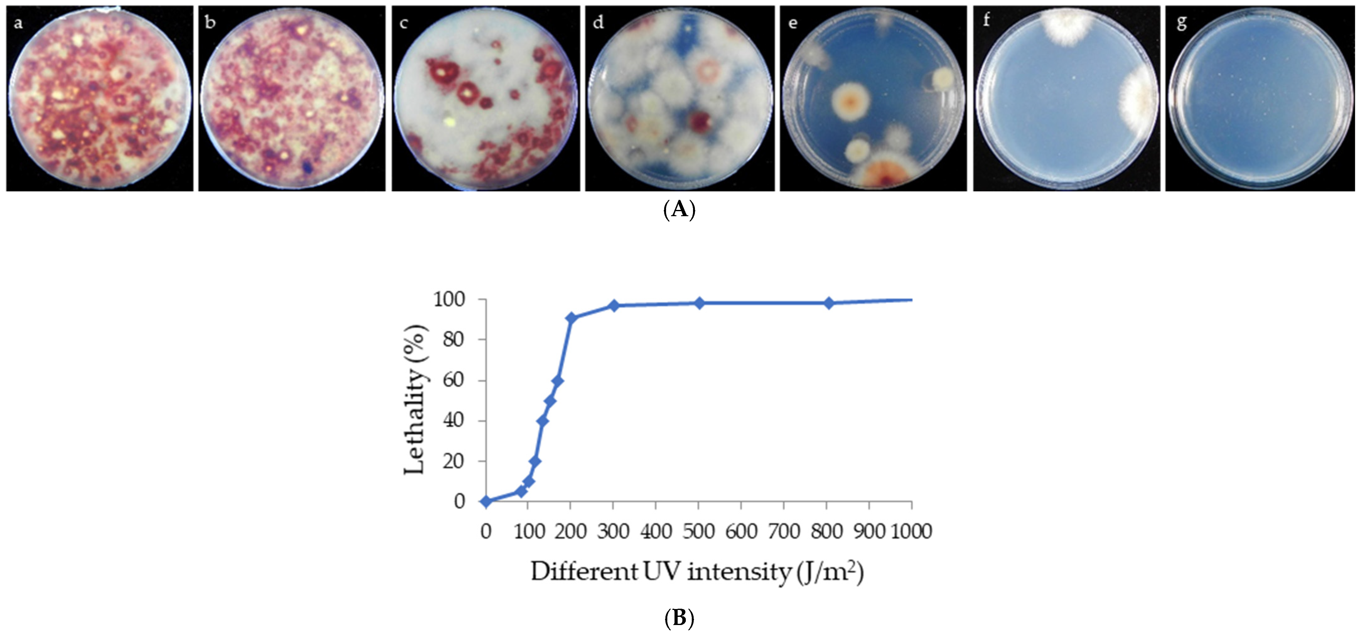
The PDA plates were separately submerged in 10 mL of sterile ultra-pure water (containing 0.02% Tween-80), and the spores were suspended to a concentration of 106 spores/mL [50]. The spore suspensions were then exposed to varying doses of UV radiation (100 J/m2, 120 J/m2, 150 J/m2, 300 J/m2, 800 J/m2, and 1000 J/m2) at room temperature. The distance between the spore suspensions and the UV lamp was 30–40 cm, and these were exposed for 60 s, 90 s, 100 s, 3 min, 8 min, and 10 min separately. After exposure, the spore suspension was uniformly spread onto PDA medium and incubated at 25 °C for 7 days in the dark [36,37,38]. Three independent biological replicates were performed for each treatment.

2.3. Lethality Assay

Spore mortality was analyzed to determine the lethality of different UV intensities. Survival data were fitted to the below equation
S = D0/D × 100%,
and the lethality data to the equation
L = (D − D0)/D × 100%,
where S, L, D, and D0 represent the survival fraction, lethality fraction, total number of viable colonies before UV treatment, and number of viable colonies after UV treatment, respectively.

2.4. Preliminary Screening of High-Yield Mutants

Following the germination of UV-treated spores, five mutant strains were randomly selected from each PDA plate exposed to different UV intensities. These strains were then inoculated onto new PDA plates and incubated at 25 °C for 7 days in the dark for preliminary screening.
Perylenequinonoid compounds are shown as red pigments under visible light, enabling direct morphological observation for screening high-yield strains. Next, five strains were individually selected for further study based on the growth rate and pigment.

2.5. Growth Rate Determination and Microscopic Observations of Mycelial Morphology

The original strain zzz816 and the mutant strains were simultaneously inoculated onto PDA plates and cultured at 25 °C in the dark for 7 days. The colony diameters of each strain were measured and the mycelial morphology of each strain was observed using a light microscope (Olympus DP71, Tokyo, Japan).

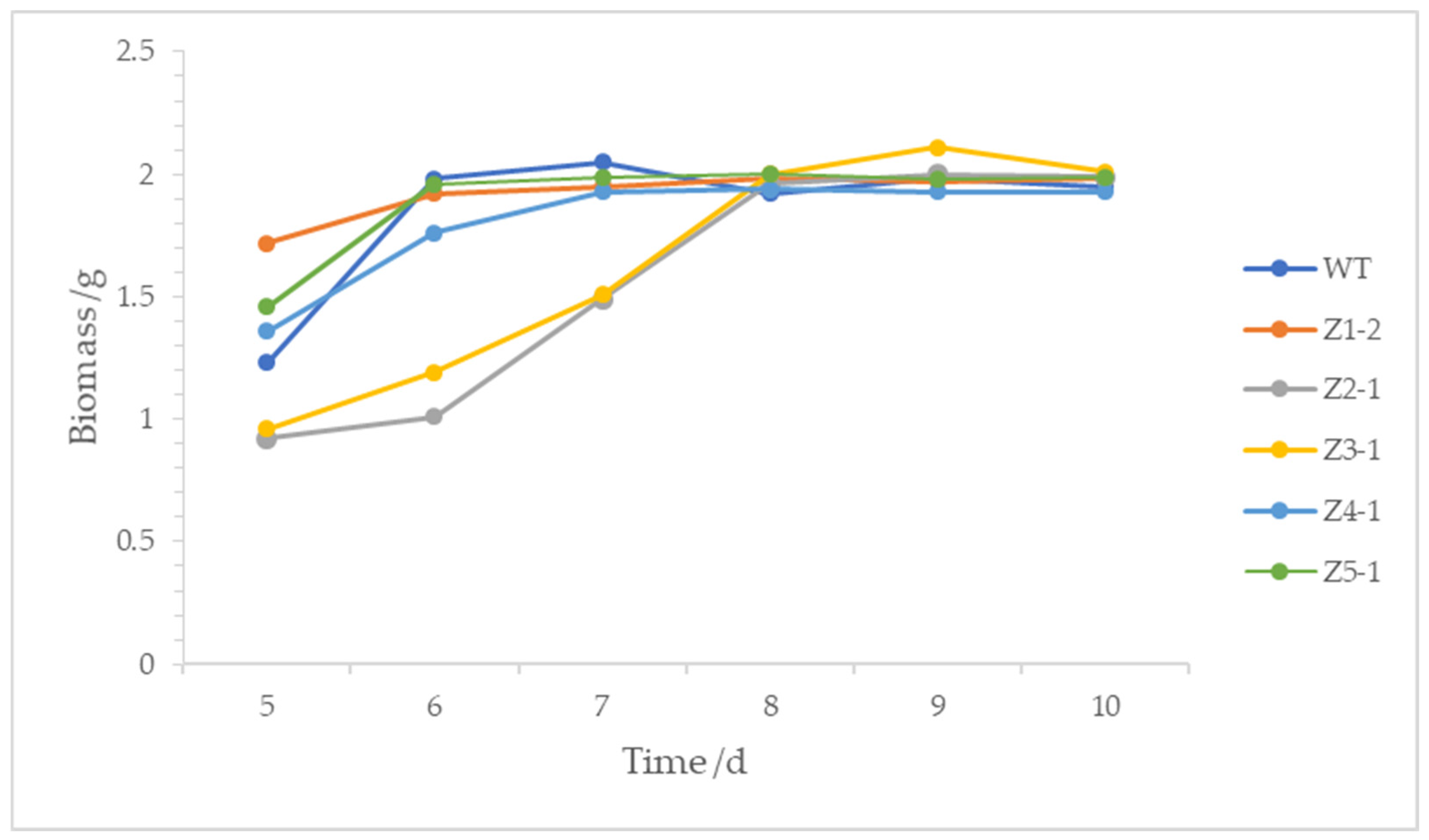
2.6. Analysis of Fermentation Time and Mycelial Biomass

The original strain zzz816 and the mutant strains were inoculated into 250-mL Erlenmeyer flasks containing 150 mL PDB liquid medium and shaken at 180 rpm at 26 °C. Mycelia were collected at different time points (5–10 days), filtered, freeze-dried, and weighed to plot the growth curves.

2.7. Extraction of Intracellular Perylenequinonoid Compounds

The original strain zzz816 and the selected mutant strains were inoculated onto PDA plates and incubated for 3 days, then transferred to PDB medium as described above. After 7 days of cultivation, the fermented mycelia were harvested by centrifugation at 12,000 rpm for 10 min at 4 °C, rinsed three times with distilled water, and vacuum freeze-dried. The mycelia were ground into powder using liquid nitrogen, and 0.5 g of the mycelium pellets was accurately weighed and chemically extracted with 150 mL of absolute ethanol via Soxhlet extraction for 12 h at 95 °C. The ethanol was evaporated under vacuum at 45 °C, and the residues were dissolved in 10 mL methanol for HPLC analysis.

2.8. HPLC Analysis Conditions

The content of perylenequinonoid compounds in the extract was analyzed using an Agilent 1200 Series high performance liquid chromatography system equipped with a Kromasil 100-5C18 (250 × 4.6 mm) column (Nouryon, Bohus, Sweden). The operating conditions included the flow rate of 1.0 mL/min, the column temperature of 35 °C, and the sample volume of 20 μL. The detection wavelength was 460 nm. The elution gradient is detailed in Table 1 [32].

2.9. Analysis of Perylenequinonoid Compounds

Standards of hypocrellin A (HA), hypocrellin B (HB), and shiraiachrome A (SA), purity ≥ 98% (HPLC), were purchased from Biopurify Phytochemicals Ltd. (Chengdu, China). Standards of elsinochrome A (EA), elsinochrome B (EB), purity ≥ 98% (HPLC), were purchased from Hangzhou Viablife Biotech Co. Ltd. (Hangzhou, China). The standard samples of perylenequinonoid compounds were analyzed by HPLC. Their identification was confirmed by comparing the retention times and spectroscopic data [32]. Quantitative analysis was performed using standard curves, prepared according to the method described by Tong et al. [48]. More detailed information about the methodology of quantification is provided in the Supplementary Materials.

2.10. Genetic Stability Test of Perylenequinonoid Compounds from Mutant Strains

To verify the stability of perylenequinonoid compound production, mutant strains were cultured continuously on PDB medium for five generations. The content of perylenequinonoid compounds was analyzed by HPLC for each generation.

3. Results

3.1. Selection of Mutant Strains and Lethality Analysis

Spores of Pseudoshiraia conidialis were treated with various UV intensities (100 J/m2, 120 J/m2, 150 J/m2, 300 J/m2, 800 J/m2, and 1000 J/m2). Lethality rates were calculated based on the number of colonies surviving after UV exposure, incubated at 25 °C for 3 days on PDA medium (Figure 2A). Figure 2B shows that spore lethality increased with the UV intensity. When the spores were treated with UV intensity from 100 J/m2 to 300 J/m2, the lethality of spores was enhanced sharply; at 150 J/m2, over half of the spores were non-viable, and no spores survived at 1000 J/m2.

3.2. Preliminary Screening of High-Yield Mutant Strains

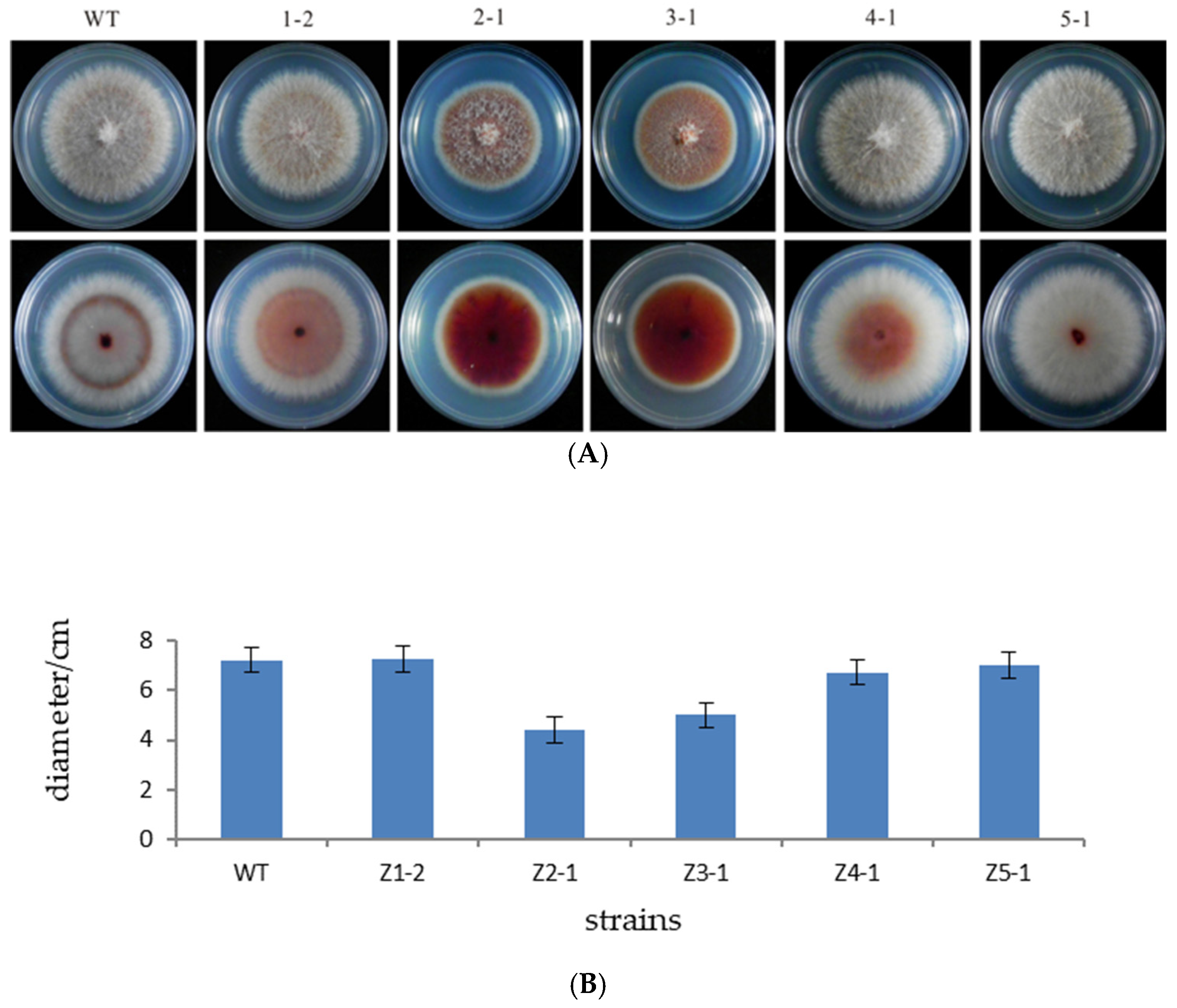
As listed in Table 2, the strains were treated with different UV intensities. Then, based on a comprehensive evaluation including the growth rate and mycelial pigment, Z1-2, Z2-1, Z3-1, and Z4-1 were selected for further study.

3.3. Determination of Growth Rate

The original strain zzz816 and the mutant strains (Z1-2, Z2-1, Z3-1, Z4-1, and Z5-1) were simultaneously inoculated onto PDA plates and cultured at 25 °C in the dark for 7 days. Colony diameters (Figure 3A) were measured to analyze the growth rates. The growth rates of the mutant strains Z2-1 and Z3-1 were significantly reduced by 39.14% and 30.84%, respectively (Figure 3B).

3.4. Microscopic Observations of Mycelium Morphology

Microscopic examination revealed that the original strain’s mycelia were more filamentous, while the mycelia of the Z2-1 and Z3-1 mutants were notably spherical, thicker, and shorter (Figure 4).

3.5. Analysis of Fermentation Time and Mycelia Biomass

As shown in Figure 5, compared with the original strain zzz816, Z1-2 exhibited a longer fermentation period, with maximum biomass on the eighth day. Z2-1 and Z3-1 also had extended fermentation periods, with the biomass increasing rapidly from the fifth to the eighth day before stabilizing. Z5-1 showed a similar pattern, reaching maximum biomass on the sixth day. Z4-1’s fermentation period remained unchanged, with maximum biomass on the seventh day. Overall, the maximum biomasses of the mutant strains did not significantly differ from the original strain.

3.6. Identification and Analysis of Perylenequinonoid Compounds

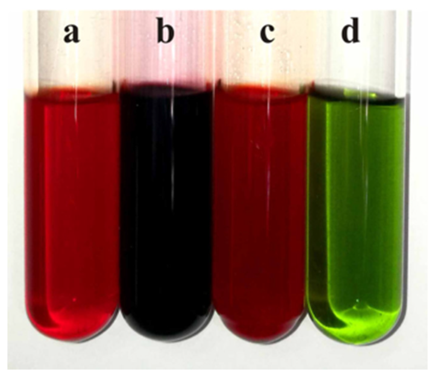
Chemical color response tests were employed to verify the perylenequinonoid compounds in mutant strains. Extracts containing such compounds turned black with FeCl3 solution, red with acidic solutions, and green with alkaline solutions (Figure 6).
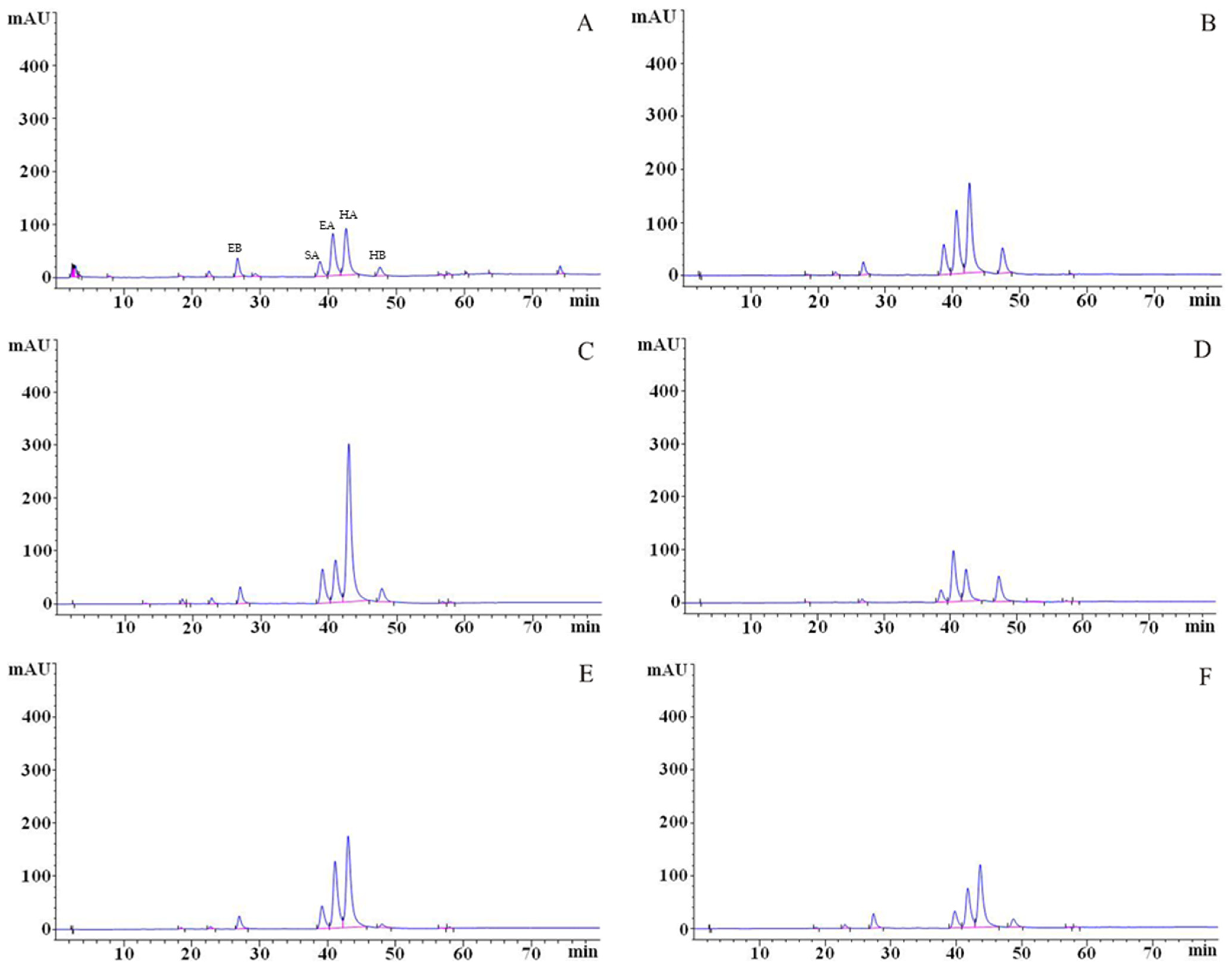
The perylenequinonoid compounds were identified through HPLC analysis (Figure 7), with characterization based on a comparative evaluation of UV–Vis spectra, chromatographic profiles, and retention times against standards [32]. Five sharp peaks were observed in the HPLC chromatogram of the original strain zzz816: elsinochrome B (EB) at 26.331 min, shiraiachrome A (SA) at 38.129 min, elsinochrome A (EA) at 39.955 min, hypocrellin A (HA) at 41.912 min, and hypocrellin B (HB) at 46.808 min.

3.7. Quantitative Determination of Perylenequinonoid Compounds

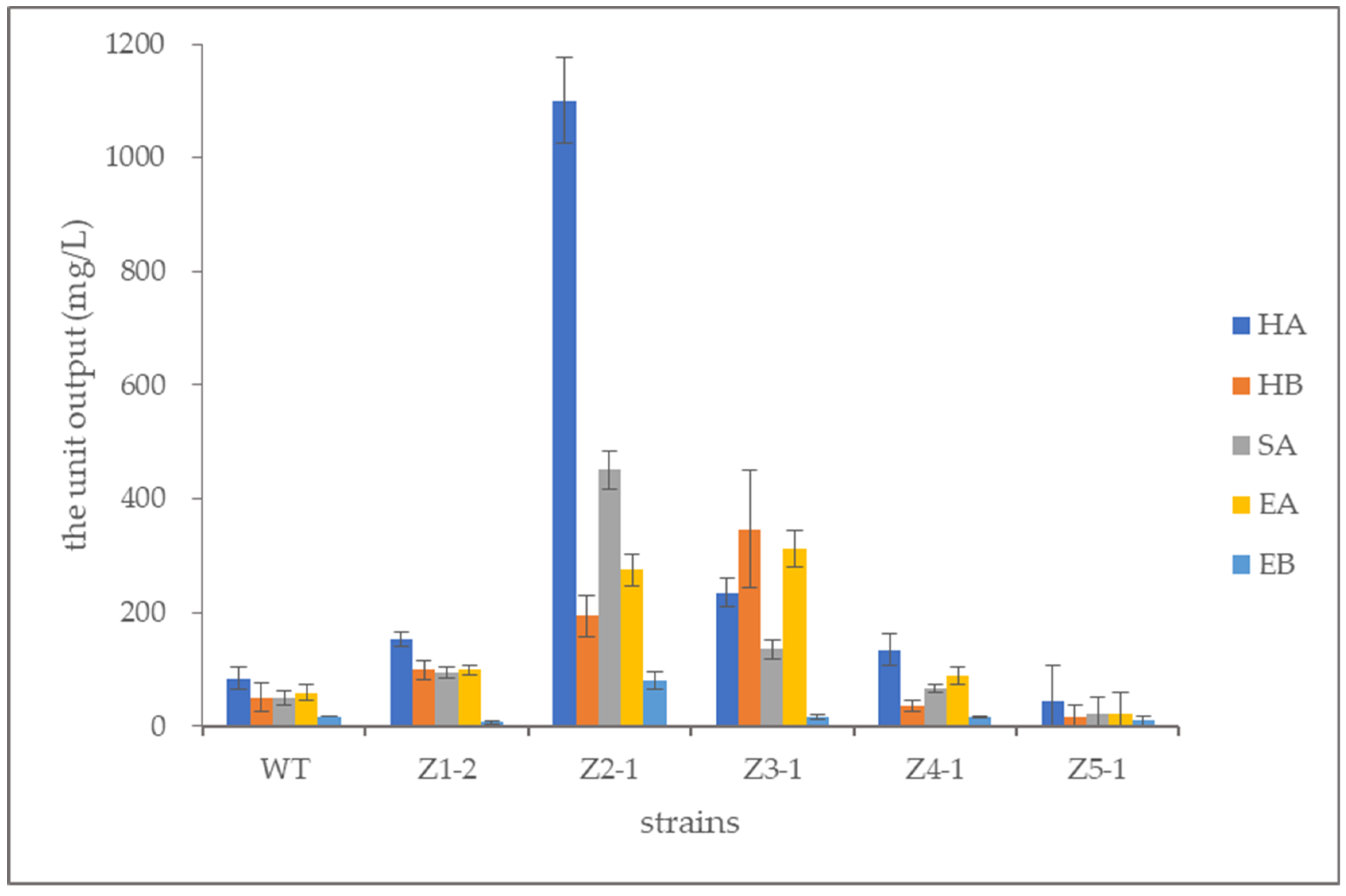
Standard curves were plotted using perylenequinonoid compound standards. For each sample, the content of each perylenequinonoid compound and the total content were calculated (Table 3 and Table 4, and Figure 8). Results showed that Z2-1 and Z3-1 produced higher amounts of perylenequinonoid compounds, corresponding to the UV treatments with intensities of 120 J/m2 and 150 J/m2, respectively.
Among the mutants, Z2-1 exhibited the highest total yield of perylenequinonoid compounds at 2101.6 mg/L, a 705.70% increase over the original strain (261.20 mg/L). Hypocrellin A production in Z2-1 reached 1100.7 mg/L, a 1208.02% increase from the original strain (84.15 mg/L). The hypocrellin B yield was 194.02 mg/L, a 286.88% increase from the original strain (50.15 mg/L). The shiraiachrome A yield was 451.20 mg/L, an 806.21% increase from the original strain (49.79 mg/L), while the elsinochrome A yield was 275.28 mg/L, a 365.95% increase from the original strain (59.08 mg/L). The elsinochrome B yield was 80.40 mg/L, a 355.01% increase from the original strain (17.67 mg/L) (Figure 8).
Z3-1 produced 312.68 mg/L of elsinochrome A, a 429.25% increase from the original strain (59.08 mg/L). Notably, elsinochrome A production in Z3-1 exceeded that of other components. Additionally, hypocrellin B production in Z3-1 significantly increased, surpassing hypocrellin A production—a phenomenon not previously reported. Through the analysis, we found not only that hypocrellin A significantly increased, but also that other types of perylenequinonoid compounds had also been greatly improved and displayed a high yield. Furthermore, the production of hypocrellin A and the total perylenequinonoid compounds in Z2-1 reached high levels (Figure 8).

3.8. Genetic Stability of Perylenequinone-Producing Mutant Strains Z2-1 and Z3-1

The high-yielding perylenequinone-producing mutant strains, designated as Z2-1 and Z3-1, were subjected to a stability evaluation through five successive subcultures. The production stability of perylenequinonoid compounds in these mutants was subsequently analyzed using HPLC. Results demonstrated that despite occasional minor fluctuations, both mutant strains maintained steady production yields through five generations, as detailed in Table 5.

4. Discussion

UV mutagenesis, known for its reliability, simplicity, and significant mutagenic results, has been widely used in microbial breeding. For example, it has been employed to enhance vitamin B12 production in Propionibacterium freudenreichii (Propionibacterium shermanii) [51] and nucleoside production in Cordyceps kyushuensis [52]. A previous study conducted complex mutagenesis and increased hypocrellin A production in Shiraia bambusicola to 80.4 mg/L, a 167.1% increase over the original strain [37]. Cobalt mutagenesis in Shiraia sp. achieved a hypocrellin A production of 2018.30 mg/L [36], and calcium/calmodulin signal transduction improved the perylenequinonoid compound yield to 1894.66 mg/L [38].
However, a significant gap still exists in strain breeding around the zzz816 group. Furthermore, a recent report revealed the distinct transcriptional regulation mechanisms between zzz816 and JAP103846 [33,39]. This suggests that zzz816-related strains would possess greater potential for genetic modification. Combined with their inherent advantages, including a higher native hypocrellin yield and the absence of inducer requirements [31,36,40], these strains indicate considerable potential for production applications.
In this study, UV irradiation was employed for the mutagenesis strategy due to its dual role in inducing DNA mutation and triggering the oxidative stress response. Crucially, it has been proven that the appearance of ROS could enhance hypocrellin biosynthesis in Shiraia-like fungi [43,44,45,46]. As anticipated, this single-factor UV mutagenesis yielded successfully multiple mutants with improved hypocrellin production. This outperformed many JAP103846 strains with complex, multi-factor mutagenesis approaches, highlighting the greater efficiency of this method in Shiraia-like fungi.
Furthermore, some promising mutants, covering Z2-1 and Z3-1, exhibited a growth pattern similar to that observed in JAP103846: enhanced intracellular hypocrellin production correlates with increased growth inhibition. This inhibitory effect may be attributed to the intrinsic photosensitizing properties of hypocrellins, which can interfere directly with normal fungal growth [53,54]. However, it is encouraging that the growth rates of the Z2-1 and Z3-1 mutants were not hindered as badly as the engineered JAP103846, and the screening of faster-growing variants during serial subculturing can effectively compensate for the deficiency.
It is noteworthy that using only a straightforward UV-mutagenesis approach, we collected several mutants with significantly enhanced performance and commercial value. This demonstrates that isolates from the zzz816 clade may more effectively utilize a metabolic regulatory mechanism distinct from the other strains. Correspondingly, the special transcription factor SbTF for hypocrellin synthesis also performs different regulatory functions across various genetic backgrounds [33], while mitochondrial genome-encoded nad and cox family proteins show distinct expression patterns [40]. Likewise, the analysis of transcriptome data from different sources highlighted substantial differences in the core genes for hypocrellin synthesis (NCBI Sequence Read Archive (SRA) database with the BioProject accession numbers PRJNA323638, PRJNA475310, PRJNA477419, and PRJNA544773). Moving forward, we plan to reorganize published omics data according to current molecular classifications and combine them with the expression changes of key genes from high-yield strains to better understand the underlying regulatory mechanism.
However, these studies focused solely on hypocrellin A or crudely estimated mixed perylenequinonoid compounds. Our results demonstrate that other perylenequinonoid compounds also play significant roles in the final products, sometimes even surpassing hypocrellin A in certain strains like Z2-1 and Z3-1. Neglecting these compounds could lead to significant waste and inconsistent product quality. In this study, a modified analytical system was used to simultaneously quantify five different perylenequinonoid compounds (HA, HB, SA, EA, and EB). The results revealed diverse proportions of these compounds in different mutants, with elsinochrome A surpassing hypocrellin A in Z3-1. Therefore, focusing solely on hypocrellin A underestimates the productive capacity of these strains. Given the distinct properties and effects of these compounds, particularly between hypocrellins and elsinochromes, it is crucial to measure each component separately. This study’s approach, simultaneously detecting each component, lays a solid foundation for evaluating diverse strains with unique properties.
In this study, all mutants achieved similar maximum biomass levels under the same fermentation conditions. From a theoretical perspective, shorter fermentation times are more beneficial. To optimize time and energy, future research should focus on improving the media composition and submerged fermentation conditions through statistically designed experiments.
UV irradiation is a conventional mutagenesis approach that induces mutations to enhance production while maintaining normal growth pattern. These strains can generally still undergo further optimization through transgenic modifications, fermentation adjustments, and EMS [33,39,49]. The UV-mutagenized variants also exhibit substantial potential for further improvement via compound mutagenesis.

Supplementary Materials

The following supporting information can be downloaded at: https://www.mdpi.com/article/10.3390/microorganisms13091999/s1, Table S1: Concentrations of the standard stock solutions, Table S2: Results of the linear regression analysis, Table S3: Precision evaluation of HPLC method for perylenequinone standard analysis, Table S4: Stability evaluation of perylenequinone standards over 24 h.

Author Contributions

Conceptualization, X.T., X.-Y.S., M.-R.H. and C.-L.H.; Data curation, X.T.; Formal analysis, X.T.; Funding acquisition, X.T., M.-R.H. and C.-L.H.; Methodology, X.T., X.-Y.S. and C.-L.H.; Project administration, X.-Y.S. and C.-L.H.; Supervision, X.-Y.S. and C.-L.H.; Writing—original draft, X.T.; Writing—review and editing, X.T., X.-Y.S., M.-R.H. and C.-L.H. All authors have read and agreed to the published version of the manuscript.

Funding

This research was supported by the BJAST Budding Talent Program (23CE-BGS-06), the Beijing Government, and the National Natural Science Foundation of China (No. 32270012).

Institutional Review Board Statement

Not applicable.

Informed Consent Statement

Not applicable.

Data Availability Statement

The original contributions presented in this study are included in the article/Supplementary Materials. Further inquiries can be directed to the corresponding author.

Acknowledgments

Bao-Dong He is acknowledged for providing valuable suggestions.

Conflicts of Interest

The authors declare no conflicts of interest.

References

  1. Arnone, A.; Camarda, L.; Nasini, G.; Merlini, L. Cheminform abstract: Secondary mold metabolites. Part 13. Fungal perylenequinones: Phleichrome, isophleichrome, and their endoperoxides. J. Chem. Soc. Perkin Trans. 1985, 16, 1387–1392. [Google Scholar] [CrossRef]
  2. Lee, J.K.; Kim, B.T.; Kim, J.A.; Chung, H.J.; Park, S.M.; Yang, M.S.; Hwang, K.J.; Kim, D.H. Cultural characteristics and extraction of the fungal pigment phleichrome from the phytopathogenic fungus Cladosporium phlei. Biotechnol. Bioprocess Eng. 2007, 12, 508–515. [Google Scholar] [CrossRef]
  3. Chen, W.X.; Cheng, Y.T.; Wan, X.Y.; Friedrichs, E.; Puff, H.; Breitmaier, E. Die Struktur des Hypocrellins und seines Photooxidationsproduktes Peroxyhypocrellin. Liebigs Ann. Chem. 1981, 1981, 1880–1885. [Google Scholar]
  4. Wan, X.Y.; Chen, Y.T. Hypocrellin A-A new drug for photochemotherapy. Chin. Sci. Bull. 1981, 26, 1040–1042. [Google Scholar]
  5. Wan, X.Y.; Zhang, W.L.; Wang, Q.F. Isolation and identification of hypocrellin B from Hypocrella bambusae. J. Yunnan Univ. 1985, 4, 461–463. [Google Scholar]
  6. Diwu, Z.J.; Lown, J.W. Hypocrellins and their use in photosensitization. Photochem. Photobiol. 1990, 52, 609–616. [Google Scholar] [CrossRef]
  7. Du, W.; Liang, J.; Han, Y.; Yu, J.; Liang, Z. Nitric oxide mediates hypocrellin accumulation induced by fungal elicitor in submerged cultures of Shiraia bambusicola. Biotechnol. Lett. 2015, 37, 153–159. [Google Scholar] [CrossRef] [PubMed]
  8. Wu, H.; Lao, X.F.; Wang, Q.W.; Lu, R.R.; Shen, C.; Zhang, F.; Liu, M.; Jia, L. The shiraiachromes: Novel fungal perylenequinone pigments from Shiraia bambusicola. J. Nat. Prod. 1989, 52, 948–951. [Google Scholar] [CrossRef]
  9. Kishi, T.; Tahara, S.; Taniguchi, N.; Tsuda, M.; Tanaka, C.; Takahashi, S. New perylenequinones from Shiraia bambusicola. Planta Med. 1991, 57, 376–379. [Google Scholar] [CrossRef]
  10. Fang, L.Z.; Qing, C.; Shao, H.J.; Yang, Y.D.; Dong, Z.J.; Wang, F.; Zhao, W.; Yang, W.Q.; Liu, J.K. Hypocrellin D, a cytotoxic fungal pigment from fruiting bodies of the ascomycete Shiraia bambusicola. J. Antibiot. 2006, 59, 351–354. [Google Scholar] [CrossRef]
  11. Su, Y.J.; Si, S.H.; Qiao, L.W.; Cai, Y.J.; Xu, Z.M.; Yang, Y.J. The effect of a hypocrellin A enriched diet on egg yolk quality and hypocrellin A distributions in the meat of laying hens. Eur. Food Res. Technol. 2011, 232, 935–940. [Google Scholar] [CrossRef]
  12. Khiralla, A.; Mohammed, A.O.; Yagi, S. Fungal perylenequinones. Mycol. Prog. 2022, 21, 38. [Google Scholar] [CrossRef]
  13. Bao, Z.; Xie, Y.; Xu, C.; Zhang, Z.; Zhu, D. Biotechnological production and potential applications of hypocrellins. Appl. Microbiol. Biotechnol. 2023, 107, 6421–6438. [Google Scholar] [CrossRef]
  14. Xie, W.; Wei, S.; Liu, J.; Ge, X.; Zhou, L.; Zhou, J.; Shen, J. Combination anticancer therapy activity studies for the complex of hypocrellin A and gallium ion. Dye. Pigment. 2014, 101, 43–50. [Google Scholar] [CrossRef]
  15. Hirayama, J.; Ikebuchi, K.; Abe, H.; Kwon, K.W.; Ohnishi, Y.; Horiuchi, M.; Shinagawa, M.; Ikuta, K.; Kamo, N.; Sekiguchi, S. Photoinactivation of virus infectivity by hypocrellin A. Photochem. Photobiol. 1997, 66, 697–700. [Google Scholar] [CrossRef]
  16. Hudson, J.B.; Zhou, J.; Chen, J.; Harris, L.; Yip, L.; Towers, G.H.N. Hypocrellin, from Hypocrella bambuase, is phototoxic to human immunodeficiency virus. Photochem. Photobiol. 1994, 60, 253–255. [Google Scholar] [CrossRef] [PubMed]
  17. Ogawara, K.I.; Higaki, K. Nanoparticle-Based Photodynamic Therapy: Current Status and Future Application to Improve Outcomes of Cancer Treatment. Chem. Pharm. Bull. 2017, 65, 637–641. [Google Scholar] [CrossRef]
  18. Guo, L.Y.; Yan, S.Z.; Li, Q.; Xu, Q.; Lin, X.; Qi, S.S.; Yu, S.Q.; Chen, S.L. Poly(lactic-co-glycolic) acid nanoparticles improve oral bioavailability of hypocrellin A in rat. RSC Adv. 2017, 7, 42073–42082. [Google Scholar] [CrossRef]
  19. Lin, X.; Yan, S.Z.; Qi, S.S.; Xu, Q.; Han, S.S.; Guo, L.Y.; Zhao, N.; Chen, S.L.; Yu, S.Q. Transferrin-Modified Nanoparticles for Photodynamic Therapy Enhance the Antitumor Efficacy of Hypocrellin A. Front. Pharmacol. 2017, 8, 815–831. [Google Scholar] [CrossRef]
  20. Mulrooey, C.A.; O’Brien, E.M.; Morgan, B.J.; Kozlowski, M.C. Perylenequinones: Isolation, Synthesis, and Biological Activity. Eur. J. Org. Chem. 2012, 21, 3887–3904. [Google Scholar] [CrossRef] [PubMed]
  21. Chang, J.E.; Cho, H.J.; Yi, E.; Kim, D.D.; Jheon, S. Hypocrellin B and paclitaxel-encapsulated hyaluronic acid-ceramide nanoparticles for targeted photodynamic therapy in lung cancer. J. Photochem. Photobiol. B 2016, 158, 113–121. [Google Scholar] [CrossRef] [PubMed]
  22. Jiang, Y.; Leung, A.W.; Wang, X.; Zhang, H.; Xu, C. Effect of photodynamic therapy with hypocrellin B on apoptosis, adhesion, and migration of cancer cells. Int. J. Radiat. Biol. 2014, 90, 575–579. [Google Scholar] [CrossRef]
  23. Ma, J.; Jiang, L. Photogeneration of singlet oxygen (1O2) and free radicals (Sen·−, O·−2) by tetra-brominated hypocrellin B derivative. Free Radic. Res. 2001, 35, 767–777. [Google Scholar] [CrossRef]
  24. Hu, Y.; Zhang, C.; Li, S.; Jiao, Y.; Qi, T.; Wei, G.; Han, G. Effects of Photodynamic Therapy Using Yellow LED-light with Concomitant Hypocrellin B on Apoptotic Signaling in Keloid Fibroblasts. Int. J. Biol. Sci. 2017, 13, 319–326. [Google Scholar] [CrossRef]
  25. Weiss, U.; Flon, H.; Burger, W.C. The photodynamic pigment of some species of Elsinoë and Sphaceloma. Arch. Biochem. Biophys. 1957, 69, 311–319. [Google Scholar] [CrossRef]
  26. Weiss, U.; Ziffer, H.; Batterham, T.J.; Blumer, M.; Hackeng, W.H.; Copier, H.; Salemink, C.A. Pigments of Elsinoë species. I. Pigment production by Elsinoë species; Isolation of pure elsinochromes A, B, C. Can. J. Microbiol. 1965, 11, 57–66. [Google Scholar] [CrossRef]
  27. Lousberg, R.J.J.C.; Paolillo, L.; Kon, H.; Weiss, U.; Salemink, C.A. Pigments of Elsinoe species. Part IV. Confirmatory evidence for the structure of elsinochrome A and its ethers from studies of nuclear magnetic resonance (solvent and overhauser effects) and electron spin resonance. J. Chem. Soc. C Org. 1970, 16, 2154–2159. [Google Scholar] [CrossRef]
  28. Ma, F.; Zhou, L.; Wang, W.; Feng, Y.Y.; Zhou, J.H.; Wang, X.H.; Shen, J. Spectroscopic studies on the interaction of Elsinochrome A with myoglobin. Spectrosc. Spect. Anal. 2011, 31, 1601–1605. [Google Scholar] [CrossRef]
  29. Li, T.; Deng, H.; Zhao, J.; Gu, Y. Elsinochrome A photosensitizers: Alternative drugs for photodynamic therapy. J. Innov. Opt. Health Sci. 2015, 8, 1530001–1530010. [Google Scholar] [CrossRef]
  30. Chooi, Y.H.; Zhang, G.; Hu, J.; Muria-Gonzalez, M.J.; Tran, P.N.; Pettitt, A.; Maier, A.G.; Barrow, R.A.; Solomon, P.S. Functional genomics-guided discovery of a light-activated phytotoxin in the wheat pathogen Parastagonospora nodorum via pathway activation. Environ. Microbiol. 2017, 19, 1975–1986. [Google Scholar] [CrossRef] [PubMed]
  31. Shen, X.Y.; Cheng, Y.L.; Cai, C.J.; Fan, L.; Gao, J.; Hou, C.L. Diversity and Antimicrobial Activity of Culturable Endophytic Fungi Isolated from Moso Bamboo Seeds. PLoS ONE 2014, 9, e95838. [Google Scholar] [CrossRef]
  32. Tong, X.; Wang, Q.T.; Shen, X.Y.; Hou, C.L.; Cannon, P.F. Phylogenetic Position of Shiraia-Like Endophytes on Bamboos and the Diverse Biosynthesis of Hypocrellin and Hypocrellin Derivatives. J. Fungi 2021, 7, 563. [Google Scholar] [CrossRef]
  33. Lu, Z.M.; Zhang, R.T.; Huang, X.B.; Cao, X.T.; Shen, X.Y.; Fan, L.; Hou, C.L. Optimisation of hypocrellin production in Shiraia-like fungi via genetic modification involving a transcription factor gene and a putative monooxygenase gene. Mycology 2023, 15, 272–281. [Google Scholar] [CrossRef]
  34. Cai, Y.; Liang, X.; Liao, X.; Ding, Y.; Sun, J.; Li, X. High-yield hypocrellin A production in solid-state fermentation by Shiraia sp. SUPER-H168. Appl. Biochem. Biotechnol. 2010, 160, 2275–2286. [Google Scholar] [CrossRef]
  35. Shen, X.Y.; Hu, Y.J.; Song, L.; Hou, C.L. Improvement of hypocrellin production by a new fungal source and optimization of cultivation conditions. Biotechnol. Biotechnol. Equip. 2016, 30, 819–826. [Google Scholar] [CrossRef]
  36. Liu, X.Y.; Shen, X.Y.; Fan, L.; Gao, J.; Hou, C.L. High-efficiency biosynthesis of hypocrellin A in Shiraia sp. using gamma-ray mutagenesis. Appl. Microbiol. Biotechnol. 2016, 100, 4875–4883. [Google Scholar] [CrossRef] [PubMed]
  37. Dong, T.; Pan, W.; Zhao, Y.; Lei, X.; Chen, K.; Wang, J. Screening of higher hypocrellin A with strains of Shiraia bambusicola by genome-shuffling. Chin. J. Bioprocess Eng. 2012, 10, 25–29. [Google Scholar] [CrossRef]
  38. Liu, B.; Bao, J.; Zhang, Z.; Yan, R.; Wang, Y.; Yang, H.; Zhu, D. Enhanced production of perylenequinones in the endophytic fungus Shiraia sp. Slf14 by calcium/calmodulin signal transduction. Appl. Microbiol. Biotechnol. 2018, 102, 153–163. [Google Scholar] [CrossRef] [PubMed]
  39. Deng, H.; Liang, X.; Liu, J.; Zheng, X.; Fan, T.P.; Cai, Y. Advances and perspectives on perylenequinone biosynthesis. Front. Microbiol. 2022, 13, 1070110. [Google Scholar] [CrossRef]
  40. Shen, X.Y.; Cao, X.T.; Huang, X.B.; Zhuo, L.; Yang, H.M.; Fan, L.; Hou, C.L. Mitochondrial genome and transcription of Shiraia-like species reveal evolutionary aspects in protein-coding genes. IMA Fungus 2025, 16, e138572. [Google Scholar] [CrossRef]
  41. Cai, Y.; Liao, X.; Liang, X.; Ding, Y.; Sun, J.; Zhang, D. Induction of hypocrellin production by Triton X-100 under submerged fermentation with Shiraia sp. SUPER-H168. New Biotechnol. 2011, 28, 588–592. [Google Scholar] [CrossRef]
  42. López-Timoner, R.; Santos-Juanes, L.; Amat, A.M.; Arfelli, F.; Cespi, D.; Passarini, F.; Polo, M.I.; Zuriaga, E.; Arques, A. Life cycle assessment of UVC-based advanced oxidation processes as quaternary treatments: Clostridium spp. inactivation and comparison with CECs removal. Sci. Total Environ. 2025, 972, 179029. [Google Scholar] [CrossRef]
  43. Menoni, M.; Alcoba, P.; Zuluaga, M.J.; Peluffo, R.D. Generation of cellular reactive oxygen and nitrogen species by exposure to ultraviolet radiation. Biophys. Rev. 2025, 17, 547–560. [Google Scholar] [CrossRef]
  44. Deng, H.; Chen, J.; Gao, R.; Liao, X.; Cai, Y. Adaptive Responses to Oxidative Stress in the Filamentous Fungal Shiraia bambusicola. Molecules 2016, 21, 1118. [Google Scholar] [CrossRef]
  45. Wang, W.J.; Li, X.P.; Shen, W.H.; Huang, Q.Y.; Cong, R.P.; Zheng, L.P.; Wang, J.W. Nitric oxide mediates red light-induced perylenequinone production in Shiraia mycelium culture. Bioresour. Bioprocess. 2024, 11, 2. [Google Scholar] [CrossRef]
  46. Wu, X.; Meng, X.; Xiao, Y.; Yang, H.; Zhang, Z.; Zhu, D. Energy Metabolism Enhance Perylenequinone Biosynthesis in Shiraia sp. Slf14 through Promoting Mitochondrial ROS Accumulation. Int. J. Mol. Sci. 2024, 25, 10113. [Google Scholar] [CrossRef] [PubMed]
  47. Beseli, A.; Noar, R.; Daub, M.E. Characterization of Cercospora nicotianae Hypothetical Proteins in Cercosporin Resistance. PLoS ONE 2015, 10, e0140676. [Google Scholar] [CrossRef]
  48. Tong, Z.W.; Mao, L.; Liang, H.; Zhang, Z.; Wang, Y.; Yan, R.; Zhu, D. Simultaneous Determination of Six Perylenequinones in Shiraiaia sp. Slf14 by HPLC. J. Liq. Chromatogr. Relat. Technol. 2017, 40, 536–540. [Google Scholar] [CrossRef]
  49. Liu, X.Y.; Fan, L.; Gao, J.; Shen, X.Y.; Hou, C.L. Global identification of alternative splicing in Shiraia bambusicola and analysis of its regulation in hypocrellin biosynthesis. Appl. Microbiol. Biotechnol. 2020, 104, 211–223. [Google Scholar] [CrossRef] [PubMed]
  50. Brandsberg, J.W.; French, M.E. In vitro susceptibility of isolates of Aspergillus fumigatus and Sporothrix schenckii to amphotericin B. Antimicrob. Agents Chemother. 1972, 2, 402–404. [Google Scholar] [CrossRef] [PubMed]
  51. Zhang, Y.; Liu, J.Z.; Huang, J.S.; Mao, Z.W. Genome shuffling of Propionibacterium shermanii for improving vitamin B12 production and comparative proteome analysis. J. Biotechnol. 2010, 148, 139–143. [Google Scholar] [CrossRef]
  52. Wang, Y.; Zhang, G.; Zhao, X.; Ling, J. Genome shuffling improved the nucleosides production in Cordyceps kyushuensis. J. Biotechnol. 2017, 260, 42–47. [Google Scholar] [CrossRef] [PubMed]
  53. Liu, X.; Fang, R.; Feng, R.; Li, Q.; Su, M.; Hou, C.; Zhuang, K.; Dai, Y.; Lei, N.; Jiang, Y.; et al. Cage-modified hypocrellin against multidrug-resistant Candida spp. with unprecedented activity in light-triggered combinational photodynamic therapy. Drug Resist. Updates 2022, 65, 100887. [Google Scholar] [CrossRef] [PubMed]
  54. Ma, G.; Khan, S.I.; Jacob, M.R.; Tekwani, B.L.; Li, Z.; Pasco, D.S.; Walker, L.A.; Khan, I.A. Antimicrobial and antileishmanial activities of hypocrellins A and B. Antimicrob. Agents Chemother. 2004, 48, 4450–4452. [Google Scholar] [CrossRef] [PubMed]
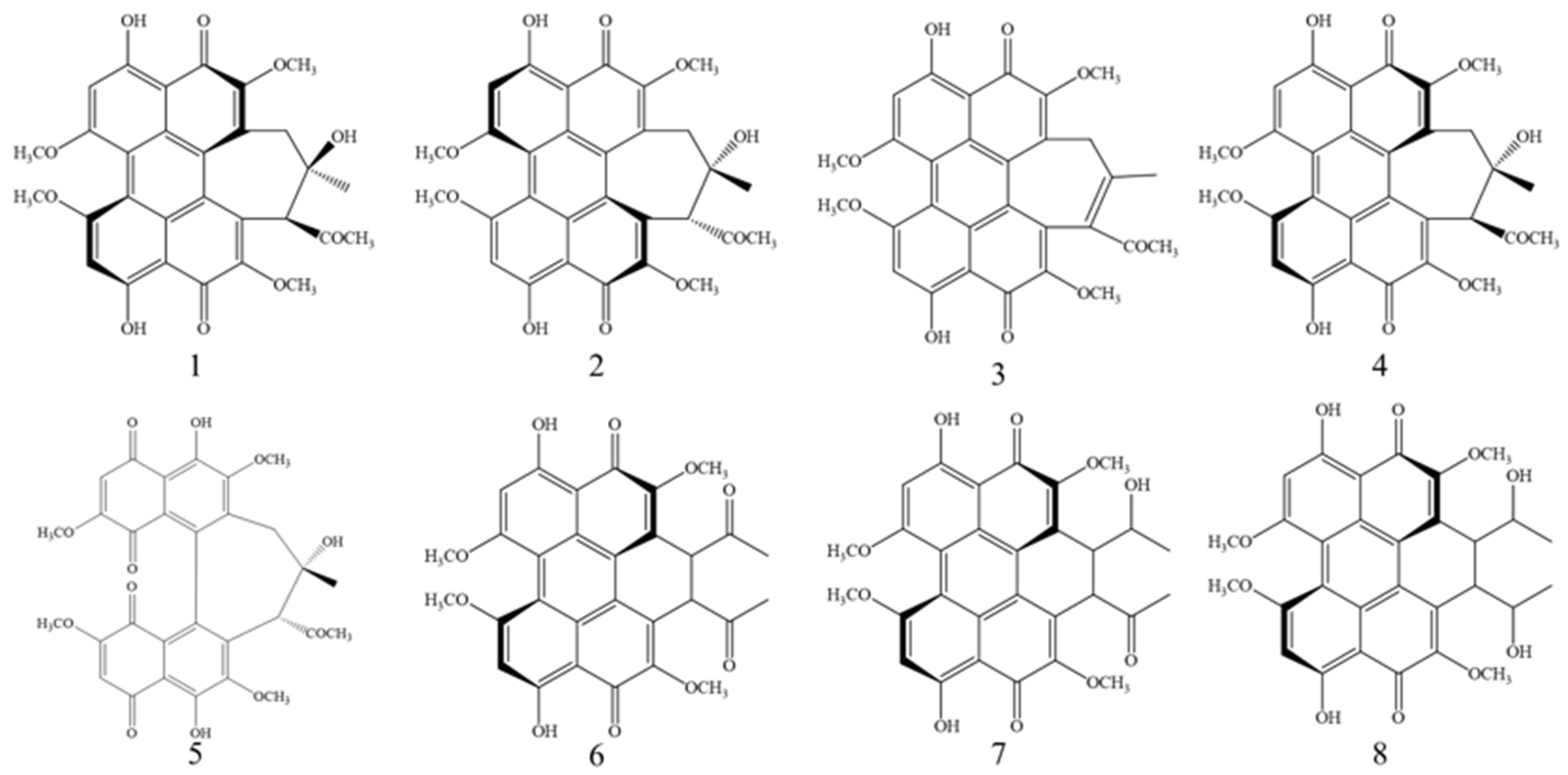
Figure 1. Chemical structures of hypocrellin (1), hypocrellin A (2), hypocrellin B (3), shiraiachrome A (4), hypocrellin D (5), elsinochrome A (6), elsinochrome B (7), and elsinochrome C (8).
Microorganisms 13 01999 g001
Figure 2. (A) Germination of spores under different UV intensities. (a) Germination of original strain zzz816. (bg) Germination of spores treated with UV intensities of 100 J/m2, 120 J/m2, 150 J/m2, 300 J/m2, 800 J/m2, and 1000 J/m2, respectively. (B) Lethality of spores treated with different UV intensities.
Microorganisms 13 01999 g002
Figure 3. Growth rate determination of the strains. (A) Growth phenotypes of the wild-type and mutant strains. The top row shows front views of 7-day-old PDA cultures of the original strain zzz816 (WT) and the mutant strains (Z1-2, Z2-1, Z3-1, Z4-1, and Z5-1). The bottom row displays the corresponding back views of the cultures. (B) Growth rate of the original strain zzz816 and the mutant strains.
Microorganisms 13 01999 g003
Figure 4. Mycelia of the original strain zzz816 (WT) and the mutant strains (Z1-2, Z2-1, Z3-1, Z4-1, and Z5-1) treated with different UV intensities under the microscope.
Microorganisms 13 01999 g004
Figure 5. Fermentation periods of the original strain zzz816 (WT) and the mutant strains (Z1-2, Z2-1, Z3-1, Z4-1, and Z5-1).
Microorganisms 13 01999 g005
Figure 6. Chemical color response test of the extracts. (a) Ethanol extract of the original strain zzz816. (b) Extract with FeCl3 solution. (c) Extract with acidic solution. (d) Extract with alkaline solution.
Microorganisms 13 01999 g006
Figure 7. HPLC chromatograms of the samples. (A) Original strain zzz816. (B) Mutant strain Z1-2. (C) The sample of mutant strain Z2-1 after fourfold dilution. (D) The sample of mutant strain Z3-1 after fourfold dilution. (E) Mutant strain Z4-1. (F) Mutant strain Z5-1.
Microorganisms 13 01999 g007
Figure 8. Perylenequinonoid compounds in the original strain and mutant strains.
Microorganisms 13 01999 g008
Table 1. The 0.1% phosphoric acid water–methanol phase of the HPLC gradient.
Time (min)0.1% Phosphoric Acid Water (%, v/v)Methanol (%, v/v)
0–104060
10–1540–3060–70
15–253070
25–4530–2570–75
45–6025–075–100
60–800100
Table 2. Strains treated with different UV intensities.
UV Intensity (J/m2)Strain Names
100Z1-1, Z1-2, Z1-3, Z1-4, Z1-5
120Z2-1, Z2-2, Z2-3, Z2-4, Z2-5
150Z3-1, Z3-2, Z3-3, Z3-4, Z3-5
300Z4-1, Z4-2, Z4-3, Z4-4, Z4-5
800Z5-1, Z5-2, Z5-3, Z5-4, Z5-5
Table 3. Perylenequinonoid compounds (mg/g) in the original and mutant strains during 7-day cultivation.
StrainsHAHBSAEAEBTotal Content
WT6.16 ± 1.383.67 ± 1.833.64 ± 0.824.32 ± 0.441.29 ± 0.0619.09 ± 4.54
Z1-211.78 ± 1.017.64 ± 1.247.26 ± 0.707.66 ± 0.630.57 ± 0.2534.91 ± 3.81
Z2-1110.81 ± 7.6919.53 ± 3.6545.42 ± 3.4327.71 ± 2.778.09 ± 1.56211.57 ± 19.10
Z3-123.35 ± 2.4734.44 ± 10.2513.41 ± 1.5631.06 ± 3.101.60 ± 0.31103.87 ± 17.69
Z4-110.49 ± 2.102.70 ± 0.785.17 ± 0.586.89 ± 1.231.26 ± 0.1326.50 ± 4.81
Z5-13.23 ± 4.791.30 ± 1.491.63 ± 2.301.72 ± 2.790.71 ± 0.648.59 ± 12.01
HA: hypocrellin A, HB: hypocrellin B, SA: shiraiachrome A, EA: elsinochrome A, EB: elsinochrome B.
Table 4. Perylenequinonoid compounds (mg/L) in the original and mutant strains during 7-day cultivation.
StrainsHAHBSAEAEBTotal Content
WT84.15 ± 18.8950.15 ± 25.0649.79 ± 11.2059.08 ± 14.3417.67 ± 0.79260.84 ± 70.28
Z1-2153.18 ± 13.1099.29 ± 16.1294.34 ± 9.0499.61 ± 8.147.39 ± 3.20453.81 ± 49.59
Z2-11100.70 ± 76.40194.02 ± 36.21451.20 ± 34.04275.28 ± 27.5080.40 ± 15.492101.60 ± 189.64
Z3-1235.08 ± 24.83346.68 ± 103.25135.04 ± 15.72312.68 ± 31.2316.12 ± 3.111045.60 ± 178.14
Z4-1134.91 ± 27.0234.75 ± 10.0666.46 ± 7.4388.59 ± 15.8216.23 ± 1.62340.94 ± 61.95
Z5-142.91 ± 63.5617.20 ± 19.7921.63 ± 30.5022.87 ± 37.099.38 ± 8.51113.99 ±159.43
HA: hypocrellin A, HB: hypocrellin B, SA: shiraiachrome A, EA: elsinochrome A, EB: elsinochrome B.
Table 5. Yield of the total perylenequinonoid compounds in mutant strains Z2-1 and Z3-1 over five generations.
Generation NumbersMutant Strain Z2-1 (mg/L)Rate of Change (%)Mutant Strain Z3-1 (mg/L)Rate of Change (%)
12101.601045.60
22198.74.621094.34.66
32237.56.471147.89.77
42045−2.69978.2−6.45
52219.25.601121.87.29
Disclaimer/Publisher’s Note: The statements, opinions and data contained in all publications are solely those of the individual author(s) and contributor(s) and not of MDPI and/or the editor(s). MDPI and/or the editor(s) disclaim responsibility for any injury to people or property resulting from any ideas, methods, instructions or products referred to in the content.