Article

Removal of Ibuprofen from Contaminated Water by Bioaugmentation with Novel Bacterial Strains Isolated from Sewage Sludge

1
Institute of Natural Resources and Agrobiology of Seville, Spanish National Research Council (IRNAS-CSIC), 41012 Seville, Spain
2
Department of Microbiology and Parasitology, Faculty of Pharmacy, University of Seville, 41012 Seville, Spain
3
Department of Analytical Chemistry, University of Seville, C/Virgen de África 7, 41011 Seville, Spain
*
Author to whom correspondence should be addressed.
Microorganisms 2025, 13(8), 1927; https://doi.org/10.3390/microorganisms13081927
Submission received: 8 July 2025 / Revised: 2 August 2025 / Accepted: 13 August 2025 / Published: 18 August 2025
(This article belongs to the Special Issue Microbes in Wastewater Treatment)

Abstract

Ibuprofen (IBP), one of the most consumed drugs in the world, is only partially removed in Wastewater Treatment Plants (WWTPs). Its presence in effluents and sewage sludge introduces IBP into the environment. It is imperative to continue research on IBP degraders that can be used in the future to eliminate IBP at the WWTP level. This study describes the use of nine specific IBP-degrading bacteria isolated from sewage sludge (Achromobacter denitrificans, Bordetella petrii, Brucella tritici, Curtobacterium flaccumfaciens, Microbacterium paraoxydans, Pseudomonas citronellolis, Pseudomonas nitroreducens, Shinella zoogloeoides, Stenotrophomonas acidaminiphila) for the removal of IBP from water. Their half-maximal inhibitory concentration of IBP for bacterial growth (IC50) revealed a high level of IBP tolerance. Degradation of IBP (10 mg L−1) was effective for all the strains using glucose as a secondary substrate. Seven of the nine strains were shown to be IBP degraders for the first time by our research group, highlighting A. denitrificans CSW15, with almost 47% IBP degraded, and C. flacumfaciens CSW18, with 32.2% after 28 days. Three IBP transformation products were identified: 1-hydroxyibuprofen (1-OH-IBP), 2-hydroxyibuprofen (2-OH-IBP), and carboxyibuprofen (CBX-IBP). A comparison of the effectiveness of IBP degradation by the nine isolates with most other IBP-degrading bacteria previously reported was carried out.

1. Introduction

Ibuprofen (IBP), a widely used non-steroidal anti-inflammatory drug (NSAID), is one of the most consumed pharmaceuticals worldwide for the treatment of pain and inflammation. After its use by humans, it is excreted and enters wastewater treatment plants (WWTPs), where, despite the application of various treatment technologies, complete removal of IBP is not achieved, and a significant amount is released into the environment [1]. Through the continuous discharge of treated effluents from WWTPs, IBP has been detected in rivers, lakes, and even groundwater, affecting water quality and the organisms that depend on it. Moreover, IBP is relatively stable and does not easily break down in the environment, which contributes to its persistence and classification as an emerging contaminant [2]. For this reason, the European Union has identified IBP as a candidate for inclusion in the priority substance list under the Water Framework Directive [3], as recognized by the Priority Substance Directive [4] and the European Commission [5]. IBP can also be found in agricultural soils due to the use of treated wastewater for irrigation, as well as the application of sewage sludge from WWTPs used as fertilizer [6], with potential implications for food chain transfer.
Given the widespread use and ecological risks associated with IBP, its removal during wastewater and sludge treatment is of critical importance, since WWTPs are the primary entry point for IBP into the environment, with the advantage that WWTPs offer more controlled conditions (pH, temperature, carbon sources, etc.) than natural environments. Various physical and chemical methods have been proposed for IBP removal, mainly for water and wastewater, but the most promising approaches are biological methods, which offer a cost-effective and environmentally sustainable alternative, particularly those employing specific microbial strains or consortia isolated from environments with long exposure to IBP, such as WWTPs [7]. However, only a limited number of IBP-degrading isolated bacterial strains have been reported and the majority of them were identified only a few years ago [8,9,10,11,12,13,14,15,16,17].
Although most degradation studies have been conducted under laboratory conditions and have not been applied in real environmental settings, it is essential to continue researching new bacteria that specifically degrade IBP to scale up these bioremediation technologies in WWTPs in the near future under the specific environmental conditions required for their microbial activity. This study aims to investigate, in depth, the ability of nine pure bacterial strains, previously isolated from sewage sludge from a WWTP by our research group [18], to degrade IBP in contaminated water. Before degradation experiments, the tolerance of each strain to IBP will be quantified through IC50 determination. Subsequently, IBP biotransformation kinetics will be evaluated and the possible transformation products formed during the process will be identified. A comparison of their degradation effectiveness with the rest of IBP-degrading bacteria in the literature will be carried out, as well as a comparative phylogenetic analysis of these novel strains based on 16S rRNA gene sequences to search for taxonomic similarities with other IBP-degrading bacteria.

2. Materials and Methods

2.1. Chemicals and Bacterial Strains

Analytical standards of IBP (C13H18O2, purity > 98%, CAS: 15687–27–1) and IBP sodium salt (C13H17O2Na, purity > 98%, CAS: 31121–93–4) were purchased from Sigma-Aldrich (Madrid, Spain). The Luria–Bertani (LB) broth and the mineral salt medium (MSM) were prepared with analytical-grade chemicals (Sigma-Aldrich, Madrid, Spain) and autoclaved at 121 °C for 20 min before use. LB broth contained (per liter of deionized water): 10 g tryptone, 5 g yeast extract, and 10 g NaCl (BD Difco™, Fisher Scientific, Pittsburgh, PA, USA). MSM was composed of the following components (per liter of deionized water): 0.5 g of KH2PO4, 0.5 g of K2HPO4, 0.01 g of NaCl, 0.2 g of MgCl2⋅6 H2O, 0.02 g of CaCl2, 1 g of (NH4)2SO4, 0.339 mg of MnSO4, 0.428 mg of ZnSO4, 0.347 mg of (NH4)6Mo7O24⋅4 H2O, 0.4 mg of CoCl2⋅6 H2O, 5 mg of FeSO4⋅7 H2O, 0.2 mg of CuSO4⋅5 H2O, and 10 mg of EDTA. All solvents used were HPLC-grade.
IBP-degrader bacterial strains used were isolated and identified by Aguilar-Romero et al. [18] from an IBP-degrader consortium C7 obtained from fresh sewage sludge collected from a WWTP in Seville (Southwest Spain), described previously by Vargas-Ordóñez et al. [19]. The nine bacterial strains under study were isolated as described by Aguilar-Romero et al. [18], with details about cultivation and isolation conditions, pure cultures acquisition, stock culture preservation and phylogenetic analysis.
These nine bacterial strains were deposited in the GenBank database with the following accession numbers: B. tritici CSW06 (OQ859973), B. petrii CSW07 (OQ859974), M. paraoxydans CSW08 (OQ859975), P. citronellolis CSW09 (OQ859976), S. acidaminiphila CSW10 (OQ859977), S. zoogloeoides CSW12 (OQ987996), P. nitroreducens CSW13 (OQ987997), A. denitrificans CSW15 (OQ987999), C. flaccumfaciens CSW18 (OQ988002). They were selected to evaluate the biotransformation of IBP since they all demonstrated previously biodegradative abilities [18].
Phylogenetic tree analyses based on the 16S rRNA sequences were performed using the maximum-likelihood and neighbour-joining algorithms with 1000 bootstrap replications in MEGA version 11 software [20] to show the evolutionary distances among the different bacterial isolates (based on best BLAST matches).

2.2. Inoculum Preparation

Bacterial strains of the isolates were cultivated in LB medium in the presence of IBP sodium salt (200 mg L−1) (30 ± 1 °C, 150 rpm) and harvested at the beginning of the stationary phase by centrifugation at 7000 rpm (10 min). The pellets obtained were washed with MSM to completely remove the IBP and LB previously added, and then resuspended in MSM.
Bacterial growth was monitored by the optical density (OD) at 600 nm at the beginning of the assay using a VWR UV-3100 spectrophotometer (Avantor, Radnor, PA, USA), and by colony-forming units (CFU) from serial dilutions on LB medium plates. The initial cell density of each strain added to the degradation experiments was 108 CFU mL−1.

2.3. Inhibitory Concentration of IBP for Bacterial Growth

The half-maximal inhibitory concentration (IC50) for bacterial growth refers to the concentration of IBP that reduces bacterial growth by 50%. This value was determined using duplicate batch cultures in sterile 100 mL Erlenmeyer flasks. Each flask contained 20 mL of LB medium, IBP sodium salt at varying concentrations (10, 100, 500, 1000, 3000, and 5000 mg L−1), and an initial bacterial inoculum with an OD600nm of 0.1. In addition, a duplicate culture without IBP was included as a reference for normal bacterial growth.
The assay was incubated under aerobic conditions with orbital shaking at 180 rpm and 30 °C for 24 h. Bacterial growth in each culture was measured by optical density (OD600nm) using a VWR UV-3100 spectrophotometer after the incubation period. Based on these measurements, bacterial viability (%) was calculated as the OD600nm of the culture in the presence of drug concentration divided by the OD600nm of the culture without IBP. IC50 values were estimated by plotting the logarithm of IBP concentration (log [IBP]) (mg L−1) against bacterial viability (%) using Microsoft Excel.

2.4. Biodegradation Assays in Solution with Isolated IBP-Degrading Bacteria

Isolated bacterial strains were inoculated in flasks with 20 mL of MSM spiked with IBP (10 mg L−1) at an initial cell density of 108 CFU mL−1. Microcosms were incubated in triplicate on a temperature-controlled rotary shaker at 30 °C and 180 rpm under aerobic conditions for 28 days.
Taking into account that none of the nine bacterial strains under study were able to degrade IBP as a monosubstrate, they were inoculated in the presence of glucose (3 g L−1). Non-inoculated sterile controls were also prepared to observe any IBP abiotic degradation. For abiotic controls, solutions containing MSM were sterilized by autoclaving at 121 °C for 40 min, and sodium azide was added at an initial concentration of 200 mg L−1 to avoid biological activity.
At periodic intervals, the corresponding flasks were removed from the incubator to determine the remaining IBP concentration in solution. To break cell walls and recover IBP accumulated in the microbial biomass, flasks were frozen and thawed three times. Samples were centrifuged at 11,000 rpm for 2 min, and IBP concentration was determined in the supernatants as explained in Section 2.5.

2.5. Analytical Quantification of Ibuprofen and Its Transformation Products

IBP concentration was determined in the supernatant solutions as described in [17]. Briefly, a HPLC analyser with UV–Vis detector (LC-2010AHT, Shimadzu, Kyoto, Japan) was used, with a C-18 column (4 × 150 mm), mobile phase methanol: water (80:20) at pH 3 with orthophosphoric acid (1%), flow rate of 1.2 mL min−1, detection wavelength 210 nm and injection volume 25 µL. The IBP detection limit was 0.05 mg L−1.
The analysis of IBP metabolites was conducted using high-performance liquid chromatography coupled with tandem mass spectrometry (HPLC-MS/MS). Chromatographic separation was performed on an Agilent 1290 Infinity II HPLC system (Agilent, Santa Clara, CA, USA), equipped with a vacuum degasser, binary pump, autosampler, and a thermostated column compartment. The analytical column used was a Halo C18 (50 mm × 4.6 mm i.d., 2.7 µm particle size) from Teknokroma (Barcelona, Spain), protected by a matching Halo C18 guard column (5 mm × 4.6 mm i.d., 2.7 µm particle size).
The mobile phase consisted of water with 0.1% formic acid (solvent A) and acetonitrile with 0.1% formic acid (solvent B), operated under a gradient elution program as follows: 0–2 min, isocratic at 10% B; 2–4 min, linear gradient from 10% to 30% B; 4–8 min, linear gradient from 30% to 100% B; held at 100% B for 7 min; and re-equilibrated to initial conditions over 5 min. The flow rate was set at 0.6 mL min−1, with an injection volume of 10 µL, and the column temperature was maintained at 35 °C.
Mass spectrometric detection was carried out using an Agilent 6495 triple quadrupole mass spectrometer equipped with an electrospray ionization (ESI) source. The ionization parameters were as follows: capillary voltage 4000 V, drying gas flow rate 11 L min−1, drying gas temperature 350 °C, nebulizer pressure 40 psi, and fragmentor voltage 166 V. The instrument operated in both positive and negative ESI modes.
Two multiple reaction monitoring (MRM) transitions were selected for each target compound to enable both quantification and confirmation. The selected transitions and their corresponding quantifier/qualifier ion ratios are presented in Table S1. Optimized collision energies for each MRM transition are also provided in Table S1. The limits of quantification (LOQs) were 1 µg L−1 for 1-hydroxyibuprofen and 2-hydroxyibuprofen, and 10 µg L−1 for carboxyibuprofen.

2.6. Models of Biodegradation Kinetics

All IBP biodegradation curves were analyzed and fitted to the most appropriate kinetic models using the Excel tool developed by the FOCUS [21] workgroup for degradation kinetics. The fitting process employed the GRG non-linear algorithm of the Solver add-in (Microsoft), and parameter optimization was carried out using the least-squares approach.
Overall, the degradation patterns aligned well with two first-order kinetic models: the Simple First-Order (SFO) model and the biphasic First-Order model known as the Hockey-Stick (HS) model, described by the following equations:
Mt = M0 e−kt                                        (SFO)
DT50 = ln2/K                                        (SFO)
Mt = M0 e−k1 tb e−k2(t-tb)                         (HS)
DT50 = (ln 100/100 − 50)/K1                    if DT50 ≤ tb         (HS)
DT50 = tb + (ln 100/100 − 50) – K1 tb)/K2      if DT50 > tb      (HS)
In these models, Mₜ represents the IBP concentration (mg L−1) at a given time t, while M0 denotes the initial concentration immediately after spiking. The degradation rate constant is expressed as K (day−1). For the Hockey-Stick (HS) model, K1 and K2 correspond to the rate constants (day−1) associated with the rapid and slower degradation phases, respectively, and tb indicates the breakpoint time at which the rate changes. The DT50 value, or half-life, signifies the time required for the IBP concentration to decrease by 50% from its initial level.

2.7. Statistical Analysis

Standard deviations were calculated using Microsoft Excel for parameters such as biodegradation, bacterial viability and transformation products, and were included in the Figures for data comparison. The kinetic model applied in biodegradation assays was implemented using the Solver tool software from Microsoft’s statistical package. The suitability of this kinetic model was evaluated using scaled residual errors and Chi-square (χ2) tests, with a significance level set at p < 0.05.

3. Results and Discussion

3.1. Ibuprofen-Induced Inhibition of Bacterial Growth

IBP is widely recognized for its anti-inflammatory properties; however, several studies have also demonstrated that it can exhibit antibacterial activity [22]. This potential effect has been supported by research showing its ability to inhibit bacterial growth, as evidenced by the calculation of IC50 values against various bacterial strains. In this study, the IC50 was determined to assess the effect of different IBP concentrations (10, 100, 1000, 3000, and 5000 mg L−1) on the bacterial strains under study. The assay involved comparing OD600 values after 24 h of incubation in media with and without IBP. A linear correlation was observed between bacterial viability and the logarithm of IBP concentration across all strains (Figure 1).
Overall, the results indicate a high level of IBP tolerance among the isolates. The highest IC50 values were observed for B. petrii CSW07, P. nitroreducens CSW13, and C. flaccumfaciens CSW18, with values of 1353, 1247, and 908 mg L−1, respectively (Table 1). These were followed by S. zoogloeoides CSW12, A. denitrificans CSW15, P. citronellolis CSW09, and M. paraoxydans CSW08, with IC50 values of 689, 601, 450, and 279 mg L−1, respectively (Table 1).
These results suggest that these strains can tolerate high concentrations of IBP under nutrient-rich conditions and may, therefore, have strong potential for its biodegradation, taking into account that the maximum IBP concentration in influent and effluent municipal WWTPs observed by Wu et al. [7] in 25 countries were 39.8 mg L−1 and 0.059 mg L−1, respectively. Previous studies support this potential. Show et al. [23] and Wittich et al. [24] demonstrated the capacity of P. citronellolis and M. paraoxydans to efficiently degrade IBP, consistent with their high IC50 values observed in this study.
In contrast, comparative data are lacking for the other species tested. Notably, only two strains exhibited significantly low IC50 values: Stenotrophomonas acidaminiphila CSW10 and Brucella tritici CSW06, with values of 38 and 48 mg L−1, respectively (Table 1). These results suggest that these bacteria may be less effective in degrading IBP at high concentrations since differences in IC50 values reflect genetic and physiological variability that is key for the selection of strains in bioremediation processes [25].
Nonetheless, all strains were selected for further experimentation, as the IBP concentration to be tested in subsequent assays will be lower (10 mg L−1). As far as we know, no previous studies have reported IC50 values for the bacterial genera analyzed in this study in the presence of IBP, which highlights the novelty of our findings regarding their tolerance to IBP.
IBP has also been demonstrated to inhibit the growth of certain bacteria in laboratory settings, suggesting a potential antibacterial effect, particularly against specific strains. Some bacteria, like Staphylococcus aureus and Paracoccus yeei, have shown susceptibility to IBP at lower concentrations, while others, like certain Enterobacter strains, have exhibited resistance [26]. Marchlewicz et al. [10] showed that in the presence of 809 mg L−1 of this drug, there is a 50% inhibition of the growth of the Bacillus thuringiensis B1(2015b) strain, showing an IC50 value quite similar (or even lower) than those obtained for some of the nine bacteria under study (Table 1).

3.2. IBP Biodegradation in Aqueous Solution by Bacteria Isolated from Sewage Sludge

From the nine selected bacterial strains under study, none of them was capable of degrading IBP 10 mg L−1 when provided as the sole carbon source [18]. Anthropogenic pollutants tend to be resistant to biological degradation, and co-metabolic conditions are frequently applied [27], since the presence of a secondary substrate increases the biomass of the degrader microorganism and/or induces the presence of enzymes required for the degradation of the target substrate [28]. Other IBP-degrader bacteria have used glucose ([10,29,30]) or other substrates such as yeast extract ([11,23,31,32,33]), dextrose and peptone ([12,34]) or n-hexane [35] to stimulate microbial growth and enzyme production.
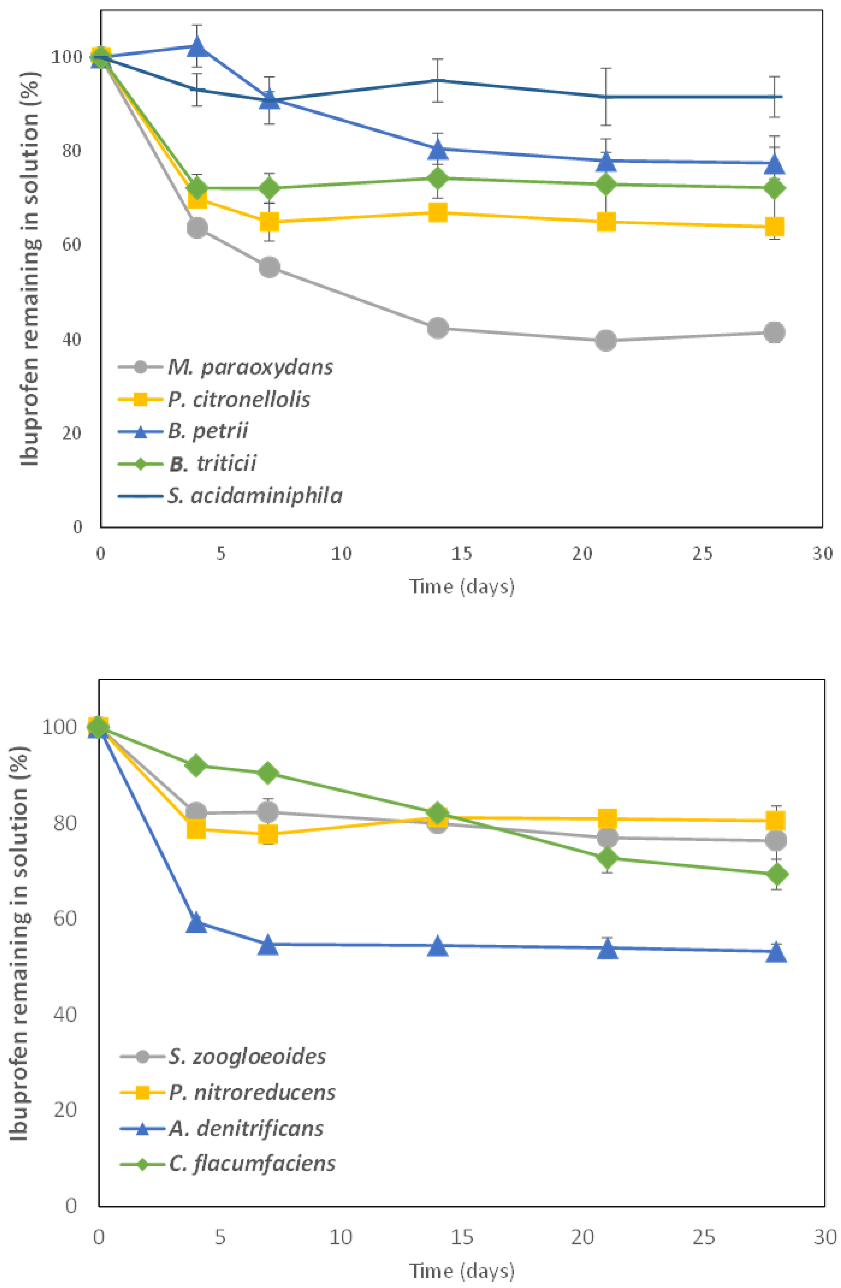
Taking into account that these nine bacterial strains were not able to degrade IBP as a monosubstrate, we selected glucose as a second substrate to be used as C and energy source. Complete IBP biodegradation curves using the different isolated bacterial strains were carried out during 28 days. The presence of 3 g L−1 glucose promoted IBP degradation for all these bacterial strains. The degradation curves obtained are shown in Figure 2 and the corresponding kinetic parameters are given in Table 2.
All the strains tested were able to degrade IBP in the presence of glucose but with a wide variation in the extent of degradation in 28 days (from 9% to 64%). Only two of the isolated bacteria, M. paraoxidans and P. citronellolis, had been previously reported to degrade IBP. M. paraoxidans CSW08 showed the best results, reaching almost 64% IBP degradation in 28 days and with a DT50 of only 12.5 days (Table 2). As previously mentioned, only recently did Show et al. [23] identify M. paraoxidans as an IBP degrader for the first time. There are not many references about M. paraoxidans as a degrader of organic contaminants [36], but there is more information about its role in removing heavy metals from different matrices [37].
P. citronellolis has also been very recently indicated as an IBP degrader [24], but only when the three isolated bacterial strains P. citronellolis RW422, 423 and 424 were added as a consortium, since none of the isolates were able to grow with IBP as the sole carbon and energy source. In the case of P. citronellolis CSW09, more than 36% IBP (10 mg L−1) was degraded in 28 days (Table 2). Another pharmaceutical compound, such as the synthetic estrogen 17α-ethynylestradiol has recently been demonstrated to be degraded by P. citronellolis SJTE-3 [38].
Our research group demonstrated for the first time that the rest of the isolated bacterial strains under study showed the ability to degrade IBP [18]. Among them, it is worth noting IBP degradation by A. denitrificans CSW15, with almost 47% degraded in 28 days and a DT50 value of 44.7 days (Table 2). This is the first time that A. denitrificans has been described as an IBP degrader, but it was demonstrated to increase the degradation of sulfamethoxazole and other sulphonamides [39,40], oxytetracycline [41], and other organic contaminants such as phenanthrene [42] or microplastics [43].
C. flaccumfaciens CSW18 was able to degrade up to 32.2% IBP but it had not been described previously as an IBP degrader. However, this bacterium is able to degrade various organic compounds, such as phenol (700 mg L−1 in 96 h) [44]. C. flaccumfaciens is a genus with a wide distribution, particularly in soils, where it is able to degrade structural polysaccharides, playing a role in the decomposition of organic matter in litter communities [45].
B. petrii is also described as an IBP degrader for the first time in this study, with CSW06 being able to degrade up to 24.5% IBP in 28 days, with a DT50 value of 61.2 days (Table 2). However, B. petrii is known for its ability to degrade a variety of organic compounds, such as aromatic hydrocarbons [46], or pesticides such as endosulfan [47] and 2,4-D [48]. B. petrii is also known to produce biosurfactants, which enhance the bioavailability of hydrophobic pollutants and help in their biodegradation [49].
S. zoogloeoides is a bacterial strain frequently isolated from sewage and activated sludge from WWTPs with the capacity to degrade organic contaminants such as pyridine [50], anthracene [51], or pharmaceuticals such as the estrogenic compound 17α-ethinylestradiol [52], but this is the first time that its ability to degrade IBP is shown (24% IBP degraded).
P. acidaminiphila is a bacterial strain frequently found in contaminated soils and also in activated sludge. Its degradative abilities have been demonstrated for a wide variety of pollutants, such as the herbicide glyphosate [53], the plasticizer dibutyl-phthalate (DBP) [54] or the antibiotic nitrofurantoin [55]. However, this is the first time that P. acidaminiphila CSW10 is reported as an IBP degrader, although with a very low degradation capacity (<10% in 28 days).
P. nitroreducens CSW13 is also reported for the first time as an IBP degrader, reaching almost 20% degradation (Table 2), although other P. nitroreducens strains have previously been reported as degraders of the insecticide chlorpyrifos [56] or the antidepressant fluoxetine [57].
The same applies to the bacterial strain B. triticii, but it is worth noting that this is a very poorly known species with few reported degradative abilities. Only recently has it been observed to degrade low-density polyethylene (LDPE) plastic [58], but B. triticii CSW06 was capable of degrading for the first time more than 27% IBP in solution in the presence of glucose (Table 2).
It should be noted in Figure 2 that those degradation curves which fit a biphasic first-order sequential model (Hockey-Stick, HS) show a value of K2 (the rate constant for the degradation of the slow fraction) that is extremely low (Table 2), indicating that from the value of tb (time at which the rate constant changes) IBP degradation is quite difficult, as in the cases of CSW09, CSW10, CSW12 or CSW15, and even null for some strains, such as B. triticii CSW06 or P. nitroreducens CSW13. This could probably be due to the toxic effects of IBP-generated biotransformation products and metabolites during the 28-day degradation process on these strains, affecting the kinetics of the degradation process, although this was not directly tested in this study.
Although the Inhibitory Concentrations of IBP for bacterial growth (IC50) values previously observed indicated a high tolerance level to increasing concentrations of IBP, metabolites were likely not present. The study of biotransformation products and/or metabolites generated will be addressed in a later section.
Table 3 provides a comparative study of the results obtained in the present study regarding the concentrations of IBP degraded by the nine bacterial strains with the results obtained by other researchers using different IBP-degrader bacteria. There are some strains whose degrading efficiency is much higher, such as those with proven ability to mineralize IBP: Sphingomonas sp. Ibu-2 (500 mg L−1 in 3d, [8,9]) or Sphingopyxis granuli RW412 (456.5 mg L−1 in 3d, [16]). Some others are not proposed as mineralizers but show a very high IBP degradation, such as Nocardia sp. NRRL 5646 (1000 mg L−1 in 5d, [59]), Variovorax sp. Ibu-1 (500 mg L−1 in 7d, [60]), or the consortium Comamonas aquatic + Bacillus sp. (100 mg L−1 in 1.4d, [61]).
Some other strains show degradative efficiencies more similar to the nine bacteria under study using initial concentrations similar to those used in the present research (10 mg L−1), such as Bacillus thuringiensis B1 (2015b) [10,29], Serratia marcescens BL1 [11], Citrobacter freundii PYI-2 and Citrobacter portucalensis YPI-2 [62], Pseudoalteromonas sp. [63], Rhizobium daejeonense IBU_18 [33] or Klebsiella pneumoniae TIBU2.1, Klebsiella variicola c, Pseudomonas aeruginosa LOIBU1.2, and Mycolicibacterium aubagnense HPB1.1 [15].
Some others show lower IBP degradation efficiency compared to the nine bacteria under study: Patulibacter sp. I11 [31], Pseudoxanthomonas sp. DIN-3 [64], Novosphingobium sp. and Pseudomonas sp. [65], or Nocardioides carbamazepine sp. [30].
Table 3. Comparison of IBP degradation by the nine strains in this study and by other reported IBP-degrading bacteria.
StrainConcentration
(mg L−1)
Degraded Concentration (mg L−1)/Time (Days)References
Nocardia sp. NRRL 564610001000/5 d[59]
Sphingomonas sp. Ibu-2500500/3 d[8,9]
Patulibacter sp. I111.0
0.25
0.28/12.5 d
0.125/12.5 d
[31]
Variovorax sp. Ibu-1500500/7 d[60]
Comamonas aquatic + Bacillus sp.100100/1.38 d[61]
Bacillus thuringiensis B1(2015b)2020/6 d[10,29]
Serratia marcescens BL13028/5 d[11]
Pseudoxanthomonas sp. DIN-30.050.02/14 d[64]
Microccocus yunnanensis0.20.18/0.5 d[12,34]
Novosphingobium sp.
Pseudomonas sp.
0.062
0.082
0.062/3 d
0.082/8 d
[65]
Rhodococcus cerastii IEGM 12780.1
100
0.1/1.25 d
100/6 d
[35]
Sphingopyxis granuli RW412456.5456.5/3.1 d[16]
Citrobacter freundii PYI-2
Citrobacter portucalensis YPI-2
8.0
8.0
8.0/15 d
8.0/15 d
[62]
Bacillus siamensis DSI-12020/1 d[66]
Nocardioides carbamazepine sp. Nov.1.51.05/49 d[30]
Pseudoalteromonas sp.1.00.89/3 d[63]
Streptomyces murinus D218
Pseudomonas alloputida M20
20
20
15.34/0.5 d
16.66/0.5 d
[67]
Rhizobium daejeonense IBU_181.51.36/28 d[33]
Microbacterium paraoxydans1515/5 d[23]
Klebsiella pneumoniae TIBU2.155/14 d[15]
Klebsiella variicola LOIBU1.153/14 d[15]
Pseudomonas aeruginosa LOIBU1.253.3/14 d[15]
Mycolicibacterium aubagnense HPB1.152.9/14 d[15]
Labrys neptuniae CSW111.0
5.0
10
100
1.0/4 d
5.0/4 d
10/7 d
48.4/28 d
[17]
M. paraoxydans CSW08106.38/14 dThis study
A. denitrificans CSW15104.69/7 dThis study
P. citronellolis CSW09103.61/7 dThis study
C. flacumfaciens CSW18103.22/28 dThis study
B. tritici CSW06102.72/7 dThis study
B. petrii CSW07102.45/28 dThis study
S. zoogloeoides CSW12102.40/21 dThis study
P. nitroreducens CSW13101.98/14 dThis study
S. acidaminiphila CSW10100.9/28 dThis study

3.3. Phylogenetic Analysis of Ibuprofen-Degrading Bacterial Strains Based on 16S rRNA Gene

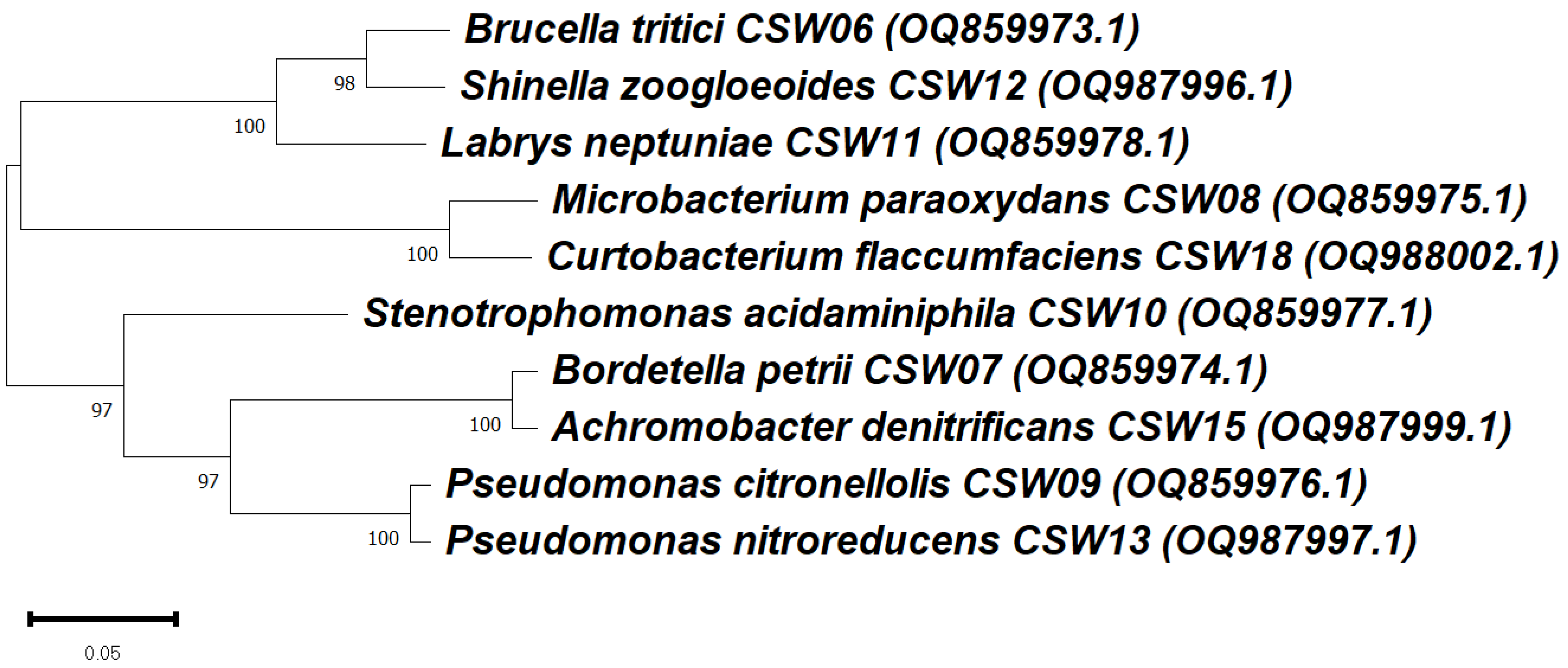
A phylogenetic analysis was conducted to evaluate the evolutionary relationships among the nine bacterial strains under study, isolated from the IBP-degrading consortium C7 (Figure 3). The strain Labrys neptuniae CSW11, which was previously studied by Aguilar-Romero et al. [18], was included because it was also isolated from the same consortium C7.
The phylogenetic tree, constructed based on 16S rRNA gene sequences, revealed several well-supported clades, with bootstrap values ≥ 97%. Despite these close genetic relationships, significant variability was observed in the IBP degradation profiles among the isolates.
Although L. neptuniae CSW11, B. tritici CSW06 and S. zoogloeoides CSW12 are phylogenetic neighbors, their degradation efficiencies were quite different, since CSW11 exhibited a high degradation capacity, reaching 100% IBP removal (10 mg L−1) in only 7 days [18], while CSW6 and CSW12 showed lower degradation efficiencies, reaching only 27.2% and 24.0% IBP removal, respectively, after 28 days (Table 2). Similarly, although M. paraoxydans CSW08 and C. flaccumfaciens CSW18 are closely related, their degradation capacities differed substantially (63.8% vs. 32.2%, respectively). Strains belonging to Pseudomonas spp. (CSW09 and CSW13) also formed a distinct clade but exhibited differing degradation efficiency (36.1%, 19.8%).
These findings suggest that IBP degradation capacity is not strictly correlated with phylogenetic proximity. Instead, the ability to degrade IBP may depend on specific catabolic pathways or genes that are not universally distributed among closely related taxa. This highlights the potential role of horizontal gene transfer and ecological adaptation in shaping the functional capabilities of environmental bacteria involved in pharmaceutical bioremediation, although no genomic analysis was performed in this study.
In order to observe the taxonomic variety of the different bacterial strains that have been found to be IBP-degraders in the literature, Table 4 was constructed, which also includes the nine IBP-degrader strains described in the present study.
Seven of the nine IBP-degrader bacteria are included in the Phylum Pseudomonadota, with the majority of strains shown to mineralize IBP belonging to the family Sphingomonadaceae [8,9,13,14,16,68]. However, Wittich et al. [24] reported for the first time some Pseudomonas strains (Pseudomonas citronellolis RW422, RW423 and RW424, Pseudomonadota/Gammaproteobacteria/Pseudomonadales/Pseudomonadaceae) with the capacity to mineralize IBP. These seven strains, which belong to the Phylum Pseudomonadota, are included in different families: Rhizobiaceae (S. zoogloeoides CSW12), the same family as Rhizobium daejeonense IBU_18 [33]; Xanthomonadaceae (S. acidaminiphila CSW10), the same family as Pseudoxanthomonas sp. DIN-3 [64]; Pseudomonadaceae (P. citronellolis CSW09 and P. nitroreducens CSW13), the same family as P. citronellolis RW422 + RW423+ RW424 [24]; P. alloputida M20 [67] and P. aeruginosa LOIBU1.2 [15]. Three of the nine strains under study belong to the Phylum Pseudomonadota, but to families to which no other IBP-degrading bacterium belongs: Brucellaceae (B. tritici CSW06), Alcaligenaceae (B. petrii CSW07 and A. denitrificans CSW15). The two other bacteria (M. paraoxydans CSW08 and C. flaccumfaciens CSW18) belong to the Phylum Actinomycetota and both are included in the family Microbacteriaceae, the same as M. paraoxydans used by Show et al. [23] as an IBP-degrader. As far as we know, the rest of the IBP-degrader bacterial strains reported in the literature belong to the phylum Bacillota (family Bacillaceae, [10,29,61,66]). Taking into account that it is estimated that around 1300 bacterial phyla exist, although only 89 are recognized on the Silva database [69] and 41 are formally accepted by the LPSN [70], it can be deduced that the bacterial strains shown to degrade IBP are, at present, representatives of a very limited number of phyla.

3.4. Detection of the Main Metabolites in IBP Biotransformation by Isolated Bacteria

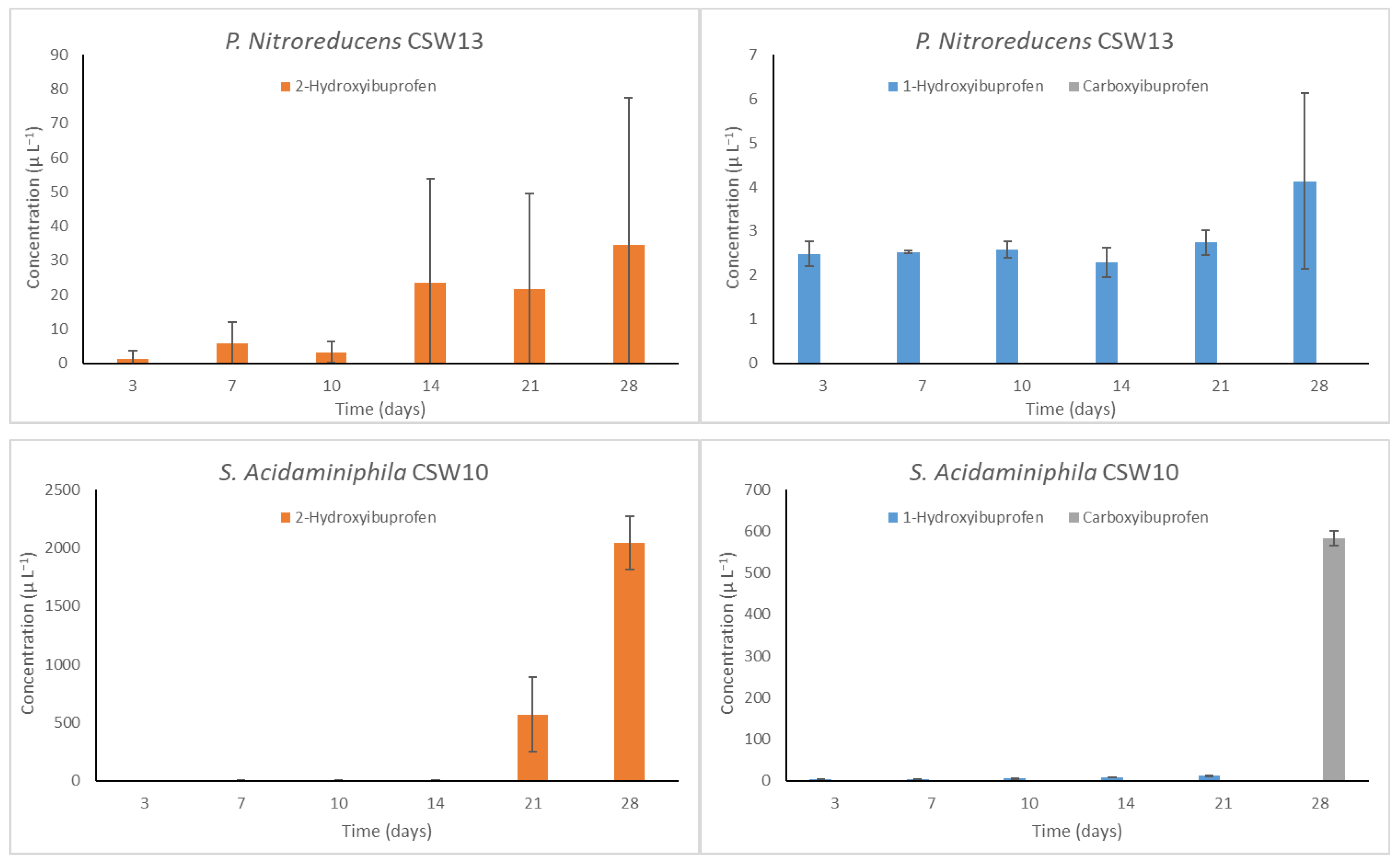
IBP biotransformation products generated throughout the degradation process have been identified and are included in Figure 4 (the strains are shown in decreasing order of IBP degradation). Three metabolites were identified: 1-hydroxyibuprofen (1-OH- IBP), 2-hydroxyibuprofen (2-OH-IBP) and carboxyibuprofen (CBX-IBP). These are the same biotransformation products observed for IBP biodegradation by L. neptuniae CSW11, a bacterial strain also isolated from the C7 consortium [17].
Observing Figure 4, no general trend in the appearance and persistence of the three products in the system was observed, although the concentration of 2-OH-IBP was generally much higher than that of 1-OH-IBP. For some strains (CSW6, CSW10, CSW15), 2-OH-IBP concentrations reached up to 1000 µg L−1, while for others (CSW07, CSW12, CSW13, CSW15), values below 50 µg L−1 were observed, and these do not appear to be related to the concentration of IBP degraded.
In the case of 1-OH-IBP, values below 8 µg L−1 were obtained, except for CSW18, which reached up to 33 µg L−1. Both are products of IBP hydroxylation through the aliphatic side chains of the ring, with hydroxylation being the first reaction in IBP biodegradation. In general, 1- and 2-OH-IBP concentrations seem to increase throughout the degradation process, remaining in the system even for those strains with the highest degradation efficiency.
On the other hand, not all the isolates under study produced CBX-IBP, at least within the studied time range. Its occurrence does not appear to be related only to those bacteria that cause the most degradation. Several pathways for IBP degradation have been proposed for different bacterial strains, each one starting with one of these three metabolites [28,71]. But since two or three of these compounds are generally present [32,64], multiple degradation pathways may occur simultaneously.
Taking into account that in almost all of the degrading processes using the nine strains under study IBP degradation reached a plateau and did not continue to increase, this could indicate that the biotransformation products obtained remained in the system, which raises concerns about possible toxicity compared to that of IBP parent compound, although further ecotoxicology testing is required to confirm this. This toxicity was demonstrated in the case of L. neptuniae CSW11 [17], where the simultaneous presence of the same three biotransformation products was observed. However, Grabarczyk et al. [72] conducted an ecotoxicity screening evaluation of IBP and other selected pharmaceuticals and their transformation products on various organisms, observing that at least 2-OH-IBP and CBX-IBP are less toxic than IBP to V. fischeri, L. minor, D. magna and R. subcapitata. Specific conditions detailed by Salgado et al. [32] to detect a wide variety of other IBP metabolites and biotransformation products were also used, but no new metabolites were found in the present study.

4. Conclusions

Biodegradation of IBP using nine bacterial strains isolated from sewage sludge from a WWTP has been carried out, evaluating the degradation kinetics of each of them as well as the formation of the most relevant transformation products. All of them showed IBP degradative capacity in the presence of glucose as a secondary substrate, with an extent of degradation ranging from 9% to 64% in 28 days. The disappearance of IBP from water, along with the appearance of three biotransformation products (1-hydroxyibuprofen (1-OH-IBP), 2-hydroxyibuprofen (2-OH-IBP), and carboxyibuprofen (CBX-IBP)) throughout the process, confirms that these nine bacteria are IBP-degraders.
The tolerance of each strain to IBP present in the medium was quantified using the IC50 parameter, with values ranging between 38 and 1353 mg L−1, which are higher than those usually found even in WWTP waters, indicating a high level of IBP tolerance for all the strains studied. This is the first time that IC50 values are reported for IBP tolerance in the bacterial genera of the nine strains under study.
Phylogenetic analysis based on 16S rRNA gene sequences was conducted to explore the taxonomic affiliations of the nine bacteria under study in comparison to other bacterial strains previously found to be IBP-degraders. It was concluded that seven of the nine strains are included in the Phylum Pseudomonadota, the same as most of the IBP-degrader bacteria previously mentioned in the literature, but they belong to five different families: Brucellaceae, Alcaligenaceae, Rhizobiaceae, Xanthomonadaceae, and Pseudomonadaceae. The two other bacteria belong to the Phylum Actinomycetota and both are included in the family Microbacteriaceae.
Considering that the experiments were conducted in vitro, under controlled laboratory conditions and using small solution volumes, the results should be regarded as preliminary. At the laboratory scale, the method needs to be improved by incorporating consortia of other bacterial strains capable of degrading the toxic metabolites produced. This would lead not only to the degradation of IBP but also to its complete mineralization. Moreover, although the results suggest that these nine bacterial strains can degrade IBP in liquid media, they may not accurately reflect their behavior in more complex natural environments. On a larger scale or in situ, the bioremediation performance could differ significantly. Factors such as nutrient availability, pH, temperature, and the presence of other organisms and contaminants can all dramatically influence bacterial growth, motility, and interactions.

Supplementary Materials

The following supporting information can be downloaded at: https://www.mdpi.com/article/10.3390/microorganisms13081927/s1, Table S1. Optimized MRM parameters of the QqQ-MS determination of ibuprofen and its metabolites.

Author Contributions

Conceptualization, I.A.-R., A.L.-M., F.M., J.V. and E.M.; Data curation, I.A.-R., A.L.-M., F.M., J.V., E.A. and J.L.S.; Funding acquisition, E.M.; Investigation, I.A.-R., A.L.-M. and F.M.; Methodology, I.A.-R., A.L.-M., F.M., E.A. and J.L.S.; Project administration, E.M.; Resources, E.M.; Supervision, F.M., J.V. and E.M.; Writing—original draft, I.A.-R., A.L.-M. and E.M.; Writing—review and editing, I.A.-R., A.L.-M., F.M., J.V. and E.M. All authors have read and agreed to the published version of the manuscript.

Funding

This work was supported by the Junta de Andalucía (Council of Economic Transformation, Industry, Knowledge and University) under the research project PY20_01069 (FEDER Andalucía PO 2014–2020).

Institutional Review Board Statement

Not applicable.

Informed Consent Statement

Not applicable.

Data Availability Statement

The original contributions presented in the study are included in the article/Supplementary Materials; further inquiries can be directed to the corresponding author.

Conflicts of Interest

The authors declare no conflicts of interest.

References

  1. Chopra, S.; Kumar, D. Ibuprofen as an emerging organic contaminant in environment, distribution and remediation. Heliyon 2020, 6, e04087. [Google Scholar] [CrossRef]
  2. Jan-Roblero, J.; Cruz-Maya, J.A. Ibuprofen: Toxicology and biodegradation of an emerging contaminant. Molecules 2023, 28, 2097. [Google Scholar] [CrossRef]
  3. WFD; EC. Directive 2000/60/EC of the European Parliament and of the Council of 23 October 2000 Establishing a Framework for Community Action in the Field of Water Policy. J 327 (L). 2000. Available online: https://www.eea.europa.eu/policy-documents/directive-2000-60-ec-of (accessed on 1 August 2025).
  4. EC. European Parliament Legislative Resolution of 2 July 2013 on the Proposal for a Directive of the European Parliament and of the Council Amending Directives 2000/60/EC and 2008/105/EC as Regards Priority Substances in the Field of Water Policy; European Commission: Brussels, Belgium, 2013. [Google Scholar]
  5. DG Env. Priority Substances Review-Next Steps. 2020. Available online: https://circabc.europa.eu/sd/a/97910629-048e-4b2b-90a2-fe520aab2adc/WGChem2020Jan(7)PrioritySubstancesReview-nextsteps.pdf (accessed on 1 August 2025).
  6. Nguyen, M.K.; Lin, C.; Nguyen, H.L.; Hung, N.T.Q.; La, D.D.; Nguyen, X.H.; Chang, S.W.; Chung, W.J. Occurrence, fate, and potential risk of pharmaceutical pollutants in agriculture: Challenges and environmentally friendly solutions. Sci. Total Environ. 2023, 899, 165323. [Google Scholar] [CrossRef] [PubMed]
  7. Wu, J.; Liu, Z.; Ma, Q.; Dai, L.; Dang, Z. Occurrence, removal and risk evaluation of ibuprofen and acetaminophen in municipal wastewater treatment plants: A critical review. Sci. Total Environ. 2023, 891, 164600. [Google Scholar] [CrossRef]
  8. Murdoch, R.W.; Hay, A.G. Formation of catechols via removal of acid side chains from ibuprofen and related aromatic acids. Appl. Environ. Microbiol. 2005, 71, 6121–6125. [Google Scholar] [CrossRef]
  9. Murdoch, R.W.; Hay, A.G. Genetic and chemical characterization of ibuprofen degradation by Sphingomonas Ibu-2. Microbiology 2013, 159, 621–632. [Google Scholar] [CrossRef]
  10. Marchlewicz, A.; Domaradzka, D.; Guzik, U.; Wojcieszyńska, D. Bacillus thuringiensis B1(2015b) is a Gram-Positive Bacteria Able to Degrade Naproxen and Ibuprofen. Water Air Soil Pollut. 2016, 227, 197. [Google Scholar] [CrossRef]
  11. Xu, B.; Xue, G.; Yang, X. Isolation and application of an ibuprofen-degrading bacterium to a biological aerated filter for the treatment of micro-polluted water. Front. Environ. Sci. Eng. 2018, 12, 15. [Google Scholar] [CrossRef]
  12. Sharma, K.; Kaushik, G.; Thotakura, N.; Raza, K.; Sharma, N.; Nimesh, S. Fate of ibuprofen under optimized batch biodegradation experiments using Micrococcus yunnanensis isolated from pharmaceutical sludge. Int. J. Environ. Sci. Technol. 2019, 16, 8315–8328. [Google Scholar] [CrossRef]
  13. Balciunas, E.M.; Kappelmeyer, U.; Harms, H.; Heipieper, H.J. Increasing ibuprofen degradation in constructed wetlands by bioaugmentation with gravel containing biofilms of an ibuprofen-degrading Sphingobium yanoikuyae. Eng. Life Sci. 2020, 20, 160–167. [Google Scholar] [CrossRef]
  14. Aulestia, M.; Flores, A.; Acosta-Jurado, S.; Santero, E.; Camacho, E.M. Genetic characterization of the ibuprofen-degradative pathway of Rhizorhabdus wittichii MPO218. App. Environ. Microbiol. 2022, 88, e00388-22. [Google Scholar] [CrossRef]
  15. Lara-Moreno, A.; Costa, M.C.; Vargas-Villagomez, A.; Carlier, J.D. New bacterial strains for ibuprofen biodegradation: Drug removal, transformation, and potential catabolic genes. Environ. Microb. Rep. 2024, 16, e13320. [Google Scholar] [CrossRef]
  16. Aguilar-Romero, I.; De la Torre-Zúñiga, J.; Quesada, J.M.; Haïdour, A.; O’Connell, G.; McAmmond, B.M.; Van Hamme, J.D.; Romero, E.; Regina-Michaela Wittich, R.M.; van Dillewijn, P. Effluent decontamination by the ibuprofen-mineralizing strain, Sphingopyxis granuli RW412: Metabolic processes. Environ. Pollut. 2021, 274, 116536. [Google Scholar] [CrossRef] [PubMed]
  17. Aguilar-Romero, I.; Madrid, F.; Villaverde, J.; Alonso, E.; Santos, J.L.; Morillo, E. Removal of ibuprofen in water by bioaugmentation with Labrys neptuniae CSW11 isolated from sewage sludge—Assessment of biodegradation pathway based on metabolite formation and genomic analysis. J. Xenobiot. 2025, 15, 5. [Google Scholar] [CrossRef] [PubMed]
  18. Aguilar-Romero, I.; Madrid, F.; Villaverde, J.; Morillo, E. Ibuprofen-enhanced biodegradation in solution and sewage sludge by an indigenous microbial consortium. Shift Assoc. Bact. Communities J. Hazard. Mater. 2024, 464, 132970. [Google Scholar] [CrossRef] [PubMed]
  19. Vargas-Ordóñez, A.; Aguilar-Romero, I.; Villaverde, J.; Madrid, F.; Morillo, E. Isolation of novel bacterial strains Pseudomonas extremaustralis CSW01 and Stutzerimonas stutzeri CSW02 from sewage sludge for paracetamol biodegradation. Microorganisms 2023, 11, 196. [Google Scholar] [CrossRef]
  20. Tamura, K.; Stecher, G.; Kumar, S. MEGA11: Molecular evolutionary genetics analysis version 11. Mol. Biol. Evol. 2021, 38, 3022–3027. [Google Scholar] [CrossRef]
  21. FOCUS. Guidance Document on Estimating Persistence and Degradation Kinetics from Environmental Fate Studies on Pesticides in EU Registration. Report of the FOCUS Work Group on Degradation Kinetics, EC Document Reference Sanco/10058/2005 version 2.0; 2006; 434 p. Available online: https://esdac.jrc.ec.europa.eu/public_path/projects_data/focus/dk/docs/finalreportFOCDegKinetics.pdf (accessed on 2 July 2025).
  22. Okpala, O.E.; Rondevaldova, J.; Kokoska, L. Anti-inflammatory drugs as potential antimicrobial agents: A review. Front. Pharmacol. 2025, 16, 1557333. [Google Scholar] [CrossRef]
  23. Show, S.; Sarkar, P.; Barman, S.; Halder, G. Microbial remediation of ibuprofen contaminated water using novel isolate Microbacterium paraoxydans. Chem. Pap. 2023, 77, 517–531. [Google Scholar] [CrossRef]
  24. Wittich, R.M.; Haïdour, A.; Aguilar-Romero, I.; de la Torre-Zúñiga, J.; van Dillewijn, P. Biodegradation of microtoxic phenylpropanoids (phenylpropanoic acid and ibuprofen) by bacteria and the relevance for their removal from wastewater treatment plants. Genes 2023, 14, 442. [Google Scholar] [CrossRef]
  25. Rajapaksha, R.M.C.P. Heavy metal tolerance of culturable bacteria and fungi in a long-term cultivated tropical ultisol. Eur. J. Soil Biol. 2011, 47, 9–15. [Google Scholar] [CrossRef]
  26. Al-janabi, A.A.H.S. In vitro antibacterial activity of Ibuprofen and acetaminophen. J. Glob. Infect. Dis. 2010, 2, 105–108. [Google Scholar] [CrossRef]
  27. Lara-Moreno, A.; Aguilar-Romero, I.; Rubio-Bellido, M.; Madrid, F.; Villaverde, J.; Santos, J.L.; Alonso, E.; Morillo, E. Novel nonylphenol-degrading bacterial strains isolated from sewage sludge: Application in bioremediation of sludge. Sci. Total Environ. 2022, 847, 157647. [Google Scholar] [CrossRef]
  28. Mulkiewicz, E.; Wolecki, D.; Świacka, K.; Kumirska, J.; Stepnowski, P.; Caban, M. Metabolism of non-steroidal anti-inflammatory drugs by non-target wild-living organisms. Sci. Total Environ. 2021, 791, 148251. [Google Scholar] [CrossRef] [PubMed]
  29. Marchlewicz, A.; Guzik, U.; Smulek, W.; Wojcieszynska, D. Exploring the degradation of ibuprofen by Bacillus thuringiensis B1 (2015b): The new pathway and factors affecting degradation. Molecules 2017, 22, 1676. [Google Scholar] [CrossRef] [PubMed]
  30. Benedek, T.; Pápai, M.; Gharieb, K.; Bedics, A.; Táncsics, A.; Tóth, E.; Daood, H.; Maróti, G.; Wirth, R.; Menashe, O.; et al. Nocardioides carbamazepini sp. nov., an ibuprofen degrader isolated from a biofilm bacterial community enriched on carbamazepine. Syst. Appl. Microbiol. 2022, 45, 126339. [Google Scholar] [CrossRef]
  31. Almeida, B.; Kjeldal, H.; Lolas, I.; Knudsen, A.D.; Carvalho, G.; Nielsen, K.L.; Barreto Crespo, M.T. Quantitative proteomic analysis of ibuprofen-degrading Patulibacter sp. strain I11. Biodegradation 2013, 24, 615–630. [Google Scholar] [CrossRef] [PubMed]
  32. Salgado, R.; Brito, D.; Noronha, J.P.; Almeida, B.; Bronze, M.R.; Oehmen, A.; Carvalho, G.; Barreto-Crespo, M.T. Metabolite identification of ibuprofen biodegradation by Patulibacter medicamentivorans under aerobic conditions. Environ. Technol. 2020, 41, 450–465. [Google Scholar] [CrossRef]
  33. Pápai, M.; Benedek, T.; Táncsics, A.; Bornemann, T.L.V.; Plewka, J.; Probst, A.J.; Hussein, D.; Maróti, G.; Menashe, O.; Kriszt, B. Selective enrichment, identification, and isolation of diclofenac, ibuprofen, and carbamazepine degrading bacteria from a groundwater biofilm. Environ. Sci. Pollut. Res. 2023, 30, 44518–44535. [Google Scholar] [CrossRef]
  34. Sharma, K.; Kaushik, G.; Thotakura, N.; Raza, K.; Sharma, N.; Nimesh, S. Enhancement effects of process optimization technique while elucidating the degradation pathways of drugs present in pharmaceutical industry wastewater using Micrococcus yunnanensis. Chemosphere 2020, 238, 124689. [Google Scholar] [CrossRef]
  35. Ivshina, I.B.; Tyumina, E.A.; Bazhutin, G.A.; Vikhareva, E.V. Response of Rhodococcus cerastii IEGM 1278 to toxic effects of ibuprofen. PLoS ONE 2021, 16, e0260032. [Google Scholar] [CrossRef]
  36. Jin, J.; Yao, J.; Liu, W.; Zhang, Q.; Liu, J. Fluoranthene degradation and binding mechanism study based on the active-site structure of ring-hydroxylatingdioxygenase in Microbacterium paraoxydans JPM1. Environ. Sci. Pollut. Res. 2017, 24, 363–371. [Google Scholar] [CrossRef]
  37. Singh, V.; Singh, J.; Singh, N.; Rai, S.N.; Verma, M.K.; Verma, M.; Singh, V.; Chivate, M.S.; Bilal, M.; Mishra, V. Simultaneous removal of ternary heavy metal ions by a newly isolated Microbacterium paraoxydans strain VSVM IIT(BHU) from coal washery effluent. BioMetals 2023, 36, 829–845. [Google Scholar] [CrossRef]
  38. Peng, W.; Lin, S.; Deng, Z.; Liang, R. Bioaugmentation removal and microbiome analysis of the synthetic estrogen 17α-ethynylestradiol from hostile conditions and environmental samples by Pseudomonas citronellolis SJTE-3. Chemosphere 2023, 317, 137893. [Google Scholar] [CrossRef]
  39. Reis, P.J.M.; Reis, A.C.; Ricken, B.; Kolvenbach, B.A.; Manaia, C.M.; Corvini, P.F.X.; Nunes, O.C. Biodegradation of sulfamethoxazole and other sulfonamides by Achromobacter denitrificans PR1. J. Haz. Mat. 2014, 280, 741–749. [Google Scholar] [CrossRef]
  40. Nguyen, P.Y.; Silva, A.F.; Reis, A.C.; Nunes, O.C.; Rodrigues, A.M.; Rodrigues, J.E.; Cardoso, V.V.; Benoliel, M.J.; Reis, M.A.M.; Oehmen, A.; et al. Bioaugmentation of membrane bioreactor with Achromobacter denitrificans strain PR1 for enhanced sulfamethoxazole removal in wastewater. Sci. Total Environ. 2019, 648, 44–55. [Google Scholar] [CrossRef] [PubMed]
  41. Zhang, S.; Hou, J.; Zhang, X.; Cheng, L.; Hu, W.; Zhang, Q. Biochar-assisted degradation of oxytetracycline by Achromobacter denitrificans and underlying mechanisms. Bioresour. Technol. 2023, 387, 129673. [Google Scholar] [CrossRef] [PubMed]
  42. Zhang, Z.; Sun, J.; Guo, H.; Wang, C.; Fang, T.; Rogers, M.J.; He, J.; Wang, H. Anaerobic biodegradation of phenanthrene by a newly isolated nitrate-dependent Achromobacter denitrificans strain PheN1 and exploration of the biotransformation processes by metabolite and genome analyses. Environ. Microbiol. 2021, 23, 908–923. [Google Scholar] [CrossRef] [PubMed]
  43. Maleki Rad, M.; Moghimi, H.; Azin, E. Biodegradation of thermo-oxidative pretreated low-density polyethylene (LDPE) and polyvinyl chloride (PVC) microplastics by Achromobacter denitrificans Ebl13. Mar. Pollut. Bull. 2022, 181, 113830. [Google Scholar] [CrossRef]
  44. Khleifat, K.; Magharbeh, M.; Alqaraleh, M.; Al-Sarayrah, M.; Alfarrayeh, I.; Al Qaisi, Y.; Alsarayreh, A.; Alkafaween, M. Biodegradation modelling of phenol using Curtobacterium flaccumfaciens as plant-growth-promoting bacteria. Heliyon 2022, 8, e10490. [Google Scholar] [CrossRef]
  45. Chase, A.B.; Arevalo, P.; Polz, M.F.; Berlemont, R.; Martiny, J.B.H. Evidence for ecological flexibility in the cosmopolitan genus Curtobacterium. Front. Microbiol. 2016, 7, 1874. [Google Scholar] [CrossRef]
  46. Al-Saleh, E.; Hassan, A. Enhanced crude oil biodegradation in soil via biostimulation. Int. J. Phytoremediation 2016, 18, 822–831. [Google Scholar] [CrossRef] [PubMed]
  47. Kong, L.; Zhang, Y.; Zhu, L.; Wang, J.; Wang, J.; Du, Z.; Zhang, C. Influence of isolated bacterial strains on the in situ biodegradation of endosulfan and the reduction of endosulfan-contaminated soil toxicity. Ecotoxicol. Environ. Saf. 2018, 160, 75–83. [Google Scholar] [CrossRef] [PubMed]
  48. Nguyen, T.L.A.; Dang, H.T.C.; Koekkoek, J.; Braster, M.; Parsons, J.R.; Brouwer, A.; de Boer, T.; van Spanning, R.J.M. Species and metabolic pathways involved in bioremediation of vietnamese soil from Bien Hoa airbase contaminated with herbicides. Front. Sustain. Cities 2021, 326, 692018. [Google Scholar] [CrossRef]
  49. Odukkathil, G.; Vasudevan, N. Biodegradation of endosulfan isomers and its metabolite endosulfate by two biosurfactant producing bacterial strains of Bordetella petrii. J. Environ. Sci. Health B 2015, 50, 81–89. [Google Scholar] [CrossRef] [PubMed]
  50. Bai, Y.; Sun, Q.; Zhao, C.; Wen, D.; Tang, X. Aerobic degradation of pyridine by a new bacterial strain Shinella zoogloeoides BC026. J. Ind. Microbiol. Biotechnol. 2009, 36, 1391–1400. [Google Scholar] [CrossRef]
  51. Ntougias, S.; Melidis, P.; Navrozidou, E.; Tzegkas, F. Diversity and efficiency of anthracene-degrading bacteria isolated from a denitrifying activated sludge system treating municipal wastewater. Int. Biodeterior. Biodegrad. 2015, 97, 151–158. [Google Scholar] [CrossRef]
  52. Palma, T.L.; Shylova, A.; Costa, M.C. Isolation and characterization of bacteria from activated sludge capable of degrading 17α-ethinylestradiol, a contaminant of high environmental concern. Microbiology 2021, 167, 001038. [Google Scholar] [CrossRef]
  53. Li, J.; Chen, W.J.; Zhang, W.; Zhang, Y.; Lei, Q.; Wu, S.; Huang, Y.; Mishra, S.; Bhatt, P.; Chen, S. Effects of free or immobilized bacterium Stenotrophomonas acidaminiphila Y4B on glyphosate degradation performance and indigenous microbial community structure. J. Agric. Food Chem. 2022, 70, 13945–13958. [Google Scholar] [CrossRef]
  54. Zhang, Y.; Tang, J.; Wu, M.; Zhou, X.; Wang, S.; Ye, H.; Xiang, W.; Zhang, Q.; Cai, T. Whole genome sequencing exploitation analysis of dibutyl phthalate by strain Stenotrophomonas acidaminiphila BDBP 071. Food Biosci. 2023, 51, 102185. [Google Scholar] [CrossRef]
  55. Pacholak, A.; Juzwa, W.; Zgoła-Grześkowiak, A.; Kaczorek, E. Multi-faceted analysis of bacterial transformation of nitrofurantoin. Sci. Total Environ. 2023, 874, 162422. [Google Scholar] [CrossRef] [PubMed]
  56. Aswathi, A.; Pandey, A.; Sukumaran, R.K. Rapid degradation of the organophosphate pesticide Chlorpyrifos by a novel strain of Pseudomonas nitroreducens AR-3. Bioresour. Technol. 2019, 292, 122025. [Google Scholar] [CrossRef]
  57. Palma, T.L.; Shylova, A.; Dias Carlier, J.; Costa, M.C. An autochthonous aerobic bacterial community and its cultivable isolates capable of degrading fluoxetine. J. Chem. Technol. Biotechnol. 2021, 96, 2813–2826. [Google Scholar] [CrossRef]
  58. Alamer, N.J.; Aldayel, M.F.; Khalifa, A. Isolation and characterization of Brucella spp., low-density polyethylene (LDPE) plastic degrading bacteria in Al-Ahsa Region, Saudi Arabia. Appl. Sci. 2023, 13, 4629. [Google Scholar] [CrossRef]
  59. Chen, Y.; Rosazza, J.P.N. Microbial transformation of ibuprofen by a Nocardia species. Appl. Environ. Microbiol. 1994, 60, 1292–1296. [Google Scholar] [CrossRef]
  60. Murdoch, R.W.; Hay, A.G. The biotransformation of ibuprofen to trihydroxyibuprofen in activated sludge and by Variovorax Ibu-1. Biodegradation 2015, 26, 105–113. [Google Scholar] [CrossRef]
  61. Fortunato, M.S.; Fuentes Abril, N.P.; Martinefski, M.; Trípodi, V.; Papalia, M.; Rádice, M.; Gutkind, G.; Gallego, A.; Korol, S.E. Aerobic degradation of ibuprofen in batch and continuous reactors by an indigenous bacterial community. Environ. Technol. 2016, 37, 2617–2626. [Google Scholar] [CrossRef]
  62. Chopra, S.; Kumar, D. Characteristics and growth kinetics of biomass of Citrobacter freundii strains PYI-2 and Citrobacter portucalensis strain YPI-2 during the biodegradation of Ibuprofen. Int. Microbiol. 2022, 25, 615–628. [Google Scholar] [CrossRef]
  63. Li, Z.; Wang, J.; Gu, C.; Guo, Y.; Wu, S. Marine bacteria-mediated abiotic-biotic coupling degradation mechanism of ibuprofen. J. Hazard. Mater. 2022, 435, 128960. [Google Scholar] [CrossRef]
  64. Lu, Z.; Sun, W.; Li, C.; Ao, X.; Yang, C.; Li, S. Bioremoval of non-steroidal anti-inflammatory drugs by Pseudoxanthomonas sp. DIN-3 isolated from biological activated carbon process. Water Res. 2019, 161, 459–472. [Google Scholar] [CrossRef]
  65. Rutere, C.; Knoop, K.; Posselt, M.; Ho, A.; Horn, M.A. Ibuprofen degradation and associated bacterial communities in hyporheic zone sediments. Microorganisms 2020, 8, 1245. [Google Scholar] [CrossRef]
  66. Chopra, S.; Kumar, D. Characterization and biodegradation of ibuprofen by Bacillus siamensis strain DSI-1 isolated from wastewater. Rend. Lincei. Sci. Fis. Nat. 2022, 33, 643–652. [Google Scholar] [CrossRef]
  67. Chen, R.; Huang, J.; Li, X.; Yang, C.; Wu, X. Functional characterization of an efficient ibuprofen-mineralizing bacterial consortium. J. Hazard. Mater. 2023, 447, 130751. [Google Scholar] [CrossRef] [PubMed]
  68. Aulestia, M.; Flores, A.; Mangas, E.L.; Pérez-Pulido, A.J.; Santero, E.; Camacho, E.M. Isolation and genomic characterization of the ibuprofen-degrading bacterium Sphingomonas strain MPO218. Environ. Microbiol. 2021, 23, 267–280. [Google Scholar] [CrossRef]
  69. Quast, C.; Pruesse, E.; Yilmaz, P.; Gerken, J.; Schweer, T.; Yarza, P.; Peplies, J.; Glöckner, F.O. The SILVA ribosomal RNA gene database project: Improved data processing and web-based tools. Nucleic Acids Res. 2013, 41, D590–D596. [Google Scholar] [CrossRef]
  70. Parte, A.C.; Carbasse, J.S.; Meier-Kolthoff, J.P.; Reimer, L.C.; Göker, M. List of prokaryotic names with standing in nomenclature (LPSN) moves to the DSMZ. Int. J. Syst. Evol. Microbiol. 2020, 70, 5607–5612. [Google Scholar] [CrossRef] [PubMed]
  71. Zur, J.; Pinski, A.; Marchlewicz, A.; Hupert-Kocurek, K.; Wojcieszynska, D.; Guzik, U. Organic micropollutants paracetamol and ibuprofen. Toxicity, biodegradation, and genetic background of their utilization by bacteria. Environ. Sci. Pollut. Res. 2018, 25, 21498–21524. [Google Scholar] [CrossRef]
  72. Grabarczyk, Ł.; Mulkiewicz, E.; Stolte, S.; Puckowski, A.; Pazda, M.; Stepnowski, P.; Białk-Bielińska, A. Ecotoxicity screening evaluation of selected pharmaceuticals and their transformation products towards various organisms. Environ. Sci. Pollut. Res. 2020, 27, 26103–26114. [Google Scholar] [CrossRef]
Figure 1. Graphical representation of the best-fitting linear curves for bacterial viability as a function of the logarithm of IBP concentration (log [IBP]) for the nine bacterial isolates. Error bars indicate standard deviation from three replicates.
Microorganisms 13 01927 g001aMicroorganisms 13 01927 g001b
Figure 2. Ibuprofen (10 mg L−1) degradation capability by several bacterial strains isolated from consortium C7 in the presence of glucose (3 g L−1). Error bars indicate standard deviation from three replicates.
Microorganisms 13 01927 g002
Figure 3. Maximum likelihood phylogenetic tree based on 16S rRNA gene sequences, showing the relationships among the different isolated bacterial strains under study. Labrys neptuniae CSW11 has also been included, as it was isolated from the same ibuprofen-degrading consortium C7. Bootstrap values at branch nodes indicate support based on 1000 replicates. The scale bar represents 0.05 substitutions per nucleotide. GenBank accession numbers are provided in parentheses.
Microorganisms 13 01927 g003
Figure 4. Ibuprofen biotransformation products detected during the degradation process. Error bars indicate standard deviation from three replicates.
Microorganisms 13 01927 g004aMicroorganisms 13 01927 g004b
Table 1. Inhibitory concentrations (IC50) of ibuprofen for specific degrading bacteria.
BacteriaIC50 (mg/L)R2
B. tritici CSW06480.99
B. petrii CSW0713530.98
M. paraoxydans CSW082790.93
P. citronellolis CSW094501
S. acidaminiphila CSW10380.93
S. zoogloeoides CSW126890.99
P. nitroreducens CSW1312470.96
A. denitrificans CSW156010.96
C. flaccumfaciens CSW189080.94
Table 2. Kinetic parameters from ibuprofen biotransformation in solution 28 days after inoculation with different bacterial degrading strains and glucose (3 g L−1).
Bacterial StrainKinetic Model K1
(d−1)
K2
(d−1)
tb
(d)
DT50
(d)
Extent of Degradation (%)R2ErrscaledCalculated
χ2 *
M. paraoxydans CSW08HS0.2740.0211.7012.563.80.9670.591.925
A. denitrificans CSW15HS0.4340.0021.3444.746.90.9930.050.225
P. citronellolis CSW09HS0.2480.0031.5211936.10.9890.140.155
C. flacumfaciens CSW18SFO0.013--51.632.20.9861.010.120
B. tritici CSW06HS0.2260.0001.4042627.20.9870.290.113
B. petrii CSW07SFO0.011--61.224.50.8573.081.029
S. zoogloeoides CSW12HS0.1340.0031.3815024.00.9950.050.020
P. nitroreducens CSW13HS0.1320.0001.3690719.80.9740.250.292
S. acidaminiphila CSW10HS0.0830.0010.836699.020.8050.370.143
* χ2 calculated values < χ2 corresponding tabulated value 12,592 (p < 0.05).
Table 4. Taxonomic classification of ibuprofen-degrading bacteria reported in the literature and those identified in this study.
Strain Phylum/Class/Order/FamilyReference
Sphingomonas sp. Ibu-2Pseudomonadota/Alphaproteobacteria/Sphingomonadales/Sphingomonadaceae[8,9]
Sphingobium yanoikuyaePseudomonadota/Alphaproteobacteria/Sphingomonadales/Sphingomonadaceae[13]
NovosphingobiumPseudomonadota/Alphaproteobacteria/Sphingomonadales/Sphingomonadaceae[65]
Rhizorhabdus wittichii MPO218Pseudomonadota/Alphaproteobacteria/Sphingomonadales/Sphingomonadaceae[14]
Sphingopyxis granuli TFAPseudomonadota/Alphaproteobacteria/Sphingomonadales/Sphingomonadaceae[68]
Sphingopyxis granuli RW412Pseudomonadota/Alphaproteobacteria/Sphingomonadales/Sphingomonadaceae[16]
Rhizobium daejeonense IBU_18Pseudomonadota/Alphaproteobacteria/Hyphomicrobiales/Rhizobiaceae[33]
Shinella zoogloeoides CSW12Pseudomonadota/Alphaproteobacteria/Hyphomicrobiales/RhizobiaceaeThis study
Labrys neptuniae CSW11Pseudomonadota/Alphaproteobacteria/Hyphomicrobiales/Xanthobacteraceae[17]
Brucella tritici CSW06Pseudomonadota/Alphaproteobacteria/Rhizobiales/BrucellaceaeThis study
Variovorax sp. Ibu-1Pseudomonadota/Betaproteobacteria/Burkholderiales/Comamonadaceae[60]
Comamonas aquatic (+ Bacillus sp.)Pseudomonadota/Betaproteobacteria/Burkholderiales/Comamonadaceae[61]
Bordetella petrii CSW07Pseudomonadota/Betaproteobacteria/Burkholderiales/AlcaligenaceaeThis study
Achromobacter denitrificans CSW15Pseudomonadota/Betaproteobacteria/Burkholderiales/AlcaligenaceaeThis study
Serratia marcescens BL1Pseudomonadota/Gammaproteobacteria/Enterobacterales/Yersiniaceae[11]
Pseudoxanthomonas sp. DIN-3Pseudomonadota/Gammaproteobacteria/Xanthomonadales/Xanthomonadaceae[64]
Stenotrophomonas acidaminiphila CSW10Pseudomonadota/Gammaproteobacteria/Xanthomonadales/XanthomonadaceaeThis study
Pseudoalteromonas sp.Pseudomonadota/Gammaproteobacteria/Alteromonadales/Pseudoalteromonadaceae[63]
Pseudomonas citronellolis RW422 + RW423+ RW424Pseudomonadota/Gammaproteobacteria/Pseudomonadales/Pseudomonadaceae[24]
Pseudomonas alloputida M20Pseudomonadota/Gammaproteobacteria/Pseudomonadales/Pseudomonadaceae[67]
Pseudomonas aeruginosa LOIBU1.2Pseudomonadota/Gammaproteobacteria/Pseudomonadales/Pseudomonadaceae[15]
Pseudomonas citronellolis CSW09Pseudomonadota/Gammaproteobacteria/Pseudomonadales/PseudomonadaceaeThis study
Pseudomonas nitroreducens CSW13Pseudomonadota/Gammaproteobacteria/Pseudomonadales/PseudomonadaceaeThis study
Citrobacter freundii PYI-2Pseudomonadota/Gammaproteobacteria/Enterobacterales/Enterobacteriaceae[62]
Citrobacter portucalensis YPI-2Pseudomonadota/Gammaproteobacteria/Enterobacterales/Enterobacteriaceae[62]
Klebsiella pneumoniae TIBU2.1Pseudomonadota/Gammaproteobacteria/Enterobacterales/Enterobacteriaceae[15]
Klebsiella variicola cPseudomonadota/Gammaproteobacteria/Enterobacterales/Enterobacteriaceae[15]
Patulibacter sp. I11Actinomycetota/Thermoleophilia/Solirubrobacterales/Patulibacteraceae[31]
Microccocus yunnanensisActinomycetota/Actinomycetes/Micrococcales/Micrococcaceae[12,34]
Patulibacter medicamentivoransActinomycetota/Thermoleophilia/Solirubrobacterales/Patulibacteraceae[32]
Rhodococcus cerastii IEGM 1278Actinomycetota/Actinomycetia/Mycobacteriales/Nocardiaceae[35]
Nocardioides carbamazepine sp. Actinomycetota/Actinomycetia/Propionibacteriales/Nocardioidaceae[30]
Streptomyces murinus D218 Actinomycetota/Streptomycetales/Streptomycineae/Streptomycetaceae[67]
Mycolicibacterium aubagnense HPB1.1Actinomycetota/Actinomycetes/Mycobacteriales/Mycobacteriaceae[15]
Nocardia sp. NRRL 5646Actinomycetota/Actinomycetes/Mycobacteriales/Nocardiaceae[59]
Microbacterium paraoxydansActinomycetota/Actinomycetes/Micrococcales/Microbacteriaceae[23]
Microbacterium paraoxydans CSW08Actinomycetota/Actinomycetes/Micrococcales/MicrobacteriaceaeThis study
Curtobacterium flaccumfaciens CSW18Actinomycetota/Actinomycetes/Micrococcales/MicrobacteriaceaeThis study
Bacillus sp. (+Comamonas aquatic)Bacillota/Bacilli/Bacillales/Bacillaceae[61]
Bacillus thuringiensis B1(2015b)Bacillota/Bacilli/Bacillales/Bacillaceae[10,29]
Bacillus siamensis DSI-1Bacillota/Bacilli/Bacillales/Bacillaceae[66]
Disclaimer/Publisher’s Note: The statements, opinions and data contained in all publications are solely those of the individual author(s) and contributor(s) and not of MDPI and/or the editor(s). MDPI and/or the editor(s) disclaim responsibility for any injury to people or property resulting from any ideas, methods, instructions or products referred to in the content.