Article

Genomic Characterization of Marine Staphylococcus shinii Strain SC-M1C: Potential Genetic Adaptations and Ecological Role

1
Department of Microbiology & Immunology, Faculty of Pharmacy, Suez Canal University, Ismailia 41522, Egypt
2
Department of Microbiology & Immunology, Faculty of Pharmacy, Sinai University—Elkantara Branches, Ismailia 41522, Egypt
3
Department of Microbiology & Immunology, Faculty of Pharmacy, Galala University, New Galala City 43511, Egypt
4
Department of Pathological Sciences, College of Medicine, Ajman University, Ajman P.O. Box 346, United Arab Emirates
5
Department of Microbiology and Immunology, Faculty of Pharmacy (Boys), Al-Azhar University, Cairo 11562, Egypt
*
Authors to whom correspondence should be addressed.
Microorganisms 2025, 13(8), 1866; https://doi.org/10.3390/microorganisms13081866
Submission received: 30 June 2025 / Revised: 1 August 2025 / Accepted: 7 August 2025 / Published: 9 August 2025
(This article belongs to the Section Environmental Microbiology)

Abstract

Staphylococcus shinii (S. shinii) is a coagulase-negative species primarily associated with the degradation of organic matter, contributing to nutrient cycling in natural environments. This species has been mainly studied in clinical and terrestrial contexts, with no previous reports of its presence in marine environments. In this study, we report the first isolation of S. shinii from a marine habitat. The strain SC-M1C was isolated from the Red Sea sponge Negombata magnifica. Whole-genome sequencing confirmed its taxonomic identity as S. shinii. The genome uncovers potential adaptive characteristics that facilitate survival in marine ecosystems, comprising genes associated with osmoregulation, nutrient acquisition, stress response, and resistance to heavy metals. Moreover, multiple genomic islands and plasmids were identified, suggesting a potential role in horizontal gene transfer and environmental adaptability. The presence of biosynthetic gene clusters linked to non-ribosomal peptides, siderophores, and terpene production indicates potential for biochemical versatility beyond traditional metabolic expectations. This study presents the first genomic insights into S. shinii in a marine context, highlighting its ecological significance and adaptive mechanisms in a high-salinity environment. These findings expand our understanding of staphylococcal ecology beyond terrestrial and clinical origins and provide a foundation for exploring the role of S. shinii in marine microbial interactions and environmental resilience.

1. Introduction

Microbes are considered key players in maintaining the health of marine hosts, forming complex symbiotic relationships with invertebrates such as sponges, corals, and mollusks. These associations often confer benefits ranging from defense against pathogens to the provision of essential nutrients [1,2]. With the increasing exploration of extreme marine environments, scientists are uncovering novel microbial lineages with biotechnological and medical potential, in addition to adaptive mechanisms that challenge traditional views of microbial physiology and evolution [3,4].
The Red Sea represents a unique marine ecosystem characterized by high salinity, elevated temperatures, and distinct seasonal variations. These extreme conditions foster a rich diversity of microbial life, including complex symbiotic communities associated with marine invertebrates [5,6,7]. Sponge-associated microorganisms play crucial roles in nutrient cycling, host defense, and environmental resilience, making them key players in marine microbial ecology [8]. Despite their ecological importance, many bacterial taxa within these sponge microbiomes remain underexplored, particularly non-pathogenic genera traditionally associated with terrestrial or clinical environments.
Unlike dominant marine species like Vibrio, Pseudomonas, and Alteromonas, which have been the major focus in research studies, the Staphylococcus species has received less attention in marine settings, despite their emerging ecological and biotechnological significance [9,10]. Staphylococcus is a low-GC Gram-positive bacterial group and is commonly found in diverse habitats ranging from soil and water to human and animal hosts [11]. While species like Staphylococcus aureus are highly pathogenic and cause various infections, many others, such as S. epidermidis, S. lugdunensis, S. saprophyticus, and S. haemolyticus, have a more restricted pathogenic potential or are primarily commensal. In addition, various Staphylococcus species, especially those isolated from environmental sources or utilized in food production (e.g., S. carnosus, S. xylosus, and S. equorum), are typically regarded as non-pathogenic [12,13,14]. In the context of marine Staphylococci, various Staphylococcus species have been previously isolated from different sponge samples under various culture conditions [15,16,17].
S. shinii has recently been described and validated as a novel species [18]. Meanwhile, 25 genomes of this species have been listed in the National Center for Biotechnology Information (NCBI) genome database (accessed June 2025). The NCBI-listed strains were found to be isolated from different sources, such as Korean fermented bean paste, cattle parasites, raw cow’s milk, river water, bovine mastitis collection, and a human nasal swab [19,20]. Unlike its pathogenic relatives, S. shinii is considered a part of microbial communities associated with organic degradation.
Currently, whole-genome sequencing has become an essential tool for understanding bacterial biology, offering insights into mechanisms of adaptation, virulence, antimicrobial resistance, and horizontal gene transfer that allow bacteria to survive under extreme conditions, interact with their environment, and evolve in response to selective pressures [21,22,23]. However, genomic studies of S. shinii have primarily focused on terrestrial or clinical isolates, leaving its genetic traits and potential ecological roles in marine environments unexplored.
This study aims to elucidate the genetic adaptations and ecological functions of the S. shinii strain SC-M1C, a marine bacterium isolated from a sponge, with a focus on its potential roles in marine ecosystems. To address the current knowledge gap, we present a comprehensive genome-wide analysis of this marine isolate. Our findings expand the understanding of Staphylococcus ecology beyond terrestrial and clinical settings, contributing to the broader exploration of microbial adaptation in extreme marine environments. This work also underscores the importance of further investigating the ecological roles of overlooked bacterial taxa within sponge-associated communities.

2. Materials and Methods

2.1. Sample Collection and Strain Isolation

The strain SC-M1C was isolated from the Red Sea sponge Negombata magnifica (Figure 1a). The sponge was collected on 10 September 2020, from a sandy bottom at a depth of 6 m in Naama Bay, Sharm El-Sheikh, Sinai, Egypt (Figure 1b). The sponge identification was undertaken morphologically, and the bioactivity of the sponge’s microbiome has been previously reported in our laboratory [6,24]. Sponge tissue samples, kept at ambient seawater temperature, were transported to the lab in sterile bags within less than 8 h of collection. Approximately 1 g of the sample was then aseptically removed, rinsed with filtered and autoclaved natural seawater (NSW), and homogenized by vortexing in 10 mL of sterilized NSW. Tenfold serial dilutions (10−1 to 10−6) were prepared in sterilized NSW, and 100 µL aliquots were plated on Reasoner’s 2A agar (R2A) (DifcoTM, Detroit, MI, USA) supplemented with 2% NaCl using a sterile spreader to disperse around 100 μL of each dilution onto R2A plates. The plates were then incubated at 30 °C. A total of twenty isolates were recovered, 19 of which were reported to have antimicrobial activity [6]. SC-M1C strain showed full growth and characteristic colony morphology within 24 h. Following isolation, the strain was further purified by repetitive sub-culturing on marine agar, and preserved as a glycerol stock at −80 °C.

2.2. DNA Extraction and Whole-Genome Sequencing

The bacterial isolate was grown overnight in R2A broth at 30 °C while shaking at 150 rpm until the log phase of growth was reached. The grown culture was centrifuged at 10,000 rpm for 10 min to pellet the bacterial cells, and the supernatant was discarded. DNA was extracted from the pelleted bacteria using the DNA extraction protocol of the FavorPrep™ Tissue Genomic DNA Extraction Mini Kit (Favorgen Biotech Corp., Hsinchu City, Taiwan) for Gram-positive bacteria. DNA quantity and purity were assessed with a NanoDropTM 1000 Spectrophotometer V3.7 (Thermo Fisher Scientific Inc., Wilmington, DE, USA). The genomic DNA was then shipped to Admera Health, LLC in the USA for library preparation using KAPA Hyper Prep PCR Free and paired-end 150 bp sequencing on a HiSeq 6000 instrument (Illumina, San Diego, CA, USA).

2.3. Reads Preprocessing and Assembly

For quality control, reads were filtered using Trimmomatic version 0.39 [25]. This process involved clipping Illumina adaptors, applying a sliding window trim of at least 4 bases, and setting a minimum average quality score of 20. Subsequently, these filtered reads were assembled with the Unicycler version 0.4.8 assembler via the BV-BRC server [26].

2.4. Strain Typing and Phylogeny

A phylogenomic-based genome analysis between the genome of the isolate SC-M1C and the top-related strain types was conducted on the Type Strain Genome Server (TYGS) [24]. To ensure the taxonomic affiliations of both isolates, the average nucleotide identity (ANI) and DNA–DNA hybridization (DDH) were calculated using the JSpeciesWS (web server) [27], employing both BLAST-based alignment (ANIb) and MUMmer-based alignment (ANIm) with default parameters and a genome-to-genome distance calculator [28], respectively.

2.5. Reference-Guided Scaffolding and SC-M1C Genome Annotation

RagTag version 2.1.0 was used for the reference-guided scaffolding using the default parameters [29]. Rapid annotations using Subsystems Technology (RAST) [30] and Prokka [31] were used to annotate the SC-M1C genome. The Comprehensive Antibiotic Resistance Database (CARD), the National Database of Antibiotic Resistant Organisms (NDARO), and PATRIC [32] were used to annotate antibiotic resistance genes [33]. The IslandViewer 4 server was used to locate the genomic islands (GIs) [34]. To investigate the main metabolic processes in the strain SC-M1C, the KEGG was used for KO annotations and reconstruction of metabolic pathways [35]. The Cluster of Orthologous Genes (COG) database was used to classify SC-M1C protein sequences into the different COG functional categories [36]. The PlasmidFinder version 2.1 was used to search for plasmid replicons within the genome [37]. The BGCs accountable for the production of secondary metabolites and their similarities to known metabolites were detected by AntiSMASH bacterial version 6.0 [38]. The VFDB server was used to identify the virulence factor-related genes [39].

3. Results

3.1. The Isolated Strain’s Phenotypic Characteristics

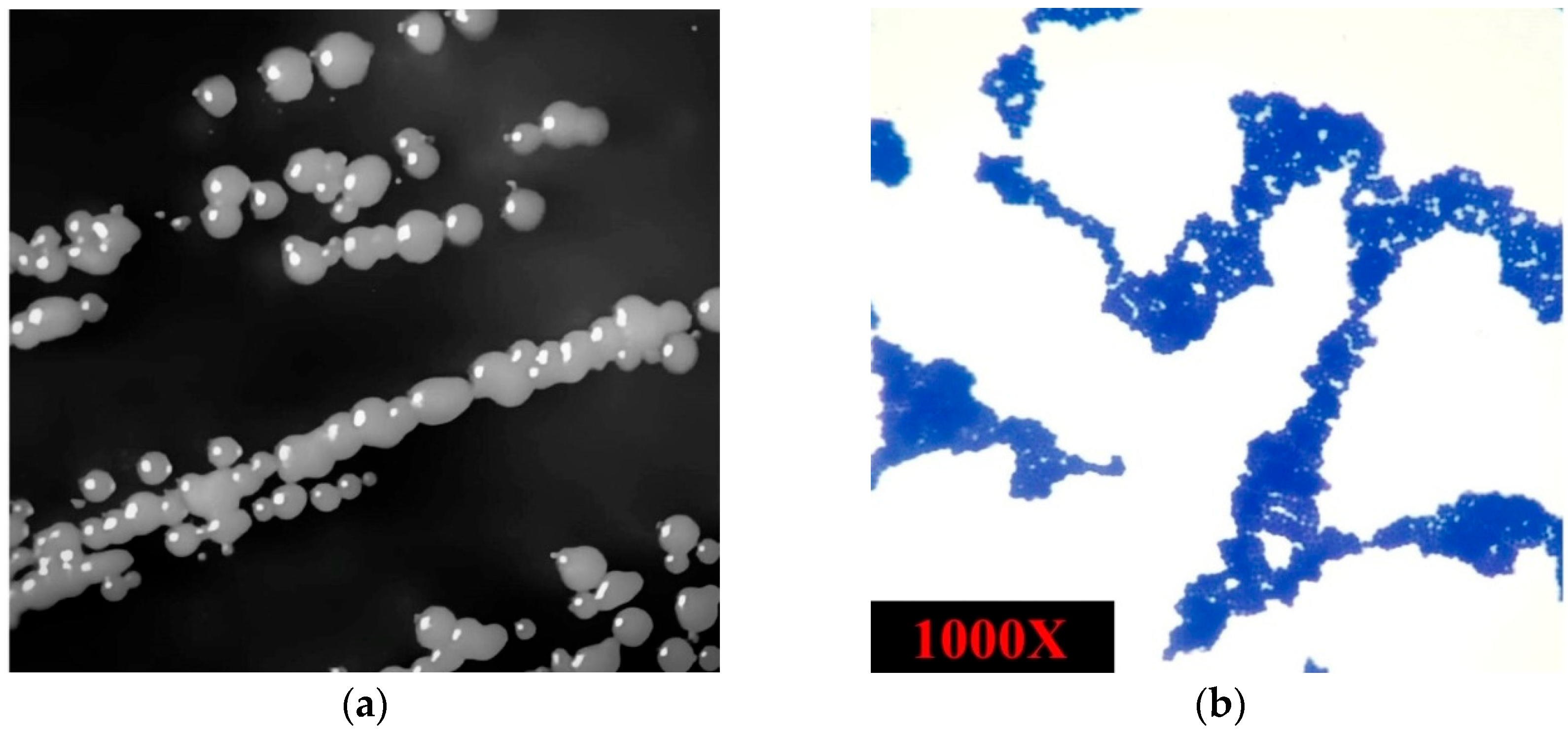
On nutrient agar, the isolate formed smooth, circular, convex, and opaque colonies with entire margins and a pale-yellow pigmentation, typically measuring 1–2 mm in diameter after 24 h of incubation at 30 °C (Figure 2a). Microscopic examination of the isolated strain revealed Gram-positive, spherical cocci arranged in irregular clusters. The microorganism was non-spore-forming, non-motile, and catalase-positive, consistent with the general characteristics of the Staphylococcus genus (Figure 2b).

3.2. Genome Characteristics and Strain Typing of Isolate SC-M1C

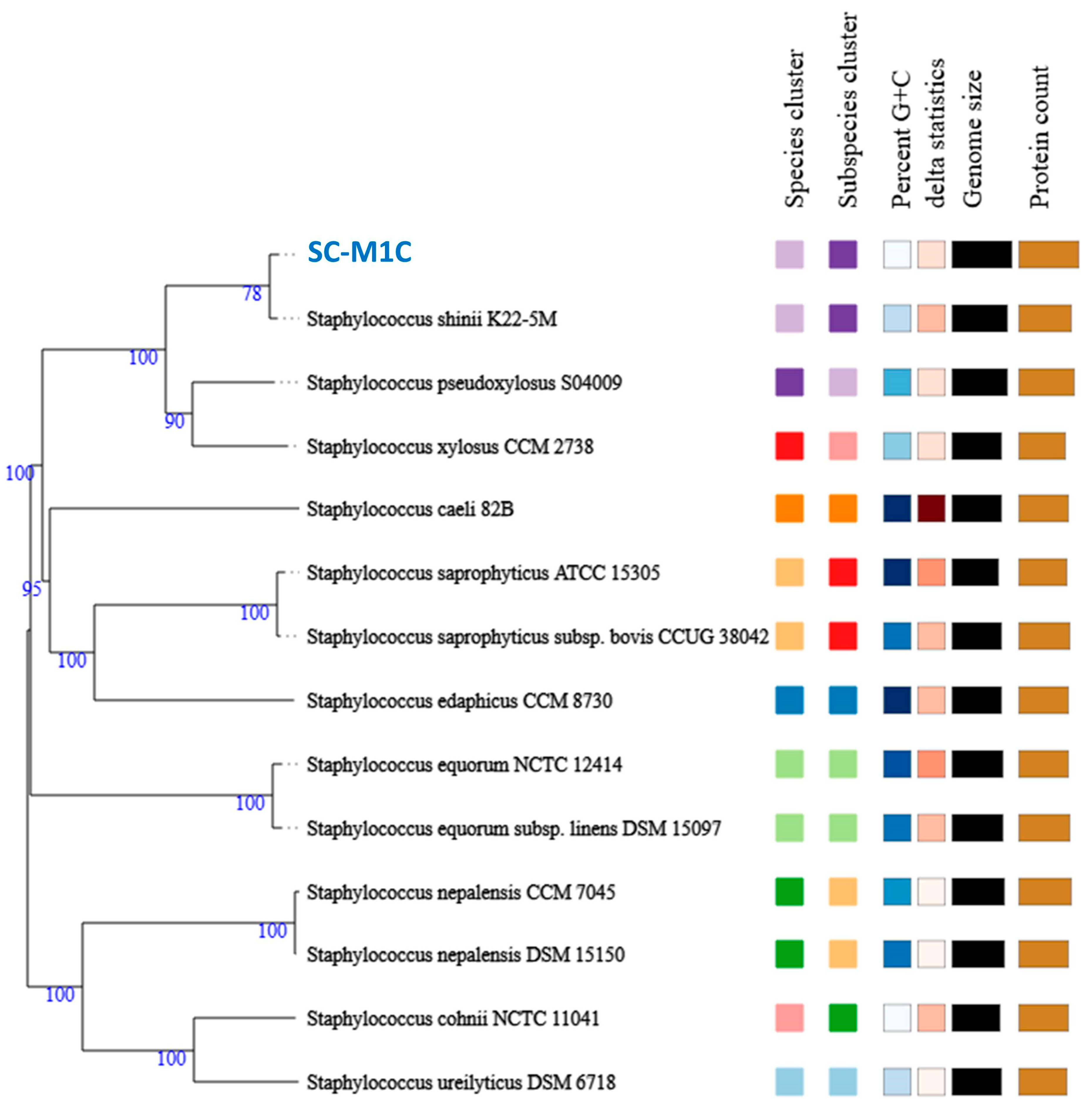
The post-filtering read statistics resulted in 18,112,185 sequences and a total length of 2.7 Gbp (average sequence length 151 bp per sequence). No sequences were flagged as poor quality. The genome sequencing yielded 36 contigs with an N50 value of 522,659 bp, a GC content of 32.34%, and a total genome length of 3,223,632 bp. Strain typing via the Type Strain Genome Server (TYGS) identified the isolated strain as S. shinii, a low GC-content, Gram-positive bacterium within the Bacillota (formerly Firmicutes), class Bacilli, and order Bacillales. To confirm the taxonomic positions of the isolate, digital DNA–DNA hybridization (dDDH), and average nucleotide identity (ANI) comparisons were performed with their closest type strains, as the type strain “S. shinii K22-5M” exhibited the closest relationship, with dDDH, ANIm, and ΔCG% values of 94.8%, 99.40% and 0.15%, respectively (Table 1). These values confirm the taxonomic position of the isolated strain. A phylogenomic tree depicting the evolutionary relationships between both strains and their closely related type strains is presented in Figure 3.

3.3. S. shinii Strain SC-M1C Genome Annotation and Genome Mapping

The Similar Genome Finder tool identified Staphylococcus pseudoxylosus strain 14AME19 (GenBank: CP068712.1) as the closest complete genome available on the public database to the strain SC-M1C, with a distance of 0.00730426, making it suitable for reference-guided scaffolding. Using RagTag, the genome of the strain SC-M1C was scaffolded into eight contigs, with the largest spanning 3,052,874 bp and the smallest 5491 bp, resulting in a coverage of 98.7% of the reference genome.
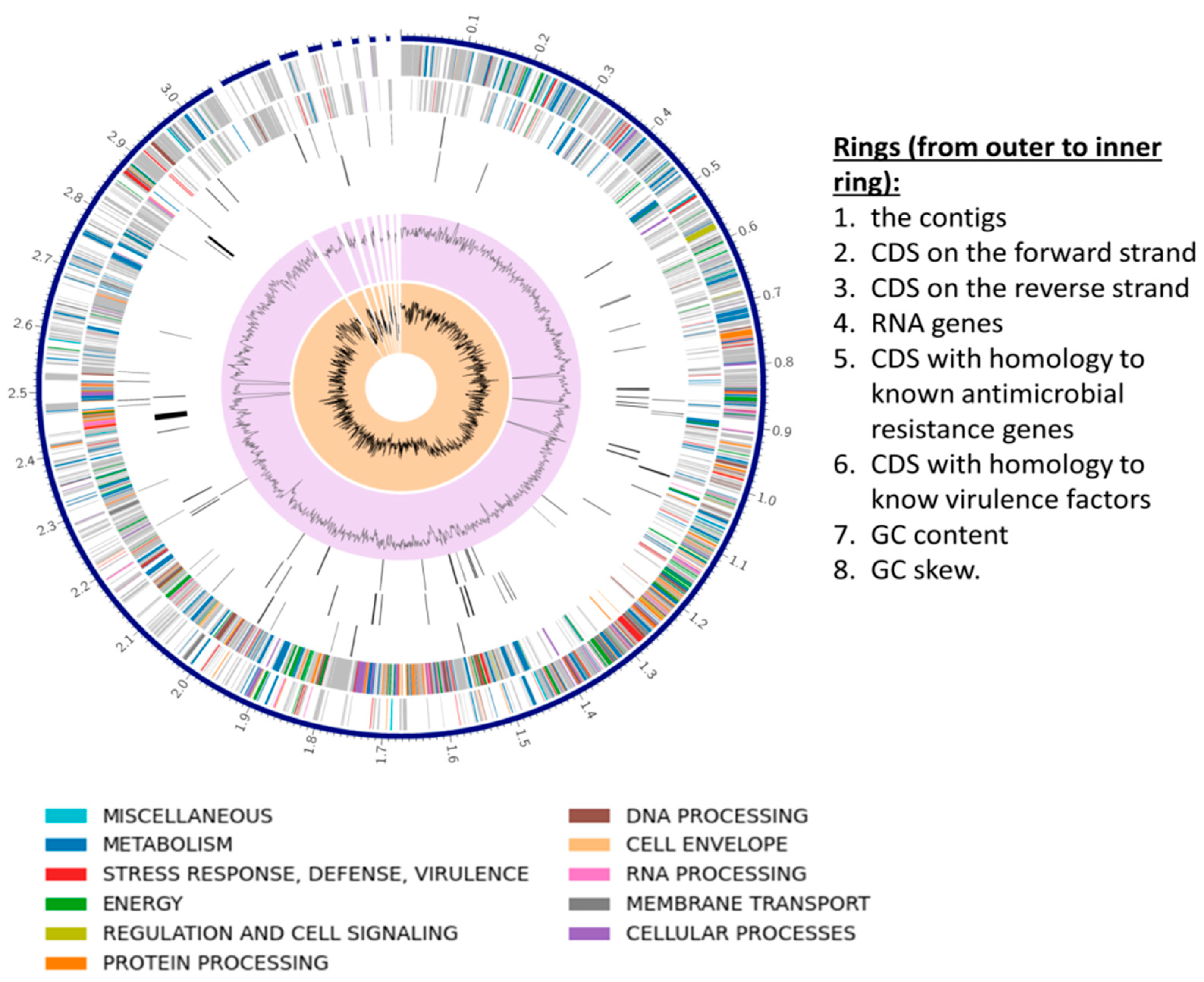
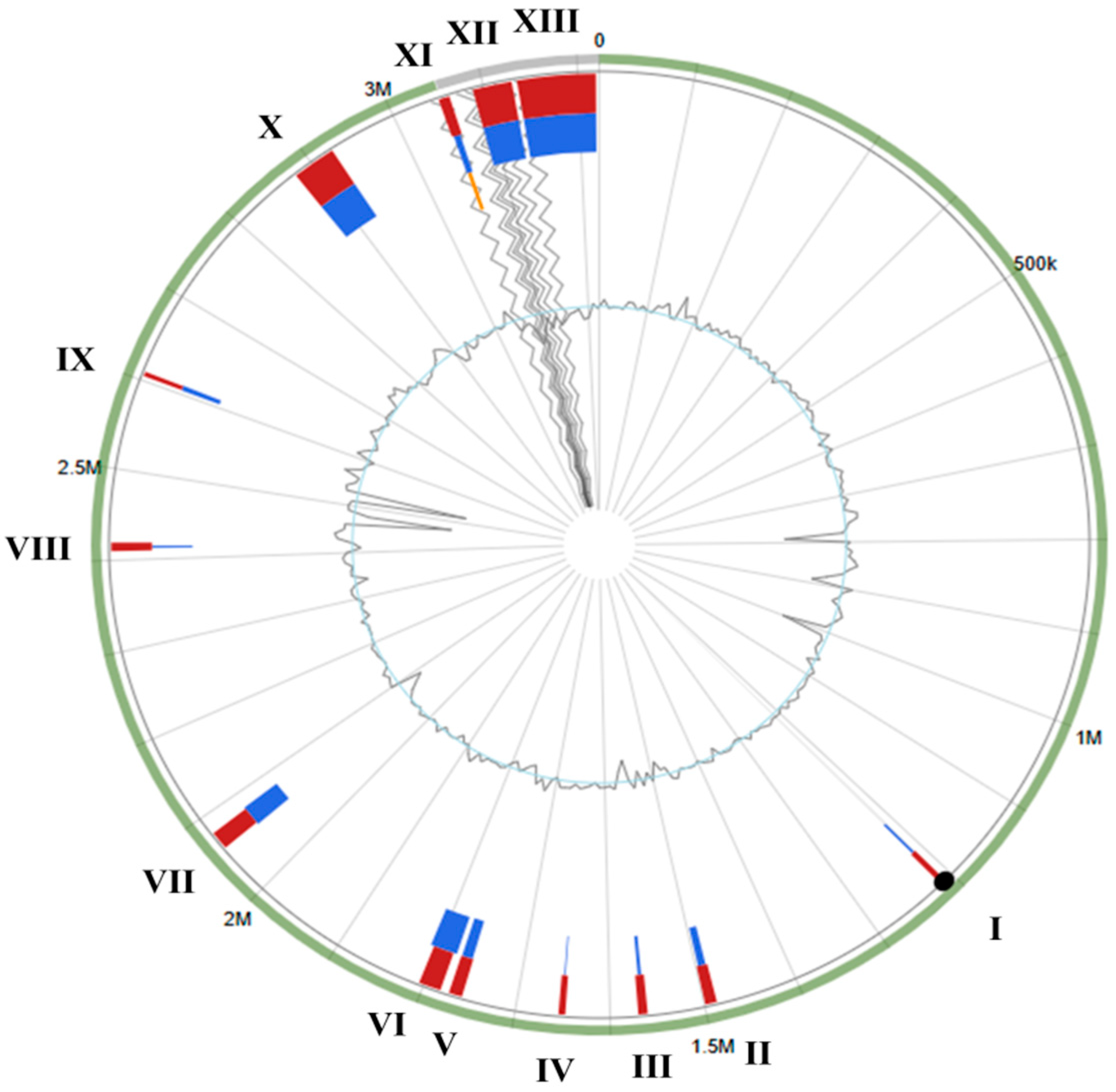
The RAST and Prokka pipelines were used to annotate the genome of S. shinii strain SC-M1C, as RAST returned more annotations with functional assignments than Prokka (Table 2). The genome map of the S. shinii strain SC-M1C is illustrated in Figure 4.
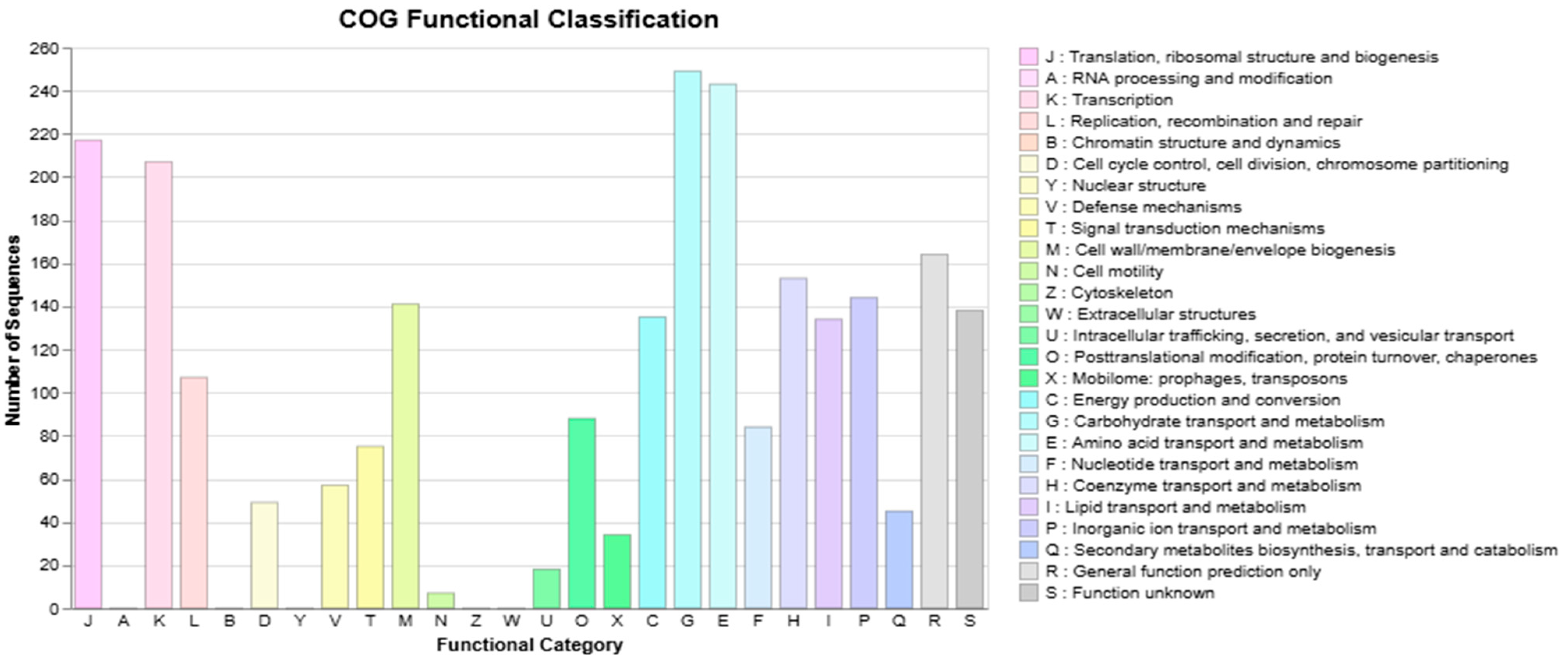
Approximately 80.89% of the coding genes within the genome of S. shinii strain SC-M1C were assigned to the COG functional category; with 5.97% related to inorganic ion transport and metabolism; 3.01% related to signal transduction mechanisms; 5.66% related to cell wall, membrane, and envelope biogenesis; and 1.81% related to secondary metabolites biosynthesis, transport, and catabolism (Figure 5).

3.4. Antimicrobial Resistance and Horizontal Gene Transfer

While the scaffolded contig 1 represents the bacterial chromosome, contigs from 2 to 8 contain plasmid-related genes. Plasmid analysis using PlasmidFinder version 2.0 identified specific plasmid replicons in these contigs. A replicon belonging to the Inc18 family was detected, with a 93.04% sequence identity to the rep16 replicon (GenBank: CP000737), located on contig number 4 at positions 3121–3637. Additionally, two replicons from the RepA-N family were identified: rep20, exhibiting 90.95% identity (GenBank: HE616681) and found on contig number 3 at positions 6541–7479, and rep24c, showing 97.38% identity (GenBank: FR687301) and located on contig number 6 at positions 422–1414. These findings indicate the presence of plasmids associated with the Inc18 and RepA-N families, which are commonly linked to functions such as antibiotic resistance, conjugative transfer, and stress response, highlighting their potential role in adaptive mechanisms within the genome. Furthermore, a total of 11 virulence factors were annotated based on the virulence factor database (VFDB) within the genome of SC-M1C (Table 3).
On the other hand, a total of 12 antibiotic resistance genes were identified in the CARD database, along with 2 in NDARO and 42 in PATRIC (Table 4).
IslandViewer 4, using the complete genome of S. shinii strain 14AME19 (GenBank: CP068712) as a reference, identified 13 GIs, ranging in size from 5152 bp to 83,760 bp, within the genome of S. shinii strain SC-M1C (Figure 6). Notably, the presence of transposases, phage-related genes, integrases, and mobilization proteins within these islands underscores their potential for horizontal gene transfer, facilitating genetic adaptability and ecological fitness in dynamic marine environments (Supplementary Table S1).

3.5. Reconstruction of S. shinii Strain SC-M1C Metabolic Pathways

To investigate the secondary metabolite potential of the studied genome, biosynthetic gene clusters (BGCs) were predicted and analyzed using the AntiSMASH server. Twelve BGCs were identified, representing diverse classes, including nonribosomal peptide synthetases (NRPS), type III polyketide synthases (T3PKS), lanthipeptides, siderophores, and terpene-related clusters. Two of the BGCs showed 100% identity with known clusters, namely staphylopine (opine-like metallophore) and staphyloferrin A (siderophore), suggesting conserved pathways for metal acquisition. The remaining clusters displayed various biosynthetic types but lacked direct matches to characterized metabolites, indicating the potential for novel compound production. The details of the identified BGCs are summarized in Table 5.
For reconstruction of the metabolic pathways in the strain SC-M1C, the KEGG was used for annotating the CDS-predicted protein products. From 3077 CDs within the genome of S. shinii strain SC-M1C, there were 1630 protein products (53.0%) were annotated on the KEGG database. The annotated proteins were incorporated into 214 KEGG pathways. There were 59 complete KEGG modules, with multiple of them being functionally and ecologically significant, such as the betaine module (M00555), the formaldehyde assimilation module (M00345), and menaquinone biosynthesis module (M00116).
To adapt to the dynamic and often extreme conditions of marine ecosystems, microorganisms have evolved a range of genomic and metabolomic strategies that collectively support ecological adaptation. Key among these is osmoregulation and salt tolerance, biofilm formation and surface adhesion, nutrient acquisition, stress response, and symbiosis.

3.5.1. Osmoregulation and Salt Tolerance

To thrive in marine environments characterized by fluctuating and often high salinity, microorganisms must tightly regulate intracellular osmotic pressure. The genome of S. shinii strain SC-M1C reveals a powerful osmo-regulatory system composed of multiple Na+/H+ antiporters and compatible solute transporters that collectively maintain ionic balance and cellular homeostasis (Table 6).
The mnhA1–G1 genes encoding the multi-subunit Na+/H+ antiporter were likely organized in a single operon-like cluster, suggesting coordinated expression. The nhaC gene was present as a single-copy gene. In contrast, five homologous copies of opuD were identified across the genome, indicating gene duplication and possible functional diversification in compatible solute uptake.

3.5.2. Nutrient Acquisition

Marine environments are often oligotrophic, demanding efficient nutrient scavenging strategies. The genome encodes transporters and metabolic enzymes that facilitate the uptake and assimilation of essential but limited nutrients, including nitrogen, phosphate, methionine, thiamine, and iron (Table 7).
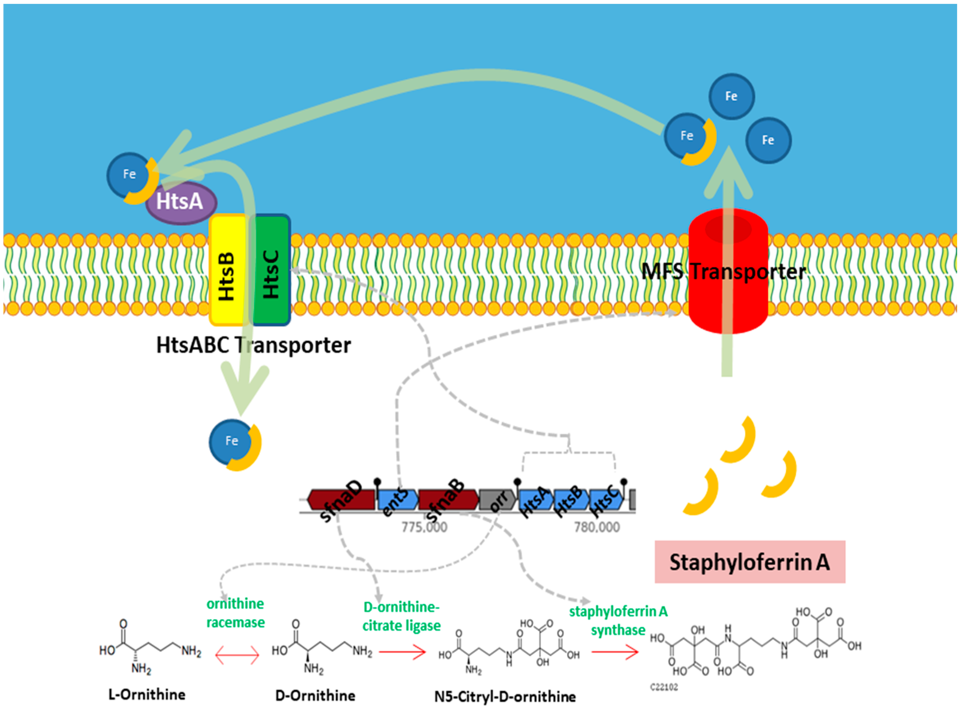
Moreover, S. shinii strain SC-M1C possesses several specialized metabolic pathways dedicated to efficient nutrient acquisition. For instance, the detection of the complete staphyloferrin A module (M00876) reveals a capacity for high-affinity iron scavenging through siderophore production, a critical advantage in iron-depleted seawater (Figure 7).
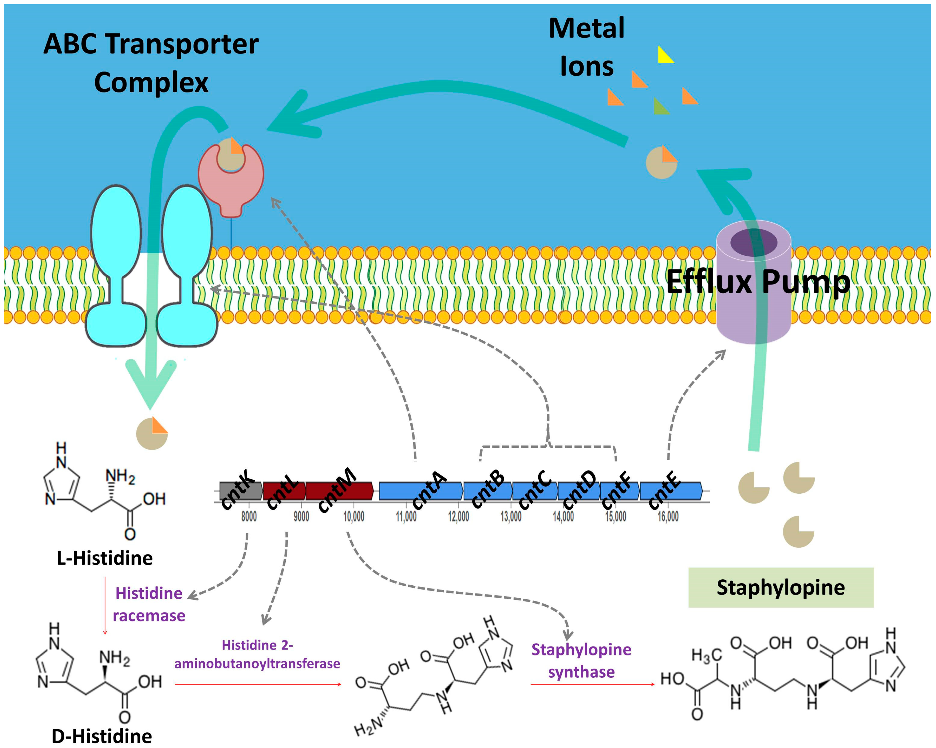
Similarly, the presence of the staphylopine biosynthesis module (M00949) indicates an ability to acquire essential trace metals such as nickel and cobalt via metallophore-mediated transport (Figure 8).

3.5.3. Stress Response and Resistance

Exposure to environmental stressors such as UV radiation, oxidative agents, and metal ions is common in marine habitats. Several genes encode stress defense mechanisms, including oxidative damage repair, redox balance maintenance, and heavy metal detoxification (Table 8).

3.5.4. Aromatic Hydrocarbon Degradation

The genome of S. shinii SC-M1C harbors genes implicated in the degradation of aromatic hydrocarbons (Table 9). These genes suggest a potential capacity for detoxifying aromatic and quinone-based compounds, which may contribute to the strain’s adaptability within the chemically complex sponge microenvironment.

3.5.5. Two-Component Systems in S. shinii Strain SC-M1C

Analysis of the S. shinii SC-M1C genome revealed the presence of a diverse array of two-component systems (TCSs), comprising a total of 30 TCS-related proteins. This included 14 histidine kinases (12 classified as classic histidine kinases and 2 possible incomplete kinases), 15 response regulators, and 1 phosphotransferase protein.

4. Discussion

Marine Firmicutes play a crucial role in marine ecosystems, contributing to nutrient cycling and the production of diverse bioactive compounds with potential pharmaceutical applications [58,59]. While genera such as Bacillus have been extensively studied, marine Staphylococcus species—particularly those associated with sponges—remain largely underexplored. To our knowledge, this study represents the first comprehensive genome-wide analysis of an S. shinii strain from the marine environment. The sponge-associated marine bacterium S. shinii strain SC-M1C represents a valuable opportunity to examine the genomic features related to the resilience to challenging conditions in the marine environment. While the genome of S. shinii strain SC-M1C was about 3 Mbp in length, previous studies of marine Staphylococci genomic analysis recorded a genome size of around 2.5 Mbp [60,61], which may suggest a more complex metabolic repertoire of the S. shinii strain, making it interesting for study.
Genomic islands (GIs) are type of mobile genetic elements that integrate within bacterial genomes. These regions often exhibit distinct nucleotide compositions, including variations in GC content and codon usage, and are frequently flanked by mobility elements such as direct repeats, insertion sequences, or integrase genes, which facilitate their integration into the host genome [62]. GIs typically encode clusters of functionally related genes that confer adaptive advantages, such as virulence factors, antibiotic resistance, metabolic versatility, or symbiotic capabilities [63,64]. GIs encode proteins involved in processes such as DNA uptake, cell morphology regulation, stress tolerance, nutrient transport, and defense mechanisms against phages and environmental stresses.
GIs I, II, and III carry genes associated with DNA uptake, cell surface modification, and stress response, suggesting roles in niche adaptation and survival under marine stressors (e.g., high salt, limited nutrients, or oxidative conditions) [65,66,67,68]. Additionally, several islands exhibit specialized potential functions that enhance the metabolic versatility and resilience of the S. shinii strain SC-M1C. For example, GI X encodes arsenical/cadmium resistance operons and two-component systems linked to heavy metal resistance, heavy metal detoxification, and stress signaling. The largest island, GI XIII, encompasses a variety of genes implicated in cell wall remodeling, protein folding, DNA repair, and stress response, highlighting its importance in maintaining genomic integrity and cellular homeostasis. Collectively, these GIs reflect a mosaic of horizontally acquired traits that likely enable S. shinii to thrive in its marine habitat by conferring adaptive advantages through enhanced metabolic capabilities, structural adaptations, and robust defense mechanisms against biotic and abiotic challenges.
The AntiSMASH-based screening uncovered an array of cryptic BGCs. With the exception of the staphylopine and staphyloferrin A BGCs, the remaining clusters displayed various biosynthetic types but lacked direct matches to characterized metabolites, indicating the potential for novel compound production. Staphylopine and staphyloferrin A are metallophores produced by Staphylococcus species that support survival in metal-limited settings. Staphyloferrin A is a citrate-based siderophore that specifically chelates ferric-iron for import via the HtsABC transporter system [69,70]. Staphylopine, on the other hand, is a broad-spectrum metallophore primarily involved in zinc (and also nickel/cobalt) acquisition under conditions of host nutritional immunity, imported via the CntABC system [71,72,73,74]. While most research focuses on host–pathogen interactions, marine microorganisms commonly produce siderophores as an adaptive strategy to acquire iron in extremely iron-poor seawater [71].
Moreover, the presence of a complete KEGG module of betaine (M00555) suggests a specialized capacity for osmoregulation. Betaine functions as a potent osmoprotectant, stabilizing cellular structures and maintaining turgor pressure under high-salinity conditions [75]. This pathway likely confers a significant survival advantage, enabling SC-M1C to withstand the osmotic stresses characteristic of marine ecosystems. The ability to synthesize betaine not only supports resilience during salinity fluctuations but may also facilitate colonization of diverse marine niches.
Additionally, the formaldehyde assimilation module (M00345) equips SC-M1C to utilize one-carbon compounds as alternative carbon sources, likely exploiting metabolic byproducts produced by surrounding microbial communities [76]. These specialized systems underscore the strain’s metabolic flexibility and its potential to dominate in competitive, nutrient-limited marine ecosystems. Together, these adaptive pathways underscore SC-M1C’s capacity to endure dynamic and challenging environmental stresses.
To withstand the osmotic pressure characteristic of marine environments, S. shinii strain SC-M1C possesses a genetic repertoire associated with osmoregulation and salt tolerance. For instance, the mnh gene cluster, widely recognized for its role in maintaining ionic balance and conferring tolerance to high salinity and alkaline conditions in Gram-positive bacteria such as Staphylococcus aureus and Bacillus species, is present in the SC-M1C genome as the mnhA1–G1 operon. Its presence likely contributes to the halotolerant phenotype of the strain [77]. Furthermore, the genome harbors five homologous copies of opuD, which encode glycine betaine transporters essential for osmoprotection under salt stress [78]. This transporter system is evolutionarily conserved and functionally active in a wide range of bacterial species, including marine-associated taxa.
In marine ecosystems, the degradation of aromatic hydrocarbons, especially polycyclic aromatic hydrocarbons (PAHs), is a critical microbial process involving numerous genes encoding hydrocarbon-degrading enzymes. The SC-M1C genome contains the catE gene, a key component of the meta-cleavage pathway involved in aromatic compound degradation [79]. The catE gene is commonly found in a wide range of hydrocarbon-degrading bacteria, including species from the genera Pseudomonas, Bacillus, and Rhodococcus, and serves as a genetic marker for monitoring aromatic hydrocarbon degradation in polluted environments [80,81]. Moreover, the genome of SC-M1C also carries the genes mhqR, mhqA, mhqO, and mhqD, which are primarily associated with the degradation of hydroquinone and other phenolic compounds. These intermediates are often generated during the breakdown of lignin-derived or other complex aromatic substrates [82].
Additionally, S. shinii has a diverse collection of TCS-related genes. TCSs are crucial for bacterial dynamism and resilience to various environmental stresses. These systems enable bacteria to sense changes in their surrounding ecosystem and respond accordingly, ensuring their ability to adapt to diverse conditions [83,84]. The overall repertoire of TCSs in SC-M1C highlights its capacity to adapt to diverse environmental conditions typically encountered in marine ecosystems, such as nutrient fluctuations, osmotic stress, and interactions with other microbial species.

5. Conclusions

S. shinii was isolated from a Red Sea sponge in a marine environment. Key adaptations for high-salinity survival are highlighted in the genome, such as genes related to heavy metal tolerance, osmoregulation, nutrition uptake, and stress response. The presence of various genomic islands and plasmids suggests robust horizontal gene transfer mechanisms and improved environmental adaptability. Furthermore, the identification of biosynthetic gene clusters for siderophores (staphyloferrin A) and metallophores (staphylopine) points to a versatile metabolism for metal acquisition. Future research should prioritize transcriptomic validation to confirm gene expression under specific marine stresses and conduct bioactivity screening of novel compounds derived from the identified biosynthetic pathways.

Supplementary Materials

The following supporting information can be downloaded at: https://www.mdpi.com/article/10.3390/microorganisms13081866/s1, Table S1. A summary of the genomic islands (GIs) identified by the IslandViewer 4 server within the Staphylococcus shinii strain SC-M1C genome.

Author Contributions

M.E.S.: Conceptualization, investigation, methodology, and writing—original draft. A.M.S.: Conceptualization, formal analysis, data curation, investigation, methodology, validation, visualization, and writing—original draft. H.L.: Conceptualization, investigation, methodology, and writing—original draft. Y.S.M.: Conceptualization, investigation, and writing—review of the final draft. S.M.S.: Conceptualization, investigation, validation, supervision, project administration, and review, edit of the final draft. All authors have read and agreed to the published version of the manuscript.

Funding

Deanship of Research and Graduate Studies, Ajman University project awarded AY 2024/2025 REF. number (2024-IRG-MED-1).

Institutional Review Board Statement

This study protocol was approved by the Ethics Committee of the Faculty of Pharmacy, Suez Canal University (protocol code 201810PHD2, 17 October 2018).

Informed Consent Statement

Not applicable.

Data Availability Statement

The original data presented in the study are available in NCBI under the following accessions: PRJNA1280784 (Bioproject), SAMN49534678 (BioSample), SRR34102925 (Sequence Read Archive), and GCF_051216755.1 (Assembled genome).

Conflicts of Interest

The authors declare no conflicts of interest.

References

  1. Ibarbalz, F.M.; Henry, N.; Brandão, M.C.; Martini, S.; Busseni, G.; Byrne, H.; Coelho, L.P.; Endo, H.; Gasol, J.M.; Gregory, A.C.; et al. Global Trends in Marine Plankton Diversity across Kingdoms of Life. Cell 2019, 179, 1084–1097.e21. [Google Scholar] [CrossRef] [PubMed]
  2. Hoshino, T.; Doi, H.; Uramoto, G.I.; Wörmer, L.; Adhikari, R.R.; Xiao, N.; Morono, Y.; D’Hondt, S.; Hinrichs, K.U.; Inagaki, F. Global Diversity of Microbial Communities in Marine Sediment. Proc. Natl. Acad. Sci. USA 2020, 117, 27587–27597. [Google Scholar] [CrossRef]
  3. Lauritano, C.; Coppola, D. Biotechnological Applications of Products Released by Marine Microorganisms for Cold Adaptation Strategies: Polyunsaturated Fatty Acids, Antioxidants, and Antifreeze Proteins. J. Mar. Sci. Eng. 2023, 11, 1399. [Google Scholar] [CrossRef]
  4. Arromrak, B.S.; Li, Z.; Gaitán-Espitia, J.D. Adaptive Strategies and Evolutionary Responses of Microbial Organisms to Changing Oceans. Front. Mar. Sci. 2022, 9, 864797. [Google Scholar] [CrossRef]
  5. Sedeek, A.M.; Elfeky, H.; Hanora, A.S.; Solyman, S.M. Genomic Insights into Biosynthesis and Adaptation in the Bioactive Marine Bacterium Streptomyces Albidoflavus VIP-1 from the Red Sea. BMC Microbiol. 2025, 25, 372. [Google Scholar] [CrossRef]
  6. Kamel, H.L.; Hanora, A.; Solyman, S.M. Metataxanomic, Bioactivity and Microbiome Analysis of Red Sea Marine Sponges from Egypt. Mar. Genom. 2022, 61, 100920. [Google Scholar] [CrossRef] [PubMed]
  7. El Samak, M.; Zakeer, S.; Hanora, A.; Solyman, S.M. Metagenomic and Metatranscriptomic Exploration of the Egyptian Red Sea Sponge Theonella Sp. Associated Microbial Community. Mar. Genom. 2023, 70, 101032. [Google Scholar] [CrossRef]
  8. Taylor, M.W.; Radax, R.; Steger, D.; Wagner, M. Sponge-Associated Microorganisms: Evolution, Ecology, and Biotechnological Potential. Microbiol. Mol. Biol. Rev. 2007, 71, 295–347. [Google Scholar] [CrossRef] [PubMed]
  9. Hentati, D.; Cheffi, M.; Hadrich, F.; Makhloufi, N.; Rabanal, F.; Manresa, A.; Sayadi, S.; Chamkha, M. Investigation of Halotolerant Marine Staphylococcus Sp. CO100, as a Promising Hydrocarbon-Degrading and Biosurfactant-Producing Bacterium, under Saline Conditions. J. Environ. Manag. 2021, 277, 111480. [Google Scholar] [CrossRef]
  10. Thirumurthy, K.; Kaliyamoorthy, K.; Kandasamy, K.; Ponnuvel, M.; Viyakarn, V.; Chavanich, S.; Dufossé, L. Antioxidant and Anti-Breast Cancer Properties of Hyaluronidase from Marine Staphylococcus Aureus (CASMTK1). J. Mar. Sci. Eng. 2023, 11, 778. [Google Scholar] [CrossRef]
  11. Heaton, C.J.; Gerbig, G.R.; Sensius, L.D.; Patel, V.; Smith, T.C. Staphylococcus Aureus Epidemiology in Wildlife: A Systematic Review. Antibiotics 2020, 9, 89. [Google Scholar] [CrossRef]
  12. Soge, O.O.; Meschke, J.S.; No, D.B.; Roberts, M.C. Characterization of Methicillin-Resistant Staphylococcus Aureus and Methicillin-Resistant Coagulase-Negative Staphylococcus Spp. Isolated from US West Coast Public Marine Beaches. J. Antimicrob. Chemother. 2009, 64, 1148–1155. [Google Scholar] [CrossRef] [PubMed]
  13. Thapaliya, D.; Hellwig, E.J.; Kadariya, J.; Grenier, D.; Jefferson, A.J.; Dalman, M.; Kennedy, K.; DiPerna, M.; Orihill, A.; Taha, M.; et al. Prevalence and Characterization of Staphylococcus Aureus and Methicillin-Resistant Staphylococcus Aureus on Public Recreational Beaches in Northeast Ohio. GeoHealth 2017, 1, 320–332. [Google Scholar] [CrossRef]
  14. Rosenstein, R.; Götz, F. What Distinguishes Highly Pathogenic Staphylococci from Medium- and Non-Pathogenic? In Between Pathogenicity and Commensalism; Dobrindt, U., Hacker, J., Svanborg, C., Eds.; Current Topics in Microbiology and Immunology; Springer: Berlin/Heidelberg, Germany, 2012; pp. 33–89. [Google Scholar]
  15. Amelia, T.S.M.; Amirul, A.-A.A.; Saidin, J.; Bhubalan, K. Identification of Cultivable Bacteria from Tropical Marine Sponges and Their Biotechnological Potentials. Trop. Life Sci. Res. 2018, 29, 187–199. [Google Scholar] [CrossRef]
  16. Anteneh, Y.S.; Yang, Q.; Brown, M.H.; Franco, C.M.M. Factors Affecting the Isolation and Diversity of Marine Sponge-Associated Bacteria. Appl. Microbiol. Biotechnol. 2022, 106, 1729–1744. [Google Scholar] [CrossRef]
  17. Anteneh, Y.S.; Yang, Q.; Brown, M.H.; Franco, C.M.M. Antimicrobial Activities of Marine Sponge-Associated Bacteria. Microorganisms 2021, 9, 171. [Google Scholar] [CrossRef]
  18. Cho, G.-S.; Li, B.; Brinks, E.; Franz, C.M.A.P. Characterization of Antibiotic-Resistant, Coagulase-Negative Staphylococci from Fresh Produce and Description of Staphylococcus Shinii Sp. Nov. Isolated from Chives. J. Microbiol. 2022, 60, 877–889. [Google Scholar] [CrossRef]
  19. Arreguin-Perez, C.A.; Cossio-Bayugar, R.; Aguilar-Diaz, H.; Miranda-Miranda, E. In Situ Localization of Staphylococcus Shinii and Staphylococcus Succinus in Infected Rhipicephalus Microplus Ticks: Implications for Biocontrol Strategies. Pathogens 2024, 13, 1025. [Google Scholar] [CrossRef] [PubMed]
  20. Sosa-Fajardo, A.; Díaz-Muñoz, C.; Van der Veken, D.; Pradal, I.; Verce, M.; Weckx, S.; Leroy, F. Genomic Exploration of the Fermented Meat Isolate Staphylococcus Shinii IMDO-S216 with a Focus on Competitiveness-Enhancing Secondary Metabolites. BMC Genom. 2024, 25, 575. [Google Scholar] [CrossRef] [PubMed]
  21. Deng, Y.; Xu, H.; Su, Y.; Liu, S.; Xu, L.; Guo, Z.; Wu, J.; Cheng, C.; Feng, J. Horizontal Gene Transfer Contributes to Virulence and Antibiotic Resistance of Vibrio Harveyi 345 Based on Complete Genome Sequence Analysis. BMC Genom. 2019, 20, 761. [Google Scholar] [CrossRef]
  22. Arias, P.M.; Butler, J.; Randhawa, G.S.; Soltysiak, M.P.M.; Hill, K.A.; Kari, L. Environment and Taxonomy Shape the Genomic Signature of Prokaryotic Extremophiles. Sci. Rep. 2023, 13, 16105. [Google Scholar] [CrossRef]
  23. Leopold, S.R.; Goering, R.V.; Witten, A.; Harmsen, D.; Mellmann, A. Bacterial Whole-Genome Sequencing Revisited: Portable, Scalable, and Standardized Analysis for Typing and Detection of Virulence and Antibiotic Resistance Genes. J. Clin. Microbiol. 2014, 52, 2365–2370. [Google Scholar] [CrossRef]
  24. Meier-Kolthoff, J.P.; Göker, M. TYGS Is an Automated High-Throughput Platform for State-of-the-Art Genome-Based Taxonomy. Nat. Commun. 2019, 10, 2182. [Google Scholar] [CrossRef] [PubMed]
  25. Bolger, A.M.; Lohse, M.; Usadel, B. Trimmomatic: A Flexible Trimmer for Illumina Sequence Data. Bioinformatics 2014, 30, 2114–2120. [Google Scholar] [CrossRef]
  26. Wick, R.R.; Judd, L.M.; Gorrie, C.L.; Holt, K.E. Unicycler: Resolving Bacterial Genome Assemblies from Short and Long Sequencing Reads. PLoS Comput. Biol. 2017, 13, e1005595. [Google Scholar] [CrossRef]
  27. Richter, M.; Rosselló-Móra, R.; Oliver Glöckner, F.; Peplies, J. JSpeciesWS: A Web Server for Prokaryotic Species Circumscription Based on Pairwise Genome Comparison. Bioinformatics 2016, 32, 929–931. [Google Scholar] [CrossRef]
  28. Meier-Kolthoff, J.P.; Auch, A.F.; Klenk, H.P.; Göker, M. Genome Sequence-Based Species Delimitation with Confidence Intervals and Improved Distance Functions. BMC Bioinform. 2013, 14, 60. [Google Scholar] [CrossRef] [PubMed]
  29. Alonge, M.; Soyk, S.; Ramakrishnan, S.; Wang, X.; Goodwin, S.; Sedlazeck, F.J.; Lippman, Z.B.; Schatz, M.C. RaGOO: Fast and Accurate Reference-Guided Scaffolding of Draft Genomes. Genome Biol. 2019, 20, 224. [Google Scholar] [CrossRef] [PubMed]
  30. Aziz, R.K.; Bartels, D.; Best, A.; DeJongh, M.; Disz, T.; Edwards, R.A.; Formsma, K.; Gerdes, S.; Glass, E.M.; Kubal, M.; et al. The RAST Server: Rapid Annotations Using Subsystems Technology. BMC Genom. 2008, 9, 75. [Google Scholar] [CrossRef]
  31. Seemann, T. Prokka: Rapid Prokaryotic Genome Annotation. Bioinformatics 2014, 30, 2068–2069. [Google Scholar] [CrossRef]
  32. Wattam, A.R.; Abraham, D.; Dalay, O.; Disz, T.L.; Driscoll, T.; Gabbard, J.L.; Gillespie, J.J.; Gough, R.; Hix, D.; Kenyon, R.; et al. PATRIC, the Bacterial Bioinformatics Database and Analysis Resource. Nucleic Acids Res. 2014, 42, D581–D591. [Google Scholar] [CrossRef]
  33. Alcock, B.P.; Huynh, W.; Chalil, R.; Smith, K.W.; Raphenya, A.R.; Wlodarski, M.A.; Edalatmand, A.; Petkau, A.; Syed, S.A.; Tsang, K.K.; et al. CARD 2023: Expanded Curation, Support for Machine Learning, and Resistome Prediction at the Comprehensive Antibiotic Resistance Database. Nucleic Acids Res. 2023, 51, D690–D699. [Google Scholar] [CrossRef]
  34. Bertelli, C.; Laird, M.R.; Williams, K.P.; Lau, B.Y.; Hoad, G.; Winsor, G.L.; Brinkman, F.S. IslandViewer 4: Expanded Prediction of Genomic Islands for Larger-Scale Datasets. Nucleic Acids Res. 2017, 45, W30–W35. [Google Scholar] [CrossRef]
  35. Kanehisa, M.; Sato, Y.; Morishima, K. BlastKOALA and GhostKOALA: KEGG Tools for Functional Characterization of Genome and Metagenome Sequences. J. Mol. Biol. 2016, 428, 726–731. [Google Scholar] [CrossRef]
  36. Tatusov, R.L.; Fedorova, N.D.; Jackson, J.D.; Jacobs, A.R.; Kiryutin, B.; Koonin, E.V.; Krylov, D.M.; Mazumder, R.; Smirnov, S.; Nikolskaya, A.N.; et al. The COG Database: An Updated Vesion Includes Eukaryotes. BMC Bioinform. 2003, 4, 41. [Google Scholar] [CrossRef]
  37. Carattoli, A.; Zankari, E.; García-Fernández, A.; Voldby Larsen, M.; Lund, O.; Villa, L.; Møller Aarestrup, F.; Hasman, H. In Silico Detection and Typing of Plasmids Using PlasmidFinder and Plasmid Multilocus Sequence Typing. Antimicrob. Agents Chemother. 2014, 58, 3895–3903. [Google Scholar] [CrossRef]
  38. Blin, K.; Shaw, S.; Kloosterman, A.M.; Charlop-Powers, Z.; van Wezel, G.P.; Medema, M.H.; Weber, T. AntiSMASH 6.0: Improving Cluster Detection and Comparison Capabilities. Nucleic Acids Res. 2021, 49, W29–W35. [Google Scholar] [CrossRef] [PubMed]
  39. Zhou, S.; Liu, B.; Zheng, D.; Chen, L.; Yang, J. VFDB 2025: An Integrated Resource for Exploring Anti-Virulence Compounds. Nucleic Acids Res. 2025, 53, D871–D877. [Google Scholar] [CrossRef]
  40. Casey, D.; Sleator, R.D. A Genomic Analysis of Osmotolerance in Staphylococcus Aureus. Gene 2021, 767, 145268. [Google Scholar] [CrossRef] [PubMed]
  41. Prágai, Z.; Eschevins, C.; Bron, S.; Harwood, C.R. Bacillus Subtilis NhaC, an Na+/H+ Antiporter, Influences Expression of the PhoPR Operon and Production of Alkaline Phosphatases. J. Bacteriol. 2001, 183, 2505–2515. [Google Scholar] [CrossRef] [PubMed]
  42. Ziegler, C.; Bremer, E.; Krämer, R. The BCCT Family of Carriers: From Physiology to Crystal Structure. Mol. Microbiol. 2010, 78, 13–34. [Google Scholar] [CrossRef]
  43. Kadaba, N.S.; Kaiser, J.T.; Johnson, E.; Lee, A.; Rees, D.C. The High-Affinity E. Coli Methionine ABC Transporter: Structure and Allosteric Regulation. Science 2008, 321, 250–253. [Google Scholar] [CrossRef] [PubMed]
  44. Ogawa, K.; Akagawa, E.; Yamane, K.; Sun, Z.W.; LaCelle, M.; Zuber, P.; Nakano, M.M. The NasB Operon and NasA Gene Are Required for Nitrate/Nitrite Assimilation in Bacillus Subtilis. J. Bacteriol. 1995, 177, 1409–1413. [Google Scholar] [CrossRef]
  45. Lin, J.T.; Goldman, B.S.; Stewart, V. The NasFEDCBA Operon for Nitrate and Nitrite Assimilation in Klebsiella Pneumoniae M5al. J. Bacteriol. 1994, 176, 2551–2559. [Google Scholar] [CrossRef]
  46. Pereira, N.; Shilova, I.N.; Zehr, J.P. Use of the High-affinity Phosphate Transporter Gene, PstS, as an Indicator for Phosphorus Stress in the Marine Diazotroph Crocosphaera watsonii (Chroococcales, Cyanobacteria). J. Phycol. 2019, 55, 752–761. [Google Scholar] [CrossRef]
  47. Moreno-Letelier, A.; Olmedo, G.; Eguiarte, L.E.; Martinez-Castilla, L.; Souza, V. Parallel Evolution and Horizontal Gene Transfer of the Pst Operon in Firmicutes from Oligotrophic Environments. Int. J. Evol. Biol. 2011, 2011, 781642. [Google Scholar] [CrossRef] [PubMed]
  48. Schyns, G.; Potot, S.; Geng, Y.; Barbosa, T.M.; Henriques, A.; Perkins, J.B. Isolation and Characterization of New Thiamine-Deregulated Mutants of Bacillus Subtilis. J. Bacteriol. 2005, 187, 8127–8136. [Google Scholar] [CrossRef] [PubMed]
  49. Josts, I.; Almeida Hernandez, Y.; Andreeva, A.; Tidow, H. Crystal Structure of a Group I Energy Coupling Factor Vitamin Transporter S Component in Complex with Its Cognate Substrate. Cell Chem. Biol. 2016, 23, 827–836. [Google Scholar] [CrossRef]
  50. Podkowa, K.J.; Briere, L.-A.K.; Heinrichs, D.E.; Shilton, B.H. Crystal and Solution Structure Analysis of FhuD2 from Staphylococcus Aureus in Multiple Unliganded Conformations and Bound to Ferrioxamine-B. Biochemistry 2014, 53, 2017–2031. [Google Scholar] [CrossRef]
  51. Sebulsky, M.T.; Heinrichs, D.E. Identification and Characterization of FhuD1 and FhuD2, Two Genes Involved in Iron-Hydroxamate Uptake in Staphylococcus Aureus. J. Bacteriol. 2001, 183, 4994–5000. [Google Scholar] [CrossRef]
  52. Weiss, B.; Cunningham, R.P. Genetic Mapping of Nth, a Gene Affecting Endonuclease III (Thymine Glycol-DNA Glycosylase) in Escherichia Coli K-12. J. Bacteriol. 1985, 162, 607–610. [Google Scholar] [CrossRef]
  53. Rosario-Cruz, Z.; Eletsky, A.; Daigham, N.S.; Al-Tameemi, H.; Swapna, G.V.; Kahn, P.C.; Szyperski, T.; Montelione, G.T.; Boyd, J.M. The CopBL Operon Protects Staphylococcus Aureus from Copper Toxicity: CopL Is an Extracellular Membrane–Associated Copper-Binding Protein. J. Biol. Chem. 2019, 294, 4027–4044. [Google Scholar] [CrossRef]
  54. Kuznetsova, M.V.; Nesterova, L.Y.; Mihailovskaya, V.S.; Selivanova, P.A.; Kochergina, D.A.; Karipova, M.O.; Valtsifer, I.V.; Averkina, A.S.; Starčič Erjavec, M. Nosocomial Escherichia Coli, Klebsiella Pneumoniae, Pseudomonas Aeruginosa, and Staphylococcus Aureus: Sensitivity to Chlorhexidine-Based Biocides and Prevalence of Efflux Pump Genes. Int. J. Mol. Sci. 2025, 26, 355. [Google Scholar] [CrossRef] [PubMed]
  55. Costa, S.S.; Viveiros, M.; Amaral, L.; Couto, I. Multidrug Efflux Pumps in Staphylococcus Aureus: An Update. Open Microbiol. J. 2013, 7, 59–71. [Google Scholar] [CrossRef] [PubMed]
  56. Morales-Blancas, G.Y.; Reyna-Terán, J.D.; Hernández-Eligio, J.A.; Ortuño-Pineda, C.; Toribio-Jiménez, J.; Rodríguez-Barrera, M.Á.; Toledo-Hernández, E.; Rojas-Aparicio, A.; Romero-Ramírez, Y. The CatE Gene of Bacillus Licheniformis M2-7 Is Essential for Growth in Benzopyrene, and Its Expression Is Regulated by the Csr System. World J. Microbiol. Biotechnol. 2023, 39, 177. [Google Scholar] [CrossRef] [PubMed]
  57. Tago, K.; Sato, J.; Takesa, H.; Kawagishi, H.; Hayatsu, M. Characterization of Methylhydroquinone-Metabolizing Oxygenase Genes Encoded on Plasmid in Burkholderia Sp. NF100. J. Biosci. Bioeng. 2005, 100, 517–523. [Google Scholar] [CrossRef]
  58. Chukwudulue, U.M.; Barger, N.; Dubovis, M.; Luzzatto Knaan, T. Natural Products and Pharmacological Properties of Symbiotic Bacillota (Firmicutes) of Marine Macroalgae. Mar. Drugs 2023, 21, 569. [Google Scholar] [CrossRef]
  59. Sedeek, A.M.; Ismail, M.M.; Elsayed, T.R.; Ramadan, M.A. Evaluation of the Marine Bacterial Population in the Great Bitter Lake, Egypt, as a Source of Antimicrobial Secondary Metabolites. Fermentation 2022, 8, 309. [Google Scholar] [CrossRef]
  60. Foster, G.; Robb, A.; Paterson, G.K. Isolation and Genome Sequencing of Staphylococcus Schleiferi Subspecies Coagulans from Antarctic and North Sea Seals. Access Microbiol. 2020, 2, e000162. [Google Scholar] [CrossRef]
  61. Jamal, M.T.; Abdulrahman, I.; Amran, R.H.; Al-Matary, M.A.; Satheesh, S. Complete Genome Sequencing and Antibiofilm Activity of an Endophytic Bacterium Associated with Marine Sponge Hyrtios Erectus Collected from the Red Sea. Front. Mar. Sci. 2025, 12, 1588772. [Google Scholar] [CrossRef]
  62. Aminov, R.I. Horizontal Gene Exchange in Environmental Microbiota. Front. Microbiol. 2011, 2, 158. [Google Scholar] [CrossRef]
  63. Carraro, N.; Rivard, N.; Burrus, V.; Ceccarelli, D. Mobilizable Genomic Islands, Different Strategies for the Dissemination of Multidrug Resistance and Other Adaptive Traits. Mob. Genet. Elem. 2017, 7, 1–6. [Google Scholar] [CrossRef] [PubMed]
  64. Tokuda, M.; Shintani, M. Microbial Evolution through Horizontal Gene Transfer by Mobile Genetic Elements. Microb. Biotechnol. 2024, 17, e14408. [Google Scholar] [CrossRef]
  65. Kaur, J.; Kaur, J. Comparative Genomics of Seven Genomes of Genus Idiomarina Reveals Important Halo Adaptations and Genes for Stress Response. 3 Biotech 2024, 14, 40. [Google Scholar] [CrossRef]
  66. Mani, I.; Singh, V.; Alzahrani, K.J.; Chu, D.-T. (Eds.) Microbial Genomic Islands in Adaptation and Pathogenicity; Springer Nature: Singapore, 2023; ISBN 978-981-19-9341-1. [Google Scholar]
  67. Zhao, Y.; Wei, H.-M.; Yuan, J.-L.; Xu, L.; Sun, J.-Q. A Comprehensive Genomic Analysis Provides Insights on the High Environmental Adaptability of Acinetobacter Strains. Front. Microbiol. 2023, 14, 1177951. [Google Scholar] [CrossRef] [PubMed]
  68. Kaufmann, H.; Salvador, C.; Salazar, V.W.; Cruz, N.; Dias, G.M.; Tschoeke, D.; Campos, L.; Sawabe, T.; Miyazaki, M.; Maruyama, F.; et al. Genomic Repertoire of Twenty-Two Novel Vibrionaceae Species Isolated from Marine Sediments. Microb. Ecol. 2025, 88, 36. [Google Scholar] [CrossRef]
  69. Hammer, N.D.; Skaar, E.P. Molecular Mechanisms of Staphylococcus Aureus Iron Acquisition. Annu. Rev. Microbiol. 2011, 65, 129–147. [Google Scholar] [CrossRef]
  70. Meiwes, J.; Fiedler, H.-P.; Haag, H.; Zähner, H.; Konetschny-Rapp, S.; Jung, G. Isolation and Characterization of Staphyloferrin A, a Compound with Siderophore Activity from Staphylococcus Hyicus DSM 20459. FEMS Microbiol. Lett. 1990, 67, 201–206. [Google Scholar] [CrossRef]
  71. Chen, J.; Guo, Y.; Lu, Y.; Wang, B.; Sun, J.; Zhang, H.; Wang, H. Chemistry and Biology of Siderophores from Marine Microbes. Mar. Drugs 2019, 17, 562. [Google Scholar] [CrossRef]
  72. Grim, K.P.; San Francisco, B.; Radin, J.N.; Brazel, E.B.; Kelliher, J.L.; Párraga Solórzano, P.K.; Kim, P.C.; McDevitt, C.A.; Kehl-Fie, T.E. The Metallophore Staphylopine Enables Staphylococcus Aureus To Compete with the Host for Zinc and Overcome Nutritional Immunity. MBio 2017, 8, e01281-17. [Google Scholar] [CrossRef] [PubMed]
  73. Zhang, J.; Wang, S.; Bai, Y.; Guo, Q.; Zhou, J.; Lei, X. Total Syntheses of Natural Metallophores Staphylopine and Aspergillomarasmine A. J. Org. Chem. 2017, 82, 13643–13648. [Google Scholar] [CrossRef] [PubMed]
  74. Ghssein, G.; Ezzeddine, Z. The Key Element Role of Metallophores in the Pathogenicity and Virulence of Staphylococcus Aureus: A Review. Biology 2022, 11, 1525. [Google Scholar] [CrossRef] [PubMed]
  75. Carini, P.; Steindler, L.; Beszteri, S.; Giovannoni, S.J. Nutrient Requirements for Growth of the Extreme Oligotroph “Candidatus Pelagibacter Ubique” HTCC1062 on a Defined Medium. ISME J. 2013, 7, 592–602. [Google Scholar] [CrossRef]
  76. Klein, V.J.; Irla, M.; Gil López, M.; Brautaset, T.; Fernandes Brito, L. Unravelling Formaldehyde Metabolism in Bacteria: Road towards Synthetic Methylotrophy. Microorganisms 2022, 10, 220. [Google Scholar] [CrossRef]
  77. Vaish, M.; Price-Whelan, A.; Reyes-Robles, T.; Liu, J.; Jereen, A.; Christie, S.; Alonzo, F.; Benson, M.A.; Torres, V.J.; Krulwich, T.A. Roles of Staphylococcus Aureus Mnh1 and Mnh2 Antiporters in Salt Tolerance, Alkali Tolerance, and Pathogenesis. J. Bacteriol. 2018, 200, e00611-17. [Google Scholar] [CrossRef]
  78. Hoffmann, T.; Bremer, E. Guardians in a Stressful World: The Opu Family of Compatible Solute Transporters from Bacillus Subtilis. Biol. Chem. 2017, 398, 193–214. [Google Scholar] [CrossRef]
  79. Gibson, D.T.; Parales, R.E. Aromatic Hydrocarbon Dioxygenases in Environmental Biotechnology. Curr. Opin. Biotechnol. 2000, 11, 236–243. [Google Scholar] [CrossRef]
  80. Widada, H.; Nojiri, K.; Kasuga, T.; Yo, J. Molecular Detection and Diversity of Polycyclic Aromatic Hydrocarbon-Degrading Bacteria Isolated from Geographically Diverse Sites. Appl. Microbiol. Biotechnol. 2002, 58, 202–209. [Google Scholar] [CrossRef]
  81. Seo, J.-S.; Keum, Y.-S.; Li, Q.X. Bacterial Degradation of Aromatic Compounds. Int. J. Environ. Res. Public Health 2009, 6, 278–309. [Google Scholar] [CrossRef]
  82. Tropel, D.; van der Meer, J.R. Bacterial Transcriptional Regulators for Degradation Pathways of Aromatic Compounds. Microbiol. Mol. Biol. Rev. 2004, 68, 474–500. [Google Scholar] [CrossRef] [PubMed]
  83. Tierney, A.R.; Rather, P.N. Roles of Two-Component Regulatory Systems in Antibiotic Resistance. Future Microbiol. 2019, 14, 533–552. [Google Scholar] [CrossRef] [PubMed]
  84. Hirakawa, H.; Kurushima, J.; Hashimoto, Y.; Tomita, H. Progress Overview of Bacterial Two-Component Regulatory Systems as Potential Targets for Antimicrobial Chemotherapy. Antibiotics 2020, 9, 635. [Google Scholar] [CrossRef] [PubMed]
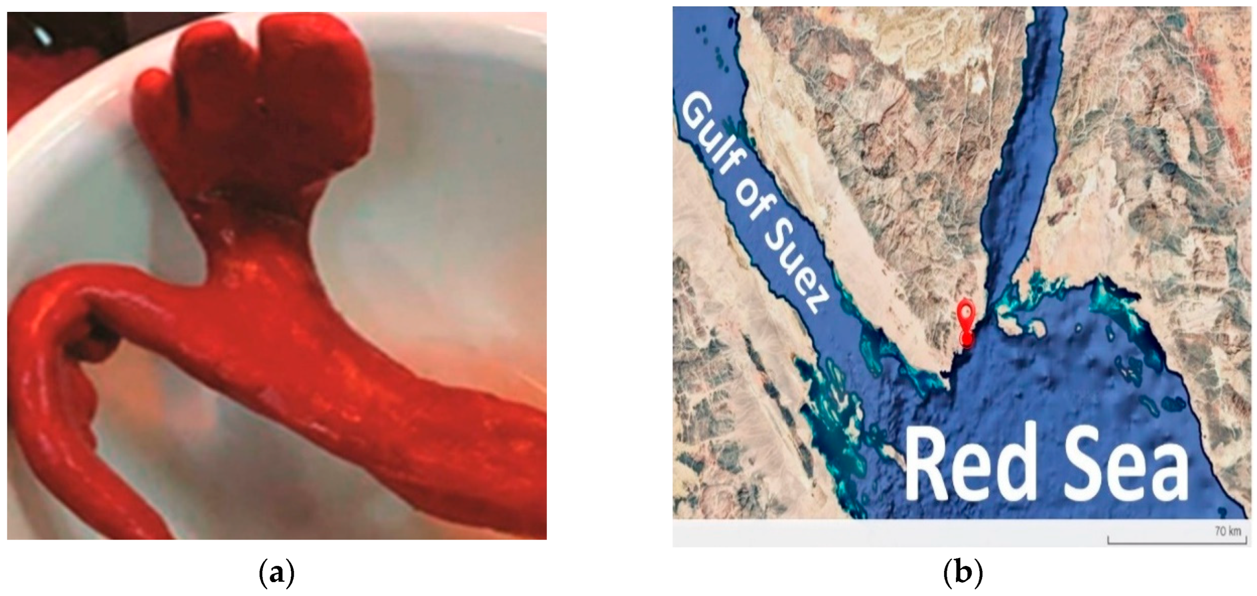
Figure 1. (a) Negombata magnifica sample collected from the Red Sea and (b) map showing the collection site of Negombata magnifica in the Red Sea.
Microorganisms 13 01866 g001
Figure 2. (a) Colony morphology of S. shinii strain SC-M1C on nutrient agar after 24 h and (b) S. shinii strain SC-M1C under microscope using 1000× magnification power.
Microorganisms 13 01866 g002
Figure 3. A phylogenomic tree constructed by the Type Strain Genome Server (TYGS). The tree is based on the genome of S. shinii strain SC-M1C and its top-related type strains. Confidence values are displayed near the nodes. The taxonomic status of these type strains were checked and corrected according to the List of Prokaryotic names with Standing in Nomenclature.
Microorganisms 13 01866 g003
Figure 4. A circular diagram represents the genome map of S. shinii strain SC-M1C.
Microorganisms 13 01866 g004
Figure 5. Clusters of orthologous genes (COG) functional classification of coding DNA sequences of the S. shinii strain SC-M1C genome.
Microorganisms 13 01866 g005
Figure 6. Genomic islands (GIs) identified by the IslandViewer 4 server within the S. shinii strain SC-M1C genome. The wavy lines represent the ends of contigs.
Microorganisms 13 01866 g006
Figure 7. Proposed diagram of the production of staphyloferrin A in S. shinii strain SC-M1C genome.
Microorganisms 13 01866 g007
Figure 8. Proposed diagram of the production of staphylopine in the S. shinii strain SC-M1C genome.
Microorganisms 13 01866 g008
Table 1. The genomic relatedness of the S. shinii strain SC-M1C to its closely related type strains. The table lists various genomic parameters, including digital DNA–DNA hybridization (dDDH), the difference in GC content (∆GC), and the average nucleotide identity (ANI) using BLAST-based alignment (ANIb) and MUMmer-based alignment (ANIm) with default parameters. The taxonomic statuses of these strains were checked and corrected according to the List of Prokaryotic Names with Standing in Nomenclature (https://lpsn.dsmz.de/, accessed on 24 May 2025).
StrainNCBI AccessionANIm (%)ANIb (%)dDDH (%)GC (%)
Staphylococcus shinii K22-5MGCF_017583065.199.4099.0994.80.15
Staphylococcus pseudoxylosus S04009GCF_003697915.191.9491.1544.30.46
Staphylococcus xylosus CCM 2738GCF_002732165.191.1690.7041.50.33
Staphylococcus saprophyticus CCUG 38042GCF_002902545.185.0480.7124.80.65
Staphylococcus saprophyticus ATCC 15305GCF_000010125.185.0080.7224.60.85
Staphylococcus edaphicus CCM 8730GCF_002614725.184.7680.34240.92
Staphylococcus caeli 82BGCF_900097965.184.7979.7023.70.95
Staphylococcus equorum NCTC 12414GCF_003970515.184.6079.7723.70.77
Staphylococcus equorum subsp. linens DSM 15097GCF_002901955.184.3879.5823.40.67
Staphylococcus nepalensis DSM 15150GCF_002902745.184.8178.5723.30.63
Staphylococcus nepalensis CCM 7045GCF_014635045.184.7078.4022.90.59
Staphylococcus ureilyticus DSM 6718GCF_002902235.184.4178.1222.60.16
Staphylococcus cohnii NCTC 11041GCF_002902365.184.2278.3222.40.1
Table 2. A summary of RAST and Prokka annotations of S. shinii strain SC-M1C genome.
RASTProkka
Total CDs30773032
CDs with functional assignment22851946
Hypothetical CDs7921086
rRNA21
tRNA4238
Table 3. Predicted virulence factors identified in the S. shinii SC-M1C genome using the virulence factor database (VFDB).
Virulence ClassVirulence FactorAssociated Gene(s)
AdherenceAutolysin; elastin-binding protein; fibronectin-binding proteinsatl; ebp; fnbA
EnzymeLipase; serine V8 protease; thermonucleasegeh; sspA; nuc
Immune evasionCapsule; polyglutamic acid capsule (Bacillus); polysaccharide capsule (Bacillus); capB, capC; galE
Iron uptakePeriplasmic-binding protein-dependent ABC transport system (Vibrio-like)vctC
Nutritional factorAllantoin utilization (Klebsiella-like)
Table 4. Antimicrobial resistance (AMR) genes identified in the genome of S. shinii strain SC-M1C, associated mechanisms, and corresponding antibiotic classes.
AMR MechanismGeneAntibiotic Class
Antibiotic inactivation enzymeBlaZ familyβ-lactams
FosBFosfomycin
Mph(C) familyMacrolides
Antibiotic resistance gene cluster, cassette, or operonTcaB, TcaB2, TcaRGlycopeptides
Antibiotic target modification or protectionAlrCell wall synthesis inhibitors
Antibiotic target modification or protectionDdlCell wall synthesis inhibitors
EF-G, EF-TuProtein synthesis inhibitors
folA, Dfr, folPAntifolates
gyrA, gyrBFluoroquinolones
inhA, fabIFatty acid synthesis inhibitors
Iso-tRNAProtein synthesis inhibitors
kasAFatty acid synthesis inhibitors
MurACell wall synthesis inhibitors
RhoTranscription inhibitors
rpoB, rpoCRifamycins
S10p, S12pAminoglycosides
Efflux pumps conferring antibiotic resistanceNorAFluoroquinolones
YkkCDMultidrug efflux
Gene conferring resistance via absencegidBAminoglycosides (loss of methylation)
Protein-altering cell wall chargeGdpD, MprF, PgsACationic antimicrobial peptides
Regulator modulating antibiotic resistance gene expressionBceR, BceS, LiaF, LiaR, LiaSGlycopeptides and antimicrobial peptides
Table 5. Predicted biosynthetic gene clusters identified in the S. shinii SC-M1C genome using AntiSMASH version 6.0.
BGCTypeSize (bp)Most Similar Known ClusterIdentity
1Opine-like metallophore, terpene37,511Staphylopine100%
2RiPP-like10,281
3T3PKS41,169
4NI siderophore32,991Staphyloferrin A100%
5Cyclic lactone autoinducer20,709
6Terpene20,818
7Terpene precursor20,890
8Terpene precursor20,953
9Lanthipeptide class i24,084
10NRPS56,796
11NRPS44,376
12NRPS44,328
Table 6. Osmoregulation and salt tolerance genes identified in S. shinii strain SC-M1C. Genes include Na+/H+ antiporters and compatible solute transporters involved in maintaining osmotic balance in marine conditions.
FunctionGenesCOG(s)RoleReferences
Na+/H+ antiport system (multi-subunit)mnhA1, mnhB1, mnhC1, mnhD1, mnhE1, mnhF1, mnhG1COG0651, COG2111, COG1006, COG1863, COG2212, COG1320Maintains intracellular pH and osmotic balance under high-salinity conditions.[40]
Na+/H+ antiporternhaCCOG1757Regulates pH and ion homeostasis in saline environments.[40,41]
Compatible solute uptakeopuDCOG1292Imports glycine betaine to protect against osmotic stress.[40,42]
Table 7. Genes related to nutrient acquisition identified in S. shinii SC-M1C genome and their potential roles.
NutrientGenesCOG(s)RoleReferences
MethioninemetP, metN2COG2011, –Active transport of methionine in nutrient-poor marine habitats.[43]
Nitrogen (nitrate/nitrite assimilation)nasDCOG1251Reduces nitrite to ammonium for nitrogen assimilation.[44,45]
PhosphatepstSScavenges inorganic phosphate in oligotrophic waters.[46,47]
ThiamineykoDCOG1122Imports thiamine, essential for metabolic processes.[48,49]
IronfhuDFacilitates acquisition of iron under limiting conditions.[50,51]
Table 8. Stress response-related genes and their potential roles in the S. shinii SC-M1C genome.
Stress TypeGenesCOG(s)RoleReferences
Oxidative stress and DNA repairnth, trxB, recA–, COG0492, –Protects from oxidative damage and promotes DNA repair.[52]
Heavy metal stress (copper)copA, copBExports excess copper to prevent toxicity.[53]
Detoxification and effluxsepA, mdtDRemoves toxic or host-derived compounds.[54,55]
Table 9. Genes associated with potential aromatic hydrocarbon degradation in S. shinii SC-M1C.
GenesCOG(s)RoleReferences
catECOG2514Acts as a key catechol 2,3-dioxygenase, performing meta-cleavage of aromatic rings, a critical step in the breakdown of petroleum hydrocarbons.[56]
mhqR, mhqA, mhqO, mhqDCOG1846, COG0346, COG0400This mhq cluster enables the degradation of a variety of aromatic compounds.[57]
Disclaimer/Publisher’s Note: The statements, opinions and data contained in all publications are solely those of the individual author(s) and contributor(s) and not of MDPI and/or the editor(s). MDPI and/or the editor(s) disclaim responsibility for any injury to people or property resulting from any ideas, methods, instructions or products referred to in the content.