Article

FlbD: A Regulator of Hyphal Growth, Stress Resistance, Pathogenicity, and Chlamydospore Production in the Nematode-Trapping Fungus Arthrobotrys flagrans

State Key Laboratory for Conservation and Utilization of Bio-Resources in Yunnan, and Key Laboratory for Microbial Resources of the Ministry of Education, School of Life Sciences, Yunnan University, Kunming 650091, China
*
Author to whom correspondence should be addressed.
Microorganisms 2025, 13(8), 1847; https://doi.org/10.3390/microorganisms13081847 (registering DOI)
Submission received: 26 June 2025 / Revised: 4 August 2025 / Accepted: 7 August 2025 / Published: 7 August 2025
(This article belongs to the Special Issue Microorganisms as Biocontrol Agents in Plant Pathology, 2nd Edition)

Abstract

Arthrobotrys flagrans is a typical nematode-trapping fungus that captures nematodes by producing three-dimensional networks. FlbD is a DNA-binding protein containing a Myb domain, which plays a significant role in fungal development. However, the biological function of FlbD in nematode-trapping fungi remains unknown. In this study, we analyzed the physicochemical properties and conserved domains of AfFlbD and constructed the AfFlbD knockout strains (ΔAfFlbD) using homologous recombination. Our functional analysis revealed that the mutants produced more cottony aerial mycelia at the colony center. Additionally, the cell length of the mutants was reduced, indicating that AfFlbD regulates cell morphology in A. flagrans. Chemical stress tolerance assays of the mutants demonstrated reduced sensitivity to NaCl and sorbitol stresses but increased sensitivity to SDS and H2O2 stresses compared to the WT strain. Interestingly, the mutants spontaneously produced traps, and its pathogenicity to nematodes was significantly enhanced, suggesting that AfFlbD negatively regulates the pathogenicity of A. flagrans. Furthermore, the number of chlamydospores produced by the mutants was markedly reduced, though their morphology remained unchanged. Fluorescence localization analysis showed that AfFlbD localizes to the nuclei of chlamydospores, thereby regulating chlamydospore formation. This study provides important theoretical insights into the biological function of the FlbD transcription factor and offers new perspectives for the application of nematode-trapping fungi as a method of controlling plant-parasitic nematodes.

1. Introduction

Spores, whether sexual or asexual, constitute a crucial mode of reproduction for filamentous fungi. Furthermore, certain fungi rely on their pathogenicity to acquire nutrients from hosts during growth and development, thereby facilitating population propagation [1]. Both spore formation and the pathogenic process are precisely regulated by multiple genes. FlbA-E are upstream developmental activators of the central regulatory pathway for asexual sporulation in various fungi, governing the formation of asexual spores [2,3]. Of these activators, the flbD gene encodes a Myb-type DNA-binding protein, a critical transcription factor required to activate the expression of the brlA gene. FlbD has been reported in fungi such as Saccharomyces cerevisiae, Aspergillus nidulans, A. fumigatus, and Magnaporthe oryzae, where it has been found to profoundly influence the formation of conidia [4,5]. In A. nidulans, FlbD activates transcription of the brlA gene, thereby promoting conidiation. Deletion of this gene delays asexual sporulation and results in a “fluffy” colony phenotype [6]. In Fusarium graminearum, FlbD ensures proper hyphal differentiation and growth by regulating the expression of relevant genes. The deletion of FlbD reduces conidia production and impairs fungal pathogenicity [7]. Collectively, FlbD serves critical functions in regulating development and life cycle progression across diverse fungi. However, its biological role in nematode-trapping fungi remains uncharacterized.
Arthrobotrys flagrans is a representative nematode-trapping (NT) fungus that captures nematodes via three-dimensional adhesive networks and produces chlamydospores, making it one of the most promising biocontrol agents among nematophagous fungi [8,9]. Key components contributing to its trap formation and nematode capture include the sofT gene [10], the sipC gene [11], the secondary metabolite 6-methylsalicylic acid (6-MSA) [12], the small secreted protein CyrA [13], and the virulence factor NipA [14]. Deletion of the global regulator AfLaeA (a histone methyltransferase) impairs fungal growth, secondary metabolism, and pathogenicity toward nematodes, with the mutant notably losing the ability to produce chlamydospores [9]. The G protein-coupled receptor GprC, localized to the plasma membrane and mitochondria, perceives nematode pheromones (ascarosides) and activates mitochondrial function during nematode predation [8]. Biocontrol agents based on A. flagrans chlamydospores (e.g., Bioverm®) have been successfully applied to control nematodes in various livestock species [15]. Furthermore, A. flagrans exhibits significant biocontrol potential against Meloidogyne spp. [16] and Xiphinema index [10,17], establishing it as a highly promising biocontrol fungus for sustainable agriculture [18]. The major difference between A. flagrans and other nematode-trapping fungi is the ability of A. flagrans to readily form chlamydospores for survival [10]. However, the genes regulating chlamydospore formation in A. flagrans remain poorly understood.
To explore whether AfFlbD is involved in the chlamydospore formation and pathogenicity, we knocked out the AfFlbD gene in A. flagrans and conducted comparative analyses between the AfFlbD gene knockout strains (ΔAfFlbD) and the wild-type (WT) strain. Key differences were assessed in hyphal growth, stress responses, pathogenicity, and chlamydospore formation. Subcellular localization of AfFlbD was determined using GFP fluorescence. These investigations reveal the regulatory roles of AfFlbD in hyphal growth, stress response, pathogenicity, and chlamydospore formation in A. flagrans. Notably, AfFlbD functions as a negative regulator of trap development while serving as a positive regulator of chlamydospore formation. This study provides novel insights into the dual functionality of FlbD in chlamydospore formation and pathogenicity, revealing its contributions to both chlamydospore development and predatory mechanisms in nematode-trapping fungi.

2. Materials and Methods

2.1. Strains and Culture Conditions

A. flagrans and Caenorhabditis elegans were stored in the Microbial Library of the Germplasm Bank of Wild Species from Southwest China. The WT strain of A. flagrans was cultivated on potato dextrose agar (PDA) medium at 28 °C [9]. The AfFlbD gene knockout strains were cultivated on PDA medium containing 100 mg/mL hygromycin B.

2.2. Gene Structure and Conserved Protein Domain Analysis of AfFlbD

We extracted the AfFlbD gene sequence from the A. flagrans genome data [9]. The gene structure of AfFlbD was analyzed using the online tool Gene Structure Display Server (http://gsds.cbi.pku.edu.cn/, accessed on 5 June 2025) [9,19]. The molecular weight and isoelectric point (pI) of the AfFlbD protein were predicted with the Expasy ProtParam tool (https://web.expasy.org/protparam/, accessed on 1 June 2025). Conserved protein domains were identified using the NCBI CD-Search tool (https://www.ncbi.nlm.nih.gov/Structure/cdd/wrpsb.cgi, accessed on 1 June 2025). Phylogenetic tree construction and analysis were performed using MEGA X software (version 64-bit) [20]. Subcellular localization was predicted with Uniprot (https://www.uniprot.org/, accessed on 1 June 2025) [21].

2.3. Genomic DNA Extraction from A. flagrans

The WT strain of A. flagrans was inoculated on PDA medium and incubated at 28 °C for 4–5 days. Mycelia were harvested using sterile pipette tips, flash-frozen in liquid nitrogen, and thoroughly ground in a mortar. Genomic DNA was extracted using a Fungal DNA Kit (Magen, Guangzhou, China) [9].

2.4. Construction of the Gene Knockout Vector

Based on the A. flagrans genome data [9], upstream and downstream homologous arms flanking the AfFlbD gene were selected. Primers were designed using CE Design Software (https://tool.vazyme.com:18002/cetool/simple.html, accessed on 6 June 2024). The homologous arms were amplified using genomic DNA as the template, while the hygromycin resistance gene (hph) was amplified from the pCSN44 plasmid [22]. The fragments (upstream arm, hph, downstream arm) were directionally assembled into the pUC19 vector using a ClonExpress Ultra One Step Cloning Kit (Vazyme, Nanjing, China).

2.5. Protoplast Preparation of A. flagrans

The WT strain was inoculated on PDA medium and incubated at 28 °C for 3–4 days. Mycelial plugs were transferred to PDB medium and cultured under conditions of 28 °C, and 180 rpm for 24 h. Hyphae were filtered through sterile filter paper and rinsed with STC buffer (1 M sorbitol, 50 mM CaCl2, 10 mM Tris-HCl). Harvested hyphae were digested in 15–20 mL of filter-sterilized cell wall lysis enzyme solution under conditions of 28 °C, and 90 rpm for 4–5 h. Protoplasts were filtered through sterile filter paper and collected using centrifugation (4 °C, 3000× g, 8 min). Subsequently, 6–10 μg DNA was added to 107 protoplasts. After 30 min incubation on ice, 1 mL PTC buffer (10 mM Tris-HCl [pH 7.5], 50 mM CaCl2, 50% [wt/vol] polyethylene glycol [PEG] 3350) was added, mixed gently by pipetting, and incubated at 28 °C for 40 min. The mixture was then plated onto PDASS regeneration medium (PDA supplement with 0.6 M of sucrose, 0.3 g/L peptone, 0.3 g/L tryptone, 0.3 g/L yeast extract, 8 g/L agar) in 9 cm dishes. Plates were sealed and incubated at 28 °C for 48 h, and transformants were selected on PDA supplemented with hygromycin B (100 μg/mL) or G418 (50 μg/mL) [9].

2.6. Screening and Verification of AfFlbD Knockout Strains

Transformants were cultured on PDA medium containing hygromycin B (100 μg/mL). Hyphae (~1.0 g) were scraped using sterile tips, mixed with quartz sand and urea extraction buffer, and homogenized in a high-throughput tissue grinder. Genomic DNA was extracted using a DNA isolation kit (Magen, Guangzhou, China). Gene-specific primers (F1/R1) amplified the target gene, while F2/R3 amplified the deletion cassette. The primers F1/R1 confirmed complete gene replacement, while F2/R2 amplified the “Up+hph” fragment for knockout validation (Table S1).

2.7. Subcellular Localization of AfFlbD Protein

A 2000 bp upstream sequence of the AfH2B gene (promoter) and a 1000 bp downstream sequence (terminator) were retrieved from the A. flagrans genome [8]. The AfH2B promoter, AfFlbD, GFP, AfH2B terminator, and G418 resistance cassette were directionally assembled into the pUC19 vector with the ClonExpress Ultra One Step Cloning Kit (Vazyme, Nanjing, China). The constructed plasmids were transformed into protoplasts of A. flagrans, and AfFlbD localization in chlamydospores was observed after G418 selection.

2.8. Hyphal Growth Rate Analysis

WT and knockout strains were cultured on PDA (supplemented with 100 μg/mL hygromycin B for mutants) for 4 days. Mycelial plugs (0.8 cm diameter) were inoculated onto PDA and TYGA media (10 g/L tryptone, 5 g/L yeast extract, 10 g/L glucose, 5 g/L molasses, 15 g/L agar), incubated at 28 °C, and photographed daily for 4 days [9,23]. Colony diameters were measured daily. Three replicates per strain were analyzed using GraphPad Prism 10 (San Diego, CA, USA).

2.9. Hyphal Morphology Analysis

WT and mutant strains were grown on PDA with embedded cover slips for 2–3 days. Hyphae were stained with 10 μg/mL Calcofluor White (CFW) (Sigma-Aldrich, St. Louis, MO, USA) for 30 min. Hyphal length was examined using fluorescence microscopy [20,23].

2.10. Chemical Stress Tolerance Assay

The WT and knockout strains were cultured on PDA (supplemented with 100 μg/mL hygromycin B for mutants) for 4 days. Mycelial plugs (0.8 cm diameter) were inoculated onto PDA plates containing graded concentrations of the following chemical stressors: sorbitol (0.25, 0.5, 1 M), NaCl (0.1, 0.2, 0.3 M), sodium dodecyl sulfate (0.01%, 0.02%, 0.03%), and H2O2 (1.75, 2.5, 5 mM). Plates were incubated at 28 °C for 5 days. Colony diameters were measured daily to calculate Relative Growth Inhibition (RGI) [9]. Three replicates per condition were analyzed.

2.11. Trap Production and Pathogenicity Analysis

WT and knockout strains were cultured on PDA (9 cm diameter) at 28 °C for 5–6 days. Mycelial plugs were transferred to 250 mL PDB and shaken (180 rpm, 28 °C, 12 h). Hyphae were filtered through sterile funnels, rinsed with sterile water, and resuspended in 7 mL sterile water. Suspensions were spread evenly on 3.5 cm WA plates and incubated at 28 °C for 2–3 days. Synchronized L3 stage C. elegans were washed with sterile water. Approximately 300 nematodes were added to fungal lawns. Plates were incubated at 28 °C to induce trap formation [9]. Trap counts and nematode mortality were recorded at 0, 3, 6, 12, and 24 h. Three biological replicates were performed per strain.

2.12. Chlamydospore Formation Assay

WT and mutant strains were synchronously cultured for 4 days on PDA (mutants on PDA supplemented with 100 μg/mL hygromycin B). Mycelial plugs (0.8 cm diameter) were inoculated onto 6 cm WA plates and incubated at 28 °C. Chlamydospore production was monitored daily until day 14, when final counts were quantified [9]. Three biological replicates were performed per strain.

2.13. Quantitative Real-Time PCR (qRT-PCR) Assay

We performed qRT-PCR assay using the ChamQ Universal SYBR qPCR Master Mix (Vazyme, Nanjing, China) on a Roche LightCycler 480 system (Roche Applied Science, Rotkreuz, Switzerland). The amplification protocol was carried out as previously described [9]. The glyceraldehyde-3-phosphate dehydrogenase (AfGpd) gene used as the reference, and relative transcript levels were calculated using the 2−ΔΔCT method [24]. All measurements were performed with at least three biological replicates. Gene-specific primers are listed in Table S1.

2.14. Data Analysis

Statistical analyses were performed using GraphPad Prism 10 (San Diego, CA, USA). Significance between groups was determined with Student’s t-test, with statistical thresholds defined as follows: * p < 0.05, ** p < 0.01, *** p < 0.001 [9]. The experimental data in figures are presented as mean ± standard deviation (Mean ± SD).

3. Results

3.1. Gene Structure and Protein Sequence Analysis of AfFlbD

The AfFlbD gene comprises a 1017 bp coding sequence lacking introns (Figure 1A), encoding a 338 amino acid protein with a molecular weight of 38.11 kDa and an isoelectric point (pI) of 10.28 (Table S2). Subcellular localization predictions indicated the nuclear localization of AfFlbD (Table S2). A phylogenetic tree constructed using the neighbor-joining method in MEGA X revealed that A. flagrans FlbD shares close evolutionary affinity with homologs from A. oligospora ATCC 24927, Dactylellina haptotyla CBS 200.50, A. nidulans FGSC A4, and A. fumigatus (Figure 1B). Conserved domain analysis via NCBI CD-Search demonstrated that all FlbD homologs contain the REB1 domain—a DNA-binding motif universally conserved in fungi. The A. flagrans FlbD additionally harbors a Myb domain, which aligns with the SANT domains present in most other homologs (Figure 1C). Moreover, protein–protein interaction predictions using the STRING database identified potential binding partners for FlbD, including histone acetyltransferase, cyclin-dependent kinase, integrase catalytic domain protein, Ty1/copia-type retrotransposon domain protein, GATA transcription factor SRE1, adenylosuccinate lyase, Mgr3-like protein, and replication factor A protein 3 (Figure 1D).

3.2. AfFlbD Regulates Growth and Stress Responses in A. flagrans

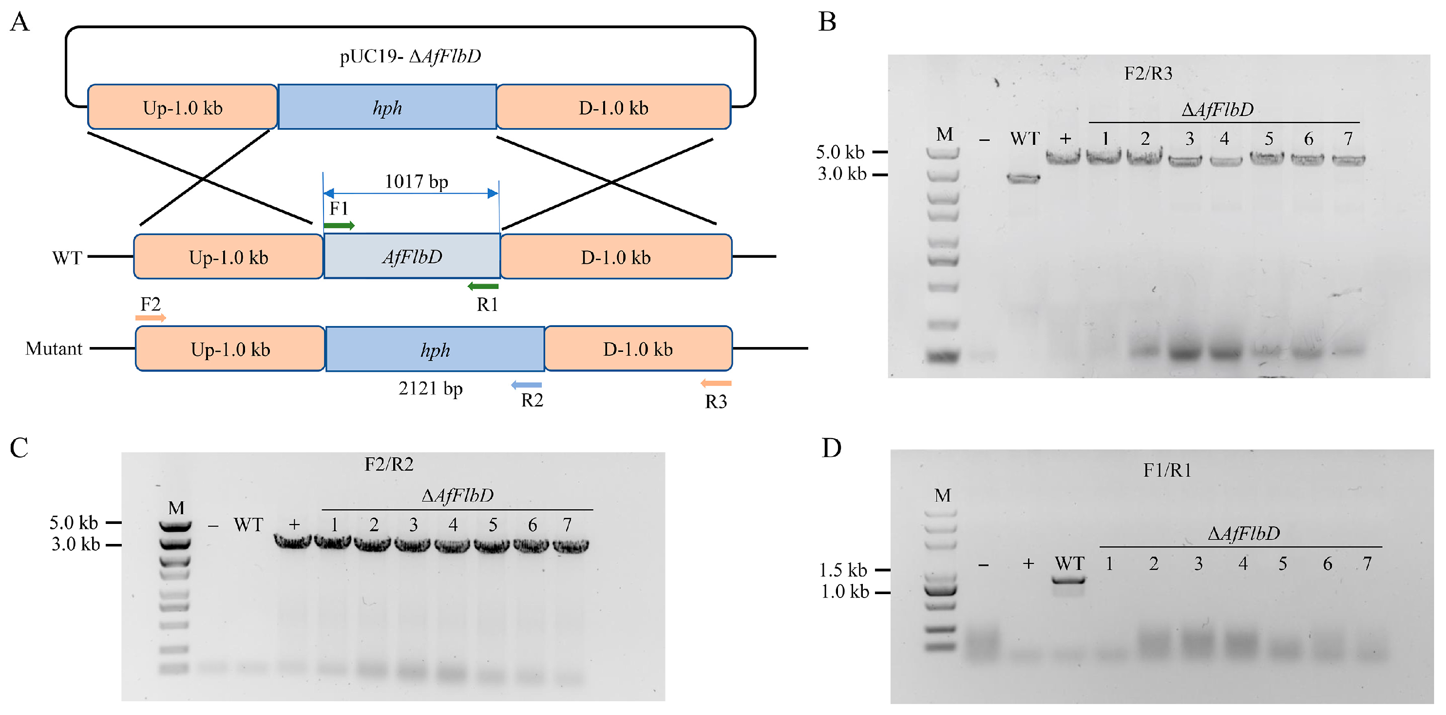
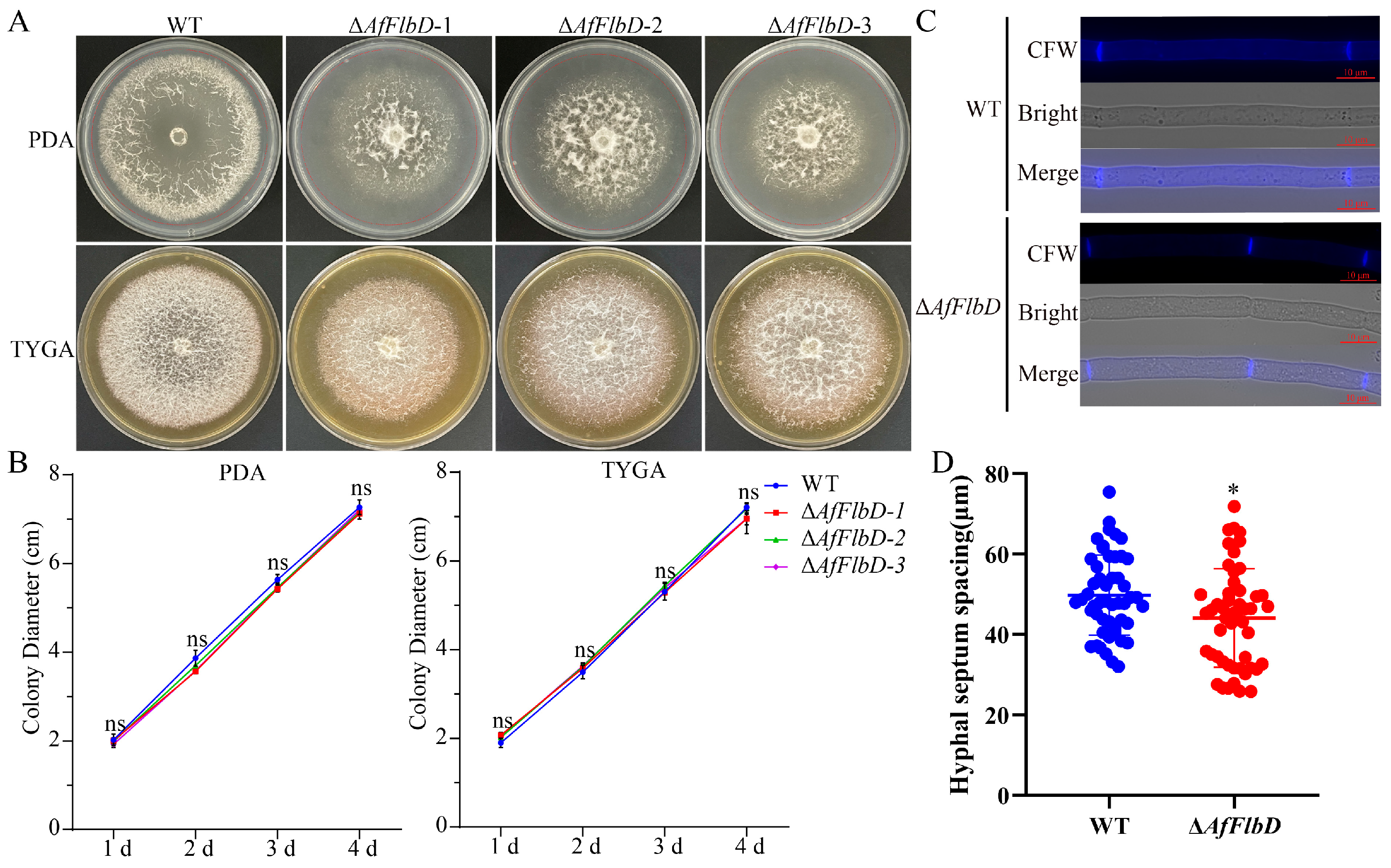
To investigate the biological function of AfFlbD, we generated AfFlbD knockout mutants via homologous recombination (Figure 2A), validated by three primer pairs (Figure 2B–D). Complete gene deletion was confirmed in seven mutant strains (Figure 2). To examine the impact of AfFlbD on hyphal growth, the WT and mutants were cultured on PDA and TYGA media for 4 days with daily diameter measurements. All three ΔAfFlbD strains showed no significant growth rate differences versus the WT (Figure 3A,B). However, the colonies of the mutants exhibited dense cottony aerial hyphae centrally and sparse peripheral hyphae on PDA (Figure 3A). Calcofluor White (CFW) staining revealed reduced septal spacing in the hyphae of the ΔAfFlbD strains (vs. WT), indicating that AfFlbD regulates hyphal cell length (Figure 3C,D).
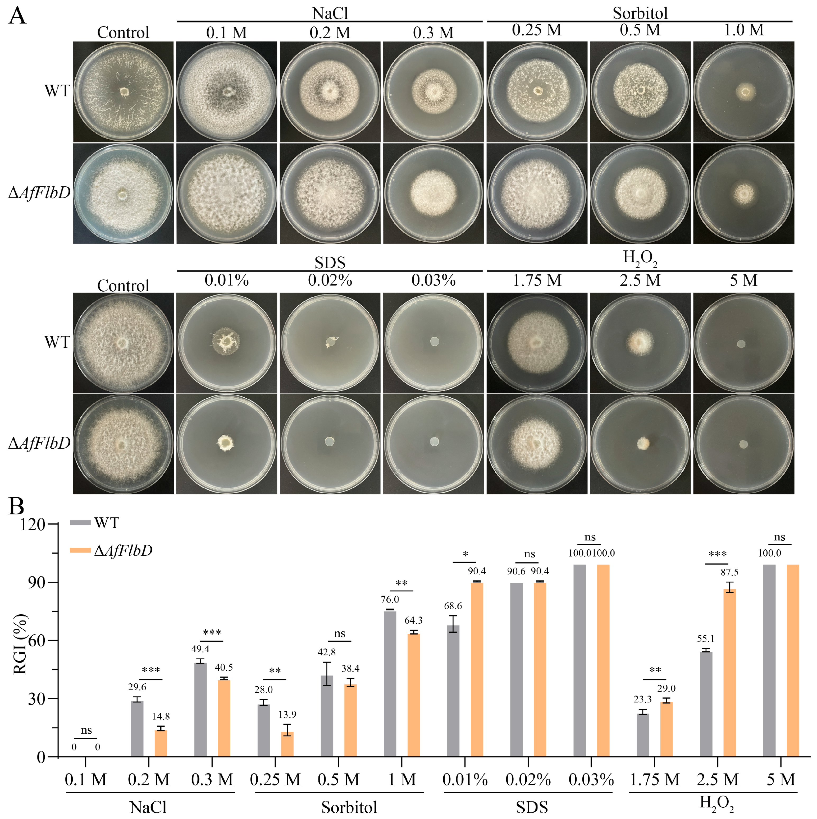
Furthermore, to investigate the role of AfFlbD in stress adaptation, A. flagrans was cultured on PDA supplemented with gradient concentrations of NaCl, sorbitol, sodium dodecyl sulfate (SDS), and H2O2. All tested stressors inhibited growth in both the WT and ΔAfFlbD strains (Figure 4A). Compared with the WT, the RGI values of the ΔAfFlbD strains on the media containing NaCl (0.2 M, 0.3 M) and sorbitol (0.25 M, 0.5 M, 1 M) were reduced, indicating that the sensitivity of the ΔAfFlbD strains to NaCl and sorbitol was significantly decreased (Figure 4B). In contrast, both the WT and ΔAfFlbD strains ceased growth on the medium containing SDS at concentrations of 0.02% and 0.03% (Figure 4A). Compared with WT, the ΔAfFlbD strains had higher RGI values on the media containing SDS (0.01%) and H2O2 (1.75 mM and 2.5 mM), indicating that the sensitivity of the ΔAfFlbD strains to SDS and H2O2 was increased (Figure 4B).

3.3. AfFlbD Negatively Regulates the Pathogenicity of A. flagrans

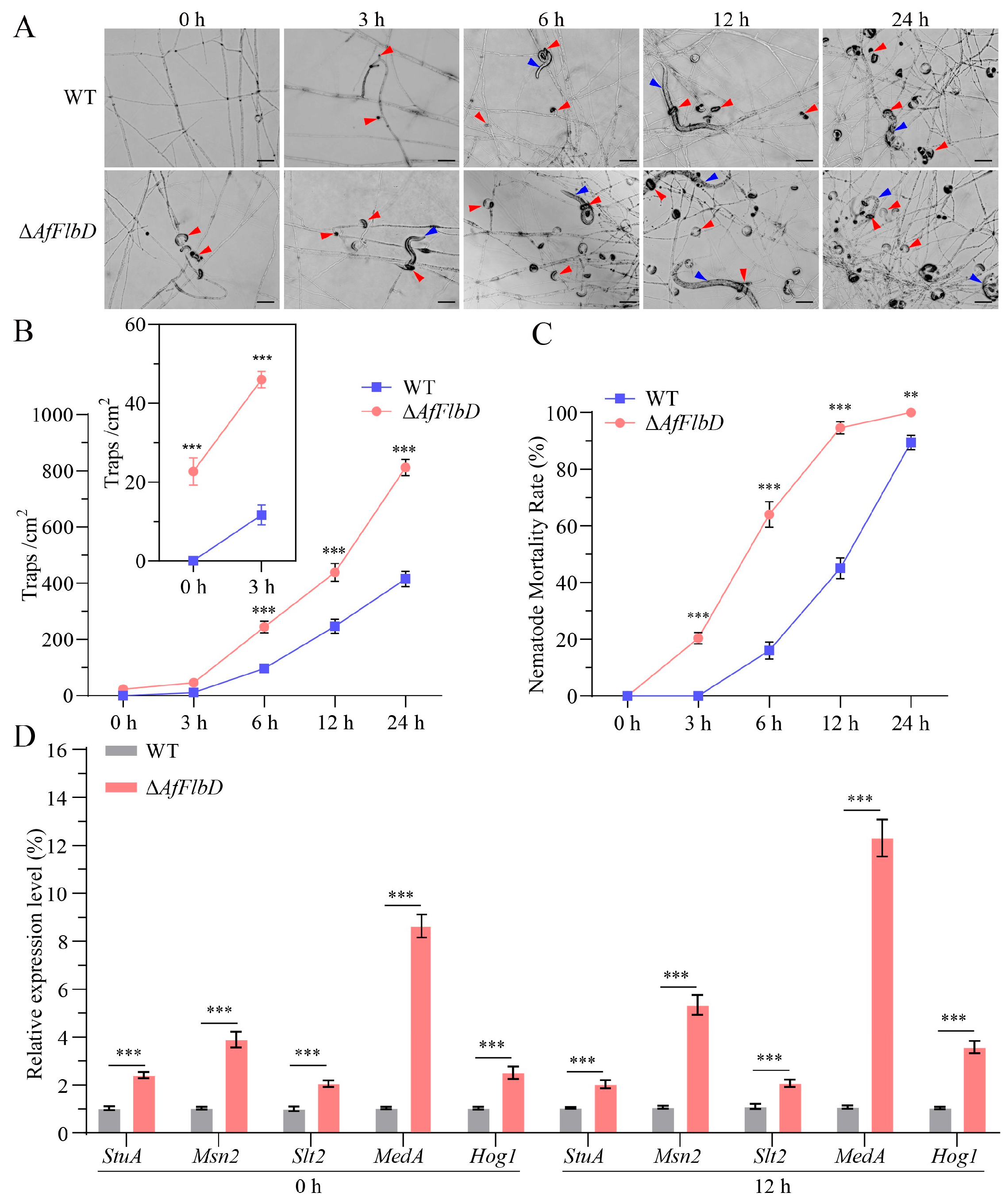
The mycelia of the WT and ΔAfFlbD strains were inoculated onto 3.5 cm WA plates and cultured in an incubator at 28 °C for 2–3 days until the mycelia covered the plates. Approximately 300 synchronized C. elegans at the L3 stage were added to each plate. The formation of traps and the mortality rate of nematodes were observed and recorded under an optical microscope at 0, 3, 6, 12, and 24 h after inoculation. The results showed that, without nematode induction, the ΔAfFlbD strains produced about 22.7 traps (/cm2) on WA medium after 3 days of culture, while the WT strain did not produce any traps (Figure 5A,B). After the addition of nematodes, it was observed that the nematodes were captured and killed by the traps produced by the ΔAfFlbD strains (Figure 5A). Both the WT and ΔAfFlbD strains could be induced to produce traps, but the ΔAfFlbD strains produced them significantly faster than the WT strain. The number of traps produced by the ΔAfFlbD strains reached about 46.0 (/cm2) in 3 h, with a mortality rate of about 20.3%, while the WT strain was almost non-lethal (Figure 5C). As time increased, the number of traps produced by the ΔAfFlbD strains was significantly higher than that of the WT strain, about twice as many as the WT strain at 24 h (Figure 5B). The nematode mortality rate of the ΔAfFlbD strains reached 94.6% in 12 h after the addition of nematodes, while the WT strain took over 24 h to achieve 89.4% (Figure 5C).
The genes related to trap formation in the nematode-trapping fungus A. oligospora have been extensively studied. To explore the mechanism by which AfFlbD negatively regulates the formation of traps, we selected several trap-related genes (StuA, Msn2, Slt2, MedA, and Hog1) and compared their expression levels at 0 h and 12 h after the production of traps. In A. oligospora, the deletion of StuA, Slt2, or MedA abolished trap formation in mutant strains [25,26,27], whereas the loss of Msn2 or Hog1 led to significantly reduced trap production [28,29]. Our results showed that, without nematode induction (0 h), the expression levels of StuA, Msn2, Slt2, MedA, and Hog1 in the ΔAfFlbD strains were 2.4-fold, 3.9-fold, 2.0-fold, 9.4-fold, and 2.5-fold higher than those in the WT strain, respectively (Figure 5D), while after 12 h of nematode induction, the same expression levels of in the ΔAfFlbD strains were 2.0, 5.3, 2.1, 12.3, and 3.6 times higher than those in the WT strain, respectively (Figure 5D). This confirms that AfFlbD negatively regulates the expression of these genes.

3.4. AfFlbD Regulates Chlamydospore Formation in A. flagrans

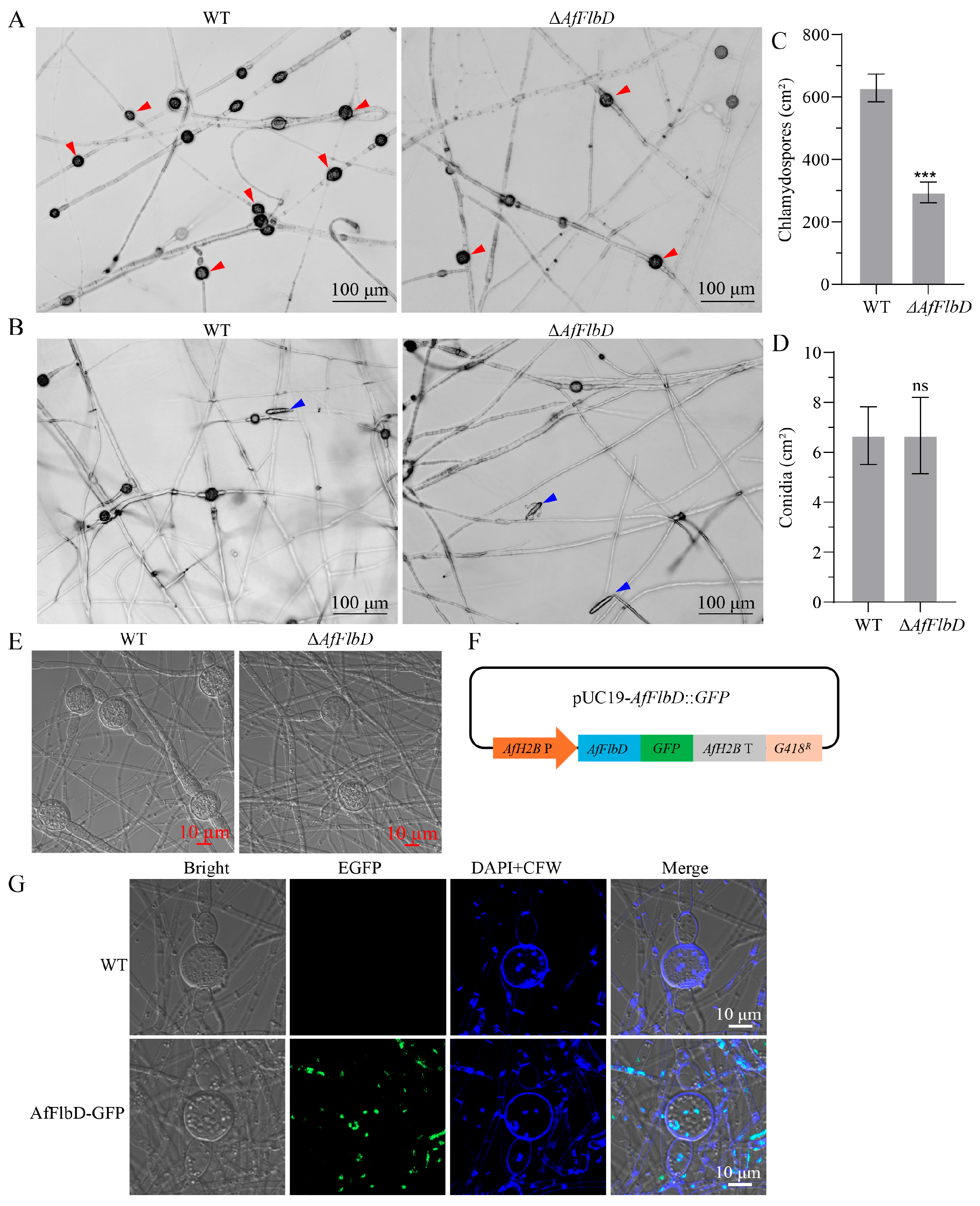
To investigate the regulatory role of AfFlbD in the asexual sporulation of A. flagrans, we synchronously inoculated both the WT and ΔAfFlbD strains onto 6 cm WA medium for 14 days and observed chlamydospore production under microscopy. The results showed that the AfFlbD knockout strains produced both chlamydospores and conidia (Figure 6A,B), but its chlamydospore yield was half that of the WT strain (Figure 6C), with no significant differences in conidial quantity or morphology compared to WT (Figure 6D). Meanwhile, confocal microscopy revealed that the deletion of AfFlbD did not alter chlamydospore morphology in A. flagrans (Figure 6E). As a transcription factor, AfFlbD was predicted to regulate gene expression in the nucleus (Table S2). Thus, an AfFlbD-GFP fusion protein was expressed in A. flagrans using the AfH2B gene promoter (Figure 6F). The results demonstrate that the AfFlbD-GFP fusion protein localizes to the nuclei of chlamydospores, thereby regulating chlamydospore development (Figure 6G).

4. Discussion

In this study, we conducted a multi-phenotypic comparative analysis of the ΔAfFlbD strains and the WT strain, revealing that AfFlbD participate in hyphal development and stress response in A. flagrans, in addition to playing crucial roles in chlamydospore formation, trap development, and pathogenicity. Intriguingly, AfFlbD is a positive regulator of chlamydospore formation and a negative regulator of trap production.
Several studies have proved that FlbD is involved in regulation of aerial hyphae morphology but not in hyphal growth rate in fungi [30]. The deletion of the AfFlbD gene in A. flagrans did not significantly affect hyphal growth rate (Figure 3). However, the ΔAfFlbD strains produced abundant cottony aerial mycelia at the colony center, while the periphery exhibited sparser aerial hyphae. Similarly, the deletion of the FlbD gene led to very “fluffy-like” hyphae on the colony surfaces of A. nidulans, A. niger, and F. graminearum [3,7,30,31]. This indicates that the functional role of AfFlbD in regulating aerial hyphae morphology is evolutionarily conserved among some fungal species. Additionally, the ΔAfFlbD strains exhibited a reduction in hyphal compartment length, indicating that AfFlbD regulates cellular dimensions in A. flagrans. Regarding stress response to chemical agents, the ΔAfFlbD strains showed reduced sensitivity to NaCl (0.2 M and 0.3 M) and sorbitol but enhanced sensitivity to 0.01% SDS and 2.5 mM H2O2 (Figure 4). Deletion of the FlbD gene in Beauveria bassiana also resulted in increased sensitivity to H2O2 in the mutant strains [32].
The WT strain of A. flagrans required nematode induction to form traps on WA medium, whereas the ΔAfFlbD strains spontaneously developed traps. After 12 h of interaction with nematodes, the ΔAfFlbD strains produced approximately twice as many traps as the WT strain, achieving a 94.6% nematode mortality rate. Within 24 h, the ΔAfFlbD strains reached 100% mortality, while the WT strain achieved 89.4%, demonstrating significantly enhanced pathogenicity of the ΔAfFlbD strains to nematodes (Figure 5). In contrast, the deletion of the FlbD gene in F. graminearum leads to a significant reduction in pathogenicity [7]. In B. bassiana, knockout of the FlbD gene does not affect the pathogenicity of the mutant strains against locusts, whereas the deletion of the FlbA or FlbC genes significantly reduces pathogenicity [32]. To elucidate the mechanism by which AfFlbD negatively regulates trap formation, this study examined the expression levels of several genes known to positively regulate trap development (StuA, Msn2, Slt2, MedA, and Hog1) using qRT-PCR. The results revealed that all five genes exhibited higher expression levels in the ΔAfFlbD strains compared to the WT strain (Figure 5D), indicating that AfFlbD negatively regulates their expression either directly or indirectly. The formation of traps in nematode-trapping fungi is regulated by multiple genes and pathways, representing a complex biological process [33]. Currently, there is limited evidence in fungi to reveal what downstream genes are regulated by FlbD. Further experiments are required to elucidate the molecular mechanism through which AfFlbD negatively regulates trap formation.
Previous studies have demonstrated that FlbD regulates conidiation: the deletion of FlbD in F. graminearum significantly reduced conidial production [7]; FlbD knockout in A. niger markedly decreased conidial yield and even completely suppressed conidiation under certain conditions [30], and FlbD deficiency in M. oryzae abolished conidia production [31], while FlbD deletion in A. nidulans substantially reduced conidial production [3]. Notably, FlbD deletion in A. flagrans did not affect conidiation, highlighting regulatory divergence across fungal species. Although existing studies consistently establish the role of FlbD in conidial regulation, no prior research has investigated its involvement in chlamydospore formation. This study reveals that AfFlbD knockout delays chlamydospore development and reduces chlamydospore yield to ∼46.7% of WT levels, indicating its critical role for chlamydospore formation in A. flagrans (Figure 6).
The molecular mechanism of FlbD-mediated conidiation is relatively well-characterized. In the model fungus A. nidulans, FlbD operates downstream of FluG (a developmental activator) but upstream of the transcription factors FlbB, FlbC, FlbE, and BrlA. FluG indirectly activates FlbD by relieving suppression by SfgA (a negative regulator), thereby promoting asexual sporulation [34]. Additionally, FlbB and FlbD form a complex that co-activates brlA expression. FlbD deletion prevents FlbB from binding to the promoter of brlA—the core conidiation regulator—inhibiting its expression and subsequent conidiation [5]. Conversely, FlbD overexpression activates brlA and induces the formation of complex conidiogenic structures (including stalks, vesicles, phialides, and conidia) [6]. Critically, the molecular mechanism whereby FlbD regulates chlamydospore formation remains to be elucidated.
Root-knot nematodes (RKNs) infect crops and cause significant agricultural losses annually, while the use of chemical pesticides leads to nematode resistance and poses environmental and food safety concerns [35,36]. Biological control represents an effective and eco-friendly strategy for RKNs management. A. flagrans produces traps and stress-resistant chlamydospores, endowing this fungus with substantial research and practical value [17]. In this study, AfFlbD exhibits dual regulatory roles in trap formation and chlamydospore development, providing insights for developing effective A. flagrans-based strategies against pathogenic nematodes. Our study provides valuable references for studying chlamydospore development in other biocontrol fungi and pathogenic fungi. Furthermore, investigation of chlamydospore biogenesis in pathogenic fungi (such as Candida albicans, Botrytis cinerea, and M. oryzae) that produce chlamydospores lays crucial molecular groundwork for developing broad-spectrum, eco-friendly control agents against diverse pathogenic fungi.

5. Conclusions

In this study, we employed homologous recombination to knockout the AfFlbD gene in A. flagrans and systematically characterized its biological functions through multi-phenotypic analysis. Our results demonstrate that AfFlbD plays crucial roles in hyphal growth, stress response, pathogenicity, and chlamydospore formation in A. flagrans. Significantly, AfFlbD functions as a negative regulator of trap development while serving as a positive regulator of chlamydospore formation. As the first investigation of the FlbD ortholog in a nematode-trapping fungus, this work provides novel insights into the biological functions of FlbD during nematode predation in these fungi.

Supplementary Materials

The following supporting information can be downloaded at: https://www.mdpi.com/article/10.3390/microorganisms13081847/s1, Table S1: Primers used for genetic manipulation; Table S2: The predicted physiochemical properties of AfFlbD in A. flagrans.

Author Contributions

Conceptualization, Y.Z., and G.-H.L.; methodology, Y.Z., S.-Q.P., W.-T.H., and G.-H.L.; software, Y.Z., F.-F.G., and Q.-F.S.; validation, G.-H.L.; data curation, Y.Z. and G.-H.L.; writing—original draft preparation, Y.Z., S.-Q.P., and G.-H.L.; writing—review and editing, Y.Z., and G.-H.L.; funding acquisition, G.-H.L. All authors have read and agreed to the published version of the manuscript.

Funding

This research was supported by the National Key Research and Development Program (2023YFD1400400), the National Natural Science Foundation of China (32160012), Science and Technology Innovation Base Construction Project (202407AB110004) and Projects from the Department of Science and Technology of Yunnan Province (202501AS070058, 202401BC070010, 202301BC070017).

Data Availability Statement

The raw data supporting the conclusions of this article will be made available by the authors on request. The Arthrobotrys flagrans genome database used in this study is available at the National Center for Biotechnology Information (NCBI) GenBank under the accession number PRJNA917252.

Acknowledgments

We are grateful to the Microbial Library of the Germplasm Bank of Wild Species from Southwest China for preserving and providing the experimental strains.

Conflicts of Interest

The authors declare no conflicts of interest.

References

  1. Ojeda-López, M.; Chen, W.; Eagle, C.E.; Gutiérrez, G.; Jia, W.L.; Swilaiman, S.S.; Huang, Z.; Park, H.S.; Yu, J.H.; Cánovas, D.; et al. Evolution of asexual and sexual reproduction in the aspergilli. Stud. Mycol. 2018, 91, 37–59. [Google Scholar] [CrossRef]
  2. Guo, C.T.; Luo, X.C.; Tong, S.M.; Zhou, Y.; Ying, S.H.; Feng, M.G. FluG and FluG-like FlrA coregulate manifold gene sets vital for fungal insect-pathogenic lifestyle but not involved in asexual development. mSystems 2022, 7, e0031822. [Google Scholar] [CrossRef]
  3. Garzia, A.; Etxebeste, O.; Herrero-García, E.; Ugalde, U.; Espeso, E.A. The concerted action of bZip and cMyb transcription factors FlbB and FlbD induces brlA expression and asexual development in Aspergillus nidulans. Mol. Microbiol. 2010, 75, 1314–1324. [Google Scholar] [CrossRef]
  4. Arratia-Quijada, J.; Sánchez, O.; Scazzocchio, C.; Aguirre, J. FlbD, a Myb transcription factor of Aspergillus nidulans, is uniquely involved in both asexual and sexual differentiation. Eukaryot. Cell 2012, 11, 1132–1142. [Google Scholar] [CrossRef]
  5. Otamendi, A.; Perez-de-Nanclares-Arregi, E.; Oiartzabal-Arano, E.; Cortese, M.S.; Espeso, E.A.; Etxebeste, O. Developmental regulators FlbE/D orchestrate the polarity site-to-nucleus dynamics of the fungal bZIP transcription factor FlbB. Cell Mol. Life Sci. 2019, 76, 4369–4390. [Google Scholar] [CrossRef] [PubMed]
  6. Wieser, J.; Adams, T.H. flbD encodes a Myb-like DNA-binding protein that coordinates initiation of Aspergillus nidulans conidiophore development. Genes. Dev. 1995, 9, 491–502. [Google Scholar] [CrossRef] [PubMed]
  7. Son, H.; Kim, M.G.; Chae, S.K.; Lee, Y.W. FgFlbD regulates hyphal differentiation required for sexual and asexual reproduction in the ascomycete fungus Fusarium graminearum. J. Microbiol. 2014, 52, 930–939. [Google Scholar] [CrossRef]
  8. Hu, X.; Hoffmann, D.S.; Wang, M.; Schuhmacher, L.; Stroe, M.C.; Schreckenberger, B.; Elstner, M.; Fischer, R. GprC of the nematode-trapping fungus Arthrobotrys flagrans activates mitochondria and reprograms fungal cells for nematode hunting. Nat. Microbiol. 2024, 9, 1752–1763. [Google Scholar] [CrossRef]
  9. Zhang, Y.; Wang, X.; Ran, Y.; Zhang, K.Q.; Li, G.H. AfLaeA, a global regulator of mycelial growth, chlamydospore production, pathogenicity, secondary metabolism, and energy metabolism in the nematode-trapping fungus Arthrobotrys flagrans. Microbiol. Spectr. 2023, 11, e0018623. [Google Scholar] [CrossRef]
  10. Youssar, L.; Wernet, V.; Hensel, N.; Yu, X.; Hildebrand, H.G.; Schreckenberger, B.; Kriegler, M.; Hetzer, B.; Frankino, P.; Dillin, A.; et al. Intercellular communication is required for trap formation in the nematode-trapping fungus Duddingtonia flagrans. PLoS Genet. 2019, 15, e1008029. [Google Scholar] [CrossRef]
  11. Wernet, V.; Wäckerle, J.; Fischer, R. The STRIPAK component SipC is involved in morphology and cell-fate determination in the nematode-trapping fungus Duddingtonia flagrans. Genetics 2022, 220, iyab153. [Google Scholar] [CrossRef]
  12. Yu, X.; Hu, X.; Pop, M.; Wernet, N.; Kirschhöfer, F.; Brenner-Weiß, G.; Keller, J.; Bunzel, M.; Fischer, R. Fatal attraction of Caenorhabditis elegans to predatory fungi through 6-methyl-salicylic acid. Nat. Commun. 2021, 12, 5462. [Google Scholar] [CrossRef] [PubMed]
  13. Wernet, N.; Wernet, V.; Fischer, R. The small-secreted cysteine-rich protein CyrA is a virulence factor participating in the attack of Caenorhabditis elegans by Duddingtonia flagrans. PLoS Pathog. 2021, 17, e1010028. [Google Scholar] [CrossRef] [PubMed]
  14. Emser, J.; Wernet, N.; Hetzer, B.; Wohlmann, E.; Fischer, R. The cysteine-rich virulence factor NipA of Arthrobotrys flagrans interferes with cuticle integrity of Caenorhabditis elegans. Nat. Commun. 2024, 15, 5795. [Google Scholar] [CrossRef] [PubMed]
  15. Mendes, L.Q.; Ferraz, C.M.; Ribeiro, N.R.C.; Ulfeldt, K.B.; Ribeiro, J.C.C.; Merizio, M.F.; Rossi, G.A.M.; Aguiar, A.; Araújo, J.V.; Soares, F.E.F.; et al. Efficacy of Duddingtonia flagrans (Bioverm®) on the biological control of buffalo gastrointestinal nematodes. Exp. Parasitol. 2023, 253, 108592. [Google Scholar] [CrossRef]
  16. Pandit, R.; Patel, R.; Patel, N.; Bhatt, V.; Joshi, C.; Singh, P.K.; Kunjadia, A. RNA-Seq reveals the molecular mechanism of trapping and killing of root-knot nematodes by nematode-trapping fungi. World J. Microbiol. Biotechnol. 2017, 33, 65. [Google Scholar] [CrossRef]
  17. Wernet, V.; Fischer, R. Establishment of Arthrobotrys flagrans as biocontrol agent against the root pathogenic nematode Xiphinema index. Environ. Microbiol. 2023, 25, 283–293. [Google Scholar] [CrossRef]
  18. Fischer, R.; Requena, N. Small-secreted proteins as virulence factors in nematode-trapping fungi. Trends Microbiol. 2022, 30, 615–617. [Google Scholar] [CrossRef]
  19. Zhang, Y.; Wu, Y.; Yang, X.; Yang, E.; Xu, H.; Chen, Y.; Chagan, I.; Yan, J. Alternative splicing of heat shock transcription factor 2 regulates the expression of laccase gene family in response to copper in Trametes trogii. Appl. Environ. Microbiol. 2021, 87, e00055-21. [Google Scholar] [CrossRef]
  20. Linghu, S.X.; Zhang, Y.; Zuo, J.F.; Mo, M.H.; Li, G.H. AfSwi6 regulates the stress response, chlamydospore production, and pathogenicity in the nematode-trapping fungus Arthrobotrys flagrans. Microorganisms 2024, 12, 1765. [Google Scholar] [CrossRef]
  21. UniProt Consortium. UniProt: The universal protein knowledgebase in 2025. Nucleic Acids Res. 2025, 53, D609–D617. [Google Scholar] [CrossRef] [PubMed]
  22. Zhao, N.; Liu, Q.; Zhu, M.; Zhu, L.; Yang, J. The Hog1-Nmd5 signaling pathway regulates asexual development, lipid metabolism, stress response, trap morphogenesis, and secondary metabolism of Arthrobotrys oligospora. Virulence 2025, 16, 2468294. [Google Scholar] [CrossRef] [PubMed]
  23. Wang, W.; Liu, Y.; Duan, S.; Bai, N.; Zhu, M.; Yang, J. Cellular communication and fusion regulate cell fusion, trap morphogenesis, conidiation, and secondary metabolism in Arthrobotrys oligospora. Microbiol. Res. 2024, 278, 127516. [Google Scholar] [CrossRef]
  24. Zhang, Y.; Dong, Z.; Luo, Y.; Yang, E.; Xu, H.; Chagan, I.; Yan, J. The manganese peroxidase gene family of Trametes trogii: Gene identification and expression patterns using various metal ions under different culture conditions. Microorganisms 2021, 9, 2595. [Google Scholar] [CrossRef]
  25. Xie, M.; Wang, Y.; Tang, L.; Yang, L.; Zhou, D.; Li, Q.; Niu, X.; Zhang, K.Q.; Yang, J. AoStuA, an APSES transcription factor, regulates the conidiation, trap formation, stress resistance and pathogenicity of the nematode-trapping fungus Arthrobotrys oligospora. Environ. Microbiol. 2019, 21, 4648–4661. [Google Scholar] [CrossRef]
  26. Zhen, Z.; Xing, X.; Xie, M.; Yang, L.; Yang, X.; Zheng, Y.; Chen, Y.; Ma, N.; Li, Q.; Zhang, K.Q.; et al. MAP kinase Slt2 orthologs play similar roles in conidiation, trap formation, and pathogenicity in two nematode-trapping fungi. Fungal Genet. Biol. 2018, 116, 42–50. [Google Scholar] [CrossRef]
  27. Bai, N.; Xie, M.; Liu, Q.; Zhu, Y.; Yang, X.; Zhang, K.Q.; Yang, J. AoMedA has a complex regulatory relationship with AoBrlA, AoAbaA, and AoWetA in conidiation, trap formation, and secondary metabolism in the nematode-trapping fungus Arthrobotrys oligospora. Appl. Environ. Microbiol. 2023, 89, e0098323. [Google Scholar] [CrossRef]
  28. Liu, Q.; Jiang, K.; Duan, S.; Zhao, N.; Shen, Y.; Zhu, L.; Zhang, K.Q.; Yang, J. Identification of a transcription factor AoMsn2 of the Hog1 signaling pathway contributes to fungal growth, development and pathogenicity in Arthrobotrys oligospora. J. Adv. Res. 2025, 68, 1–15. [Google Scholar] [CrossRef]
  29. Kuo, C.Y.; Chen, S.A.; Hsueh, Y.P. The high osmolarity glycerol (HOG) pathway functions in osmosensing, trap morphogenesis and conidiation of the nematode-trapping fungus Arthrobotrys oligospora. J. Fungi 2020, 6, 191. [Google Scholar] [CrossRef]
  30. Chen, X.; Moran Torres, J.P.; Wösten, H.A.B. The role of the Flb protein family in the life cycle of Aspergillus niger. Antonie Van. Leeuwenhoek 2024, 117, 58. [Google Scholar] [CrossRef]
  31. Matheis, S.; Yemelin, A.; Scheps, D.; Andresen, K.; Jacob, S.; Thines, E.; Foster, A.J. Functions of the Magnaporthe oryzae Flb3p and Flb4p transcription factors in the regulation of conidiation. Microbiol. Res. 2017, 196, 106–117. [Google Scholar] [CrossRef]
  32. Guo, C.T.; Luo, X.C.; Ying, S.H.; Feng, M.G. Differential roles of five fluffy genes (flbA-flbE) in the lifecycle in vitro and in vivo of the insect-pathogenic fungus Beauveria bassiana. J. Fungi 2022, 8, 334. [Google Scholar] [CrossRef] [PubMed]
  33. Zhu, M.C.; Li, X.M.; Zhao, N.; Yang, L.; Zhang, K.Q.; Yang, J.K. Regulatory mechanism of trap formation in the nematode-trapping fungi. J. Fungi 2022, 8, 406. [Google Scholar] [CrossRef]
  34. Seo, J.A.; Guan, Y.; Yu, J.H. FluG-dependent asexual development in Aspergillus nidulans occurs via derepression. Genetics 2006, 172, 1535–1544. [Google Scholar] [CrossRef]
  35. Mei, X.; Wang, X.; Li, G. Pathogenicity and volatile nematicidal metabolites from Duddingtonia flagrans against Meloidogyne incognita. Microorganisms 2021, 9, 2268. [Google Scholar] [CrossRef]
  36. Elling, A.A. Major emerging problems with minor meloidogyne species. Phytopathology 2013, 103, 1092–1102. [Google Scholar] [CrossRef]
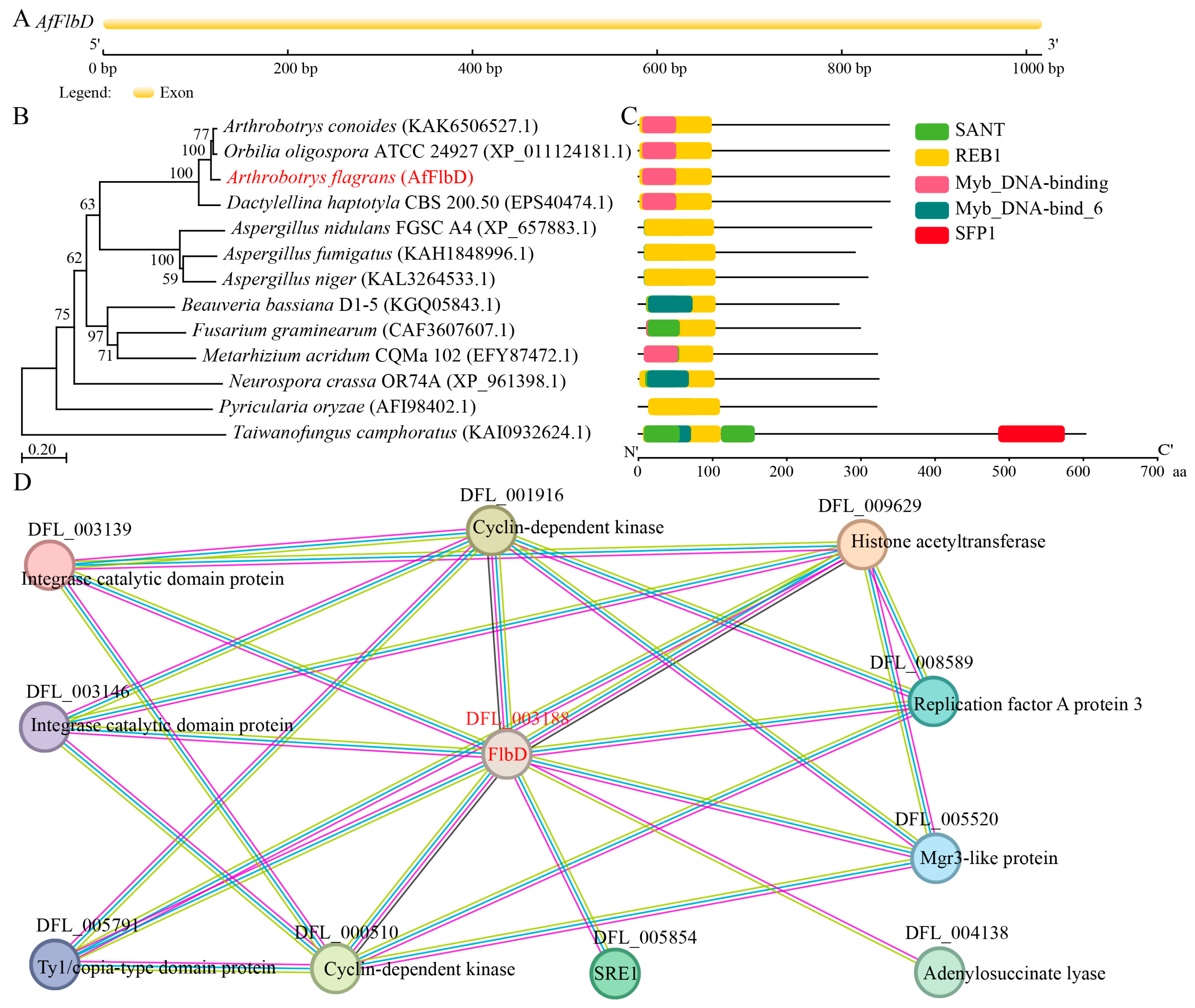
Figure 1. Bioinformatic analysis of AfFlbD gene structure, protein sequence, and interacting proteins in A. flagrans. (A) Gene structure analysis of AfFlbD. (B) AfFlbD protein phylogenetic analysis. (C) Conserved domains of FlbD protein. (D). Predicted protein interactions with AfFlbD using the STRING database.
Microorganisms 13 01847 g001
Figure 2. Knockout and validation of AfFlbD in A. flagrans. (A) Schematic diagram of AfFlbD gene knockout in A. flagrans. (BD) PCR verification of AfFlbD knockout mutants in A. flagrans.
Microorganisms 13 01847 g002
Figure 3. Comparisons of mycelial growth and cell length of the WT and ΔAfFlbD strains. (A) Colony morphologies of fungal strains cultured at 28 °C for 4 days. (B) Comparisons of mycelial growth rates. (C) CFW staining of hyphae of WT and ΔAfFlbD strains. (D) Hyphal septum spacing statistics. (Student’s t-test; * p < 0.05).
Microorganisms 13 01847 g003
Figure 4. Comparisons of stress responses for NaCl, SDS, sorbitol, and H2O2 treatment. (A) Growth of WT and mutant strains under different concentrations of NaCl, SDS, sorbitol, and H2O2. (B) RGI values of WT and mutant strains under NaCl, SDS, sorbitol, and H2O2 treatment. (Student’s t-test; * p < 0.05; ** p < 0.01; *** p < 0.001).
Microorganisms 13 01847 g004
Figure 5. Comparisons of trap formation and nematode predation efficiency of the WT and ΔAfFlbD strains. (A) Trap formation at different time points (0 h, 3 h, 6 h, 12 h, and 24 h); scale bar: 50 μm. Red triangles: traps; blue triangles: nematodes. (B) Numbers of traps at different time points. (C) Nematode mortality. (D) Analysis of the relative expression levels of five trap-positive regulatory genes in WT and ΔAfFlbD strains using qRT-PCR. (Student’s t-test; ** p < 0.01; *** p < 0.001.).
Microorganisms 13 01847 g005
Figure 6. Functional analysis of AfFlbD in chlamydospore and conidia formation. (A) The chlamydospores of the WT and ΔAfFlbD strains after 14 days of culturing on WA medium. Red triangles: chlamydospores. (B) The conidia of WT and ΔAfFlbD strains after 14 days of culturing on WA medium. Blue triangles: conidia. (C) The number of chlamydospores. (D) The number of conidia. (E) Comparisons of chlamydospore morphology. (F) Schematic diagram of plasmid construction for AfFlbD-GFP fusion protein expression. (G) Confocal microscopy analysis of AfFlbD subcellular localization. (Student’s t-test; *** p < 0.001).
Microorganisms 13 01847 g006
Disclaimer/Publisher’s Note: The statements, opinions and data contained in all publications are solely those of the individual author(s) and contributor(s) and not of MDPI and/or the editor(s). MDPI and/or the editor(s) disclaim responsibility for any injury to people or property resulting from any ideas, methods, instructions or products referred to in the content.