Article

Fluoroquinolone Residues in Piglet Viscera and Their Impact on Intestinal Microbiota Resistance: A One Health Approach

by
Olga Cardoso
1,
Maria Manuel Donato
2,
Sara Carolina Henriques
3 and
Fernando Ramos
4,*
1
Chemical Engineering and Renewable Resources for Sustainability (CERES), Department of Chemical Engineering, Faculdade de Farmácia, Universidade de Coimbra, Azinhaga de Santa Comba, 3000-548 Coimbra, Portugal
2
CIMAGO (Centro de Investigação em Meio Ambiente, Genética e Oncobiologia), Faculdade de Medicina, Universidade de Coimbra, Azinhaga de Santa Comba, 3000-548 Coimbra, Portugal
3
Research Institute for Medicines (iMed.ULisboa), Faculty of Pharmacy, Universidade de Lisboa, 1649-003 Lisboa, Portugal
4
Universidade de Coimbra, Faculdade de Farmácia, REQUIMTE/LAQV, Azinhaga de Santa Comba, 3000-548 Coimbra, Portugal
*
Author to whom correspondence should be addressed.
Microorganisms 2025, 13(6), 1389; https://doi.org/10.3390/microorganisms13061389
Submission received: 10 May 2025 / Revised: 4 June 2025 / Accepted: 12 June 2025 / Published: 14 June 2025

Abstract

:
The presence of fluoroquinolone (FQ) residues in food-producing animals has raised concerns regarding antimicrobial resistance. This study evaluated the occurrence of FQ residues in the liver and kidneys of piglets and their association with resistance patterns in Escherichia coli and Enterococcus species from piglets’ intestinal microbiota. FQ residues were found in 44% of livers and 23% of kidneys. Among 340 E. coli isolates from feces, resistance to ciprofloxacin (CIP) (43.2%) and multidrug resistance (MDR) (82.7%) were prominent. The presence of FQ in kidneys significantly increased the odds of CIP-resistant E. coli (OR = 2.94, p = 0.0013) and MDR (OR = 2.70, p = 0.0047). Enterococci (n = 130) were evenly distributed among the species E. faecalis, E. faecium, and Enterococcus spp. and presented resistance to CIP (34.6%). FQ in kidneys were associated with higher odds of CIP-resistant enterococci (OR = 3.78, p = 0.015). Interaction models revealed species-dependent responses, with Enterococcus spp. showing high resistance in the presence of FQ in kidneys (OR = 18, p = 0.006), while E. faecium exhibited lower resistance compared to E. faecalis. These findings emphasize the role of FQ residues in promoting bacterial resistance and underscore the need for the stricter regulation and routine surveillance of antimicrobial use in livestock to curb the spread of bacterial resistance to clinical drugs, and mitigate public health risks—One Health.

1. Introduction

The use of antibiotics has been of such an order of magnitude since their discovery by Fleming in 1921 that bacteria have organized themselves to avoid this stress inflicted on them. The World Health Organization (WHO) considers antibiotic resistance a global plague [1]. These resistant bacteria can appear anywhere, such as soil, water, animals, and plants (the One Health concept is very evident in this context), and when they reach humans by any means, they can cause serious illnesses, prolonged hospitalizations, etc. Antibiotics have been widely used worldwide, especially in modern animal husbandry, where they are often used as veterinary drugs for treating infectious diseases, like sulfonamides, quinolones, and macrolides [2]. Some antibiotics are even utilized as growth promoters, like small sub-therapeutic doses of tetracycline, to improve the animal’s immunity and increase production. However, the overconsumption of these veterinary antibiotics promotes the occurrence of antibiotic resistance in food animals and accelerates the dissemination process of antibiotic resistance [2].
Pig farming systems subject animals to significant stress, leading to various physiological disturbances, particularly in the gastrointestinal tract of piglets. Postweaning diarrhea is one of the most common and economically damaging conditions in pig farming. Antibiotics have been extensively used in pig production for decades to mitigate these adverse effects. However, the widespread use of antibiotics contributed to the emergence of resistant bacterial strains, raising serious public health concerns. In response, the European Union banned the inclusion of antibiotics in livestock feed in 2003.
The WHO has classified certain antimicrobial classes as critically important for human medicine, like fluoroquinolones. Despite their significance in human healthcare, these antibiotics are widely used in veterinary medicine, including swine farming, where enrofloxacin is a common choice. However, the fluoroquinolones used in animals can lead to the selection of resistance determinants to this class of antimicrobials, potentially compromising the effectiveness of ciprofloxacin in human therapy [3].
Once administered, fluoroquinolones exhibit rapid absorption with wide tissue dissemination and are excreted through urine and bile. Higher concentrations of drug residues are usually found in the liver and kidney, considering that the hepatobiliary system and the kidneys are the main routes by which drugs and their metabolites leave the body. Due to fluoroquinolones’ lipophilic characteristics, they have a long half-life and slow metabolization, increasing the risk of drug residues in animal-derived food products, which can pose health hazards to consumers [4].
A systematic review of fluoroquinolone resistance in isolates collected from healthy pigs showed that resistance to this group of antibiotics was variable in Europe. The highest resistance rates were observed in Portugal, Spain, Romania, and North Macedonia. The use of fluoroquinolones in pig farming has been banned in some countries; however, the persistence of high resistance levels in these regions indicates a strong correlation between antibiotic use in food animals and the development of resistance [3]. These findings highlight the ongoing challenges of antimicrobial resistance in animal production and reinforce the need for stringent monitoring and control measures to mitigate its spread.
Escherichia coli and the genus Enterococcus are commonly used as indicator organisms in many antimicrobial resistance surveillance programs [5]. E. coli and enterococci are ubiquitous and found in various environments, including the gastrointestinal tract, water, soil, and food. These properties make them valuable sentinel organisms for monitoring antimicrobial resistance trends and environmental transmission. Hence, surveillance programs that track the presence and patterns of antimicrobial resistance in E. coli and enterococci could provide the development of combating strategies for these global health threats [6,7,8].
This study aimed to detect fluoroquinolone (FQ) residues in the livers and kidneys of piglets collected in a Portuguese abattoir and assess whether bacterial representatives of their intestinal microbiota—such as E. coli and Enterococcus species—exhibit resistance to antibiotics used in human medicine and determine if there is a relationship between FQ residues in viscera and bacterial ciprofloxacin (CIP) resistance. Given the potential for these resistant bacteria to enter the environment and impact human health, the results of this study will be examined through the lens of the “One Health” approach.

2. Materials and Methods

2.1. Sampling and Escherichia coli and Enterococcus spp. Isolation

In this study, feces, liver, and kidney samples were collected from randomly selected healthy piglets (weighing 5–8 kg) between October 2018 and May 2019. The piglets came from an abattoir in Mealhada, which receives piglets from different regions of Portugal due to the high demand for roasting piglets (‘leitão à bairrada’), a local gastronomic specialty [9]. Sampling was carried out under a veterinarian’s supervision.
The samples collected from each animal consisted of one whole kidney; 200 g of the liver (right lobe); and a minimum of 10 g of feces. All samples were individually placed in sterile plastic bags and immediately transported to the laboratory. Liver and kidney samples were stored at −18 °C until analysis.
The isolation of E. coli was performed on the day of collection as described elsewhere using Lauryl Sulphate Agar [9], incubated at 37 °C/18 h, with the yellow-colored colonies on the agar being E. coli. Any doubts regarding identification were resolved with API20E (BioMérieux, Marcy-l-Étoile, France). In 2019, given the results obtained with Gram-negative bacteria, enterococci (Gram-positive bacteria) were also isolated to extend the study to another bacteria representative of the piglet microbiota. The isolation of Enterococcus species in the feces samples was performed [10] utilizing Slanetz Bartley Agar (Oxoid, Basingstoke, UK) and incubation at 37 °C, for 24–48 h. The red-colored colonies were representative of Enterococcus and for further identification, catalase, Bile Esculin Agar (Oxoid), and Trypticase Soy broth+6.5% NaCl tests were performed. If there was any doubt about the identification, API STREP (BioMérieux, France) was used.

2.2. Identification of Enterococcus Species

Real-time PCR (LightCycler, Roche Diagnostics, Germany) on crude bacterial DNA identified Enterococcus spp., E. faecalis, and E. faecium (Table 1).
PCR was performed in a volume of 20 µL containing 4.0 µL of LightCycler FastStart DNA MasterPLUS SYBR Green I® (Roche Diagnostics, Mannheim, Germany). An initial denaturation cycle at 95 °C for 10 min was performed in all cases followed by 45 amplification cycles. Specific denaturation, annealing, and extension conditions for each set of primers are shown in Table 1. Melting curves were also plotted automatically and analyzed with the LightCycler software version 4.1. PCR products were checked on 2% agarose gels stained with ethidium bromide and visualized with UV light. In comparing both results, it was possible to establish specific melting temperatures to identify each gene, and positive and negative controls were always included. To avoid methodology errors, a different technician, blinded to previous results, repeated all procedures.

2.3. Antimicrobial Susceptibility Assays

The isolates were tested for antimicrobial susceptibility using the agar disk diffusion method on Mueller–Hinton agar according to the guidelines provided by the European Committee for Antimicrobial Susceptibility Testing [12].
For the antibiogram of E. coli isolates, the following antimicrobial agents (Liofilchem®s.r.l., Roseto degli Abruzzi TE, Italy) were used and included the following four families of beta-lactams: amoxicillin–clavulanic acid (AMC) (20–10 µg); piperacillin (PIP) (30 µg); cefoxitin (FOX) (30 µg); ceftazidime (CAZ) (10 µg); cefepime (FEP) (30 µg); imipenem (IP) (10 µg); and aztreonam (AZT) (30 µg). The remaining 3 antimicrobials were from other families and included sulfamethoxazole-trimethoprim (SXT) (23.75–1.25 µg); ciprofloxacin (CIP) (5 µg); and amikacin (AK) (30 µg). E. coli ATCC 25922 was used as a control.
In the antibiogram of Enterococcus species, the antimicrobials were vancomycin (VAN), ciprofloxacin (CIP), linezolid (LNZ), tigecycline (TIG), ampicillin (AMP), and imipenem (IP). E. faecalis ATCC 29212 was used as a control.

2.4. Quantification of Antimicrobial Residues

UHPLC-ToF-MS was used to determine the six fluoroquinolones (FQs) (enrofloxacin, ciprofloxacin, danofloxacin, ofloxacin, norfloxacin, and marbofloxacin) in the liver and kidney. Antimicrobial and internal standards of ≥98% purity were purchased from Sigma-Aldrich (Madrid, Spain). The extraction procedure and UHPLC-ToF-MS conditions have been described previously [13].

2.5. Statistical Analysis

Data are presented as absolute and/or relative frequencies. Associations were evaluated using logistic regression models, with a significance level of 5%. Analyses were performed in R software version 4.3.2 (R Foundation for Statistical Computing, Vienna, Austria).

3. Results

3.1. Occurrence of Fluoroquinolone Residues in Piglets’ Liver and Kidney

In 2018, 32 liver and kidney samples were collected from piglets, of which 13 livers and 7 kidneys presented FQs. In 2019, 43 samples were obtained, and 20 livers and 10 kidneys presented FQs. In total, 44% of the livers and 23% of the kidneys had FQ residues. Piglets in which FQ was detected in the kidney also had FQ detected in the liver (n = 17). In total, 75 piglets were analyzed: 33 presented FQ residues in the liver (44%), and 17 in the kidney (23%).

3.2. Escherichia coli and Antibiotic Resistance

A total of 340 E. coli isolates were identified from 75 piglets’ feces. Table 2 presents the bacteria’s resistance profiles to antibiotics used in human therapeutics.
Higher resistance was observed for penicillins (AMC and PIP), followed by SXT and CIP. A total of 321 (82.7%) isolates exhibited resistance to two or more antibiotics, while 208 (61.2%) isolates were resistant to three or more antibiotics, classifying them as multidrug-resistant (MDR) bacteria.

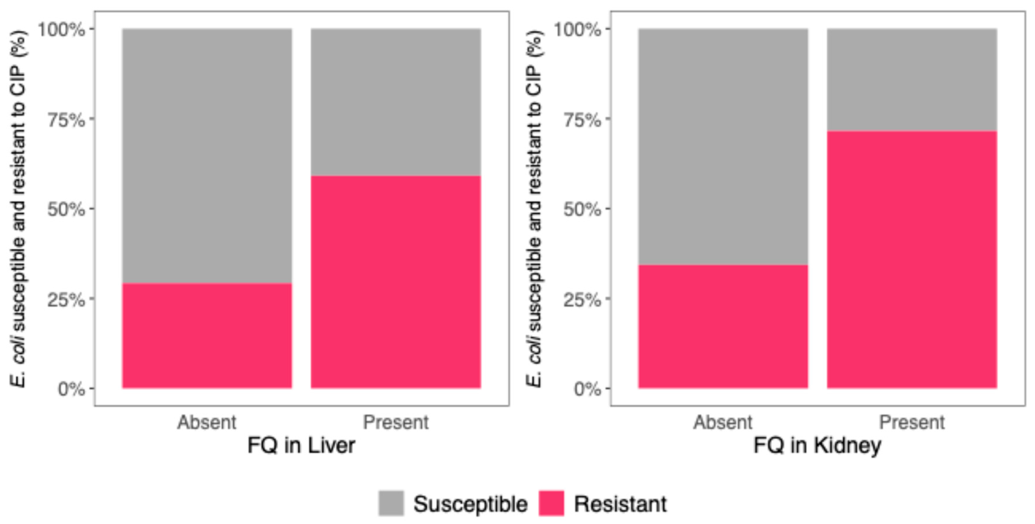
3.3. Association Between FQ Residues in Viscera and E. coli CIP Resistance

In the 33 piglets with FQ residues in the liver, 159 E. coli isolates were identified, of which 94 (59.1%) exhibited resistance to CIP, while 65 (40.9%) were susceptible. When FQ was absent from the liver, 181 E. coli isolates were identified, with 53 (29.3%) showing resistance to CIP and 128 (70.7%) being susceptible.
Among the 17 piglets in which FQ residues were detected in the kidney, 81 E. coli isolates were identified. Of these, 58 (71.6%) were resistant to CIP, while 23 (28.4%) were susceptible. In contrast, when FQ was absent from the kidney, 259 E. coli isolates were identified, of which 89 (34.4%) were resistant to CIP and 170 (65.6%) were susceptible (Figure 1).
A mixed effects logistic regression model was used to assess the relationship between FQ residues in piglet organs and E. coli resistance to CIP. The final model included the FQ residues in the liver and kidney as predictors, with the piglet from which the residues were taken used as a random effect to account for the clustering of isolates within the animals.
The model showed that the presence of FQ residues in the kidney was associated with a 2.94-fold increase in the odds of E. coli CIP resistance (odds ratio [OR] 95% CI: 1.57–5.50, p = 0.0013). Likewise, FQ residues in the liver were associated with a 2.07-fold increase in the odds of E. coli CIP resistance (OR 95% CI: 1.20–3.57, p = 0.0093) (Figure 1, Table 3).
These results suggest that FQ residues in the liver and kidney contribute to developing or maintaining CIP resistance in E. coli.

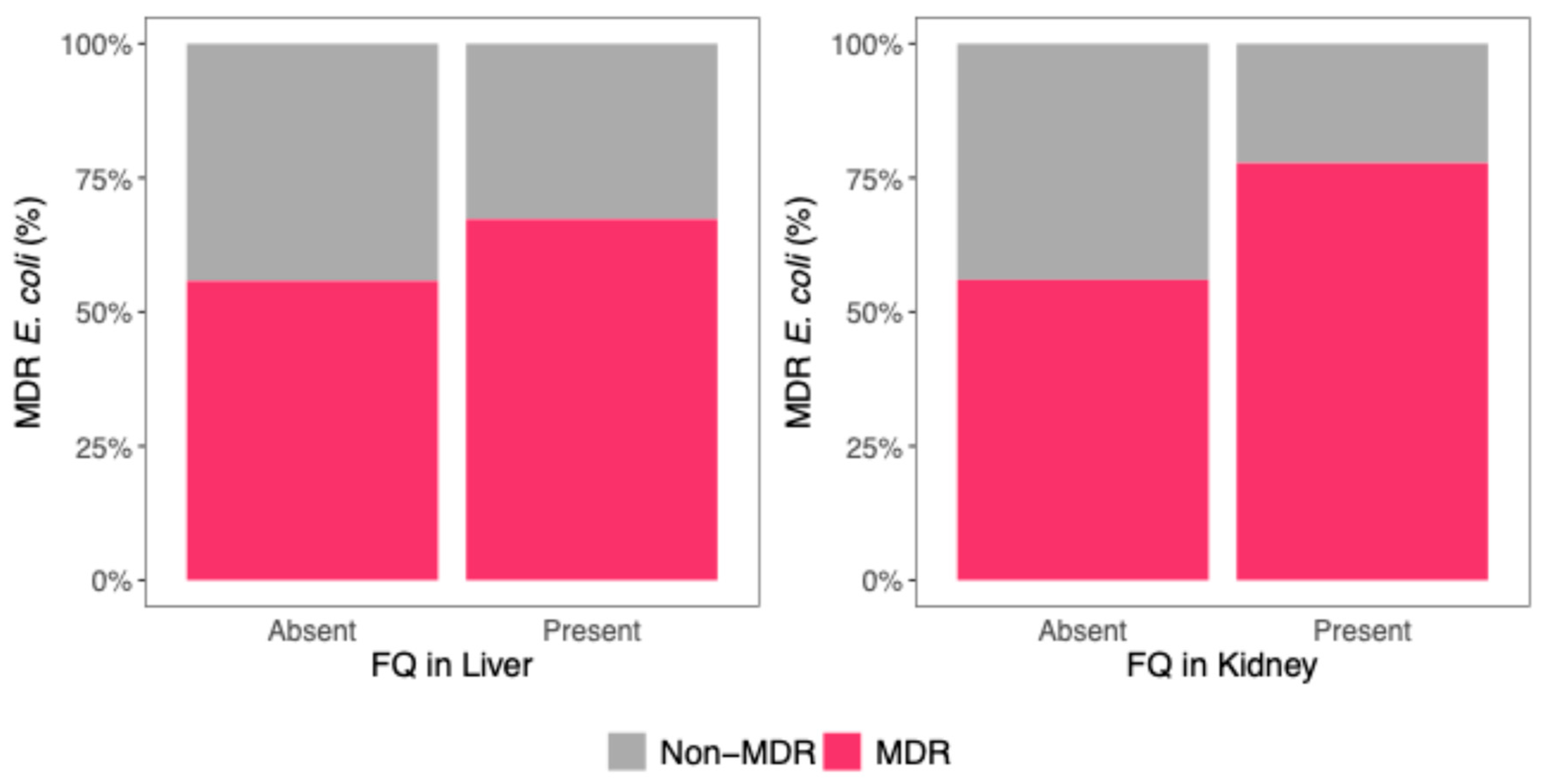
3.4. Association Between FQ Residues and Multidrug Resistance in E. coli

The relationship between FQ residues in piglet organs and MDR in E. coli isolates was also evaluated.
In piglets with FQ residues in the liver, 159 E. coli isolates were identified, of which 107 (67.3%) were MDR, while 52 (32.7%) were non-MDR. When FQs were absent from the liver, 181 isolates were identified: 101 (55.8%) MDR and 80 (44.2%) non-MDR (Figure 2 left).
Among the 17 piglets with FQ residues in the kidney, 81 E. coli isolates were identified. Of these, 63 (77.8%) were MDR, while 18 (22.2%) were non-MDR. In contrast, when FQ was absent from the kidney, 259 isolates were identified, of which 145 (56.0%) were MDR and 114 (44.0%) were non-MDR (Figure 2 right).
A logistic regression model was fitted to evaluate the association between FQ residues in the liver and kidney and the MDR E. coli. The presence of FQ in the kidney was significantly associated with a 2.70-fold increase in the likelihood of MDR (OR 95% CI: 1.37–5.47, p = 0.0047). However, FQ residues in the liver were not significantly associated with MDR (OR: 1.03, 95% CI: 0.60–1.76, p = 0.93) (Figure 2, Table 3).

3.5. Enterococcus Species Identification and Antibiotic Resistance

From 43 piglet feces, a total of 130 enterococci isolates were identified and distributed among the Enterococci species as follows: 40 (30.8%) Enterococcus faecalis; 45 (34.6%) Enterococcus faecium; and 45 (34.6%) Enterococcus spp. The distribution was relatively even, with each species represented in similar proportions. The estimated Shannon diversity index of 1.097, close to the maximum possible value of ln(3) = 1.0986, indicates a diverse and balanced community, with no dominant species.
Table 4 presents the resistance profiles of enterococci to the different antibiotics utilized in human clinical therapeutic strategies. These species showed the highest resistance to CIP and IP, while VAN, LNZ, and AMP showed much lower levels of resistance and no resistance to TIG.

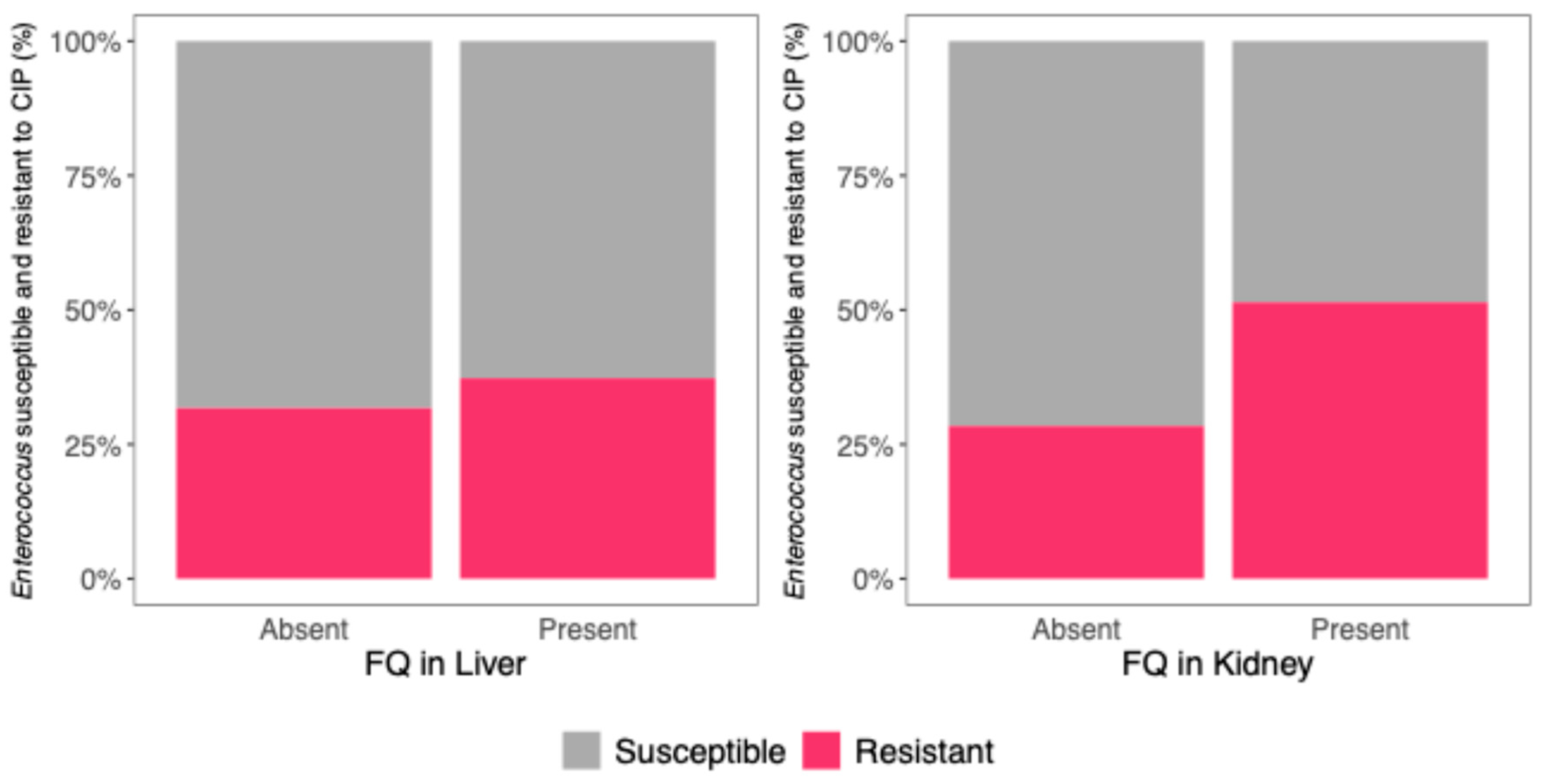
3.6. Association Between FQ Residues in Viscera and Enterococci CIP Resistance

In the 20 piglets with FQ residues in the liver, 67 enterococci isolates were identified, with 25 (37.3%) showing resistance to CIP and 42 (62.7%) being susceptible. When FQ residues were absent from the liver, 63 isolates were identified, of which 20 (31.7%) were resistant and 43 (68.3%) were susceptible.
Among the 10 piglets in which FQ residues were detected in the kidney, 35 enterococci isolates were identified. Of these, 18 (51.4%) were resistant to CIP, while 17 (48.6%) were susceptible. In contrast, when FQ residues were absent from the kidney, 95 isolates were identified, of which 27 (28.4%) were resistant and 68 (71.6%) were susceptible.
A mixed effects logistic regression model was used to evaluate the association between the presence of FQ residues in piglet organs and bacterial resistance to CIP. The model included the presence of FQs in the liver and kidney as predictors, with piglet identification as a random effect.
The model showed that the presence of FQ residue in the kidney was significantly associated with a 3.78-fold increase in the odds of enterococci CIP resistance (OR 95% CI: 1.34–11.60, p = 0.015). In contrast, FQ residues in the liver were not significantly associated with enterococci CIP resistance (OR = 0.60, 95% CI: 0.21–1.57, p = 0.316) (Figure 3, Table 3).
These results suggest that FQ residues in the kidney played a critical role in developing and maintaining CIP resistance in enterococci.
Moreover, species-specific resistance patterns were also observed for CIP: for E. faecalis, 18 isolates (45.0%) were resistant to CIP, while 22 (55.0%) were susceptible. For E. faecium, 7 isolates (15.6%) were resistant, whereas 38 (84.4%) were susceptible. For Enterococcus spp., 20 isolates (44.4%) were resistant, and 25 (55.6%) were susceptible.
Among the 35 enterococci isolates from piglets with FQ residues detected in the kidney, 18 (51.4%) were resistant to CIP, while 17 (48.6%) were susceptible: E. faecalis: 4 resistant, 7 susceptible; E. faecium: 2 resistant, 7 susceptible; Enterococcus spp.: 12 resistant, 3 susceptible.
Among the 95 enterococci isolates from piglets where FQ residues were not detected in the kidney, 27 (28.4%) were resistant to CIP, while 68 (71.6%) were susceptible: E. faecalis: 14 resistant, 15 susceptible; E. faecium: 5 resistant, 31 susceptible; Enterococcus spp.: 8 resistant, 22 susceptible.
To assess whether CIP resistance varied among Enterococcus species, a new logistic regression model was fitted, incorporating an interaction term between FQ presence in the kidney and Enterococcus species. This model showed that E. faecium had significantly lower odds of resistance compared to E. faecalis (OR = 0.17, 95% CI: 0.05–0.54, p = 0.004). No significant difference was observed between Enterococcus spp. and E. faecalis in terms of CIP resistance (OR = 0.39, 95% CI: 0.13–1.14, p = 0.090). However, the presence of FQ residues in the kidney significantly increased CIP resistance in Enterococcus spp. The interaction term showed that when FQ residues were present in the kidney, Enterococcus spp. had an approximately 18-fold higher likelihood of CIP resistance compared to E. faecalis without FQ residues in the kidney (OR 95% CI: 2.43–160.56, p = 0.006).
These results suggest that FQ residues in the kidney enhance CIP resistance in certain Enterococcus species, particularly Enterococcus spp.
Among the 67 enterococci isolates from piglets with FQ residues in the liver, 25 (37.3%) were resistant to ciprofloxacin, while 42 (62.7%) were susceptible: E. faecalis: 6 resistant, 11 susceptible; E. faecium: 2 resistant, 17 susceptible; Enterococcus spp.: 17 resistant, 14 susceptible.
Among the 63 enterococci isolates from piglets where FQ residues were not detected in the liver, 20 (31.7%) were resistant to CIP, while 43 (68.3%) were susceptible: E. faecalis: 12 resistant, 11 susceptible; E. faecium: 5 resistant, 21 susceptible; Enterococcus spp: 3 resistant, 11 susceptible.
A logistic regression model was used to evaluate the association between FQ residues in the liver and CIP resistance, including an interaction term for Enterococcus species. This model showed that E. faecium had significantly lower odds of resistance compared to E. faecalis (OR = 0.22, 95% CI: 0.06–0.75, p = 0.019). No significant difference was observed between Enterococcus spp. and E. faecalis in terms of CIP resistance (OR = 0.25, 95% CI: 0.05–1.05, p = 0.073).
However, the interaction term showed that when FQ residues were present in the liver, Enterococcus spp. had an approximately nine-fold higher likelihood of CIP resistance compared to E. faecalis without FQ residues (OR 95% CI: 1.35–69.51, p = 0.028). These results suggest that FQ residues in the liver had a species-dependent impact on enterococci CIP resistance, particularly in increasing in Enterococcus spp.

4. Discussion

This study highlights the presence of FQ residues in piglet livers and kidneys and their association with antimicrobial resistance in E. coli and enterococci. These findings have important implications for antibiotic stewardship in livestock production and the potential risks of antimicrobial resistance transmission to humans [3,14].
FQ residues in piglet liver and kidney were high, with 44% of livers and 23% of kidneys containing detectable levels. These results are consistent with previous studies demonstrating the persistence of FQ in animal tissues [4,15]. The detection of these residues indicates significant exposure of piglets to FQ [16]. The co-occurrence of FQ residues in both liver and kidney in 17 piglets suggests systemic exposure, possibly due to prolonged administration or inadequate withdrawal periods before slaughter [4,14,17].
This study also suggests that the presence of FQ residues in piglet organs is associated with increased resistance to CIP in both E. coli and Enterococcus species, raising concerns about the role of antibiotic residues in promoting antimicrobial resistance [18,19].
The observed association between FQ residues and CIP-resistant E. coli was of particular concern. The logistic regression model confirmed that the presence of FQ residues in the kidney significantly increased the odds of CIP resistance (OR = 2.94, p = 0.0013), with a slightly lower but still significant effect in the liver (OR = 2.07, p = 0.0093). These data were consistent with other studies [18]. The results highlight the selective pressure exerted by FQ residues that might drive the emergence and persistence of resistant bacterial strains [3,20,21]. This study also demonstrated an association between FQ residues and MDR E. coli. FQ residues in the kidney were significantly associated with MDR E. coli (OR = 2.70, p = 0.0047), but the effect was insignificant for residues in the liver (OR = 1.03, p = 0.93). These results are consistent with previous findings in [22], which reported that FQ exposure in livestock contributes to the emergence of MDR strains. The higher MDR rate in kidney-associated E. coli isolates suggests that FQ residues may exert more selective pressure in this organ, possibly due to higher drug concentrations in renal tissue [23].
This study further evaluated the relationship between FQ residues and CIP resistance in Enterococcus species. While FQ residues in the kidney significantly increased the odds of enterococci CIP resistance (OR = 3.93, p = 0.016), the presence of FQ in the liver was not significantly associated with this resistance (OR = 0.50, p = 0.196). This suggests that renal accumulation of FQ might have a greater impact on enterococcal resistance selection [23].
The identification of 130 enterococcal isolates with a balanced species distribution indicates a diverse microbial population in piglet feces [24]. Enterococcal species showed different levels of CIP resistance, with E. faecium showing significantly lower odds of resistance compared to other species (OR = 0.24, p = 0.006); these results parallel the findings of [25,26]. E. faecium isolates showed high resistance to imipenem, a carbapenem of hospital use, but this species has an intrinsic tolerance to beta-lactams due to the presence of PBPs (specifically PBP5) with low binding affinity for this class of antibiotics [27]. All the enterococcus were susceptible to tigecycline, as stated by other authors [28].
The results of this study underscore the risks posed by antibiotic residues in livestock production. The significant associations between FQ residues and antibiotic-resistant bacteria reinforce the need for strict regulations on the use of antibiotics in food-producing animals. The European Food Safety Authority (EFSA) and the World Health Organization (WHO) have previously emphasized the threat of antimicrobial resistance posed by antibiotic residues in food of animal origin [5,16]. These findings reinforce these concerns and suggest that monitoring and limiting the use of FQs in piglets could help reduce the spread of bacterial resistance.

5. Conclusions

This study demonstrates that FQ residues in piglet livers and kidneys are significantly associated with increased resistance to CIP in E. coli and Enterococcus species. The presence of FQ residues in the kidney was particularly influential, considerably improving the odds of CIP resistance and MDR in E. coli. These findings highlight the urgent need for antibiotic stewardship in livestock production to reduce the risk of antimicrobial resistance transmission to humans in line with the One Health concept.

Author Contributions

All authors contributed to the study’s conception and design. O.C., M.M.D., S.C.H., and F.R. performed material preparation, data collection, and analysis. O.C., M.M.D., and F.R. wrote the first draft of the manuscript, and all authors commented on previous versions. All authors have read and agreed to the published version of the manuscript.

Funding

This work was supported by the Portuguese public agency that supports science, technology and innovation, FCT (Fundação para a Ciência e a Tecnologia), through the LAQV—Laboratório Associado para a Química Verde, which is part of the REQUIMTE, Network of Chemistry and Technology [UIDB/50006/2025]; and CERES—Chemical Engineering and Renewable Resources for Sustainability [UIDB/00102/2020 DOI:10.54499/UIDB/00102/2020].

Institutional Review Board Statement

Not applicable. This study did not need approval from the ethics committee, as the animals were slaughtered for consumption in restaurants and a veterinarian collected a kidney, some liver, and feces from these carcasses.

Informed Consent Statement

Not applicable.

Data Availability Statement

No new data were created or analyzed in this study. Data sharing is not applicable to this article.

Acknowledgments

The authors wish to acknowledge the invaluable contribution of Sofia Osório in the sample collection.

Conflicts of Interest

The authors declare no conflicts of interest.

References

  1. White, A.; Hughes, J.M. Critical Importance of a One Health Approach to Antimicrobial Resistance. Ecohealth 2019, 16, 404–409. [Google Scholar] [CrossRef]
  2. Yang, F.; Han, B.; Gu, Y.; Zhang, K. Swine Liquid Manure: A Hotspot of Mobile Genetic Elements and Antibiotic Resistance Genes. Sci. Rep. 2020, 10, 15037. [Google Scholar] [CrossRef] [PubMed]
  3. Hayer, S.S.; Casanova-Higes, A.; Paladino, E.; Elnekave, E.; Nault, A.; Johnson, T.; Bender, J.; Perez, A.; Alvarez, J. Global Distribution of Fluoroquinolone and Colistin Resistance and Associated Resistance Markers in Escherichia coli of Swine Origin—A Systematic Review and Meta-Analysis. Front. Microbiol. 2022, 13, 834793. [Google Scholar] [CrossRef]
  4. Fei, Z.; Song, S.; Yang, X.; Jiang, D.; Gao, J.; Yang, D. Occurrence and Risk Assessment of Fluoroquinolone Residues in Chicken and Pork in China. Antibiotics 2022, 11, 1292. [Google Scholar] [CrossRef]
  5. European Food Safety Authority (EFSA). Technical specifications for a EU-wide baseline survey of antimicrobial resistance in bacteria from aquaculture animals. EFSA J. 2024, 22, e8928. [Google Scholar] [CrossRef]
  6. Jaleta, M.; Junker, V.; Kolte, B.; Börger, M.; Werner, D.; Dolsdorf, C.; Schwenker, J.; Hölzel, C.; Zentek, J.; Amon, T.; et al. Improvements of Weaned Pigs Barn Hygiene to Reduce the Spread of Antimicrobial Resistance. Front. Microbiol. 2024, 15, 1393923. [Google Scholar] [CrossRef]
  7. Zaheer, R.; Cook, S.R.; Barbieri, R.; Goji, N.; Cameron, A.; Petkau, A.; Polo, R.O.; Tymensen, L.; Stamm, C.; Song, J.; et al. Surveillance of Enterococcus spp. Reveals Distinct Species and Antimicrobial Resistance Diversity across a One-Health Continuum. Sci. Rep. 2020, 10, 3937. [Google Scholar] [CrossRef]
  8. Ladyhina, V.; Sternberg-Lewerin, S.; Andersson, L.; Rajala, E. Antimicrobial Resistance among Indicator Enterococcus faecium and Escherichia coli in Swedish Pig Farms. Acta Vet. Scand. 2024, 66, 34. [Google Scholar] [CrossRef] [PubMed]
  9. Rasschaert, G.; Van Elst, D.; Colson, L.; Herman, L.; de Carvalho Ferreira, H.C.; Dewulf, J.; Decrop, J.; Meirlaen, J.; Heyndrickx, M.; Daeseleire, E. Antibiotic Residues and Antibiotic-Resistant Bacteria in Pig Slurry Used to Fertilize Agricultural Fields. Antibiotics 2020, 9, 34. [Google Scholar] [CrossRef]
  10. Donato, M.M.; Assis, G.; Cardoso, O.; Oliveiros, B.; Freitas, A.; Ramos, F. Assessment of Zn and Cu in Piglets’ Liver and Kidney: Impact in Fecal Enterococcus spp.? Environ. Sci. Pollut. Res. 2024, 31, 20941–20952. [Google Scholar] [CrossRef]
  11. Igbinosa, E.O.; Beshiru, A. Antimicrobial Resistance, Virulence Determinants, and Biofilm Formation of Enterococcus Species from Ready-to-Eat Seafood. Front. Microbiol. 2019, 10, 728. [Google Scholar] [CrossRef] [PubMed]
  12. The European Committee on Antimicrobial Susceptibility Testing. Breakpoint tables for interpretation of MICs and zone diameters. Version 09.0. 2019. Available online: https://www.eucast.org (accessed on 9 May 2025).
  13. Gamboa-Cruz, C.; Barros, S.; Vila Pouca, A.S.; Barbosa, J.; Freitas, A.; Ramos, F. Assessing Antibiotic Residues in Piglet Liver and Kidney Samples: How to Manage the Results Obtained. Food Control 2021, 122, 107819. [Google Scholar] [CrossRef]
  14. Sodhi, K.K.; Singh, D.K. Insight into the Fluoroquinolone Resistance, Sources, Ecotoxicity, and Degradation with Special Emphasis on Ciprofloxacin. J. Water Process Eng. 2021, 43, 102218. [Google Scholar] [CrossRef]
  15. Hoelzer, K.; Wong, N.; Thomas, J.; Talkington, K.; Jungman, E.; Coukell, A. Antimicrobial Drug Use in Food-Producing Animals and Associated Human Health Risks: What, and How Strong, Is the Evidence? BMC Vet. Res. 2017, 13, 211. [Google Scholar] [CrossRef]
  16. Antimicrobial Resistance Surveillance in Europe 2023–2021 Data. Stockholm: European Centre for Disease Prevention and Control and World Health Organization; 2023. Cataloguing-in-Publication (CIP) Data. CIP Data. Available online: http://apps.who.int/iris (accessed on 9 May 2025).
  17. Aarestrup, F.M.; Duran, C.O.; Burch, D.G.S. Antimicrobial Resistance in Swine Production. Anim. Health Res. Rev. 2008, 9, 135–148. [Google Scholar] [CrossRef]
  18. Amsler, M.; Zurfluh, K.; Hartnack, S.; Sidler, X.; Stephan, R.; Kümmerlen, D. Occurrence of Escherichia coli Non-Susceptible to Quinolones in Faecal Samples from Fluoroquinolone-Treated, Contact and Control Pigs of Different Ages from 24 Swiss Pig Farms. Porc. Health Manag. 2021, 7, 29. [Google Scholar] [CrossRef]
  19. Cebeci, T. Species Prevalence, Virulence Genes, and Antibiotic Resistance of Enterococci from Food-Producing Animals at a Slaughterhouse in Turkey. Sci. Rep. 2024, 14, 13191. [Google Scholar] [CrossRef]
  20. Tang, K.L.; Caffrey, N.P.; Nóbrega, D.B.; Cork, S.C.; Ronksley, P.E.; Barkema, H.W.; Polachek, A.J.; Ganshorn, H.; Sharma, N.; Kellner, J.D.; et al. Restricting the Use of Antibiotics in Food-Producing Animals and Its Associations with Antibiotic Resistance in Food-Producing Animals and Human Beings: A Systematic Review and Meta-Analysis. Lancet Planet. Health 2017, 1, e316–e327. [Google Scholar] [CrossRef]
  21. Ardakani, Z.; Canali, M.; Aragrande, M.; Tomassone, L.; Simoes, M.; Balzani, A.; Beber, C.L. Evaluating the Contribution of Antimicrobial Use in Farmed Animals to Global Antimicrobial Resistance in Humans. One Health 2023, 17, 100647. [Google Scholar] [CrossRef]
  22. Li, X.; Hu, H.; Zhu, Y.; Wang, T.; Lu, Y.; Wang, X.; Peng, Z.; Sun, M.; Chen, H.; Zheng, J.; et al. Population Structure and Antibiotic Resistance of Swine Extraintestinal Pathogenic Escherichia coli from China. Nat. Commun. 2024, 15, 5811. [Google Scholar] [CrossRef]
  23. Zhou, X.; Chen, C.; Yue, L.; Sun, Y.; Ding, H.; Liu, Y. Excretion of Enrofloxacin in Pigs and Its Effect on Ecological Environment. Environ. Toxicol. Pharmacol. 2008, 26, 272–277. [Google Scholar] [CrossRef] [PubMed]
  24. Hammerum, A.M. Enterococci of Animal Origin and Their Significance for Public Health. Clin. Microbiol. Infect. 2012, 18, 619–625. [Google Scholar] [CrossRef]
  25. Hammerum, A.M.; Baig, S.; Kamel, Y.; Roer, L.; Pinholt, M.; Gumpert, H.; Holzknecht, B.; Røder, B.; Justesen, U.S.; Samulioniené, J.; et al. Emergence of VanA Enterococcus faecium in Denmark, 2005–2015. J. Antimicrob. Chemother. 2017, 72, 2184–2190. [Google Scholar] [CrossRef] [PubMed]
  26. Kim, M.H.; Moon, D.C.; Kim, S.J.; Mechesso, A.F.; Song, H.J.; Kang, H.Y.; Choi, J.H.; Yoon, S.S.; Lim, S.K. Nationwide Surveillance on Antimicrobial Resistance Profiles of Enterococcus faecium and Enterococcus faecalis Isolated from Healthy Food Animals in South Korea, 2010 to 2019. Microorganisms 2021, 9, 925. [Google Scholar] [CrossRef] [PubMed]
  27. Miller, W.R.; Munita, J.M.; Arias, C.A. Mechanisms of Antibiotic Resistance in Enterococci. Expert Rev. Anti-Infect. Ther. 2014, 12, 1221–1236. [Google Scholar] [CrossRef]
  28. de Jong, A.; Simjee, S.; El Garch, F.; Moyaert, H.; Rose, M.; Youala, M.; Dry, M. Antimicrobial Susceptibility of Enterococci Recovered from Healthy Cattle, Pigs and Chickens in Nine EU Countries (EASSA Study) to Critically Important Antibiotics. Vet. Microbiol. 2018, 216, 168–175. [Google Scholar] [CrossRef]
Figure 1. E. coli isolates (%) susceptible (gray) and resistant (pink) to CIP, as a function of the presence or absence of FQ residues in the liver (left) and kidney (right).
Microorganisms 13 01389 g001
Figure 2. MDR E. coli (%) (pink) as a function of the presence or absence of FQ residues in the liver (left) and kidney (right).
Microorganisms 13 01389 g002
Figure 3. Enterococcus species isolates (%) susceptible (gray) and resistant (pink) to CIP, as a function of the presence or absence of FQ residues in the liver (left) and kidney (right).
Microorganisms 13 01389 g003
Table 1. Primers and PCR conditions used in the identification of Enterococcus species.
SpeciesPrimer SequencePCR ConditionsAmplicon Size (bp)Reference
Enterococcus spp.TCA ACC GGG GAG GGT95 °C, 10 s; 60 °C, 5 s; 72 °C, 29 s733[11]
ATT ACT AGC GAT TCC GG
E. faecalisATC AAG TAC AGT TAG TCT95 °C, 10 s; 44 °C, 5 s; 72 °C, 38 s941[11]
ACG ATT CAA AGC TAA CTG
E. faeciumTTG AGG CAG ACC AGA TTG ACG95 °C, 10 s; 55 °C, 5 s; 72 °C, 26 s658[11]
TAT GAC AGC GAC TCC GAT TCC
Table 2. Number (%) of E. coli isolates with resistance to antibiotics.
AntibioticTotal
Amikacin (AMK)115 (33.8%)
Ciprofloxacin (CIP)147 (43.2%)
Sulfamethoxazole-Trimethoprim (SXT)197 (57.9%)
Aztreonam (AZT)64 (18.8%)
Cefoxitin (FOX)42 (12.4%)
Cefepime (FEP)59 (17.4%)
Ceftazidime (CAZ)39 (11.5%)
Amoxicillin + Clavulanic Acid (AMC)228 (67.1%)
Piperacillin (PIP)229 (67.4%)
Imipenem (IP)9 (2.6%)
Table 3. Odds ratio in the relationship between FQ residues in piglet organs and bacterial CIP resistance.
FQ Residues
Kidney OR (95% CI), p-ValueLiver OR (95% CI, p-Value)
E. coli resistance to CIP2.94 (1.57–5.50), p = 0.00132.07 (1.20–3.57), p = 0.0093
MDR E. coli2.70 (1.37–5.47), p = 0.00471.03 (0.60–1.76), p = 0.930
Enterococcus resistance to CIP3.78 (1.34–11.60), p = 0.0150.60 (0.21–1.57), p = 0.316
FQ—fluoroquinolone; CIP—ciprofloxacin; MDR—multi-drug resistance; OR—odds ratio; CI—confidence interval.
Table 4. Number of enterococci isolates with resistance to antibiotics.
AntibioticE. faecalis (n = 40)E. faecium (n = 45)Enterococcus spp. (n = 45)Total (n = 130)
Vancomycin (VAN)1135
Ciprofloxacin (CIP)1872045
Linezolid (LNZ)0112
Tigecycline (TIG)0000
Ampicillin (AMP)1214
Imipenem (IP)629641
Disclaimer/Publisher’s Note: The statements, opinions and data contained in all publications are solely those of the individual author(s) and contributor(s) and not of MDPI and/or the editor(s). MDPI and/or the editor(s) disclaim responsibility for any injury to people or property resulting from any ideas, methods, instructions or products referred to in the content.

