Article

Unlocking Nature’s Microbial Defenders: Genetic Mechanisms and Potential Against Monilinia spp. Pathogens

by
Augustina Kolytaitė
,
Ingrida Mažeikienė
*,
Monika Kurgonaitė
,
Saulė Raklevičiūtė
,
Gabija Paškevičiūtė
and
Birutė Frercks
Institute of Horticulture, Lithuanian Research Centre for Agriculture and Forestry, Kaunas Str. 30, 54333 Babtai, Lithuania
*
Author to whom correspondence should be addressed.
Microorganisms 2025, 13(4), 818; https://doi.org/10.3390/microorganisms13040818
Submission received: 10 March 2025 / Revised: 31 March 2025 / Accepted: 2 April 2025 / Published: 3 April 2025

Abstract

:
Monilinia spp., which causes brown rot, is one of the most damaging pathogens in stone fruits. Researchers are exploring epiphytic and endophytic microorganisms with the potential to suppress pathogens, control pathogenic microorganisms, and/or promote plant growth. In this study, microorganisms with antagonistic activity against three Monilinia species were isolated from plum orchard soil and plum fruits. Antagonism tests in vitro showed strong antagonistic properties of six strains of bacteria and two yeast-like fungi against M. fructigena, M. fructicola, and M. laxa, with growth inhibition from 45.5 to 84.6%. The antagonists were identified and characterized at the genetic level using whole genome sequencing (WGS). Genes involved in antibiotic resistance, virulence, secondary metabolite synthesis, and plant growth promotion were identified and characterized through genome mapping, gene prediction, and annotation. None of the microorganisms studied were predicted to be pathogenic to humans. The results of this study indicate that the bacteria Bacillus pumilus, B. velezensis, two strains of Lysinibacillus agricola, Pseudomonas chlororaphis isolated from stone fruit orchard soil, and the yeast-like fungus Aureobasidium pullulans, isolated from plums, are promising candidates for the biological control of Monilinia spp.

1. Introduction

The diversity of plant microbiomes can directly or indirectly provide nutrients, help relieve stress, and resist disease [1,2,3]. Microorganisms can regulate plant growth through nitrogen fixation [4], phosphate-solubilization [5], plant hormones secretion [4,6], or siderophore synthesis [7]. Microorganisms are already used for biological control in agriculture. The soil and plant surface are characterized by an endless number of microorganisms with advantageous activities that support plant health [8,9,10]. The bacteria Pseudomonas, Bacillus, Burkholderia, Streptomyces, Serratia, and Stephanobacteria, as well as fungi from the Trichoderma genus, are recognized as suitable for biological control, and their synthesized antimicrobial properties and bioactive compounds have been widely studied [11,12,13]. Secondary metabolites, low-molecular-weight antibiotics, and high-molecular-weight antimicrobial proteins or cell wall enzymes—hydrolases—can act as antimicrobial agents [14,15,16,17].
Monilinia spp. can cause brown rot, blossom blight, twig cankers, and fruit rot diseases, and are among the world’s most important dangers to stone fruit [18]. The identification of new biological control agents (BCAs) that effectively inhibit disease development is a major challenge for organic horticultural management [19]. Various biocontrol products based on bacterial or yeast antagonists (Candida oleophila, Aureobasidium pullulans, Pantoea agglomerans) have advanced through the development or commercialization stages, and several are successful [20]. In agricultural research, finding novel native biologically active compounds is relevant [21], but processes take too long when classical microbiology methods are used. Whole genome sequencing (WGS) is a technique that provides deep insights into the biosynthetic potential of selected microbial species. WGS analysis of microorganisms allows us to categorize genes associated with antagonistic and plant growth-promoting properties, understand their molecular and functional mechanisms, and thus reduce resources and time in developing biological products [20,21,22,23,24,25].
The aim of this study was to identify microorganisms isolated from soil and plum fruits and describe their properties at the genetic potential level and their antagonistic abilities to inhibit Monilinia spp. pathogens. In summary, we provide essential insights into the eight microbial isolates and their antagonistic and plant growth-promoting (PGP) mechanisms, which will benefit future applications aiming at enhancing the biocontrol of Monilinia spp. spread in stone orchards.

2. Materials and Methods

2.1. Isolation of Microorganisms

Microorganisms were isolated from the surrounding soil and fruits of Prunus domestica trees, maintained at the collection site of genetic resources for stone fruit orchards (Lithuanian Research Centre for Agriculture and Forestry, Institute of Horticulture, map for 55.0562814 23.8088851) in 2023. The procedure for isolation is as follows. Six genotypes with different resistances to Monilinia spp. were chosen: highly resistant No. 293 (free pollination of Cacanska najbolja), resistant No. 202 (Amitar × Jure), No. 219 (free pollination of Cacanska najbolja), No. 242 (Cacanska najbolja × Jure), susceptible No. 212 (Amitar × Jure), and No. 250 (Harmonija × Jure). Isolate names are assigned by tree number. The soil was collected at three places, with the distance from the trunk being 50 cm and the depth being 20 cm, and processed immediately for microbe isolation. The same trees were chosen for the collection of fruits. Five fruits of plums of approximately the same size and with no visual defects were selected at a similar height (around 150 cm) and sun exposure. The fruits were picked wearing sterile gloves, placed into clean plastic bags, and processed to further steps for microbe isolation. The soil and fruits (flesh with skin) were weighed (5 g for each sample). Additionally, fruit samples were homogenized. After adding 10 mL of NaCl 0.9%, the samples were shaken for 30 min at 200 rpm at room temperature. A total of 20 μL of suspension was spread on Petri dishes with lysogeny broth (LB) with agar media in five replicates and incubated for 7 days at 22 °C. Emerging colonies from these plates were further re-streaked until pure colonies were attained. The microbial stock cultures, containing LB and 30% glycerol, were kept at −80 °C until further analysis.
M. fructigena MFS01 was isolated, identified, and described by Kolytaite et al. [26]. The cultures of M. fructicola CBS 101512 (USA) and M. laxa CBS 489.50 (The Netherlands, Baarn) were obtained from Westerdijk Fungal Biodiversity Institute (The Netherlands, Utrecht) (https://wi.knaw.nl/Collection (accessed on 17 June 2024)).
The collection of isolates was stored in deep-freezing conditions according to the description of the rules of the Garden Plant Genetics and Biotechnology Laboratory in the Cryopreservation laboratory of LAMMC in liquid nitrogen.

2.2. Evaluation of Morphological Characteristics of Microorganism Isolates

Visual characteristics of all eight plum microbial isolates were evaluated to assess how groups of cells appear in pure cultures on LB agar plates at 22 °C. After 48 h of incubation, the colony shape, elevation, margin, color, smoothness, opacity, consistency, and overall appearance were assessed.
A rub or string test was performed as an alternative to the Gram stain as a confirmatory test [27] for bacterial isolates. A 3% potassium hydroxide solution was used. A small bacterial scrape was placed on a glass slide and, subsequently, one to two drops of the potassium hydroxide (KOH) solution was carefully pipetted on top of the specimen. The resulting suspension was mixed using a sterile plastic loop. Gram-negative bacteria cell walls undergo lysis with 3% KOH, whereas the cell walls of gram-positive bacteria remain intact. Upon lysis of the cell walls of gram-negative bacteria, the release of intracellular DNA occurs, resulting in a viscous (stringy) mixture. The formation of a string in KOH within 60 s is interpreted as a positive test result and indicates that an isolate is a gram-negative organism [27].

2.3. In Vitro Inhibition Assay for Monilinia spp.

The inhibition of pathogens M. fructigena, M. fructicola, and M. laxa was assessed using 88 isolated microbial strains. The mycelium plug from the actively growing zone of a 1-week-old fungus (5 mm in diameter) was placed in the middle of the Petri dish containing potato dextrose agar (PDA). Fresh bacterial or yeast-like fungal isolates were streaked in a square pattern around the mycelium plug. Five replicates were created for every microbial isolate. As a control, a plate containing only the fungus disc was used. The dual culture plates were incubated at 22 °C until the mycelium in the control plates reached the edge. Three sites on each plate were used to measure radial growth inhibition, and were determined as follows:
% I = C T C · 100
where I is the percent of the inhibition of growth, C is the average radius of the fungus on the control plates (5 replicates), and T is the average radius of the fungus on the plates with the isolated microbes (5 replicates).
Antagonistic microorganisms were selected according to the percentage of growth inhibition > 30%. The test was performed using Fisher’s LSD test, followed by Duncan’s multiple range test to test differences between different antagonists.

2.4. Evaluation of Genetic Profiles of Potential Antagonists

Only bacteria showing more than 30% inhibition of Monilinia spp. were selected for taxonomic identification. Genomic DNA was extracted from an overnight LB liquid cell suspension of isolated microbial antagonists using DNeasy PowerSoil Pro Kit according to the manufacturer’s instructions (Qiagen, Hilden, Germany). DNA quality and concentration were evaluated using NanoPhotometer™ sUV/Vis Spectrophotometer (Implen GmBH, Munich, Germany). Microbial whole genome sequencing was performed at Novogene (Cambridge, UK) using the NovaSeq X Plus Series (PE150) sequencing platform (1 G raw data per sample). Sequences were assembled, and contigs were obtained using Unicycler software (v0.5.1) [28]. BUSCO analysis was used to assess the completeness of the genome assembly [29]. Species were identified using BLAST and the National Center for Biotechnology Information (NCBI) database [30].
The complete genome sequences (cleaned data) of the isolates have been submitted to GenBank under the accession numbers: SAMN44109744, SAMN44109745, SAMN44109747, SAMN44109746, SAMN44109750, SAMN44109751, SAMN44109752, SAMN44109754. Plant growth-promoting genes were predicted using the Kyoto Encyclopedia of Genes and Genomes (KEGG) database [31]. The web-based tool, Antibiotics and Secondary Metabolites Analysis SHell (antiSMASH 7.0) software was used to predict the biosynthesis gene clusters of secondary metabolites in selected isolates [32]. The Comprehensive Antibiotic Resistance Database (CARD) was used to collect information on the putative antimicrobial resistance genes [33]. A web server PathogenFinder [34] and the virulence factor database (VFDB, [35]) were used to predict bacterial pathogenicity. To evaluate the relationships between predicted gene clusters involved in the synthesis of secondary metabolites in antagonists to Monilinia spp., a Sankey diagram was generated using SRplot software on web https://sankeymatic.com/ (accessed on 27 January 2025) [36].

3. Results

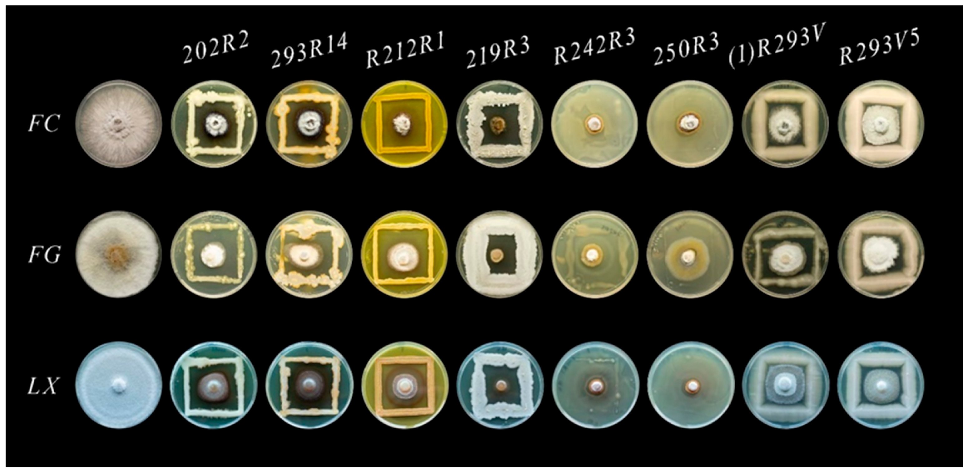
3.1. Morphology of Antagonistic Microorganisms

In total, 88 monocultures were isolated and tested against Monilinia spp. in vitro. Eight tested microorganisms were selected as strong antagonists against M. fructicola, M. fructigena, and M. laxa, which were used further in our research. Six bacterial isolates were obtained from the soil of the plum orchard. Two microbial strains were isolated from plum fruits and appeared to be yeast-like fungi.
The bacterial isolates with strong antifungal properties were separated into four distinct morphotypes, as shown in Table 1. The isolates could be divided based on several traits: color, form, margin, opacity, appearance, elevation, consistency, and smoothness. Two creamy circular isolates, 202R2 and 293R14, were grouped into the A morphotype. Orange circular isolate R212R1 and white circular isolate 219R3 were placed separately into two different morphotypes, B and C, respectively. Two creamy irregular isolates, R242R3 and 250R3, were grouped into the D morphotype. Most colonies were circular, but R242R3 and 250R3 were irregular. The margin of isolates was entire or undulate. Both isolates 202R2 and 293R14 were opaque. The R212R1 and 219R3 colonies were opaque as well. Both R242R3 and 250R3 isolates were translucent. Morphotypes B and D had a glistening appearance, while morphotypes A and C did not. The investigated isolates were characterized by flat or convex elevation and had butyrous or mucoid consistency. Isolates R212R, R242R3, and 250R3 were smooth, but 202R2, 293R14, and 219R3 had a coarse surface. All isolates were gram-positive, except isolate R212R1, which appeared to be gram-negative.
Two yeast-like fungi isolates (1)R293V2 and R293V5, were separated into distinct morphotype E. They both were creamy, circular, opaque, flat, butyrous, and smooth. The margin was entire, and the appearance was glistening.

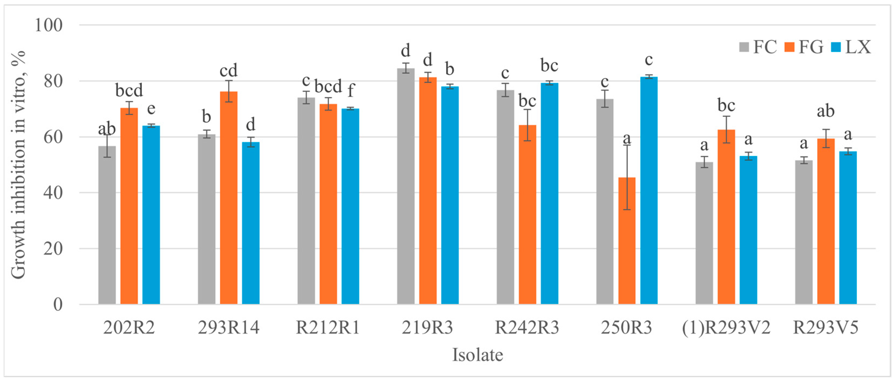
3.2. Antagonistic Activity of Microorganisms Against Monilinia spp. In Vitro

The Monilinia spp. growth inhibition level varied from 45.5% to 84.6% for eight antagonists (Figure 1 and Figure 2). The highest inhibition percentage rates—81.3% and 84.6% against M. fructigena and M. fructicola, respectively—were obtained with bacteria 219R3. The highest inhibition rate of 81.5% against M. laxa was reached using bacteria 250R3. Both isolates (1)R293V2 and R293V5 with a morphological similarity to yeast-like fungi showed similar antagonism levels for each pathogen. Strong antifungal activity against M. fructigena was observed using 202R2 (70.3%) and 293R14 (76.3%) bacteria. The strongest effect on the M. fructicola—74.1% and 84.6%—was observed using R212R1 and 219R3 bacteria, respectively. The strongest antagonistic activity against M. laxa—79.3% and 81.6%—was reached with R242R3 and 250R3 bacteria, respectively.

3.3. Genetic Characteristics of the Isolated Microbial Strains

All morphotypes were identified as bacteria, and the other two isolates proved to be yeast-like fungi upon WGS analysis (Table 2). The morphotype A (202R2 and 293R14) had an identity with the Bacillus pumilus reference genome of 95.65% and 95.64%, respectively. The morphotype C (219R3) had 98.87% identity with the B. velezensis reference genome. The morphotype D (R242R3, 250R3) was identified as Lysinibacillus agricola with identities of 90.65% and 90.71% to the reference genome. Gram-negative morphotype B (R212R1), forming orange colonies, showed the identity of 95.11% with the Pseudomonas chlororaphis subsp. chlororaphis. Two yeast-like fungi strains, (1)R293V2 and R293V5, were identified as Aureobasidium pullulans fungi, with 97% identity to the reference genome.
B. pumilus strains 202R2, 293R14, and B. velezensis 219R3 genome sizes ranged from ~3.7 to ~3.9 Mb (Table 1). The P. chlororaphis isolate had a genome size of ~6.9 Mb, and L. agricola isolates’ genome sizes were ~4.9 and ~4.9 Mb, respectively. Eucaryotes A. pullulans (isolates (1)R293V2 and R293V5) genome sizes were ~30,364 and 28,396 Mb, respectively. The overall GC content, protein-coding genes’ number, and other genome components (RNAs, repeat regions, and CRISPR) are shown in Table 2. Only B. pumilus 202R2, B. pumilus 293R14, and L. agricola R242R3 were predicted to have 3, 1, and 2 CRISPRs, respectively. The number of coding sequences (CDS) assigned in the NR, Gene Ontology (GO), and KEGG databases of the isolates are shown in Table 2. The CDS differences between isolates of the same species were determined.

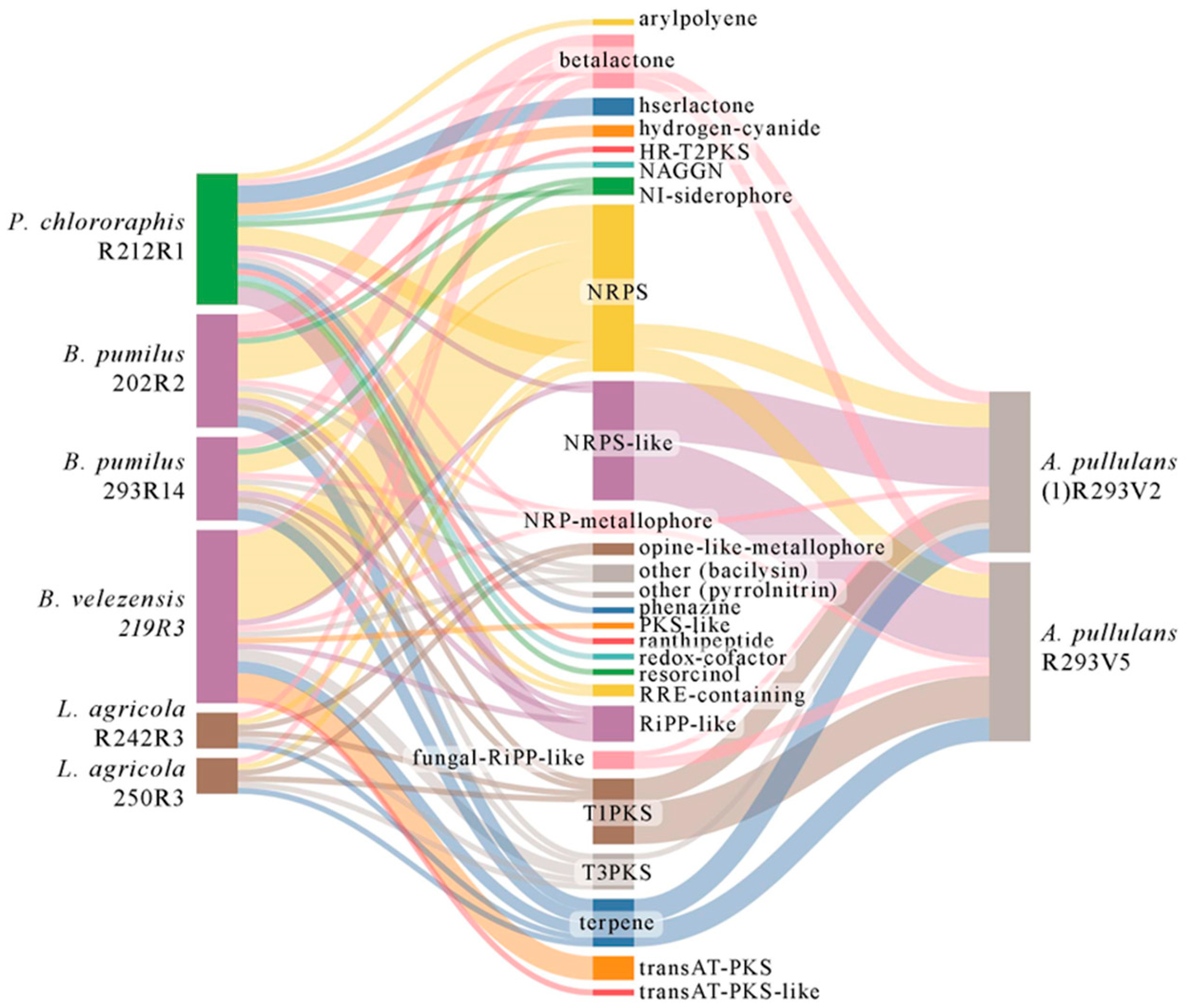
3.4. Genetic Background for Synthesizing Secondary Metabolites

The types of secondary metabolites of each isolate are presented in a Sankey diagram (Figure 3). All analyzed microorganisms were characterized by the synthesis of nonribosomal peptides (NRPSs) and betalactone. NPRS-like enzymes were mostly abundant in fungi A. pullulans. They were also detected in bacteria P. chlororaphis and B. velezensis. A large part (in total 18) of the secondary metabolite types were specific only to bacteria, and one type, the fungal-ribosomally synthesized and post-translationally modified peptides (RiPP)-like gene cluster, was inherent only for fungi. The characteristics of secondary metabolites and comparative analysis with NCBI of the gene clusters are shown in Tables S1–S8. B. velezensis 219R3 was predicted to have the highest number of gene clusters responsible for secondary metabolites’ synthesis among selected bacterial isolates. Fungal isolates were identified with an even higher number of gene clusters, but their diversity was greater in bacteria. A. pullulans R293V5 was determined to have 26, and A. pullulans (1)R293V2 to have 24. B. velezensis 219R3 was identified with 21 gene clusters involved in synthesizing secondary metabolites, while P. chlororaphis R212R1 had 18, B. pumilus 202R2 had 14, and B. pumilus 293R14 had 11. L. agricola R242R3 and 250R3 were predicted to have five gene clusters involved in synthesizing secondary metabolites, but only this species was characterized by non-ribosomal peptides (NRP)-metallophore synthesis. Both B. pumilus isolates and P. chlororaphis R212R1 had NI-siderophore clusters, an important factor that helps the plant in nitrogen metabolism.
There were few unique gene clusters specific for isolate B. pumilus 202R2 encoded HR-T2PKS; B. velezensis 219R3—PKS-like, transAT-PKS and transAT-PKS-like; P. chlororaphis R212R1—arylpolyene, hserlactone, hydrogen-cyanide, NAGGN, phenazine, ranthipeptide, redox-cofactor, and resorcinol. After comparison with known secondary metabolite gene clusters, it was predicted that all Bacillus spp. isolates might be able to produce bacillibactin and bacilysin. Additionally, B. pumilus 293R14 was predicted to synthesize lichenysin and B. velezensis 219R3 to synthesize macrolactin H, bacillaene, fengycin, and difficidin. P. chlororaphis R212R1 was predicted to synthesize pyrrolnitrin, hydrogen cyanide, and pyocyanine. Both A. pullulans isolates were predicted to produce choline and UNII-YC2Q1O94PT. Additionally, it was estimated that A. pullulans R293V5 should synthesize chaetoglobosin.

3.5. Genes Linked with Plant Growth Promotion (PGP) and Antagonism

Genes related to PGP activity and antagonism in the isolated microbial genomes were screened (Table 3). Phosphate transporters encoded genes involved in phosphate metabolism and tryptophan biosynthesis genes (trp) related to IAA synthesis were found in all isolates. Nitrogen metabolism genes were identified for all eight isolates, including gudB and rocG, which catalyze the reduction of L-glutamate to ammonia; nasD and nasE—the reduction of nitrite to ammonia; gltD—glutamate synthase (NADPH) small chain. The nitrogen-fixation-related genes nifS and nifU were annotated in Bacillus spp. genomes. Additionally, salA and sufU were identified in B. velezensis 219R3. Only nifU was found in both L. agricola isolates. Siderophore production genes were not found in P. chlororaphis, however, in other species they were identified. Hydrolase genes were annotated only for Bacillus spp. and fungi A. pullulans genomes. Chitinase activity was predicted only for isolates B. velezensis 219R3 and A. pullulans R293V5. All eight isolated microorganisms were identified as having biofilm synthesis genes as well as some key genes for the synthesis of volatile substances. Homologs of the ilv gene for 2,3-butanediol synthesis, or genes metH and ispE for methanethiol and isoprene synthesis involved in the biocontrol mechanism of antagonistic isolates, were characteristic of all antagonistic microorganisms in this study.

3.6. Pathogenicity and Virulence Annotation of Isolated Bacteria

The bacteria pathogenicity was analyzed using PathogenFinder. Both B. pumilus isolates matched 16 non-pathogenic families, whereas B. velezensis matched 101, P. chlororaphis matched 84, L. agricola R242R3 matched 26, and L. agricola 250R3 matched 28. All Bacillus and Lysinibacillus spp. isolates did not have any pathogenic families, while P. chlororaphis had two of them. All six bacterial isolates were predicted as non-pathogenic microorganisms.
The genome sequences of isolated bacterial isolates were analyzed in the virulence factor database (VFDB), and 30–32 virulence factor genes were identified for B. pumilus isolates, 39 for B. velezensis, 211 for P. chlororaphis, and 29 and 32 for L. agricola R242R3 and 250R3 isolates, respectively (Table 4 and Tables S9–S11). These genes were mainly involved in bacterial adhesion, antiphagocytosis, host immune system evasion, iron uptake, regulation, secretion system, and bacterial toxins production (Table 4).

4. Discussion

Each class of chemical fungicides against Monilinia spp. has a specific target [37,38,39,40], and pathogen resistance develops if used repeatedly [41,42]. Thus, fungicide rotation and combination strategies are essential in treating fungal diseases. A promising agrochemical protection strategy is microbial products and the search for new components for them [43]. The genetic diversity of microorganisms is infinite due to their high adaptability to the environment and the natural interaction between microorganisms. The multicomponent impact of antagonists prevents the pathogen from adapting [44]. This research showed that 8 out of 88 isolated microorganisms had strong antagonistic properties to Monilinia spp. pathogens (grow inhibition 45% to 85%) in the microbiota surrounding the plums in the rhizosphere or on the fruit surface (Figure 1 and Figure 2).
According to WGS analysis, three microorganisms were identified as members of the Bacillus genus (B. pumilus 202R2 and 293R14, B. velezensis 219R3) (Table 2). Bacillus spp. is widely distributed worldwide and applied in various fields. The isolates R242R3 and 250R3 showed genetic identity with L. agricola (access. No. NZ_CP14476 in NCBI)—90.65 and 90.71%, respectively. A cutoff of 97–99% similarity is often used to distinguish species within bacterial taxonomy, but this varies based on the specific group of organisms being studied. This is a novel species of the Lysinibacillus genus, first isolated from farmland soil, identified and described in China in 2021 [45]. There are limitations with the reference genome for this species, which means that using it as the standard we cannot yet determine genetic variations. Thus, the one genotype (NZ_CP067341) as a reference genome might not represent the full genetic diversity of a species. We confirmed that both isolates identified in this study have 100% identity among 16s rRNA genes and assigned them to the L. agricola species. Lysinibacillus shares similarities with Bacillus species, but has a unique peptidoglycan composition and fatty acid profile [46]. Morphological characteristics of the isolated R242R3 and 250R3 distinguished them from the others as irregular in form and translucent in opacity (Table 1). Bigger genome sizes with less GC content and fewer gene percentages in the genomes have been identified among these genera in general genome features (Table 2). It is established that L. agricola isolates contained 180 and 156 ncRNA regions (R242R3 and 250R3, respectively), while the Bacillus genomes contained 16, 14, and 20 ncRNA regions. The ncRNA regions determine a higher complexity of the regulation system and thus better adaptation to stress, increased virulence, and more efficient metabolism in bacteria. Bacillus spp. and Lysinibacillus spp. have broader applications in agriculture, such as biofertilizers with PGP properties, biopesticides, biofungicides, and bioremediation [46]. Bacillus spp. and Lysinibacillus spp. are gram-positive and form endospores that can survive in a wide range of environmental conditions and can be formulated into stable dry powders as a product [47]. The gram-negative isolate R212R1 (Table 1) was identified as bacteria P. chlororaphis subsp. chlororaphis with the homology of 95.11%. P. chlororaphis is a plant-associated bacterium with a fast growth rate, wide usage, and various applications, including biocontrol activity, plant growth promotion, and secondary metabolite production [48]. Two fungal isolates—(1)R293V2 and R293V5—were yeast-like fungi A. pullulans. This is a black yeast-like fungus of a ubiquitous genus that can survive in diverse environments [49]. A. pullulans are found in most phyllosphere and carposphere habitats and possess high antagonistic activity [50].
All isolated antagonists—B. pumilus, B. velezensis, L. agricola, P. chlororaphis, and A. pullulans—are known as species with antifungal capacity and positive effects on plant growth in agriculture [21,46,51,52,53]. There are several reported strains of B. pumilus that showed an antifungal effect on the mycelial growth of M. fructigena and M. fructicola, by up to ~60% in dual culture assays. M. laxa in the same study was inhibited only by up to 30% [21]. In this study, B. pumilus isolates showed a similar inhibition effect against M. fructicola, but it was stronger against M. frutigena and M. laxa. There were no reports of B. velezensis antagonism against brown rot pathogens, but it is indicated as a broad antagonistic spectrum against various phytopathogenic fungi [51]. In our research, B. velezensis 219R3 reached around an 80% antagonism level against all tested Monilinia species. Lysinibacillus species are known for antagonistic activities against numerous plant pathogens [46,54]. L. agricola isolates showed a similar pattern of antagonism against all Monilinia spp. pathogens in this study. P. chlororaphis is well known for inhibiting various phytopathogens (Fusarium, Colletotrichum, Phytophthora, Pythium, Sclerotinia, Magnaporthe oryzae, Rhizoctonia) [52]. P. chlororaphis R212R1 inhibition results were ~70% against different Monilinia spp. A. pullulans washed cells inhibited all three Monilinia species on nectarine fruits: M. laxa was suppressed completely, and M. fructicola and M. fructigena growth was reduced by 70–90% [53]. This does not fully align with the results obtained in this research, as A. pullulans (1)R293V2 and R293V5 inhibited M. fructigena’s growth by ~60%, and M. fructicola’s and M. laxa’s growth was inhibited by ~50%.
The genetic analysis revealed that identified microorganisms have great genetic potential in synthesizing antifungal compounds like lipopeptides and volatile organic compounds (Figure 3, Tables S1–S8). It was also characterized and confirmed by predicted genes related to PGP activity and antagonism (Table 3). The unique genetic setup of some species in Bacillus spp. enables them to produce bacilysin more efficiently than others [54,55]. Bacilysin synthesis is mainly determined by the bac operon and its role in converting prephenate to bacilysin. B. velezensis 219R3 was predicted with a high variety of secondary metabolites coding regions. It had 21 clusters, including NRPSs, transAT-PKS, NRP-metallophore, RiPP-like, PKS-like, and terpene. Some of them were identified as locillomycin, surfactin, fengycin, macrolactin H, bacillibactin, bacillaene, bacilysin, butirosin, difficidin, and pliplastin coding metabolites. Studies have reported inhibition of fungal growth using some of these substances through mechanisms such as the disruption of pathogen morphology and physiology [56]. P. chlororaphis R212R1 was predicted to synthesize phenazine and resorcinol. Phenazines are pigmented antimicrobial compounds [57]. Both molecules inhibit the growth of various phytopathogens [52]. Additionally, alkyl resorcinol (dar gene cluster) produced by P. chlororaphis isolate PCL 1606 promotes root colonization by this bacterium and functions antagonistically. They are part of the allelochemical plant-to-plant signaling machinery documented for rice and sorghum [58]. A. pullulans (1)R293V2 was predicted to have UNII-YC2Q1O94PT, which encodes melanin [59]. Another isolate, A. pullulans R293V5, was predicted with the synthesis of chaetoglobosin A. It was synthesized by Chaetomium globosum and has potential antifungal activity [60]. Plant growth-promoting traits might have indirect modes of action against Monilinia spp. pathogenesis. All eight isolates were predicted with phosphate, nitrogen metabolism, and IAA production (Table 3). PGP traits are common in the B. pumilus, B. velezensis, L. agricola, P. chlororaphis, and A. pullulans species [46,52,54,61,62,63] and improve plant metabolism, absorption, and adaptation. All identified isolates had coding genes for biofilm and VOCs (2,3-butanediol, methanethiol, isoprene). In the previous study, the VOCs released by B. amyloliquefaciens FZB42 and the formation of biofilms by B. velezensis in the plant rhizosphere promoted growth and protected from infectious microbes through systemic resistance [47,64]. Only B. velezensis isolate and A. pullulans R293V5 were predicted to synthesize chitinase. Additionally, B. velezensis and A. pullulans R293V5 isolates were predicted with hydrolase activity. In another study, A. pullulans antagonistic yeast-like fungi showed enzymatic activity (chitinases, xylanase, urease) [65].
Virulence factors in biofungicides are essential against fungal pathogens. The predicted virulence factors according to VFDB were disclosed in our research (Table 4 and Tables S9–S11). The highest number of virulence factors (211) was found in the P. chlororaphis R212R1 genome. The majority (73) were related to adherence, which allows P. chlororaphis R212R1 to attach to plant surfaces (roots, leaves, or fungal pathogens). Immune evasion strategies present in three identified Bacillus isolates (202R2, 293R14, and R212R1) help bacteria avoid detection, resist immune attacks, and persist within the host. Virulence factors work by breaking down fungal structures, competing for resources, and boosting plant immunity [66]. The eight isolates identified in this study show promise as biocontrol strains for managing Monilinia-related infections. B. velezensis and P. chlororaphis isolates were predicted to have the highest number of secondary metabolites, which might be the reason for the most effective inhibition observed of selected pathogens. For precise application rates and conditions, further studies focusing on Monilinia spp. specifically in orchard trials are needed.

Supplementary Materials

The following supporting information can be downloaded at: https://www.mdpi.com/article/10.3390/microorganisms13040818/s1. Tables S1–S8: Predicted gene clusters involved in the synthesis of secondary metabolites. Tables S9–S11: Predicted virulence factors in isolates.

Author Contributions

Conceptualization, I.M. and A.K.; methodology, I.M., A.K. and B.F.; software, M.K., I.M. and A.K.; validation, A.K. and I.M.; investigation, I.M., A.K., G.P. and S.R.; resources, A.K., I.M., M.K. and B.F.; data curation, I.M. and A.K.; writing—original draft preparation, A.K.; writing—review and editing, I.M., M.K. and B.F.; visualization, A.K. and I.M.; supervision, I.M. and B.F. All authors have read and agreed to the published version of the manuscript.

Funding

This study was supported by the long-term research program ‘Advances in genetics, biotechnology and breeding for improved plant diversity and technological innovations’ implemented by Lithuanian Research Centre for Agriculture and Forestry.

Institutional Review Board Statement

Not applicable.

Informed Consent Statement

Not applicable.

Data Availability Statement

Data are contained within the article or Supplementary Materials.

Conflicts of Interest

The authors declare no conflicts of interest.

References

  1. Pageni, B.B.; Lupwayi, N.Z.; Akter, Z.; Larney, F.J.; Kawchuk, L.M.; Gan, Y. Plant growth-promoting and phytopathogen-antagonistic properties of bacterial endophytes from potato (Solanum tuberosum L.) cropping systems. Can. J. Plant Sci. 2014, 94, 835–844. [Google Scholar] [CrossRef]
  2. Gouda, S.; Das, G.; Sen, S.K.; Shin, H.S.; Patra, J.K. Endophytes: A treasure house of bioactive compounds of medicinal importance. Front. Microbiol. 2016, 7, 1538. [Google Scholar] [CrossRef]
  3. Ghadamgahi, F.; Tarighi, S.; Taheri, P.; Saripella, G.V.; Anzalone, A.; Kalyandurg, P.B.; Catara, V.; Ortiz, R.; Vetukuri, R.R. Plant growth-promoting activity of Pseudomonas aeruginosa FG106 and its ability to act as a biocontrol agent against potato, tomato and taro pathogens. Biology 2022, 11, 140. [Google Scholar] [CrossRef] [PubMed]
  4. Santoyo, G.; Moreno-Hagelsieb, G.; Orozco-Mosqueda Mdel, C.; Glick, B.R. Plant growth-promoting bacterial endophytes. Microbiol. Res. 2016, 183, 92–99. [Google Scholar] [CrossRef]
  5. Li, N.; Wang, S.; Ma, L.; Liu, Y.; Xiu, Y.; Li, X. Phosphate-solubilizing capacity of two bacteria strains and its effect on maize growth and the phosphorus fractions in red soil. J. Plant Nutr. Fertil. 2021, 27, 275–283. [Google Scholar] [CrossRef]
  6. Wang, Y.; Sun, Z.; Zhao, Q.; Yang, X.; Li, Y.; Zhou, H.; Zhao, M.; Zheng, H. Whole-Genome Analysis revealed the growth-promoting and biological control mechanism of the endophytic bacterial strain Bacillus halotolerans Q2H2, with strong antagonistic activity in potato plants. Front. Microbiol. 2024, 14, 1287921. [Google Scholar] [CrossRef]
  7. Garzón-Posse, F.; Quevedo-Acosta, Y.; Mahecha-Mahecha, C.; Acosta-Guzmán, P. Recent progress in the synthesis of naturally occurring siderophores. Eur. J. Org. Chem. 2019, 2019, 7747–7769. [Google Scholar] [CrossRef]
  8. Waksman, S.A.; Woodruff, B.H. The soil as a source of microorganisms antagonistic to disease-producing bacteria. J. Bacteriol. 1940, 40, 581–600. [Google Scholar]
  9. Berendsen, R.L.; Vismans, G.; Yu, K.; Song, Y.; de Jonge, R.; Burgman, W.P. Disease-induced assemblage of a plant-beneficial bacterial consortium. ISME J. 2018, 12, 1496–1507. [Google Scholar] [CrossRef]
  10. Babalola, O.O. Beneficial bacteria of agricultural importance. Biotechnol. Lett. 2010, 32, 1559–1570. [Google Scholar] [CrossRef]
  11. Beneduzi, A.; Ambrosini, A.; Passaglia, L. Plant growth-promoting rhizobacteria (PGPR): Their potential as antagonists and biocontrol agents. Genet. Mol. Biol. 2012, 35, 1044–1051. [Google Scholar] [CrossRef] [PubMed]
  12. Arrebola, E.; Tienda, S.; Vida, C.; de Vicente, A.; Cazorla, F.M. Fitness features involved in the biocontrol interaction of Pseudomonas chlororaphis with host plants: The case study of PcPCL1606. Front. Microbiol. 2019, 10, 719. [Google Scholar] [CrossRef] [PubMed]
  13. Boro, M.; Sannyasi, S.; Chettri, D.; Verma, A.K. Microorganisms in biological control strategies to manage microbial plant pathogens: A review. Arch. Microbiol. 2022, 204, 666. [Google Scholar] [CrossRef] [PubMed]
  14. Arrebola, E.; Jacobs, R.; Korsten, L. Iturin A is the principal inhibitor in the biocontrol activity of Bacillus amyloliquefaciens PPCB004 against postharvest fungal pathogens. J. Appl. Microbiol. 2010, 108, 386–395. [Google Scholar] [CrossRef]
  15. Zeriouh, H.; Romero, D.; Garcia-Gutierrez, L.; Cazorla, F.M.; de Vicente, A.; Perez-Garcia, A. The Iturin-like lipopeptides are essential components in the biological control arsenal of Bacillus subtilis against bacterial diseases of cucurbits. Mol. Plant-Microbe Interact. 2011, 24, 1540–1552. [Google Scholar] [CrossRef]
  16. Bonaterra, A.; Badosa, E.; Daranas, N.; Francés, J.; Roselló, G.; Montesinos, E. Bacteria as biological control agents of plant diseases. Microorganisms 2022, 10, 1759. [Google Scholar] [CrossRef]
  17. Zhou, J.; Xie, Y.; Liao, Y.; Li, X.; Li, Y.; Li, S.; Ma, X.; Lei, S.; Lin, F.; Jiang, W.; et al. Characterization of a Bacillus velezensis strain isolated from Bolbostemmatis rhizoma displaying strong antagonistic activities against a variety of rice pathogens. Front. Microbiol. 2022, 13, 983781. [Google Scholar] [CrossRef]
  18. Berrie, A.M.; Holb, I. Brown rot diseases. In Compendium of Apple and Pear Diseases and Pests, 2nd ed.; Sutton, T.B., Aldwinckle, H.S., Agnello, A.M., Walgenbach, J.F., Eds.; American Phytopathological Society: St Paul, MN, USA, 2014; pp. 43–45. [Google Scholar]
  19. Tsalgatidou, P.C.; Boutsika, A.; Papageorgiou, A.G.; Dalianis, A.; Michaliou, M.; Chatzidimopoulos, M.; Delis, C.; Tsitsigiannis, D.I.; Paplomatas, E.; Zambounis, A. Global transcriptome analysis of the peach (Prunus persica) in the interaction system of fruit–chitosan–Monilinia fructicola. Plants 2024, 13, 567. [Google Scholar] [CrossRef]
  20. Fenta, L.; Mekonnen, H. Microbial biofungicides as a substitute for chemical fungicides in the control of phytopathogens: Current perspectives and research directions. Scientifica 2024, 2024, 5322696. [Google Scholar] [CrossRef]
  21. Hrustic, J.; Medic, O.; Beric, T.; Mihajlovic, M.; Milijaševic-Marčic, S.; Stankovic, S.; Pešic, B. Suppression of Monilinia brown rot by Bacillus spp. strains. Agronomy 2023, 13, 2839. [Google Scholar] [CrossRef]
  22. Franco-Sierra, N.D.; Posada, L.F.; Santa-Maria, G.; Romero-Tabarez, M.; Villegas-Escobar, V.; Alvarez, J.C. Bacillus subtilis EA-CB0575 genome reveals clues for plant growth promotion and potential for sustainable agriculture. Funct. Integr. Genom. 2020, 20, 575–589. [Google Scholar] [CrossRef] [PubMed]
  23. Coulson, T.J.D.; Patten, C.L. Complete genome sequence of Enterobacter cloacae UW5, a Rhizobacterium capable of high levels of Indole-3-acetic acid production. Genome Announc. 2015, 3, e00843-15. [Google Scholar] [CrossRef] [PubMed]
  24. Asaf, S.; Khan, A.L.; Khan, M.A.; Al-Harrasi, A.; Lee, I.J. Complete genome sequencing and analysis of endophytic Sphingomonas sp. LK11 and its potential in plant growth. 3 Biotech 2018, 8, 389. [Google Scholar] [CrossRef] [PubMed]
  25. Iqbal, S.; Ullah, N.; Janjua, H.A. In vitro evaluation and genome mining of Bacillus subtilis strain RS10 reveals its biocontrol and plant growth-promoting potential. Agriculture 2021, 11, 1273. [Google Scholar] [CrossRef]
  26. Kolytaitė, A.; Vaitiekūnaitė, D.; Antanynienė, R.; Baniulis, D.; Frercks, B. Monilinia fructigena suppressing and plant growth promoting endophytic Pseudomonas spp. bacteria isolated from plum. Microorganisms 2022, 10, 2402. [Google Scholar] [CrossRef]
  27. Powers, E.M. Efficacy of the Ryu nonstaining KOH technique for rapidly determining Gram reactions of food-borne and waterborne bacteria and yeasts. Appl. Environ. Microbiol. 1995, 61, 3756–3758. [Google Scholar] [CrossRef]
  28. Wick, R.R.; Judd, L.M.; Gorrie, C.L.; Holt, K.E. Unicycler: Resolving bacterial genome assemblies from short and long sequencing reads. PLoS Comput. Biol. 2017, 13, e1005595. [Google Scholar] [CrossRef]
  29. Manni, M.; Berkeley, M.R.; Seppey, M.; Zdobnov, E.M. BUSCO: Assessing genomic data quality and beyond. Curr. Protoc. 2021, 1, e323. [Google Scholar] [CrossRef]
  30. NCBI Resource Coordinators. Database resources of the National Center for Biotechnology Information. Nucleic Acids Res. 2018, 46, D8–D13. [Google Scholar] [CrossRef]
  31. Kanehisa, M.; Goto, S. KEGG: Kyoto Encyclopedia of Genes and Genomes. Nucleic Acids Res. 2000, 1, 27–30. [Google Scholar] [CrossRef]
  32. Blin, K.; Shaw, S.; Kloosterman, A.M.; Charlop-Powers, Z.; van Weezel, G.P.; Medema, M.H.; Weber, T. antiSMASH 6.0: Improving cluster detection and comparison capabilities. Nucleic Acids Res. 2021, 48, W29–W35. [Google Scholar] [CrossRef] [PubMed]
  33. Alcock, B.P.; Huynh, W.; Chalil, R.; Smith, K.W.; Raphenya, A.R.; Wlodarski, M.A.; Edalatmand, A.; Petkau, A.; Syed, S.A.; Tsang, K.K.; et al. CARD 2023: Expanded curation, support for machine learning, and resistome prediction at the Comprehensive Antibiotic Resistance Database. Nucleic Acids Res. 2023, 51, D690–D699. [Google Scholar] [CrossRef] [PubMed]
  34. Cosentino, S.; Voldby, L.M.; Møller, A.F.; Lund, O. PathogenFinder—Distinguishing friend from foe using bacterial whole genome sequence data. PLoS ONE 2013, 8, e77302. [Google Scholar] [CrossRef]
  35. Liu, B.; Zheng, D.; Jin, Q.; Chen, L.; Yang, J. VFDB 2019: A comparative pathogenomic platform with an interactive web interface. Nucleic Acids Res. 2019, 47, D687–D692. [Google Scholar] [CrossRef]
  36. Tang, D.; Chen, M.; Huang, X.; Zhang, G.; Zeng, L.; Zhang, G.; Wu, S.; Wang, Y. SRplot: A free online platform for data visualization and graphing. PLoS ONE 2023, 18, e0294236. [Google Scholar] [CrossRef]
  37. Price, C.L.; Parker, J.E.; Warrilow, A.G.; Kelly, D.E.; Kelly, S.L. Azole fungicides—Understanding resistance mechanisms in agricultural fungal pathogens. Pest. Manag. Sci. 2015, 71, 1054–1058. [Google Scholar] [CrossRef]
  38. Selim, R.E.; Khalil, M.S. Strobilurins: New group of fungicides. J. Plant Sci. Phytopathol. 2021, 5, 63–64. [Google Scholar] [CrossRef]
  39. Chen, F.; Everhart, S.E.; Bryson, P.K.; Luo, C.; Song, X.; Liu, X.; Schnabel, G. Fungicide-induced transposon movement in Monilinia fructicola. Fungal Genet. Biol. 2015, 85, 38–44. [Google Scholar] [CrossRef]
  40. Martini, C.; Mari, M. Monilinia fructicola, Monilinia laxa (Monilinia Rot, Brown Rot). In Postharvest Decay; Bautista-Baños, S., Ed.; Academic Press: Cambridge, MA, USA, 2014; Chapter 7; pp. 233–265. ISBN 9780124115521. [Google Scholar] [CrossRef]
  41. Jayawardana, M.A.; Fernando, W.G.D. The mechanisms of developing fungicide resistance in Fusarium graminearum causing Fusarium head blight and fungicide resistance management. Pathogens 2024, 13, 1012. [Google Scholar] [CrossRef]
  42. Luo, C.-X.; Schnabel, G. Adaptation to fungicides in Monilinia fructicola isolates with different fungicide resistance phenotypes. Phytopathology 2008, 98, 230–238. [Google Scholar] [CrossRef]
  43. Van Lenteren, J.C.; Bolckmans, K.; Köhl, J. Biological control using invertebrates and microorganisms: Plenty of new opportunities. BioControl 2018, 63, 39–59. [Google Scholar] [CrossRef]
  44. Shi, A.; Fan, F.; Broach, J.R. Microbial adaptive evolution. J. Indust. Microbiol. Biotechnol. 2022, 49, kuab076. [Google Scholar] [CrossRef]
  45. Lu, J.R.; Liu, G.H. Lysinibacillus agricola sp. nov., isolated from soil. Arch. Microbiol. 2021, 203, 4173–4178. [Google Scholar] [CrossRef] [PubMed]
  46. Jamal, Q.M.S.; Ahmad, V. Lysinibacilli: A biological factories intended for bio-insecticidal, bio-control, and bioremediation activities. J. Fungi 2022, 8, 1288. [Google Scholar] [CrossRef]
  47. Rabbee, M.F.; Ali, M.S.; Choi, J.; Hwang, B.S.; Jeong, S.C.; Baek, K.H. Bacillus velezensis: A valuable member of bioactive molecules within plant microbiomes. Molecules 2019, 16, 1046. [Google Scholar] [CrossRef]
  48. Liu, J.; Wang, X.; Dai, G.; Zhang, Y.; Bian, X. Microbial chassis engineering drives heterologous production of complex secondary metabolites. Biotech. Adv. 2022, 59, 107966. [Google Scholar] [CrossRef]
  49. Samson, R.A.; Hoekstra, E.S.; Frisvad, J.C. Introduction to Food and Airborne Fungi, 7th ed.; Centralalbureau voor Schimmellcultures; Institute of the Royal Netherlands Academy of Arts and Sciences: Utrecht, The Netherlands, 2004; 389p. [Google Scholar]
  50. Bozoudi, D.; Tsaltas, D. The multiple and versatile roles of Aureobasidium pullulans in the vitivinicultural sector. Fermentation 2018, 4, 85. [Google Scholar] [CrossRef]
  51. Zhang, Y.; Li, Q.; Wang, C.; Liu, S. Transcriptomic and metabolomic analyses reveal the antifungal mechanism of the compound phenazine-1-carboxamide on Rhizoctonia solani AG1IA. Front. Plant Sci. 2022, 13, 1041733. [Google Scholar] [CrossRef]
  52. Bertani, I.; Zampieri, E.; Bez, C.; Volante, A.; Venturi, V.; Monaco, S. Isolation and characterization of Pseudomonas chlororaphis strain ST9; Rhizomicrobiota and in planta studies. Plants 2021, 10, 1466. [Google Scholar] [CrossRef]
  53. Mari, M.; Martini, C.; Guidarelli, M.; Neri, F. Postharvest biocontrol of Monilinia laxa, Monilinia fructicola and Monilinia fructigena on stone fruit by two Aureobasidium pullulans strains. Biol. Contr. 2012, 60, 132–140. [Google Scholar] [CrossRef]
  54. Naureen, Z.; Rehman, N.U.; Hussain, H.; Hussain, J.; Gilani, S.A.; Al Housni, S.K.; Mabood, F.; Khan, A.L.; Farooq, S.; Abbas, G.; et al. Exploring the potentials of Lysinibacillus sphaericus ZA9 for plant growth promotion and biocontrol activities against phytopathogenic fungi. Front. Microbiol. 2017, 8, 1477. [Google Scholar] [CrossRef]
  55. Islam, T.; Rabbee, M.F.; Choi, J.; Baek, K.H. Biosynthesis, molecular regulation, and application of bacilysin produced by Bacillus species. Metabolites 2022, 27, 397. [Google Scholar] [CrossRef] [PubMed]
  56. Ye, Q.; Zhong, Z.; Chao, S.; Liu, L.; Chen, M.; Feng, X.; Wu, H. Antifungal effect of Bacillus velezensis ZN-S10 against plant pathogen Colletotrichum changpingense and its inhibition mechanism. Int. J. Mol. Sci. 2023, 24, 16694. [Google Scholar] [CrossRef] [PubMed]
  57. Scales, B.S.; Dickson, R.P.; LiPuma, J.J.; Huffnagle, G.B. Microbiology, genomics, and clinical significance of the Pseudomonas fluorescens species complex, an unappreciated colonizer of humans. Clin. Microbiol. Rev. 2014, 27, 927–948. [Google Scholar] [CrossRef]
  58. Anderson, A.J.; Young Cheol, K. Biopesticides produced by plant-probiotic Pseudomonas chlororaphis isolates. Crop Prot. 2018, 105, 62–69, ISSN 0261-2194. [Google Scholar] [CrossRef]
  59. Jong, H.; Arshed, S.; Yu, J.; Cornish, D.; Vanneste, J.L.; Reglinski, T.; Elmer, P.A.G.; Templeton, M.D. A high-quality genome assembly for Aureobasidium pullulans CG163, the active ingredient in Aureo Gold, a bio-bactericide registered for control of bacterial canker of kiwifruit caused by Pseudomonas syringae pv. actinidiae Biovar 3. PhytoFrontiers 2024, 4, 486–490. [Google Scholar]
  60. Cheng, M.; Zhao, S.; Liu, H.; Liu, Y.; Lin, C.; Song, J.; Thawai, C.; Charoensettasilp, S.; Yang, Q. Functional analysis of a chaetoglobosin A biosynthetic regulator in Chaetomium globosum. Fungal Biol. 2021, 125, 201–210. [Google Scholar] [CrossRef]
  61. Pudova, D.S.; Toymentseva, A.A.; Gogoleva, N.E.; Shagimardanova, E.I.; Mardanova, A.M.; Sharipova, M.R. Comparative genome analysis of two Bacillus pumilus strains producing high level of extracellular hydrolases. Genes 2022, 13, 409. [Google Scholar] [CrossRef]
  62. Dobrzyński, J.; Jakubowska, Z.; Dybek, B. Potential of Bacillus pumilus to directly promote plant growth. Front. Microbiol. 2022, 13, 1069053. [Google Scholar] [CrossRef]
  63. Dobrzyński, J.; Jakubowska, Z.; Kulkova, I.; Kowalczyk, P.; Kramkowski, K. Biocontrol of fungal phytopathogens by Bacillus pumilus. Front. Microbiol. 2023, 14, 1194606. [Google Scholar] [CrossRef]
  64. Rabbee, M.F.; Hwang, B.S.; Baek, K.H. Bacillus velezensis: A beneficial biocontrol agent or facultative phytopathogen for sustainable agriculture. Agronomy 2023, 13, 840. [Google Scholar] [CrossRef]
  65. Zajc, J.; Cernosa, A.; Francesco, A.J.; Castoria, R.; Curtis, F.D.; Lima, G. Characterization of Aureobasidium pullulans isolates selected as biocontrol agents against fruit decay pathogens. Fungal Genom. Biol. 2020, 10, 163. [Google Scholar]
  66. Liu, F.; Hu, M.; Tan, X.X.; Li, C.; Wang, S. Pseudomonas chlororaphis L5 and Enterobacter asburiae L95 biocontrol Dickeya soft rot diseases by quenching virulence factor modulating quorum sensing signal. Microb. Biotechnol. 2023, 16, 2145–2160. [Google Scholar] [CrossRef] [PubMed]
Figure 1. Dual culture antagonism assay of M. fructicola (FC), M. fructigena (FG), and M. laxa (LX) and microbial isolates on PDA, as compared with the control plates (shown on the left).
Microorganisms 13 00818 g001
Figure 2. Inhibition of the growth of M. fructicola (FC), M. fructigena (FG), and M. laxa (LX) in vitro on PDA medium as percentages ± SE. The test was performed using Fisher’s LSD and Duncan’s multiple range test. The different letters (a, b, c, d, e and f) above the columns represent significance within the group of antagonists against the same fungal pathogen. Columns, representing average growth inhibition, with the same letters (ab, bcd, bc and cd) are not significantly different (p > 0.05). Only columns marked with the same color are comparable for significant differences. LSD 0.05 = 6.77 (for FC); LSD 0.05 = 15,226 (for FG); LSD 0.05 = 2811 (for LX).
Microorganisms 13 00818 g002
Figure 3. Sankey’s diagram of predicted gene clusters involved in the synthesis of secondary metabolites in the isolated antagonists to Monilinia spp., where the different colors represent different gene clusters.
Microorganisms 13 00818 g003
Table 1. Morphological characteristics of isolated antagonistic bacteria.
MorphotypeABCDE
Isolate202R2293R14R212R1219R3R242R3250R3(1)R293V2R293V5
Sourcesoilsoilsoilsoilsoilsoilfruitsfruits
Colorcreamyorangewhitecreamycreamy
Formcircularcircularcircularirregularcircular
Marginentireundulateundulateentire
Opacityopaquetranslucentopaque
Appearancenon-glisteningglisteningnon-glisteningglisteningglistening
Elevationflatconvexflatflat
Consistencybutyrousmucoidbutyrousbutyrous
Smoothnesscoarsesmoothcoarsesmoothsmooth
Gram+++++n.a.
Table 2. The general genome features of isolated microbial antagonists.
Characteristics of Microorganisms—Antagonists of Monilinia spp.
B. pumilusB. pumilusB. velezensisP. chlororaphis subsp. chlororaphisL. agricolaL. agricolaA. pullulansA. pullulans
Accession number in NCBI database and isolate strain in current research
SAMN
44109744
SAMN
44109745
SAMN
44109747
SAMN
44109746
SAMN
44109750
SAMN
44109751
SAMN
44109752
SAMN
44109754
202R2293R14219R3R212R1R242R3250R3(1)R293V2R293V5
Mapping statistics with the reference genome (accession number in NCBI and identity with the reference genome (%)
NC_009848NC_009725NZ_CP144767NZ_CP067341NZ_CP067341GCF_000721785
95.6595.6498.8795.1190.6590.7197.0697.00
Statistics of sequencing data and annotation
Genome size (bp)
3,733,4533,721,4083,976,1326,995,0874,901,2104,923,66430,364,19728,396,646
GC content (%)
41.5541.5246.3662.6936.7436.6650.2650.33
Gene length (bp)
3,312,8023,313,6723,520,9426,125,6213,922,5383,964,60321,770,73520,714,412
Genes % in genome
88.7389.0488.5587.5780.0380.5271.7072.95
Total number of genes
37643753383562334776476483375784
Transfer RNA/Ribosomal RNA (5S, 16S, 23S)/Transfer-messenger RNA
70/4/170/6/183/5/161/4/180/8/175/5/1158/59/N156/59—N
Noncoding RNA
161420621801567876
Repeat region/CRISPR
3/31/1N/NN/N3/2N/NN/NN/N
Genes assigned according to NR
34253654348956724346433568866256
Genes assigned according to GO
33213415335643874091421048563066
Genes assigned according to KEGG
30783002332141284005384537983088
Table 3. Predicted genes related to PGP activity and antagonism in the isolated microbial genomes.
PGP Activities
Description
B. pumilusB. velezensisP. chlororaphisL. agricolaA. pullulans
202R2293R14219R3R212R1R242R3R250R3(1)R293V2R293V5
Phosphate metabolismpstA, pstC, pstSpstA, pstB, pstC, pstS, iolU, mmsApstA, pstB, pstC, pstS, gdh, hxlB, glpX, pfk, hxlA, kdgK, rbsKpstA, pstB, pstS, iolG, iolE, iolD, mmsA, iolCpstA, pstB, pstC, pstS, mmsApstA, pstB, pstC, pstS, mmsApstB, iolG, pstA, pstC, pstS, iolUpstB, iolG, pstA, pstC, iolU
Nitrogen fixationnifS, nifUnifS, nifUnifS, salA, sufU-nifUnifU--
Nitrogen metabolismgltB, ltD, glnAgltB, ltD, glnAnasE, nasD, gudB, rocG, narG, narH, narI, glnA, cah, gltBcynS, cynT, arcC, gltB, gltD, glnA, nirB, nirD, norBcynTcynT, glnAncd2, nirB, mbtB, cah, cynT, nirD, glnA, gltB, gltDncd2, nirB, mbtB, cah, cynT, glnA, gltB
SiderophoremenF, entE, ntAfhuC, enFmenF-fhuC, menFfhuC, menFfhuC, fhuG, fhuB, fhuDfhuC, fhuG, huB, fhuD
IAA productiontrpA, rpB, trpC, rpD, trpEtrpA, rpB, trpC, rpD, trpEtrpA, trpB, trpC, trpD, trpE, trpFtrpA, trpB, trpC, trpD, trpE, trpFtrpA, trpB, trpEtrpA, trpB, trpEtrpA, trpB, trpD, trpEtrpA, trpE
HydrolaseeglSgmuD, ganBeglS---trpE, eglS, pqsH, pelFeglS, pqsH, pelF
Chitinase activity--ydhD----sleL, ydhD
BiofilmtrpE, flgM, fliA, csrAtrpE, flgM, fliA, csrAtasA, bslA, bslB, trpE, flgM, csrAcrp, cpdA, trpG, trpE, flgMtrpE, fliAtrpE, fliA, csrAtasA, bslA, bslBtasA, bslA, bslB
2,3-butanediolilvE, ilvA, ilvD, ilvC, ilvBilvE, ilvA, ilvD, ilvC, ilvBilvE, ilvA, ilvD, ilvC, ilvBilvE, ilvA, ilvD, ilvC, ilvBilvA, ilvD, ilvC, ilvBilvA, ilvD, ilvC, ilvBilvY, ilvA, ilvD, ilvC, ilvHilvK, ilvE, ilvY, ilvD, ilvC, ilvH, ilvB
Table 4. Predicted virulence factors in bacterial antagonists according to the virulence factor database (VFDB).
Predicted Virulence FactorsB. pumilusB. velezensisP. chlororaphisL. agricola
202R2293R14219R3R212R1R242R3250R3
Acid resistance001000
Adherence2227335
Antimicrobial activity000600
Antiphagocytosis1122421
Biofilm formation000400
Biosurfactant000100
Cell surface components002001
Copper uptake000100
Efflux pump000100
Enzyme110100
Glycosylation system000101
Immune evasion101118841
Intracellular survival000011
Invasion111100
Iron acquisition555000
Iron uptake2213177
Lipid and fatty acid metabolism110111
Magnesium uptake000100
Others000200
Peptidoglycan modification110001
Protease000200
Quorum sensing000500
Regulation222733
Secretion system1112822
Serum resistance000100
Serum and immune evasion000010
Stress adaptation001411
Surface protein anchoring221011
Toxin122836
Predicted VF genes3032392112932
Total genes384538424029621248115020
Disclaimer/Publisher’s Note: The statements, opinions and data contained in all publications are solely those of the individual author(s) and contributor(s) and not of MDPI and/or the editor(s). MDPI and/or the editor(s) disclaim responsibility for any injury to people or property resulting from any ideas, methods, instructions or products referred to in the content.

