Article

Identification of Released Bacterial Extracellular Vesicles Containing Lpp20 from Helicobacter pylori

by
Aoi Okamoto
1,
Tatsuki Shibuta
2,
Nanaka Morita
3,
Ryota Fujinuma
3,
Masaya Shiraishi
1,
Reimi Matsuda
1,
Mayu Okada
1,
Satoe Watanabe
3,
Tsukuru Umemura
2 and
Hiroaki Takeuchi
1,3,*
1
Medical Laboratory Science, Graduate School of Health and Welfare Sciences, International University of Health and Welfare, 4-3 Kozunomori, Narita 286-8686, Japan
2
Department of Medical Science Technology, School of Health Science at Fukuoka, International University of Health and Welfare, 137-1 Enokiz, Okawa 831-8501, Japan
3
Department of Medical Science Technology, School of Health Science at Narita, International University of Health and Welfare, 4-3 Kozunomori, Narita 286-8686, Japan
*
Author to whom correspondence should be addressed.
Microorganisms 2025, 13(4), 753; https://doi.org/10.3390/microorganisms13040753
Submission received: 21 January 2025 / Revised: 22 March 2025 / Accepted: 23 March 2025 / Published: 26 March 2025
(This article belongs to the Special Issue Editorial Board Members’ Collection Series: Bacterial Infection)

Abstract

:
Helicobacter pylori is a pathogenic bacterium that causes gastric and extragastric diseases. We have previously demonstrated that one of the mechanisms of H. pylori-associated chronic immune thrombocytopenia involves immune complexes of platelets, a H. pylori protein Lpp20 and an anti-Lpp20 antibody. However, it remains unclear how Lpp20 enters the body. We hypothesize that bacterial extracellular vesicles (bEVs) transport Lpp20. Thus, this study assessed Lpp20 in the bEVs released from seven clinical H. pylori isolates, using immunoprecipitation (IP), immunoblotting (IB), and surface plasmon resonance imaging (SPRi), with anti-GroEL (a marker of bEVs) and anti-Lpp20 antibodies. Lpp20 and bEVs were each detected in lysates of all seven strains. IP–IB experiments demonstrated that bEVs containing Lpp20 were produced by five of the strains (J99, SS1, HPK5, JSHR3, and JSHR31). SPRi using an anti-Lpp20 antibody demonstrated significantly higher reflectance from the strain HPK5 than from its lpp20-disrupted strains (p < 0.01), indicating localization of Lpp20 on the bEVs’ surface; Lpp20 may also be contained within bEVs. The bEVs containing Lpp20 were not detected from two clinical H. pylori strains (26695 and JSHR6) or from two lpp20-disrupted strains (26695ΔLpp20 and HPK5ΔLpp20). Differences in Lpp20 detection in bEVs are likely due to variations in bEV production resulting from strain diversity.

1. Introduction

Helicobacter pylori is a Gram-negative spiral-shaped pathogenic bacterium that was first reported in 1983 [1]. Many studies have shown that H. pylori infection is associated not only with upper gastrointestinal diseases [2] but also with various extragastric diseases, including autoimmune [3], hematological [4], cardiovascular [5], neurologic [6], skin [7], metabolic-related [8], hepatobiliary [9], and eye diseases [10]. Extragastric diseases caused by H. pylori infection can be improved by H. pylori eradication, but with geographical/regional differences [11]. In Japan, improvement in platelet counts has been reported in >50% of H. pylori-infected patients with chronic immune thrombocytopenia (cITP) [12,13,14]. We previously reported that one of the mechanisms underlying the development of H. pylori-associated cITP is that the H. pylori outer membrane protein Lpp20 binds to platelets, forms an immune complex with anti-Lpp20 antibodies, and induces platelet destruction and thrombocytopenia [15]. Furthermore, platelets bound to Lpp20 are aggregated and activated, suggesting a role of this protein in the development of other extragastric diseases such as thrombosis-mediated acute coronary syndrome (ACS) and chronic urticaria [15,16,17]. However, there is no case report to date of bacteremia caused by H. pylori. It is unclear how the bacterial component Lpp20 is transferred from the stomach into the bloodstream to contribute to the development of H. pylori-associated extragastric diseases.
Extracellular vesicles (EVs) released by various types of cells have been a rapidly expanding focus of research in recent years. However, the classification and nomenclature of EVs derived from bacteria lack consistency, highlighting the need for standardization. Thus, in accordance with guidelines issued by the International Society for Extracellular Vesicles (ISEV), EVs produced and released by bacteria are now collectively referred to as bacterial EVs (bEVs) [18]. bEVs released from bacteria in animals have demonstrated that bEVs transport bacterial components throughout the body. The bEVs are lipid bilayer vesicles with a diameter of 20–300 nm that contain DNA, RNA, proteins, and other components from the parent bacteria. They are mainly secreted during the growth stage [19,20,21,22]. The bEVs serve as carriers that protect their molecular cargoes from the external environment. Interestingly, bEVs have double-sided properties—an aggressive aspect, delivering virulence factors to host cells, and a defensive aspect, mediating interbacterial communication via quorum sensing [22,23,24,25,26,27,28], whose features function to protect the bacteria. H. pylori bEVs were first analyzed by double silver staining in 1997 [29], and it has been shown that H. pylori bEVs containing vacuolating cytotoxin (VacA) are delivered to gastric epithelial cells [21]. High-performance liquid chromatography–mass spectrometry (HPLC–MS/MS) analysis of two strains showed that H. pylori bEVs contain >400 molecules, including Lpp20, and that the contents of bEVs are strain-dependent [20,28]. However, there is no direct evidence regarding identification of bEVs containing Lpp20. Thus, this study was performed using seven clinical H. pylori strains isolated in geographically different regions; immunoblotting (IB), immunoprecipitation (IP), and surface plasmon resonance imaging (SPRi) analysis were used to assess Lpp20 in H. pylori bEVs.

2. Materials and Methods

2.1. Bacterial Strains and Growth Conditions

In this study, we used Escherichia coli (E. coli) and nine H. pylori strains (Table 1). The H. pylori strains included seven clinical isolates with diverse geographical origins (strains 26695/ATCC700392, J99/ATCC700824, SS1/ATCC43504, HPK5, JSHR3, JSHR6, and JSHR31), and two lpp20 gene-disrupted strains (26695ΔLpp20 and HPK5ΔLpp20), derived from strains 26695 and HPK5, respectively, generated in our laboratory. The whole genome sequences of three isolates from Japan (JSHR3, JSHR6, and JSHR31) have been registered [30], and they are recommended as standard strains for susceptibility testing in Japan.
H. pylori was cultured in Brucella broth (BB; Becton Dickinson, Franklin Lakes, NJ, USA) supplemented with 10% horse serum (HS) and 10 μg/mL vancomycin (Sawai Pharmaceutical, Osaka, Japan). This medium is referred to as BB-liquid medium (BBL); BBL supplemented with 1.4% agar was used to prepare agar plates (BB plates). H. pylori were cultured at 37 °C under 10% CO2 for 72 h according to a previous report [15]. The bacterial strains and plasmids (for lpp20 gene disruption) used in this study are shown in Table 1.

2.2. Generation of lpp20 Gene (HP1456) Disruption Strains

The lpp20-disrupted strains were prepared using constructed plasmids and homologous recombination, according to a previous report [35]. Briefly, the genomic DNAs of H. pylori strains 26695 and HPK5 were used for the amplification of the full-length lpp20 gene (528 bp) and its flanking open reading frames (HP1455 and HP1457) by PCR (Table 2). The resulting 1.3 kb PCR product was cloned into the pGEM-Teasy vector (Promega, Madison, WI, USA), yielding plasmids containing the lpp20 gene (plpp20E-1 and plpp20E-2, respectively). Next, these plasmids (“the plpp20E plasmids”) were digested with BamHI (New England Biolabs, Ipswich, MA, USA), a cleavage site for what is within the lpp20 gene. The 1.3 kb kanamycin resistance gene (kanr) obtained from BamHI-digested pUK4k was ligated into the BamHI-digested plpp20E plasmids using T4 DNA ligase (Promega, USA), followed by plasmid transformation into E. coli DH5α. As a result, the transformants selectively grew on Luria–Bertani broth (Nacalai Tesque, Kyoto, Japan)-agar plates containing 10 μg/mL kanamycin and yielded the constructed plasmids plpp20E-km-1 and plpp20E-km-2 in which the H. pylori lpp20 gene was disrupted by kanr. The plpp20E-km plasmids were used for homologous recombination to construct lpp20-disrupted H. pylori strains 26695ΔLpp20 and HPK5ΔLpp20, which selectively grew on BB plate containing 10 μg/mL kanamycin. The lpp20-disrupted strains and the direction of the Kmr insert in the lpp20 genes were confirmed by PCR with specific primers (Table 2). In addition, loss of Lpp20 was confirmed by IB with an anti-Lpp20 antibody generated in our laboratory.

2.3. Preparation of H. pylori Cell Lysates

H. pylori was cultured overnight in BBL at 37 °C under 10% CO2 with shaking at 100–115 rpm (Shaker SRR-2, AS ONE, Osaka, Japan). An aliquot of well-grown bacteria in BBL was subcultured in fresh BBL overnight. This process was repeated twice to more precisely match the growth conditions of individual bacteria. Finally, the bacterial culture was adjusted to OD600 nm = 0.3 in fresh BBL, incubated overnight, and collected. The culture was centrifuged at 7000× g for 30 min at 4 °C to separate the bacterial cells and the supernatant. The supernatant was processed for bEV preparation as described in Section 2.4. The bacterial cells were suspended in 400 μL of phosphate-buffered saline (PBS), homogenized on ice using an Omni tissue homogenizer (Omni, Dallas, TX, USA), and centrifuged at 12,000× g for 3 min at 4 °C. We used the supernatant as the H. pylori lysate. The protein concentration of lysates was measured using a Protein Assay Rapid Kit Wako II (Fujifilm Wako, Osaka, Japan). The lysate was stored at −20 °C until use.

2.4. Preparation of H. pylori bEVs

H. pylori bEVs were prepared according to previously described methods [36,37,38], with minor modifications. Briefly, the culture supernatant collected as described in Section 2.3 was further centrifuged at 12,000× g for 45 min at 4 °C to eliminate residual bacterial cells and debris. Subsequently, the cleared supernatant was passed through 0.45 and 0.22 μm filter membranes to remove other large molecules, and the cleared sample was prepared. The sample prepared was used in this study as H. pylori bEVs; bEVs were stored at −20 °C until use.

2.5. Visualization and Quantitative Analysis of bEVs’ Size and Concentration

We performed transmission electron microscopy (TEM) and nanoparticle tracking analysis (NTA) according to the ISEV’s recommended methods [18] to identify bEVs in the prepared samples. TEM analysis was performed by JEOL JEM-1400Flash electron microscopy at 100 kV at the Hanaichi UltraStructure Research Institute (Japan; https://www.kenbikyo.com/ (accessed on 17 February 2025)). Briefly, a droplet of prepared sample was placed on a carbon-film grid for 10 s. Staining solution, 2% uranyl acetate in water, was added to the grid and allowed to stain for 10 s, then the dried grid was subjected to electron microscopic observation with negative staining. NTA was performed using a NanoSight analyzer by Fujifilm Wako (Japan; https://labchem-wako.fujifilm.com/jp/custom_service/products/95163.html (accessed on 6 March 2025)). Briefly, bEV samples prepared from the strains HPK5 and HPK5ΔLpp20 were diluted in ultrapure water (Milli-Q, Merck, Darmstadt, Germany), and Mili-Q water was used as a blank. NTA was performed in five 60 s reads. The average of the five reads was calculated and plotted as particle size versus number of particles per milliliter. The recordings were processed by NTA 2.3 build 0033 software using a detection threshold of 8.

2.6. Immunoprecipitation

To analyze the properties of bEVs prepared from nine H. pylori strains, IP using anti-rabbit IgG-coated-magnetic beads (Thermo Fisher Scientific, Waltham, MA, USA) was conducted according to the manufacturer’s instructions. Briefly, each primary antibody (an anti-GroEL antibody by Sigma-Aldrich or the anti-Lpp20 antibody prepared in our laboratory [15]) was added to a bEV suspension and incubated at 4 °C for 1 h. Then, anti-rabbit IgG-coated magnetic beads washed with a buffer [0.1% bovine serum albumin (BSA)–PBS containing 2 mM ethylenediaminetetraacetic acid (EDTA)] were added into the mixtures and incubated at 4 °C overnight. After the reaction, the anti-rabbit IgG-coated-magnetic beads were washed with buffer (0.1% BSA–PBS containing 2 mM EDTA and 0.1% Tween 20), suspended in 1× sodium dodecyl sulfate (SDS) buffer and then heated at 90 °C for 5 min. The magnetic beads were removed using a magnet, and the resultant suspensions were stored at −20 °C until use. The bEV suspensions were subjected to IP–IB. IP without primary antibodies was performed as a negative control. In this study, we performed immunoprecipitation (IP) and immunoblotting (IB) to investigate whether Lpp20 is present on the surface and/or inside of bEVs. IP with an anti-Lpp20 antibody specifically captured Lpp20, and the precipitates were subjected to IB with an anti-GroEL antibody to detect bEVs, providing evidence that Lpp20 is localized on the surface of bEVs. Furthermore, we performed IP–IB with swapped antibodies (IP with an anti-GroEL antibody and IB with an anti-Lpp20 antibody) to confirm the presence of bEVs containing Lpp20.

2.7. Immunoblotting

The bacterial lysates and bEV suspensions treated without and bEV suspensions not subjected to IP, and bEV suspensions treated by IP were subjected to IB according to a previous report [15]. Briefly, samples mixed in an equal volume of 2× SDS buffer were heated at 90 °C for 5 min and then subjected to SDS–PAGE using an XV PANTERA GEL MP system (15% and 5–20%; DRC, Osaka, Japan). The proteins were transferred onto polyvinylidene fluoride membranes (Merck Millipore, Burlington, MA, USA) by using a MINICA-MP blotting module with Tris-glycine buffer (43 V, 30 min). The membrane was blocked with 0.5% skim milk and reacted with the primary antibody and then the secondary antibody. Finally, the membrane was treated with a Pierce ECL Plus Western Blotting Substrate (Thermo Fisher Scientific, USA), and bands were visualized by using an LAS-4000mini Luminescent Image Analyzer (Fujifilm Life Science, Tokyo, Japan). We used two primary antibodies: an anti-GroEL antibody for detection of BEVs [39,40,41] and an anti-Lpp20 antibody for detection of H. pylori Lpp20 [15]. As the secondary antibody, we used horseradish peroxidase-labeled goat anti-rabbit IgG (Abcam, Cambridge, UK).

2.8. Surface Plasmon Resonance Imaging (SPRi) Analysis

To evaluate the binding interactions of bEVs, SPRi analysis was performed using the OpenPleX system (HORIBA, Kyoto, Japan). First, 1 mL of a miRCURY Exosome Cell/Urine/CSF Kit (Qiagen, Venlo, The Netherlands) was added to 4 mL of the bEV suspensions of four samples (from strains HPK5, HPK5ΔLpp20, 26695, and 26695ΔLpp20), followed by vortexing and incubation at 4 °C overnight. The samples were centrifuged at 3000× g for 30 min at 25 °C to collect the bEVs as a pellet. The supernatant was discarded, and the pellet was washed twice with PBS with centrifugation at 3000× g for 5 min at 25 °C. After removing the supernatant, the EV pellet was resuspended in 0.1% casein in Dulbecco’s phosphate-buffered saline without calcium and magnesium [D-PBS (−); Fujifilm Wako, Japan], then diluted 50–200-fold and used for SPRi analysis. The biochip had a gold film activated through esterification, and when antibodies (anti-GroEL and anti-Lpp20 antibodies) were dropped onto the gold surface, they were immobilized on the surface through an amine coupling reaction. An antibody-bound biochip was incubated overnight with 1% casein–PBS solution for blocking. The prepared bEV samples were introduced into the SPRi system at a flow rate of 25 μL/min at 25 °C for 8 min. After each measurement, the sensor surface was regenerated with 10 mM glycine–HCl (pH 2.5) to remove bound bEVs. Data analysis was performed using Scrubber software version 2.0g (BioLogic Software, Canberra, Australia). A blank spot served as a negative control, and the signal intensities for analysis were calculated by subtracting the average signal from the blank spot. SPRi analysis for detection of bacterial-derived bEVs enhances the potential for future clinical applications.

3. Results

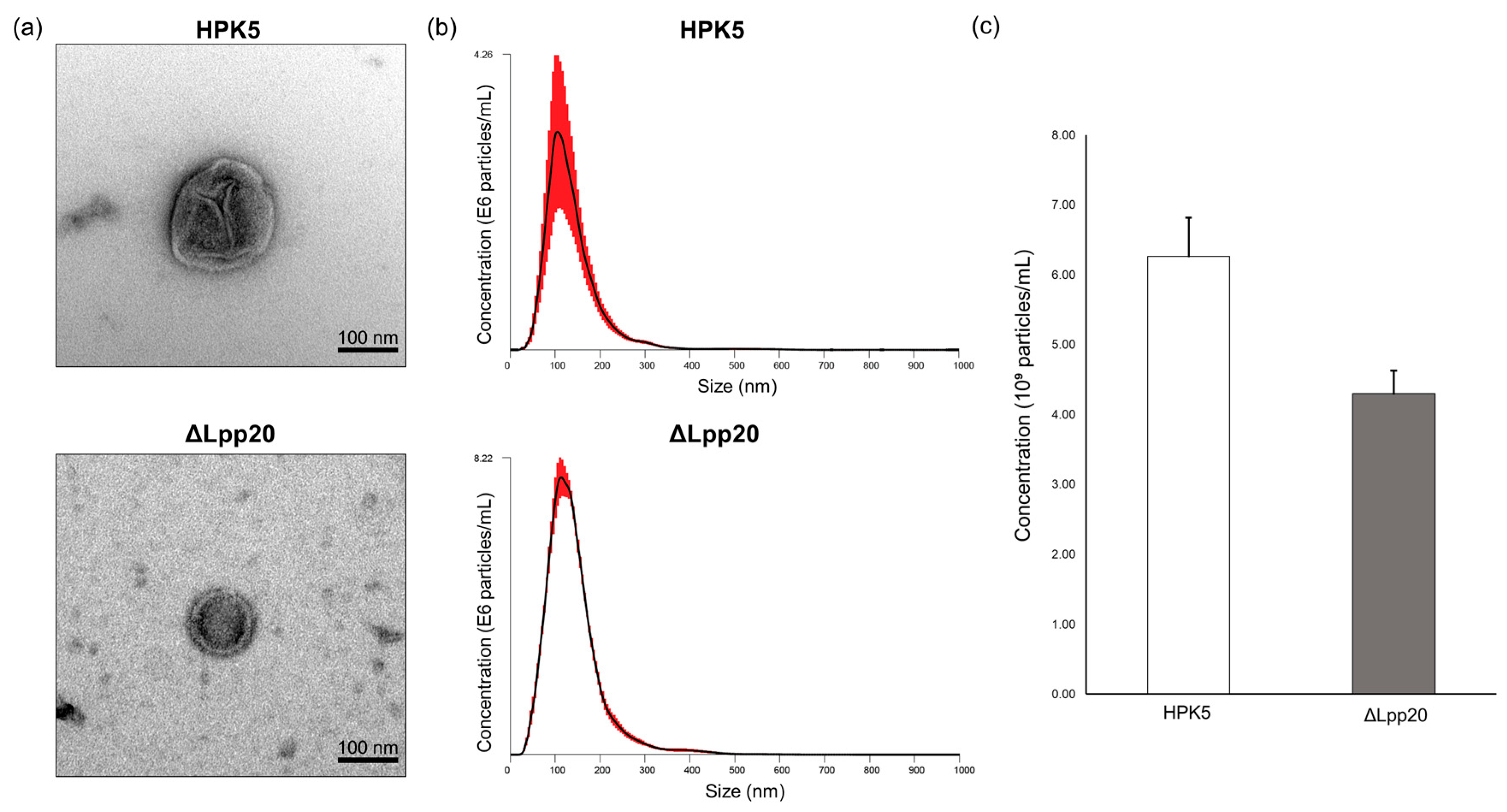
3.1. Identification and Characterization of bEVs of the Samples Used in This Study

We observed the ultrastructural morphology of H. pylori bEVs by TEM (Figure 1a). NTA revealed that the mean particle diameter of the bEVs from strains HPK5 and HPK5ΔLpp20 was 142.47 ± 8.91 nm and 139.34 ± 2.11 nm, respectively. The bEV concentration was (6.26 ± 1.78) × 109 particles/mL for the strain HPK5 and (4.30 ± 0.19) × 109 particles/mL for the strain HPK5ΔLpp20 (Figure 1b,c). An average of 2–3 bEVs per field (×10,000) was observed in the samples. These findings confirmed the presence of bEVs in the samples prepared for use in this study. Furthermore, no significant differences in particle size or concentration were observed between the strain HPK5 and its lpp20-disrupted derivative strain, HPK5ΔLpp20.

3.2. Analysis of Lpp20 in Cell Lysates and bEVs from Seven Clinical H. pylori Strains and Two lpp20-Disrupted Strains
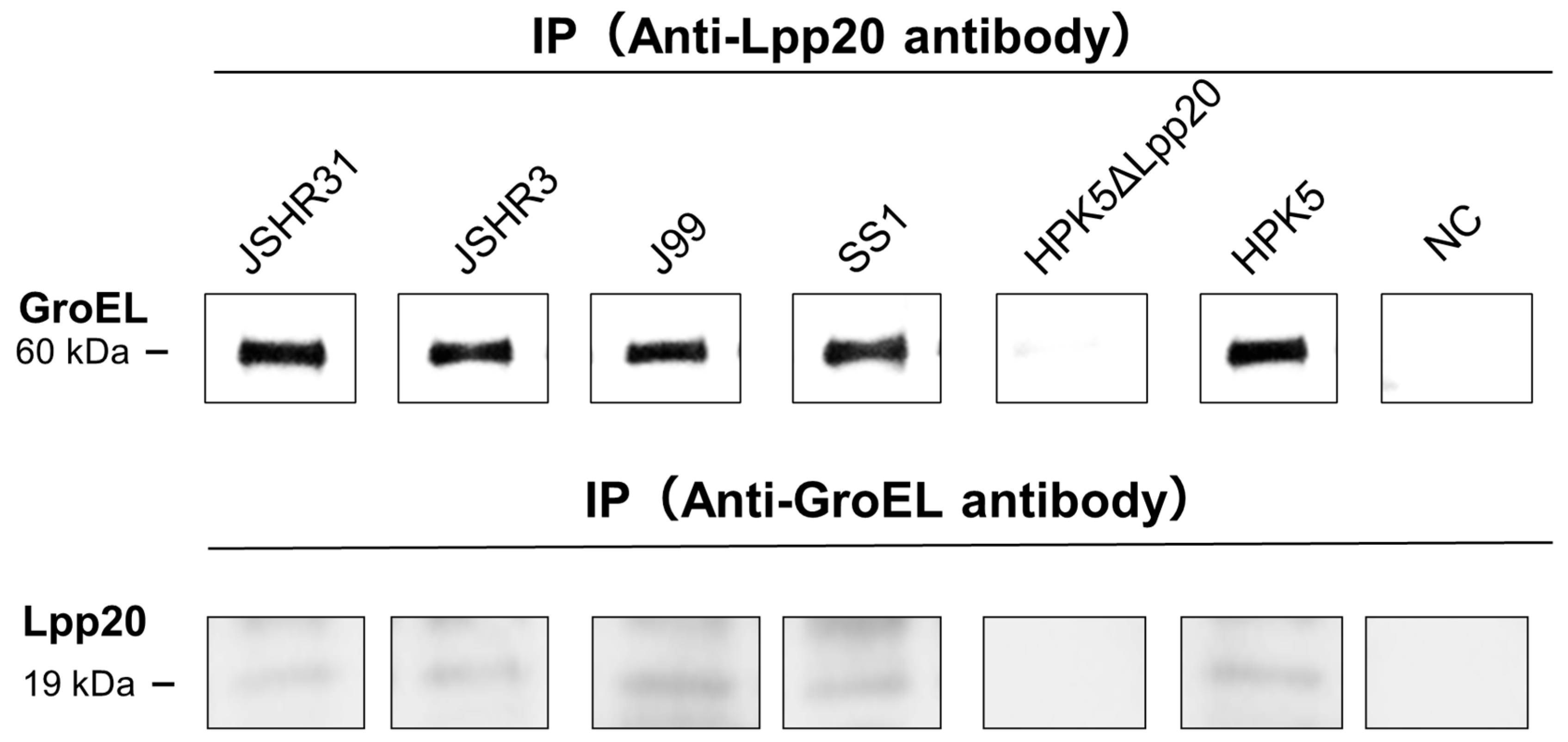
By IB with an anti-Lpp20 antibody, Lpp20 was detected in the cell lysates of seven clinical H. pylori strains (26695, J99, SS1, HPK5, JSHR3, JSHR6, and JSHR31) (Figure 2). The bEVs were detected by IB with an anti-GroEL antibody for all seven clinical strains without IP (Figure 3).
Next, bEV suspensions without IP were subjected to IB with an anti-Lpp20 antibody. bEVs containing Lpp20 were detected from five strains (J99, SS1, HPK5, JSHR3, and JSHR31), but not from the strains 26695 or JSHR6 (Figure 3).
Lpp20 was not detected in the cell lysate or bEVs from the two lpp20-disrupted strains (26695ΔLpp20 and HPK5ΔLpp20); these strains released bEVs (Figure 2 and Figure 3).

3.3. IP–IB Analysis

We performed IP–IB analysis to confirm the bEVs containing Lpp20 released from H. pylori clinical strains J99, SS1, HPK5, JSHR3, and JSHR31. IP (anti-Lpp20 antibody)–IB (anti-GroEL antibody) analysis enabled detection of bEVs from the five strains (Figure 4). Next, we performed IP (anti-GroEL antibody)–IB (anti-Lpp20 antibody) analysis to confirm the presence of Lpp20 in the bEVs from these five strains. Lpp20 was not detected in bEVs from the strain HPK5ΔLpp20, in which the lpp20 gene was disrupted (Figure 4). These data are consistent with the results obtained using bEV suspensions without IP (Section 3.2). The bEVs contained surface-localized Lpp20; this protein may also be contained within bEVs.

3.4. SPRi Analysis

In SPRi analysis with an anti-Lpp20 antibody, significantly more spots were detected for bEVs of the strain HPK5 than for the strain HPK5ΔLpp20 (p < 0.01). Minor signals were observed in a BBL medium including 10% HS (BBL + HS, no bacteria), which was used as the blank (Figure 5a); thus, the results were adjusted on the basis of the reflectivity of the blank to evaluate the binding interactions of bEVs (Figure 5b). No signal was observed in the BBL medium.
In the results from SPRi analysis with an anti-GroEL antibody, numerous spots were detected for the bEVs of both strains HPK5 and HPK5ΔLpp20 without a significant difference (Figure 5a). The binding interactions of bEVs were evaluated in a similar manner as described above (Figure 5b).

4. Discussion

H. pylori infection is thought to be related to gastric and extragastric disorders such as cITP and ACS, but these have geographically different occurrence rates [3,11]. The bEVs released from bacteria, including H. pylori, are suggested to be involved in the delivery of bacterial molecules/pathogens throughout the body [20,21,28]. Here, we investigated H. pylori bEVs containing Lpp20 in seven clinical strains originating from geographically different areas of the world. H. pylori Lpp20 is associated with cITP and thrombosis-mediated ACS. In this study, Lpp20 was expressed in and bEVs were released by all seven clinical strains (26695, J99, SS1, HPK5, JSHR3, JSHR6, and JSHR31) irrespective of their geographic origin. However, bEVs containing Lpp20 were detected from only five of the strains (HPK5, JSHR3, and JSHR31 isolated in Japan, J99 from the United States, and SS1 from Australia), but not from the strains 26695 (isolated in the United Kingdom) and JSHR6 (from Japan). These data show a difference in the content of bEVs generated and released from individual H. pylori strains, indicating different mechanisms of EV packaging between strains. The different properties of bEVs containing Lpp20 may influence the development of cITP associated with H. pylori infection. In fact, the effectiveness of H. pylori eradication therapy for cITP varies depending on the geographic region: In East Asia, including Japan, as well as Central and South America and Italy, efficacy rates are ≥50%, whereas the rate is <20% elsewhere in Europe (the United Kingdom, France, and Spain) and the United States [42,43,44,45]. In this study, bEVs containing Lpp20 were not detected from the strains 26695 and JSHR6, even though these strains expressed Lpp20 within the cells. At least, such diversity of bEVs may influence the development of various extragastric diseases associated with H. pylori.
IP–IB analysis confirmed the properties of the H. pylori bEVs containing Lpp20 described above. Lpp20 was present on the surface of the bEVs from the strains HPK5, JSHR3, JSHR31, J99, and SS1, and it may also be contained within the bEVs from these strains. Comprehensive analysis by HPLC–MS/MS of two strains (26695 and 11637) showed that H. pylori bEVs contain many molecules including Lpp20 and that the bEV content varies between strains [20,28]. Interestingly, bEVs containing Lpp20 could not be detected from 26695 in the present study, probably because of the non-ultracentrifugation-based bEVs preparation method we used. However, bEVs collected by ultracentrifugation may include other molecules and bacterial cell debris. Proteomics analysis of H. pylori bEVs collected by ultracentrifugation revealed that the most abundant protein among the unique peptides was the 60-kDa chaperonin GroEL [40]. In this study, we confirmed bEV existence in the samples prepared without ultracentrifugation and detected H. pylori bEVs using an anti-GroEL antibody. Given the variation of bEVs, more investigations with other antibodies, such as anti-outer membrane proteins and anti-urease antibodies, are needed to understand the individual properties of H. pylori bEVs. Furthermore, H. pylori possesses high genetic diversity, which enables it to persistently infect the stomach and adapt to changing conditions and environmental stresses [46]. It is necessary to clarify how the environmental conditions reflect and affect bacterial behaviors including the contents of released bEVs. Nevertheless, we directly identified bEVs containing Lpp20 and found different contents of bEVs among the seven clinical H. pylori strains that we tested, consistent with the results of earlier HPLC–MS/MS analyses.
In SPRi analysis using an anti-Lpp20 antibody, the binding interactions of bEVs meant that the reflectance ratio was significantly higher for the strain HPK5 than for the strain HPK5ΔLpp20 (p < 0.01). Meanwhile, the reflectance ratio using an anti-GroEL antibody-bound biochip did not show a significant difference between the two strains. These data indicated that the loss of Lpp20 did not significantly affect the production and release of bEVs from the strain HPK5ΔLpp20. Furthermore, consistent with the results from IP–IB, the SPRi data showed that Lpp20 was present at least on the surface of the released bEVs. However, the reflectivity observed using the anti-GroEL antibody-bound biochip did appear to be lower for the strain HPK5ΔLpp20 than for the strain HPK5. It cannot be excluded that Lpp20, an outer membrane protein of H. pylori, influences the bEVs production process in this species. It is necessary to elucidate the molecular function of Lpp20 in H. pylori.
To our knowledge, bEVs from bacteria (including H. pylori) have not been detected in blood or body fluid. Nevertheless, we consider it a possibility that bEVs deliver pathogenic factors throughout the body via the bloodstream. Released bEVs are taken up by host cells such as gastric epithelial cells [21,47], and these cells eventually produce exosomes including bEV contents and deliver them to extragastric tissues via the bloodstream. The CagA protein, encoded by cytotoxin-associated gene A (cagA), a pathogenic factor specific to H. pylori, has been strongly implicated in the development of gastric cancer [4]. The bEVs containing CagA were detected in the blood of patients with CagA-positive H. pylori infection [4,48]. Thus, we speculate that bEVs containing Lpp20 are involved in extragastric diseases. A further investigation into the properties of bEVs and/or bEVs-related transport systems in humans is important to elucidate the pathophysiology of extragastric H. pylori-related diseases and for the development of liquid biopsies for clinical practice. We are planning clinical trials using IB and SPRi analysis to address these issues.

5. Conclusions

In this study, we assessed the properties of H. pylori bEVs using seven clinical isolates from geographically distinct regions and laboratory-generated lpp20-disrupted strains. Lpp20 and bEVs were detected for all seven clinical strains. bEVs containing Lpp20 were detected from five of the seven clinical strains. Lpp20 was found on the surface of bEVs and may also be contained within bEVs. The results suggest that the mechanism by which proteins are processed and packed into bEVs depends on the individual H. pylori strain. By elucidating the strain diversity affecting bEV production, we could better understand the discrepancy between H. pylori infection rates and disease onset rates, paving the way for clinical trials. Further research is needed to elucidate the bEV packaging and transport systems for understanding the pathophysiology of extragastric diseases associated with H. pylori.

Author Contributions

Conceptualization, A.O. and H.T.; methodology, A.O., T.S., N.M., R.F., T.U. and H.T.; validation, A.O., T.S. and R.F.; formal analysis, T.S.; investigation, A.O., R.F., M.S., R.M., M.O. and S.W.; resources, A.O., T.S., T.U. and H.T.; data curation, A.O., M.S. and S.W.; writing—original draft preparation, A.O. and H.T.; writing—review and editing, A.O. and H.T.; visualization, A.O., R.M. and M.O.; supervision, H.T.; project administration, A.O. and H.T.; funding acquisition, A.O. and H.T. All authors have read and agreed to the published version of the manuscript.

Funding

This research was funded by the Japanese Society for Helicobacter Research, Grant Number 23002, and JSPS KAKENHI, Grant Number 24K10603.

Institutional Review Board Statement

Not applicable.

Informed Consent Statement

Not applicable.

Data Availability Statement

The original contributions presented in this study are included in the article. Further inquiries can be directed to the corresponding author.

Acknowledgments

We thank all the researchers for their helpful suggestions during this project and for revising the manuscript text for grammar and style.

Conflicts of Interest

The authors declare no conflicts of interest.

References

  1. Warren, J.R.; Marshall, B. Unidentified curved bacilli on gastric epithelium in active chronic gastritis. Lancet 1983, 321, 1273–1275. [Google Scholar]
  2. Fischbach, W.; Malfertheiner, P. Helicobacter pylori infection: When to eradicate, how to diagnose and treat. Dtsch. Arztebl. Int. 2018, 115, 429–436. [Google Scholar] [PubMed]
  3. Takeuchi, H.; Okamoto, A. Helicobacter pylori Infection and Chronic Immune Thrombocytopenia. J. Clin. Med. 2022, 11, 4822. [Google Scholar] [CrossRef] [PubMed]
  4. Shimoda, A.; Ueda, K.; Nishiumi, S.; Murata-Kamiya, N.; Mukai, S.A.; Sawada, S.I.; Azuma, T.; Hatakeyama, M.; Akiyoshi, K. Exosomes as nanocarriers for systemic delivery of the Helicobacter pylori virulence factor CagA. Sci. Rep. 2016, 6, 18346–18351. [Google Scholar]
  5. Qiang, L.; Hu, J.; Tian, M.; Li, Y.; Ren, C.; Deng, Y.; Jiang, Y. Extracellular vesicles from helicobacter pylori-infected cells and helicobacter pylori outer membrane vesicles in atherosclerosis. Helicobacter 2022, 27, e12877. [Google Scholar] [CrossRef]
  6. Wang, F.; Yao, Z.; Jin, T.; Mao, B.; Shao, S.; Shao, C. Research progress on Helicobacter pylori infection related neurological diseases. Ageing Res. Rev. 2024, 99, 102399. [Google Scholar]
  7. Zawada, A.E.; Naskręt, D.; Piłaciński, S.; Adamska, A.; Grzymisławski, M.; Eder, P.; Grzelka-Woźniak, A.; Zozulińska-Ziółkiewicz, D.; Dobrowolska, A. Helicobacter pylori infection is associated with increased accumulation of advanced glycation end products in the skin in patients with type 1 diabetes: A preliminary study. Adv. Clin. Exp. Med. 2023, 32, 1009–1016. [Google Scholar]
  8. Ye, J.; Feng, T.; Su, L.; Li, J.; Gong, Y.; Ma, X. Interactions between Helicobacter pylori infection and host metabolic homeostasis: A comprehensive review. Helicobacter 2023, 28, e13030. [Google Scholar]
  9. Pellicano, R.; Ianiro, G.; Fagoonee, S.; Settanni, C.R.; Gasbarrini, A. Review: Extragastric diseases and Helicobacter pylori. Helicobacter 2020, 25, e12741. [Google Scholar] [CrossRef]
  10. Pérez-Cano, H.J.; Ceja-Martínez, J.; Tellezgiron-Lara, V.; Voorduin-Ramos, S.; Morales-López, O.; Somilleda-Ventura, S.A. Relationship between Helicobacter pylori and undifferentiated non-granulomatous anterior uveitis. Infection 2023, 51, 765–768. [Google Scholar] [CrossRef]
  11. de Korwin, J.; Ianiro, G.; Gibiino, G.; Gasbarrini, A. Helicobacter pylori infection and extragastric diseases in 2017. Helicobacter 2017, 22, e12411. [Google Scholar]
  12. Takahashi, T.; Yujiri, T.; Shinohara, K.; Inoue, Y.; Sato, Y.; Fujii, Y.; Okubo, M.; Zaitsu, Y.; Ariyoshi, K.; Nakamura, Y.; et al. Molecular mimicry by Helicobacter pylori CagA protein may be involved in the pathogenesis of H. pylori-associated chronic idiopathic thrombocytopenic purpura. Br. J. Haematol. 2004, 124, 91–96. [Google Scholar] [PubMed]
  13. Kashiwagi, H.; Kuwana, M.; Hato, T.; Takafuta, T.; Fujimura, K.; Kurata, Y.; Murata, M.; Tomiyama, Y. Reference guide for management of adult immune thrombocytopenia in Japan: 2019 Revision. Int. J. Hematol. 2020, 111, 329–351. [Google Scholar] [PubMed]
  14. Fujimura, K.; Kuwana, M.; Kurata, Y.; Imamura, M.; Harada, H.; Sakamaki, H.; Teramura, M.; Koda, K.; Nomura, S.; Sugihara, S.; et al. Is eradication therapy useful as the first line of treatment in Helicobacter pylori-positive idiopathic thrombocytopenic purpura? Analysis of 207 eradicated chronic ITP cases in Japan. Int. J. Hematol. 2005, 81, 162–168. [Google Scholar]
  15. Takeuchi, H.; Islam, J.M.; Kaneko, A.; Kimura, A.; Shida, T.; Oboshi, W.; Katayama, H.; Oishi, T.; Fujieda, M.; Morimoto, N. Helicobacter pylori protein that binds to and activates platelet specifically reacts with sera of H. pylori-associated chronic immune thrombocytopenia. Platelets 2021, 32, 1120–1123. [Google Scholar] [CrossRef]
  16. Morimoto, N.; Takeuchi, H.; Takahashi, T.; Ueta, T.; Tanizawa, Y.; Kumon, Y.; Kobayashi, M.; Sugiura, T. Helicobacter pylori-associated chronic idiopathic thrombocytopenic purpura and low molecular weight H. pylori proteins. Scand. J. Infect. Dis. 2007, 39, 409–416. [Google Scholar]
  17. Bakos, N.; Fekete, B.; Prohászka, Z.; Füst, G.; Kalabay, L. High prevalence of IgG and IgA antibodies to 19-kDa Helicobacter pylori-associated lipoprotein in chronic urticaria. Allergy 2003, 58, 663–667. [Google Scholar]
  18. Welsh, J.A.; Goberdhan, D.C.; O’Driscoll, L.; Buzas, E.I.; Blenkiron, C.; Bussolati, B.; Cai, H.; Di Vizio, D.; Driedonks, T.A.; Erdbrügger, U.; et al. Minimal information for studies of extracellular vesicles (MISEV2023): From basic to advanced approaches. J. Extracell. Vesicles 2024, 13, e12404. [Google Scholar]
  19. Zavan, L.; Bitto, N.J.; Johnston, E.L.; Greening, D.W.; Kaparakis-Liaskos, M. Helicobacter pylori Growth Stage Determines the Size; Protein Composition; and Preferential Cargo Packaging of Outer Membrane Vesicles. Proteomics 2019, 19, e1800209. [Google Scholar]
  20. Wei, S.; Li, X.; Wang, J.; Wang, Y.; Zhang, C.; Dai, S.; Wang, X.; Deng, X.; Zhao, L.; Shan, B. Outer Membrane Vesicles Secreted by Helicobacter pylori Transmitting Gastric Pathogenic Virulence Factors. ACS Omega 2021, 7, 240–258. [Google Scholar]
  21. Parker, H.; Chitcholtan, K.; Hampton, M.B.; Keenan, J.I. Uptake of Helicobacter pylori outer membrane vesicles by gastric epithelial cells. Infect. Immun. 2010, 78, 5054–5061. [Google Scholar] [CrossRef] [PubMed]
  22. Jarzab, M.; Posselt, G.; Meisner-Kober, N.; Wessler, S. Helicobacter pylori-Derived Outer Membrane Vesicles (OMVs): Role in Bacterial Pathogenesis? Microorganisms 2020, 8, 1328. [Google Scholar] [CrossRef] [PubMed]
  23. Guerrero-Mandujano, A.; Hernández-Cortez, C.; Ibarra, J.A.; Castro-Escarpulli, G. The outer membrane vesicles: Secretion system type zero. Traffic 2017, 18, 425–432. [Google Scholar] [CrossRef] [PubMed]
  24. Furuyama, N.; Sircili, M.P. Outer Membrane Vesicles (OMVs) Produced by Gram-Negative Bacteria: Structure, Functions, Biogenesis, and Vaccine Application. Biomed. Res. Int. 2021, 2021, 1490732. [Google Scholar] [CrossRef]
  25. Rueter, C.; Bielaszewska, M. Secretion and Delivery of Intestinal Pathogenic Escherichia coli Virulence Factors via Outer Membrane Vesicles. Front. Cell Infect. Microbiol. 2020, 10, 91. [Google Scholar] [CrossRef]
  26. Eletto, D.; Mentucci, F.; Voli, A.; Petrella, A.; Porta, A.; Tosco, A. Helicobacter pylori Pathogen-Associated Molecular Patterns: Friends or Foes? Int. J. Mol. Sci. 2022, 23, 3531. [Google Scholar] [CrossRef]
  27. Schwechheimer, C.; Kuehn, M.J. Outer-membrane vesicles from Gram-negative bacteria: Biogenesis and functions. Nat. Rev. Microbiol. 2015, 13, 605–619. [Google Scholar] [CrossRef]
  28. Parker, H.; Keenan, J.I. Composition and function of Helicobacter pylori outer membrane vesicles. Microbes Infect. 2012, 14, 9–16. [Google Scholar] [CrossRef]
  29. Keenan, J.I.; Allardyce, R.A.; Bagshaw, P.F. Dual silver staining to characterise Helicobacter spp. outer membrane components. J. Immunol. Methods 1997, 209, 17–24. [Google Scholar] [CrossRef]
  30. Yokota, K.; Osaki, T.; Hayashi, S.; Yokota, S.I.; Takeuchi, H.; Rimbara, E.; Ojima, H.; Sato, T.; Yonezawa, H.; Shibayama, K.; et al. Establishment of a reference panel of Helicobacter pylori strains for antimicrobial susceptibility testing. Helicobacter 2022, 27, e12874. [Google Scholar] [CrossRef]
  31. Tomb, J.F.; White, O.; Kerlavage, A.R.; Clayton, R.A.; Sutton, G.G.; Fleischmann, R.D.; Ketchum, K.A.; Klenk, H.P.; Gill, S.; Dougherty, B.A.; et al. The complete genome sequence of the gastric pathogen Helicobacter pylori. Nature 1997, 388, 539–547. [Google Scholar] [CrossRef] [PubMed]
  32. Alm, R.A.; Ling, L.-S.L.; Moir, D.T.; King, B.L.; Brown, E.D.; Doig, P.C.; Smith, D.R.; Noonan, B.; Guild, B.C.; Dejonge, B.L.; et al. Genomic-sequence comparison of two unrelated isolates of the human gastric pathogen Helicobacter pylori. Nature 1999, 397, 176–180. [Google Scholar] [CrossRef] [PubMed]
  33. Lee, A.; O’Rourke, J.; De Ungria, M.; Robertson, B.; Daskalopoulos, G.; Dixon, M. A standardized mouse model of Helicobacter pylori infection: Introducing the Sydney strain. Gastroenterology 1997, 112, 1386–1397. [Google Scholar] [CrossRef] [PubMed]
  34. Karita, M.; Kouchiyama, T.; Okita, K.; Nakazawa, T. New small animal model for human gastric Helicobacter pylori infection: Success in both nude and euthymic mice. Am. J. Gastroenterol. 1991, 86, 1596–1603. [Google Scholar]
  35. Takeuchi, H.; Shirai, M.; Akada, J.K.; Tsuda, M.; Nakazawa, T. Nucleotide sequence and characterization of cdrA, a cell division-related gene of Helicobacter pylori. J. Bacteriol. 1998, 180, 5263–5268. [Google Scholar]
  36. Pasqua, M.; Zennaro, A.; Trirocco, R.; Fanelli, G.; Micheli, G.; Grossi, M.; Colonna, B.; Prosseda, G. Modulation of OMV Production by the Lysis Module of the DLP12 Defective Prophage of Escherichia coli K12. Microorganisms 2021, 9, 369. [Google Scholar] [CrossRef]
  37. Kosgodage, U.S.; Trindade, R.P.; Thompson, P.R.; Inal, J.M.; Lange, S. Chloramidine/Bisindolylmaleimide-I-Mediated Inhibition of Exosome and Microvesicle Release and Enhanced Efficacy of Cancer Chemotherapy. Int. J. Mol. Sci. 2017, 18, 1007. [Google Scholar] [CrossRef]
  38. Zhang, P.; Yeo, J.C.; Lim, C.T. Advances in Technologies for Purification and Enrichment of Extracellular Vesicles. SLAS Technol. 2019, 24, 477–488. [Google Scholar]
  39. Henriquez, T.; Santoro, F.; Medaglini, D.; Pallecchi, L.; Clemente, I.; Bonechi, C.; Magnani, A.; Paccagnini, E.; Gentile, M.; Lupetti, P.; et al. Analysis of the utility of a rapid vesicle isolation method for clinical strains of Pseudomonas aeruginosa. Microbiol. Spectr. 2024, 12, e0064924. [Google Scholar] [CrossRef]
  40. Palacios, E.; Lobos-González, L.; Guerrero, S.; Kogan, M.J.; Shao, B.; Heinecke, J.W.; Quest, A.F.; Leyton, L.; Valenzuela-Valderrama, M. Helicobacter pylori outer membrane vesicles induce astrocyte reactivity through nuclear factor-κappa B activation and cause neuronal damage in vivo in a murine model. J. Neuroinflammation 2023, 20, 66. [Google Scholar]
  41. Pernitzsch, S.R.; Alzheimer, M.; Bremer, B.U.; Robbe-Saule, M.; De Reuse, H.; Sharma, C.M. Small RNA mediated gradual control of lipopolysaccharide biosynthesis affects antibiotic resistance in Helicobacter pylori. Nat. Commun. 2021, 12, 4433. [Google Scholar]
  42. Jarque, I.; Andreu, R.; Llopis, I.; De la Rubia, J.; Gomis, F.; Senent, L.; Jiménez, C.; Martín, G.; Martínez, J.A.; Sanz, G.F.; et al. Absence of platelet response after eradication of Helicobacter pylori infection in patients with chronic idiopathic thrombocytopenic purpura. Br. J. Haematol. 2001, 115, 1002–1003. [Google Scholar] [PubMed]
  43. Michel, M.; Khellaf, M.; Desforges, L.; Lee, K.; Schaeffer, A.; Godeau, B.; Bierling, P. Autoimmune thrombocytopenic Purpura and Helicobacter pylori infection. Arch. Intern. Med. 2002, 162, 1033–1036. [Google Scholar]
  44. Michel, M.; Cooper, N.; Jean, C.; Frissora, C.; Bussel, J.B. Does Helicobater pylori initiate or perpetuate immune thrombocytopenic purpura? Blood 2004, 103, 890–896. [Google Scholar] [PubMed]
  45. Stasi, R.; Rossi, Z.; Stipa, E.; Amadori, S.; Newland, A.C.; Provan, D. Helicobacter pylori eradication in the management of patients with idiopathic thrombocytopenic purpura. Am. J. Med. 2005, 118, 414–419. [Google Scholar]
  46. Islam, J.M.; Yano, Y.; Okamoto, A.; Matsuda, R.; Shiraishi, M.; Hashimoto, Y.; Morita, N.; Takeuchi, H.; Suganuma, N.; Takeuchi, H. Evidence of Helicobacter pylori heterogeneity in human stomachs by susceptibility testing and characterization of mutations in drug-resistant isolates. Sci. Rep. 2024, 14, 12066. [Google Scholar]
  47. Chitcholtan, K.; Hampton, M.B.; Keenan, J.I. Outer membrane vesicles enhance the carcinogenic potential of Helicobacter pylori. Carcinogenesis 2008, 29, 2400–2405. [Google Scholar]
  48. Xia, X.; Zhang, L.; Chi, J.; Li, H.; Liu, X.; Hu, T.; Li, R.; Guo, Y.; Zhang, X.; Wang, H.; et al. Helicobacter pylori Infection Impairs Endothelial Function Through an Exosome-Mediated Mechanism. J. Am. Heart Assoc. 2020, 9, e014120. [Google Scholar]
Figure 1. The characterization of bEVs from HPK5 and HPK5ΔLpp20 strains. (a) TEM image (×50,000) of bEVs derived from HPK5 (upper) and HPK5ΔLpp20 (lower) (scale bar = 100 nm); (b) NTA shows the average concentration of bEVs (particles/mL) of a particular size (nm) for HPK5 (upper) and HPK5ΔLpp20 (lower). The black peak represents the mean values from five independent measurements (n = 5). The red lines on the peaks indicate the standard error of the mean ± S.E.M.; (c) The graph illustrates the particle concentrations (109 particles/mL) of HPK5 and HPK5ΔLpp20, as determined by NTA (mean ± S.E.M., n = 5). HPK5, a clinical strain in Japan (wild type); ΔLpp20, an HPK5-derived lpp20-disrupted strain.
Microorganisms 13 00753 g001
Figure 2. Analysis of Lpp20 in cell lysates from seven clinical H. pylori strains and two lpp20-disrupted strains (HPK5ΔLpp20 and 26695ΔLpp20). By immunoblotting (IB) with an anti-Lpp20 antibody, Lpp20 was detected in all seven H. pylori lysates (JSHR31, JSHR6, JSHR3, J99, SS1, HPK5, and 26695). Lpp20 was not detected in two lpp20-disrupted strains (HPK5ΔLpp20 and 26695ΔLpp20).
Microorganisms 13 00753 g002
Figure 3. Analysis of Lpp20 in bEVs from seven clinical H. pylori strains and two lpp20-disrupted strains. The bEVs were detected by IB with an anti-GroEL antibody in all nine bEV suspensions without IP. Next, the bEV suspensions without IP were subjected to IB with an anti-Lpp20 antibody, demonstrating that bEVs containing Lpp20 were detected in five strains (JSHR31, JSHR3, J99, SS1, and HPK5). The bEVs containing Lpp20 were not detected in other two bEV suspensions (from the strains JSHR6 and 26695). Lpp20 was not detected in bEVs from HPK5ΔLpp20 and 26695ΔLpp20 strains that release bEVs.
Microorganisms 13 00753 g003
Figure 4. Confirmation of bEVs containing Lpp20 by IP–IB. “NC” denotes negative control without a primary antibody, the HPK5ΔLpp20 strain represents the lpp20-disrupted strain derived from H. pylori HPK5. The upper panel shows the detection of bEVs (60 kDa) by IB (Anti-GroEL antibody) followed by IP (Anti-Lpp20 antibody). The lower panel shows the detection of Lpp20 (19 kDa) by IB (Anti-Lpp20 antibody) followed by IP (Anti-GroEL antibody).
Microorganisms 13 00753 g004
Figure 5. Evaluation of binding interaction of bEVs by SPRi analysis. (a) SPRi analysis is performed in triplicate, and the signals are shown. The bEVs signals were detected using a biochip immobilized with an anti-Lpp20 antibody (upper) and an anti-GroEL antibody (lower); (b) On one hand, the evaluation for the binding interactions of bEVs. The reflectance ratio is significantly higher in the strain HPK5 than in the strain HPK5ΔLpp20 (**, p < 0.01) using an anti-Lpp20 antibody. On the other hand, the reflectance ratio is even in two strains using an anti-GroEL antibody. HPK5, the strain HPK5; ΔLpp20, the lpp20-disrupted strain HPK5; BBL + HS, a Brucella broth liquid medium including 10% horse serum; BBL, Brucella broth liquid medium only.
Microorganisms 13 00753 g005
Table 1. Bacterial strains and plasmids.
Bacterial Strain or PlasmidGenotype or Characteristics αRegionReference
H. pylori
 26695Wild typethe United Kingdom[31]
 J99Wild typethe United States[32]
 SS1Wild typeAustralia[33]
 HPK5Wild typeJapan[34]
 JSHR3Wild typeJapan[30]
 JSHR6Wild typeJapan[30]
 JSHR31Wild typeJapan[30]
 26695ΔLpp2026695 derivative; kan in lpp20; Kmr This study
HPK5ΔLpp20 HPK5 derivative; kan in lpp20; Kmr This study
E. coli
 DH5α F φ80dlacZΔM15 Δ(argF-lac)U169 deoR recA1 endA1 hsdR17(rK mK+) supE44 thi-1 gyrA96 relA1 GIBCO-BRL
Plasmids
 pGEM-Teasy 3.0 kb cloning vector; Apr Promega
 plpp20E-1 lpp20 and its flanking ORFs (1.3 kb) of 26695 in pGEM-Teasy; Apr This study
 plpp20E-2 lpp20 and its flanking ORFs (1.3 kb) of HPK5 in pGEM-Teasy; Apr This study
 plpp20E-km-1 1.3 kb kan in lpp20 of plpp20E-1; Apr, Kmr This study
 plpp20E-km-2 1.3 kb kan in lpp20 of plpp20E-2; Apr, Kmr This study
α kan, kanamycin resistance gene; ORF, open reading frame; Apr, ampicillin resistance; Kmr, kanamycin resistance.
Table 2. Primers used for lpp20 gene disruption.
Target GenesPrimer NameSequence (5′-3′)
HP1455-HP14571457F-231TTCAGATGTGATTAACGACACC
1455R-390CTCATTCATTAAAGCGACATGC
lpp201456F-241BamTTGGATCCCTACTAACCAAGCTACAGCG
1456R-224ATTAGTGATCAAATCTTCAGCC
F, forward primer; R, reverse primer.
Disclaimer/Publisher’s Note: The statements, opinions and data contained in all publications are solely those of the individual author(s) and contributor(s) and not of MDPI and/or the editor(s). MDPI and/or the editor(s) disclaim responsibility for any injury to people or property resulting from any ideas, methods, instructions or products referred to in the content.

