Article

Analysis of the Culturable Skin Microbiome of Horses from Southern Germany

1
Microbiology Division, Department of Biology, Faculty of Sciences, Friedrich-Alexander-Universität Erlangen-Nürnberg, Staudtstr. 5, 91058 Erlangen, Germany
2
Mymicrobiome GmbH, Nürnberger Str. 108e, 96050 Bamberg, Germany
*
Authors to whom correspondence should be addressed.
Microorganisms 2025, 13(3), 623; https://doi.org/10.3390/microorganisms13030623
Submission received: 30 January 2025 / Revised: 27 February 2025 / Accepted: 3 March 2025 / Published: 8 March 2025
(This article belongs to the Special Issue Microbiome Research for Animal, Plant and Environmental Health)

Abstract

:
Horses have close interactions with humans and are important as working animals and livestock. In contrast to smaller companion animals like cats and dogs, there is only little information available about their skin microbiome. The objective of this study was to identify and characterize the culturable cutaneous microbiome of healthy horses. Samples were taken from 14 horses from Southern Germany which were randomly enrolled in this study. Matrix-assisted laser desorption/ionization time-of-flight mass spectrometry (MALDI-TOF MS) was used as a method to detect the culturable microorganisms of horse skin. The most abundant culturable species of horse skin identified in this study include Bacillus cereus, Bacillus pumilus, Carnobacterium inhibens, Exiguobacterium sibiricum, Macrococcus carouselicus, Macrococcus goetzii and Staphylococcus equorum. Analyses of the bacteria across different body regions indicated the specific preferences of species for certain skin areas. In addition, our data hinted to an influence of the age of the horses tested and an influence between the four stables studied.

1. Introduction

Horses, like dogs and cats, have close interactions with humans, e.g., as working animals or livestock, but research on their skin microbiome remains scarce in comparison to that of smaller domesticated animals such as cats and dogs [1,2,3,4,5,6,7,8,9,10,11]. One of the earliest studies focusing on wounded horses identified an unknown bacterium from the Acidobacteria phylum, which was also prevalent in the healthy control horses of the study [12]. The first notable study on the healthy equine cutaneous microbiome, which indicated seasonal variation in the equine skin microbiota, identified 38 phyla and 1665 genera. The most common phyla across all samples were Proteobacteria, Firmicutes, Actinobacteria and Bacteroidetes, but their abundance varied across different body sites. At the genus level, nine genera were consistently found across different sites: Psychrobacter, Macrococcus, Pseudomonas, Acinetobacter, Planomicrobium, Arthrobacter, Carnobacterium, Desemzia and Corynebacterium, and it was indicated that Gram-negative bacteria are prevalent in the equine skin microbiome [13]. In the latest study on healthy horses, Proteobacteria was again identified as the most abundant phylum across all sites. Actinobacteria, Firmicutes, Deinococcota and Bacteroidota were also present, with varying abundances depending on the body site. At the genus level, Corynebacterium was the most abundant, followed by Deinococcus and Macrococcus. Additionally, within this study, the genera Pseudomonas, Psychrobacter, Acinetobacter, Desemzia and Carnobacterium were identified [11].
In the two most recent studies on the cutaneous microbiome of healthy horses [11,13], it was concluded that the skin site is a primary factor influencing the composition of the skin microbiota. The studies differed in respect to the conclusion of whether the individual horse is a major factor in shaping the skin microbiome or not. An additional key difference was the presence of Deinococcota, aerobic Gram-positive rods known for their high resistance to environmental stresses, which were abundant on the back of the horses in one study [11], but not in the other [13].
The differences between studies on the cutaneous microbiome of horses highlight the significant lack of research in this area. Furthermore, none of the studies identified bacteria at the species level, which is a crucial gap. Additionally, the use of 16S rRNA sequencing as the primary method in the mentioned studies may contribute to differences in the results obtained, since this method relies on the analysis of DNA isolated from the samples and detects dead and alive organisms. Taken together, this underscores the need for studies that employ diverse and concurrent methodologies to provide a more comprehensive understanding of the equine skin microbiome.

2. Materials and Methods

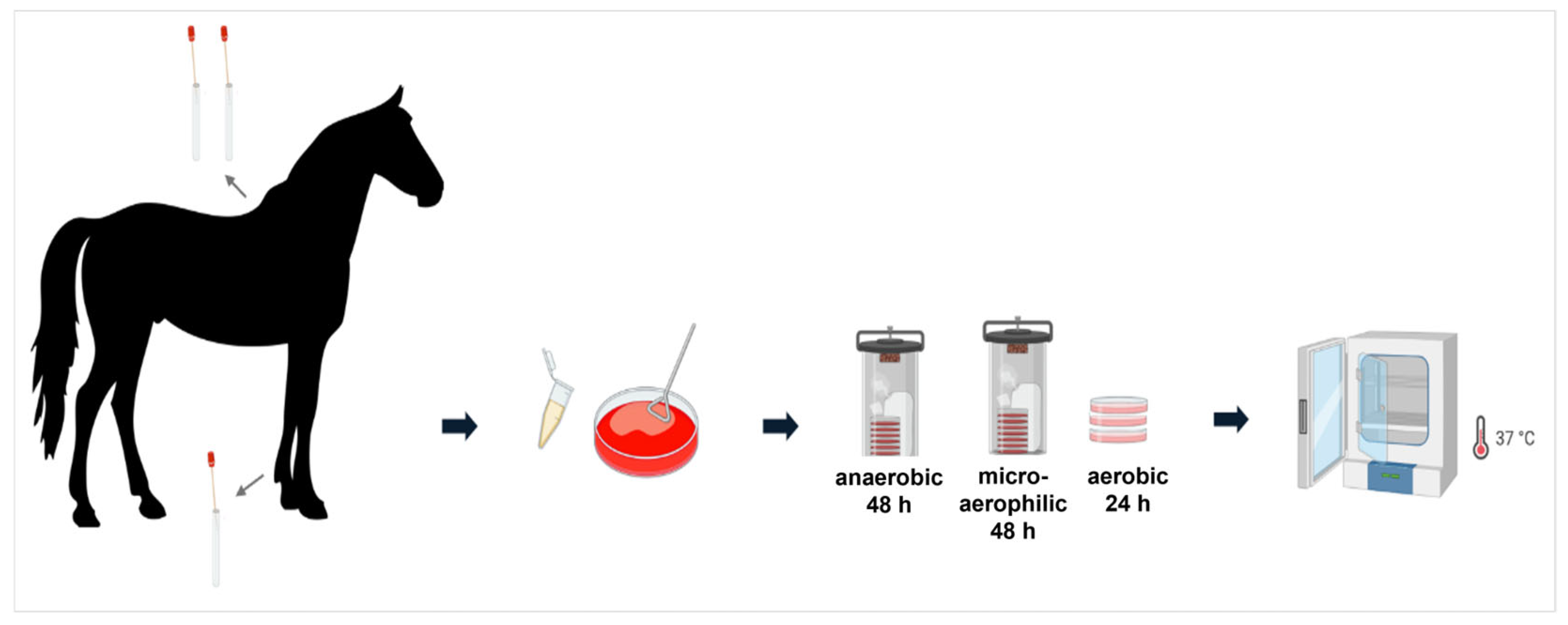
2.1. Sample Collection

eSwab®-Liquid Amies Elution Swabs (Copan, Brescia, Italy) were used to collect microorganisms from back and pastern (Figure 1) of 14 horses, which were randomly enrolled in this study from four different stables located in Southern Germany (Table 1). Sampling was carried out by the owners of the horses during routine skin care after careful training by and under the supervision of one of the authors (experienced microbiologists). To ensure sterility and minimize environmental contamination, particularly from the human microbiome, medical gloves and disinfectant spray were used during the sampling process. The hair at each site was parted to allow the swab to directly contact the skin. Each site was swabbed a minimum of five times using rotational and lateral movements with sterile swabs containing 1 mL of liquid Amies medium in specimen transport tubes. The tubes containing the swabs were subsequently stored at 4 °C until further processing and were transferred within 24 h to the laboratory where they were streaked out immediately. The participants were instructed carefully and gave written consent to participate in this study. According to the University’s ethical guidelines, approval was not required for this study.

2.2. Cultivation of Samples and Isolation of Pure Cultures

A total of 500 μL of phosphate-buffered saline (PBS; 137 mM NaCl, 2.7 mM KCl, 10 mM Na2HPO4 × 12 H2O, 2 mM KH2PO4, adjusted to pH 7.4 with phosphoric acid) was added to the tubes containing the swabs and Amies medium. Subsequently, 50 µL of this suspension from each tube was plated onto Columbia blood agar plates (Oxoid, Wesel, Germany). For each horse body site, the samples were cultured under three different conditions: aerobic, anaerobic and microaerophilic. Each condition was tested in triplicate on plates. The plates were incubated at 37 °C for 24 h under aerobic conditions, for 48 h under anaerobic conditions using a sample transport box with two Oxoid AnaeroGen sachets (Oxoid, Germany) and for 48 h under microaerophilic conditions also using a sample transport box with two Oxoid CampyGen sachets (Oxoid, Germany). For plates exhibiting excessive bacterial growth, 1:10 dilutions with PBS were performed to isolate individual colonies. From the diluted suspension, 50 µL was plated onto fresh blood agar plates.
From the mixed cultures derived from the samples, bacterial colonies were carefully selected and labeled based on their morphological, color, size and hemolytic properties. Individual bacterial colonies were subsequently isolated from the agar plates using a sterile inoculation loop and transferred to fresh blood agar plates using the three-sector streaking method.

2.3. Identification of Bacteria

For optimal results with MALDI-TOF-MS, bacterial isolates from pure cultures were streaked on new blood agar plates and incubated at 37 °C under the required conditions (aerobic plates for 24 h, microaerophilic and anaerobic plates for 48 h). The plates were wrapped with Parafilm and shipped on the same day. The MALDI-ToF MS analysis was carried out by Ripac-Labor GmbH (Potsdam, Germany) and Labor Dr. Risch (Buchs, Switzerland).
In short, fresh bacterial colonies dissolved in formic acid, sterile water and ethanol were applied to polished or ground steel MALDI target plates and a matrix, typically consisting of α-cyano-4-hydroxycinnamic acid dissolved in 50% acetonitrile, and 2.5% trifluoroacetic acid was placed on top, and the plates were left to air dry at room temperature. As the solvent evaporated, the sample and matrix mixture co-crystallized, forming a solid deposit. The plate was loaded into the MALDI-TOF mass spectrometer. The MALDI process caused the sublimation and ionization of both the sample and matrix. Each spectrum was the sum of the ions obtained from 200 laser pulses taken in five distinct areas of the same spot. Using a TOF analyzer, these spectra were examined in the 1000–11,000 mass-to-charge ratio (m/z) range. The presence or absence of peaks within the generated spectacle serves as a fingerprint for a particular isolate, creating an MS profile. The software database is then used to compare and analyze these profiles [14,15,16].

3. Results

3.1. Identification of Culturable Bacteria by MALDI-ToF MS

A total of 146 bacterial species, species-like taxonomical units and species clusters from seven phyla and seventy-two genera were identified (Table 2). The most prevalent phyla across all samples were Proteobacteria (33 genera), Firmicutes (22 genera) and Actinobacteria (12 genera). The identified genera with the most members were Bacillus, Pseudomonas, Macrococcus and Staphylococcus.

3.2. Identification of Dominant Culturable Bacterial Species on Equine Skin

MALDI-TOF analysis of swab samples from the 14 horses tested revealed B. cereus as the most dominant species, being present on all horses. M. carouselicus was detected on 13 horses, while C. inhibens and S. equorum were found on 11 horses, respectively. E mexicanum, M. epidermidis/goetzii and P. agglomerans were present on nine horses. E. artemiae/sibiricum was found on eight horses. S. parauberis and B. pumilus appeared on seven horses (Figure 2).
Bacillus cereus is a Gram-positive, spore-forming, rod-shaped bacterium, isolated from a wide range of sources such as air, soil, water and food. B. cereus is a producer of cereulid and different enterotoxins, making it a widely distributed food-poisoning pathogen [17]. Due to the formation of highly resistant endospores, it is difficult to define a natural habitat for this species. In contrast to B. cereus, the closely related Bacillus pumilus is non-pathogenic. The species is found in water, sediments and soil and has plant growth promoting activity [18].
Macrococcus species are coagulase-negative, catalase- and oxidase-positive cocci. They belong to the Staphylococcaceae family and are closely related to the Staphylococcus genus. These bacteria are typically found as commensals in animals and are generally considered non-pathogenic in their hosts. However, limited information exists on the ecological distribution of Macrococcus among different mammalian species, with few studies documenting their presence on animal skin. M. carouselicus colonies grow to 5–7 mm in diameter on various media, appearing slightly convex, entire, buttery, glistening or opaque, and exhibiting a cream-to-light beige pigmentation. M. goetzii cells can be irregular or regular spherical cocci, depending on the medium. Their colonies have whole margins, are slightly convex, smooth, shiny, 1–2 mm in diameter and are non-pigmented [19].
The genus Carnobacterium has been isolated from cold, low nutrient environments, including Antarctic lake water, Arctic soil, pufferfish, permanently cold seawater, human faces and tomb mural paintings. They are known to tolerate high pressure environments, such as the vacuum packing process in food preservation. The use of lactic acid-producing bacteria like Carnobacterium spp. in food and bio-preservation is a growing area of research in the meat, dairy and seafood industries. Bacteriocins produced by these bacteria have antimicrobial properties that limit or inhibit the growth of foodborne pathogens. C. inhibens are catalase-negative and facultatively anaerobic, with small, gray colored, round colonies measuring 1–2 mm in diameter. Growth occurs within a pH range of 5.8–9.0 [20,21].
Staphylococci are part of the normal microbiota of the skin and mucous membranes of mammals and are also widely distributed in various environments, including soil, water and air, and various foodstuffs such as meat, cheese and raw milk. They are catalase-positive and salt-tolerant. S. equorum is coagulase-negative and is frequently detected in food processing environments and fermented foods, where it can inhibit the growth of undesirable microorganisms. S. equorum forms small colonies, and the subspecies equorum was originally isolated from healthy horses [22,23].
Members of the genus Exiguobacterium have been isolated from a wide variety of environments and are catalase- and oxidase-positive. E. mexicanum colonies are orange-, yellow- and cream-colored, with rods varying from 0.5 to 0.75 mm in diameter and 0.8–3.0 mm in length. E. sibiricum is adapted to cold environments, with mass mapping visualizing the differences in protein expression at various growth temperatures. The cells vary in shape and size depending on the temperature, being 0.8 mm long and 0.6 mm in diameter at 30 °C, but reaching up to 15 mm long at temperatures between 0 and 12 °C. The colonies are bright orange, convex, entire and shiny [24,25].
P. agglomerans is a mesophilic, Gram-negative, motile bacterium that was isolated from knee lacerations [26].

3.3. Major Bacterial Species at Different Anatomical Sites

To identify the most prevalent species under varying skin conditions, their occurrence at specific body sites, namely the back and pastern, was analyzed. The analysis revealed that B. cereus was the most common species on the back, found on 12 individual horses, followed by M. carouselicus on 11 horses. Additionally, C. inhibens was identified on the back of eight horses, and S. equorum on seven horses (Figure 3a). In the pastern area, M. carouselicus was the dominant species, detected on 11 different horses. C. inhibens was identified on the pasterns of 10 horses. Moreover, B. cereus and E. mexicanum were found on nine out of fourteen horses. E. artemiae/sibiricum, P. agglomerans, M. epidermidis/goetzii and S. equorum were present on the backs of eight horses (Figure 3b).

3.4. Comparison of Dominant Culturable Bacterial Species in Different Stables

The analysis of horses from different stables (Table 3) revealed that B. cereus was present in all horses across all stables. Additionally, M. carouselicus and C. inhibens were common species in three out of the four stables.
In stable A, a total of 28 different species were identified. Among these, B. cereus, E. acetylicum, M. carouselicus and S. aureus were common among the horses. Stable B had a total of 48 different bacterial species, with B. cereus, C. inhibens, E. artemiae/sibiricum and M. carouselicus being prevalent. Stable C exhibited the highest diversity, with a total of 92 different bacterial species. Common species in this stable included B. cereus, B. pumilus, C. inhibens, E. mexicanum, M. carouselicus, P. agglomerans and S. parauberis. In stable D, 41 species were identified, with B. cereus, C. inhibens and S. equorum being common in all horses.

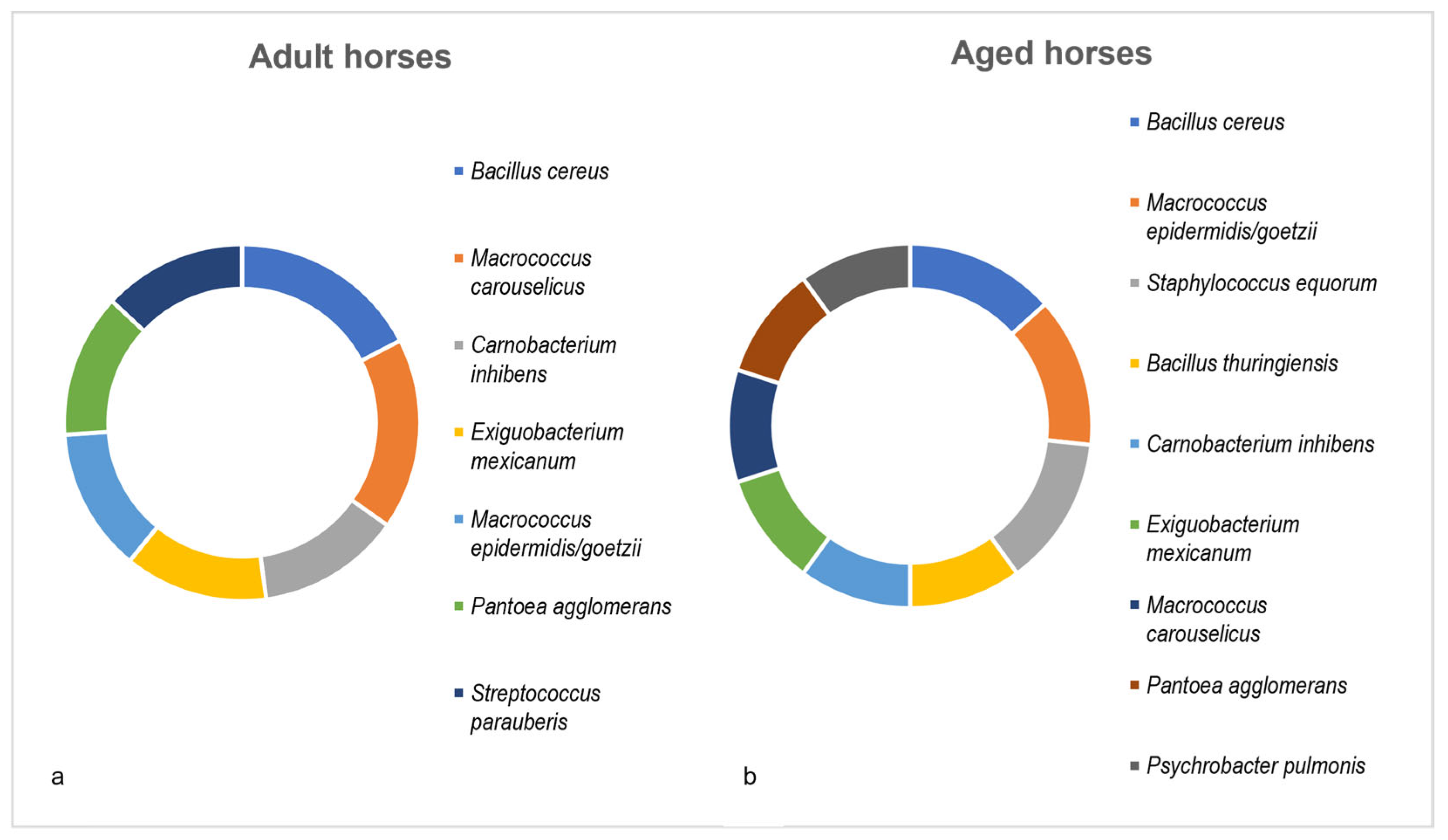
3.5. Age-Dependency of Dominant Culturable Bacterial Species

For the age-related analysis, the horses were separated into two age groups (each with n = 4): adult horses between 2 and 9 years old and aged horses between 14 and 35 years old. The bacterial species had to be present in at least three out of four horses within each group. In the adult horses, a total of 99 different species were identified. B. cereus and M. carouselicus were found in all four horses. Other common bacterial species included C. inhibens, E. mexicanum, M. epidermidis/goetzii, P. agglomerans and S. parauberis (Figure 4a).
In aged horses, a total of 58 species were identified. B. cereus, M. epidermidis/goetzii and S. equorum were present in all horses. Additionally, B. thuringiensis, C. inhibens, E. mexicanum, M. epidermidis/goetzii, P. agglomerans and P. pulmonis were found in at least three out of four horses (Figure 4b).

4. Discussion

In this study, the most frequently identified genera in all samples were Bacillus (Firmicutes), Pseudomonas (Proteobacteria), Macrococcus (Firmicutes) and Staphylococcus (Firmicutes). Among these, the Bacillus genus demonstrated the highest species diversity. B. cereus was identified as the most abundant species in all horses, followed by M. carouselicus, C. inhibens, S. equorum, E. mexicanum, M. goetzii, S. parauberis and B. pumilus, all belonging to the Firmicutes phylum. In addition, P. agglomerans, a member of the Proteobacteria, and E. sibiricum, belonging to the Actinobacteria, were observed with a high frequency. In contrast, previous studies on the skin microbiome of healthy horses reported Proteobacteria and Bacteroidota to be the predominant phyla, with Proteobacteria being the most abundant one [11,13]. Notably, Strompfová and Štempelová [11] identified Deinococcota as one of the most abundant phyla, which was not found in a previous study [13] and was only less abundant here. Differences in results may be attributed to different geographical regions and the influence of different environmental factors. As shown previously, for example, high humidity and low temperature conditions were associated with a higher frequency of bacteria and variability in the skin microbiota and could be correlated with longitudinal and latitudinal variation in UV exposure (for a review, see [27]). As observed before [7,13], non-human mammals generally have a higher number of soil-associated taxa than humans. This difference is likely due to the fact that the animal’s skin, which is largely covered in dense fur, comes into more frequent contact with soil bacteria. This suggests a potential relationship between the skin microbiome of animals and the soil microbiome of their habitats. Additionally, the spores of certain species may be present in the fur of the studied horses, leading to their reactivation during cultivation in the lab.
The analysis of the dominant culturable bacterial species on the different body regions of horses, specifically the pastern and back, revealed similar bacterial populations, in general, but a greater diversity was observed on the pastern compared to the back. B. cereus, M. carouselicus, C. inhibens and S. equorum were identified as dominant species on both body sites, while E. mexicanum, E. sibiricum, P. agglomerans and M. goetzii were found only on the pastern. A lower diversity on the back was reported previously [11,13]. It was suggested that the reduced bacterial diversity on the back of horses might be due to factors such as the presence of sweat containing alkaline minerals, less exposure to the environment and frequent coverage by saddles. The higher diversity in the pastern area may be attributed to direct contact with soil and the horse’s environment, which influences the skin microbiome. However, it is important to note the need for more research and repeated studies to fully understand the differences in skin microbiome across anatomical sites in healthy horses. Furthermore, the microbiome of the pastern would be interesting in respect to its influence on equine pastern dermatitis [28,29,30,31].
The analysis of the equine skin microbiome in horses from different stables showed the common distribution of B. cereus in all four stables, and the presence of C. inhibens and M. carouselicus in three stables. These bacteria were identified as part of the dominant species on horse skin, making their detection across multiple stables plausible. In contrast, certain bacteria were specific to individual stables. This variation may be attributed to differences in hygiene and feeding routines, the population density of horses and the frequency of their interactions and their contact with humans and other animals, such as dogs, cats and goats within each stable. Research on the dog skin microbiome has shown that humans and dogs living together can impact each other’s skin microbiome [13]. Similarly, a sheep population’s bacterial communities are influenced by their handlers, with human biology and lifestyle playing significant roles [32]. This interaction could also apply to horses in different stables, potentially explaining the observed differences.
In addition, age-dependent influences were observed. Adult horses exhibited greater overall microbiome diversity, while older horses displayed more similar bacterial communities. This observation suggests that younger horses have more diverse microbiomes, whereas the microbiomes of older horses are more uniform. These findings align with those reporting a decrease and stabilization in the skin microbiota diversity with age in donkeys [33]. The uniformity observed in aged horses could be due to their less frequent riding routines, leading to reduced contact with humans and the environment. Additionally, it might be related to changes in the functionality and metabolism of the various microbiomes within the skin ecosystem, a hypothesis that warrants further comprehensive research to fully understand these differences, especially in light of the limitation of this study, the very low number of horses included and the rather broad age groups. Follow-up studies may include newborn or very young horses in addition to involving higher numbers of animals.
Despite these insights, the impact of age on the horse skin microbiome remains underexplored. The limited sample size in this study underscores the need for larger scale research to validate these findings and to provide a more comprehensive understanding of age-related changes in the equine skin microbiome. Future studies may include quantitative analyses of culturable microbiomes, approaches for the better detection of fastidious bacteria as well as yeasts and fungi.

5. Conclusions

The study presented here focused on the analysis of the culturable microbiome of healthy horses. The results obtained are complementary to recently published studies using 16S rRNA sequencing for the taxonomic identification of species, and provide further experimental evidence that species identified as dominant by sequencing methods are detectable by culturing as well.

Author Contributions

Conceptualization, A.B. and L.B.; methodology, L.B.; investigation, M.M. and N.Z.; resources, A.B.; data curation, L.B.; writing—original draft preparation, M.M.; writing—review and editing, A.B. and L.B.; visualization, M.M.; supervision, L.B.; project administration, A.B.; funding acquisition, K.N. All authors have read and agreed to the published version of the manuscript.

Funding

This research was funded by Bayern Innovativ, grant number 2024-6459-MY-08.

Institutional Review Board Statement

This study was waived by the University’s regulations due to no experimental animal facility was involved, and all animals were fully acclimatized.

Informed Consent Statement

We obtained informed consent for this study from the owners of the horses.

Data Availability Statement

The raw data supporting the conclusions of this article will be made available by the authors on request.

Conflicts of Interest

K.N. and L.B. were employed by the company Mymicrobiome GmbH. The remaining authors declare that the research was conducted in the absence of any commercial or financial relationships that could be construed as a potential conflict of interest.

References

  1. Kil, D.Y.; Swanson, K.S. Companion animals symposium: Role of microbes in canine and feline health. J. Anim. Sci. 2011, 89, 1498–1505. [Google Scholar] [CrossRef] [PubMed]
  2. Weese, J.S. The canine and feline skin microbiome in health and disease. Vet. Dermatol. 2013, 24, 137–145.e31. [Google Scholar] [CrossRef] [PubMed]
  3. Rodrigues Hoffmann, A.; Patterson, A.P.; Diesel, A.; Lawhon, S.D.; Ly, H.J.; Elkins Stephenson, C.; Mansell, J.; Steiner, J.M.; Dowd, S.E.; Olivry, T.; et al. The skin microbiome in healthy and allergic dogs. PLoS ONE 2014, 9, e83197. [Google Scholar] [CrossRef]
  4. Noli, C. The microbiome of dogs and cats–what do we know in 2017. Rev. Vet. Clin. 2017, 52, 93–98. [Google Scholar] [CrossRef]
  5. Older, C.E.; Diesel, A.; Patterson, A.P.; Meason-Smith, C.; Johnson, T.J.; Mansell, J.; Rodrigues Hoffmann, A. The feline skin microbiota: The bacteria inhabiting the skin of healthy and allergic cats. PLoS ONE 2017, 12, e0178555. [Google Scholar] [CrossRef]
  6. Meason-Smith, C.; Diesel, A.; Patterson, A.P.; Older, C.E.; Johnson, T.J.; Mansell, J.M.; Suchodolski, J.S.; Rodrigues Hoffmann, A. Characterization of the cutaneous mycobiota in healthy and allergic cats using next generation sequencing. Vet. Dermatol. 2017, 28, 71-e17. [Google Scholar] [CrossRef]
  7. Hoffmann, A.R. The cutaneous ecosystem: The roles of the skin microbiome in health and its association with inflammatory skin conditions in humans and animals. Vet. Dermatol. 2017, 28, 60-e15. [Google Scholar] [CrossRef] [PubMed]
  8. Older, C.E.; Diesel, A.B.; Lawhon, S.D.; Queiroz, C.R.R.; Henker, L.C.; Rodrigues Hoffmann, A. The feline cutaneous and oral microbiota are influenced by breed and environment. PLoS ONE 2019, 14, e0220463. [Google Scholar] [CrossRef]
  9. Pereira, A.M.; Clemente, A. Dogs’ Microbiome from Tip to Toe. Top. Companion Anim. Med. 2021, 45, 100584. [Google Scholar] [CrossRef]
  10. Whittle, M.J.; Castillo-Fernandez, J.; Amos, G.C.A.; Watson, P. Metagenomic characterisation of canine skin reveals a core healthy skin microbiome. Sci. Rep. 2024, 14, 20104. [Google Scholar] [CrossRef]
  11. Strompfová, V.; Štempelová, L. Composition and diversity of 16S rRNA based skin bacterial microbiome in healthy horses. Vet. Res. Commun. 2024, 48, 2847–2855. [Google Scholar] [CrossRef]
  12. Kamus, L.J.; Theoret, C.; Costa, M.C. Use of next generation sequencing to investigate the microbiota of experimentally induced wounds and the effect of bandaging in horses. PLoS ONE 2018, 13, e0206989. [Google Scholar] [CrossRef]
  13. O’Shaughnessy-Hunter, L.C.; Yu, A.; Rousseau, J.D.; Foster, R.A.; Weese, J.S. Longitudinal study of the cutaneous microbiota of healthy horses. Vet. Dermatol. 2021, 32, 467-e128. [Google Scholar] [CrossRef] [PubMed]
  14. Bizzini, A.; Durussel, C.; Bille, J.; Greub, G.; Prod’Hom, G. Performance of matrix-assisted laser desorption ionization-time of flight mass spectrometry for identification of bacterial strains routinely isolated in a clinical microbiology laboratory. J. Clin. Microbiol. 2010, 48, 1549–1554. [Google Scholar] [CrossRef]
  15. Croxatto, A.; Prod’hom, G.; Greub, G. Applications of MALDI-TOF mass spectrometry in clinical diagnostic microbiology. FEMS Microbiol. Rev. 2012, 36, 380–407. [Google Scholar] [CrossRef] [PubMed]
  16. Clark, A.E.; Kaleta, E.J.; Arora, A.; Wolk, D.M. Matrix assisted laser desorption ionization time of flight mass spectrometry: A fundamental shift in the routine practice of clinical microbiology. Clin. Microbiol. Rev. 2013, 26, 547–603. [Google Scholar] [CrossRef]
  17. Dietrich, R.; Jeßberger, N.; Ehling-Schulz, M.; Märtlbauer, E.; Granum, P.E. The food poisoning toxins of Bacillus cereus. Toxins 2021, 13, 98. [Google Scholar] [CrossRef] [PubMed]
  18. Dobrzyński, J.; Jakubowska, Z.; Dybek, B. Potential of Bacillus pumilus to directly promote plant growth. Front. Microbiol. 2022, 13, 1069053. [Google Scholar] [CrossRef]
  19. Mazhar, S.; Hill, C.; McAuliffe, O. The genus Macrococcus: An insight into its biology, evolution, and relationship with Staphylococcus. Adv. Appl. Microbiol. 2018, 105, 1–50. [Google Scholar]
  20. Nicholson, W.L.; Zhalnina, K.; de Oliveira, R.R.; Triplett, E.W. Proposal to rename Carnobacterium inhibens as Carnobacterium inhibens subsp. inhibens subsp. nov. and description of Carnobacterium inhibens subsp. gilichinskyi subsp. nov., a psychrotolerant bacterium isolated from Siberian permafrost. Int. J. Sys. Evol. Microbiol. 2015, 65, 556–561. [Google Scholar]
  21. Lo, C.K.L.; Sheth, P.M. Carnobacterium inhibens isolated in blood culture of an immunocompromised, metastatic cancer patient: A case report and literature review. BMC Infect. Dis. 2021, 21, 403. [Google Scholar] [CrossRef]
  22. Nováková, D.; Sedláček, I.; Pantůček, R.; Štětina, V.; Švec, P.; Petráš, P. Staphylococcus equorum and Staphylococcus succinus isolated from human clinical specimens. J. Med. Microbiol. 2006, 55, 523–528. [Google Scholar] [CrossRef] [PubMed]
  23. Jeong, D.W.; Kim, H.R.; Han, S.; Jeon, C.O.; Lee, J.H. A proposal to unify two subspecies of Staphylococcus equorum: Staphylococcus equorum subsp. equorum and Staphylococcus equorum subsp. linens. Antonie Van Leeuwenhoek 2013, 104, 1049–1062. [Google Scholar] [CrossRef] [PubMed]
  24. López-Cortés, A.; Schumann, P.; Pukall, R.; Stackebrandt, E. Exiguobacterium mexicanum sp. nov. and Exiguobacterium artemiae sp. nov., isolated from the brine shrimp Artemia franciscana. Sys. Appl. Microbiol. 2006, 29, 183–190. [Google Scholar] [CrossRef] [PubMed]
  25. Rodrigues, D.F.; Goris, J.; Vishnivetskaya, T.; Gilichinsky, D.; Thomashow, M.F.; Tiedje, J.M. Characterization of Exiguobacterium isolates from the Siberian permafrost. Description of Exiguobacterium sibiricum sp. nov. Extremophiles 2006, 10, 285–294. [Google Scholar] [CrossRef]
  26. Gavini, F.; Mergaert, J.; Beji, A.; Mielcarek, C.; Izard, D.; Kersters, K.; De Ley, J. Transfer of Enterobacter agglomerans (Beijerinck1888) Ewing & Fife 1972 to Pantoea gen. nov. as Pantoea agglomerans comb. nov. and description of Pantoea dispersa sp. nov. Int. J. Syst. Bacteriol. 1989, 39, 337–345. [Google Scholar]
  27. Grice, E.A.; Segre, J.A. The skin microbiome. Nat. Rev. Microbiol. 2011, 9, 244–253. [Google Scholar] [CrossRef]
  28. Kaiser-Thom, S.; Hilty, M.; Axiak, S.; Gerber, V. The skin microbiota in equine pastern dermatitis: A case-control study of horses in Switzerland. Vet. Dermatol. 2021, 32, 646-e172. [Google Scholar] [CrossRef]
  29. Sangiorgio, D.B.; Hilty, M.; Kaiser-Thom, S.; Epper, P.G.; Ramseyer, A.A.; Overesch, G.; Gerber, V.M. The influence of clinical severity and topical antimicrobial treatment on bacteriological culture and the microbiota of equine pastern dermatitis. Vet. Dermatol. 2021, 32, 173-e41. [Google Scholar] [CrossRef]
  30. Kaiser-Thom, S.; Hilty, M.; Ramseyer, A.; Epper, P.; Gerber, V. The relationship between equine pastern dermatitis, meteorological factors, and the skin microbiota. Vet. Dermatol. 2022, 33, 165-e48. [Google Scholar] [CrossRef]
  31. Gerber, V.; Kaiser-Thom, S.; Oesch, S. Equine pastern dermatitis: A narrative review on clinical presentation, diagnosis, risk factors, prevention, and therapeutic approaches. J. Am. Vet. Med. Assoc. 2023, 261, S58–S65. [Google Scholar] [CrossRef] [PubMed]
  32. Jost, S.M.; Cardona, L.; Rohrbach, E.; Mathis, A.; Holliger, C.; Verhulst, N.O. Environment rather than breed or body site shapes the skin bacterial community of healthy sheep as revealed by metabarcoding. Vet. Dermatol. 2024, 35, 273–283. [Google Scholar] [CrossRef] [PubMed]
  33. Ma, Q.; Yue, Y.; Kou, X.; Hou, W.; Wang, M.; Yang, X.; Wang, C. Dynamic Distribution of Skin Microorganisms in Donkeys at Different Ages and Various Sites of the Body. Animals 2023, 13, 1566. [Google Scholar] [CrossRef] [PubMed]
Figure 1. Sampling procedure. Samples were taken from the pastern and back of the horse using eSwab®-Liquid Amies Elution Swabs. Tubes containing the swabs were subsequently stored at 4 °C until further processing and samples were subsequently plated on Columbia blood agar plates and incubated at 37 °C under aerobic, microaerophilic and anaerobic conditions, respectively (created with BioRender; https://app.biorender.com; 6 February 2025).
Microorganisms 13 00623 g001
Figure 2. Overview of the dominant culturable bacterial species from equine skin samples. The bar chart provides a comprehensive visualization of the dominant bacterial species found in equine skin samples, independent of the anatomical body sites. The X-axis represents the different bacterial species, while the Y-axis shows the number of horses in which each bacterial species was found. Each bar is color-coded according to the bacterial species, with the count numbers displayed on top of each bar for clarity. The legend on the right provides the full names of the bacterial species corresponding to each color.
Microorganisms 13 00623 g002
Figure 3. Distribution of major bacterial species at two anatomical sites on equine skin. Pie charts represent the prevalence of dominant bacterial species on the back (a) and pastern (b) of horses. Each color represents a specific bacterial species, with the number of horses harboring each species displayed inside its corresponding color-coded segment. The legend on the right provides the full names of the bacterial species corresponding to each color.
Microorganisms 13 00623 g003
Figure 4. Dominant culturable bacterial species depending on age. (a) adult and (b) aged horses. Bacterial species presented were present in at least three out of four horses within each group.
Microorganisms 13 00623 g004
Table 1. Overview of horses acquired for this study. Abbreviations: m, male; f, female.
No.StableBreedSexAgeSterilizedRoutine Riding
1AIrish Tinkerf3.5noyes
2AIrish Tinkerm12yesyes
3AIrish Tinkerm17yesyes
4BSpeed Rackingm1.5nono
5BCurleym7yesyes
6BShetland Ponym10yesno
7BConnemara Ponym10yesyes
8CAndalusierm15yesyes
9CAmerican Paintm8yesyes
10CMoritzburgerm5yesyes
11DBayerisches Warmblutf19noyes
12DPintof15yesyes
13DQuarterm31nono
14DQuarterm14yesyes
Table 2. Equine skin bacteria identified by MALDI-ToF MS.
PhylumGenusSpecies
FirmicutesAerococcusAerococcus viridans
BacillusBacillus altitudinis
Bacillus amyloliquefaciens
Bacillus cereus
Bacillus infantis
Bacillus licheniformis
Bacillus mycoides
Bacillus pumilus
Bacillus safensis
Bacillus subtilis
Bacillus subtilis/amyloliquefaciens
Bacillus thuringiensis
Bacillus xiamenensis
ButyrivibrioButyrivibrio MabrTax32
CarnobacteriumCarnobacterium gallinarum
Carnobacterium inhibens
Carnobacterium jeotgali
Carnobacterium maltaromaticum
Carnobacterium viridans
DesemziaDesemzia incerta
EnterococcusEnterococcus mundtii
LactococcusLactococcus raffinolactis
LatilactobacillusLatilactobacillus fuchuensis
LysinibacillusLysinibacillus sp.
MacrococcusMacrococcus bohemicus/epidermidis/goetzii
Macrococcus bovicus/brunensis
Macrococcus canis
Macrococcus carouselicus
Macrococcus caseolyticus
Macrococcus epidermidis/goetzii
Macrococcus equipercicus
Macrococcus flavus
Macrococcus hajekii
Macrococcus spp.
MammaliicoccusMammaliicoccus vitulinus
MicrococcusMicrococcus sp.
Micrococcus flavus
Micrococcus luteus
NeobacillusNeobacillus bataviensis
NialliaNiallia circulans
PaenibacillusPaenibacillus amylolyticus
Paenibacillus beijingensis
Paenibacillus MabrTax5
Paenibacillus illinoisensis
Paenibacillus lautus
Paenibacillus saccharophilum
ParaclostridiumParaclostridium benzoelyticum/bifermentans
PeribacillusPeribacillus simplex
Peribacillus simplex/butanolivorans/muralis/frigoritolerans
PriestiaPriestia aryabhattai/megaterium
Priestia megaterium
PsychrobacillusPsychrobacillus lasiicapitis
Psychrobacillus MabrTax3
Psychrobacillus psychrodurans
Psychrobacillus lasiicapitis
SolibacillusSolibacillus silvestris
StaphylococcusStaphylococcus aureus
Staphylococcus chromogenes
Staphylococcus delphini/intermedius/pseudintermedius
Staphylococcus equorum
Staphylococcus sciuri
Staphylococcus succinus
Staphylococcus vitulinus
StreptococcusStreptococcus equinus
Streptococcus equorum
Streptococcus gallolyticus
Streptococcus parauberis
ProteobacteriaAcinetobacterAcinebacter gandavensis
Acinetobacter lwoffii
Acinetobacter radioresistens
Acinetobacter schindleri
Acinetobacter terrestris
Acinetobacter towneri
AcetobacterAcetobacter ascendens
AeromonasAeromonas aquatica/encheleia
Aeromonas bestiarum/salmonicida
Aeromonas encheleia
Aeromonas media/rivipollensis
Aeromonas spp.
AlcaligenesAlcaligenes faecalis
AliidiomarinaAliidiomarina haloalkalitolerans
BowmanellaBowmanella yangjiangensis
ColwelliaColwellia MabrTax21
DyellaDyella amyloliquefaciens
ErwiniaErwinia billingiae
EscherichiaEscherichia vulneris
HaemophilusHaemophilus parahaemolyticus
KosakoniaKosakonia sacchari
LeclerciaLeclercia adecarboxylata
LelliottiaLelliottia amnigena
MarinomonasMarinomonas arctica/shanghaiensis
MassiliaMassilia aurea
MycobacteriumMycobacterium MabrTax33
NeisseriaNeisseria musculi
PannonibacterPannonibacter indicus
PantoeaPantoea agglomerans
Pantoea sp.
Pantoea vagans
PetrocellaPetrocella atlantisensis
PhotorhabdusPhotorhabdus heterorhabditis
PlanococcusPlanococcus glaciei
PseudomonasPseudomonas alloputida/capeferrum/hunanensis
Pseudomonas azotoformans/carnis/lactis/paralactis
Pseudomonas frederiksbergensis
Pseudomonas koreensis
Pseudomonas lundensis
Pseudomonas MabrTax108
Pseudomonas MabrTax304
Pseudomonas MabrTax342
Pseudomonas proteolytica
Pseudomonas spp.
Pseudomonas tolaasii
Pseudomonas umsongensis
PsychrobacterPsychrobacter pulmonis
RahnellaRahnella aquatilis complex
RhizobiumRhizobium MabrTax114
SerratiaSerratia myotis/quinivorans
ShewanellaShewanella sp.
StenotrophomonasStenotrophomonas rhizophila
TestudinibacterTestudinibacter aquarius
ThaueraThauera sedimentorum
ThioalkalivibrioThioalkalivibrio versutus
ActinobacteriaAgrococcusAgrococcus citreus
ArthrobacterArthrobacter citreus
CorynebacteriumCorynebacterium gallinarum
Corynebacterium kalinowskii
CurtobacteriumCurtobacterium flaccumfaciens
ExiguobacteriumExiguobacterium acetylicum
Exiguobacterium artemiae/sibiricum
Exiguobacterium mexicanum
Exiguobacterium oxidotolerans
Exiguobacterium soli
Exiguobacterium sp.
GlutamicibacterGlutamicibacter spp.
KocuriaKocuria carniphila
LuteococcusLuteococcus japonicus
MicrobacteriumMicrobacterium sp.
PaenarthrobacterPaenarthrobacter aurescens
PseudarthrobacterPseudarthrobacter chlorophenolicus
SanguibacterSanguibacter inulinus
BacteroidetesChryseobacteriumChryseobacterium indoltheticum
MyroidesMyroides odoratimimus
DeinococcotaDeinococcusDeinococcus budaensis/piscis
PseudomonadotaButtiauxellaButtiauxella agrestis
Buttiauxella MabrTax2
VerrucomicrobiotaFaecalibacteriumFaecalibacter rhinopitheci
Table 3. Most common 1 culturable bacterial species in different stables.
StableTypeDominant Species
ASmall private stableBacillus cereus
Exiguobacterium acetylicum
Macrococcus carouselicus
Staphylococcus aureus
BLarge riding schoolBacillus cereus
Carnobacterium inhibens
Exiguobacterium artemiae/sibiricum
Macrococcus carouselicus
CLarge riding schoolBacillus cereus
Bacillus pumilus
Carnobacterium inhibens
Exiguobacterium mexicanum
Macrococcus carouselicus
Pantoea agglomerans
Streptococcus parauberis
DLarge riding schoolBacillus cereus
Carnobacterium inhibens
Staphylococcus equorum
1 Bacterial species present in at least three horses within a stable, i.e., three out of four horses in stables B and C, and all three horses in stables A and D.
Disclaimer/Publisher’s Note: The statements, opinions and data contained in all publications are solely those of the individual author(s) and contributor(s) and not of MDPI and/or the editor(s). MDPI and/or the editor(s) disclaim responsibility for any injury to people or property resulting from any ideas, methods, instructions or products referred to in the content.

