Communication

Antifungal Efficacy of Selected Plant Essential Oils Against Clinical Canine Isolates Malassezia pachydermatis

Department of Pharmacology and Toxicology, University of Veterinary Medicine and Pharmacy, 041 81 Košice, Slovakia
*
Author to whom correspondence should be addressed.
Microorganisms 2025, 13(12), 2675; https://doi.org/10.3390/microorganisms13122675
Submission received: 29 September 2025 / Revised: 13 November 2025 / Accepted: 20 November 2025 / Published: 24 November 2025
(This article belongs to the Special Issue Fungal Infections and Antifungal Agents)

Abstract

Malassezia pachydermatis, an important opportunistic secondary pathogen, is often associated with atopic dermatitis or otitis externa in dogs. Recent studies indicate an increase in resistance of this yeast to commonly used antifungal agents. Therefore, the search for new antifungal agents is a challenge. In the present study, the susceptibility of M. pachydermatis strains to 10 plant essential oils—EOs (bergamot, grapefruit, coriander, hyssop, lavender, tea tree, nutmeg, oregano, rosemary, and Spanish sage) was determined using the broth microdilution method. All 15 clinical strains tested were susceptible to coriander (100%). A good antifungal activity was shown for EO from nutmeg (93.33%), bergamot (86.66), Spanish sage and hyssop (73.33%) and rosemary (66.67%). Lower antifungal efficacy was identified in EOs from grapefruit, lavender, tea tree and oregano (53.33%). The obtained results indicate promising prospects for the clinical use of essential oils in the treatment of M. pachydermatis infections.

1. Introduction

Malassezia pachydermatis is considered an important opportunistic secondary pathogen, often detected in atopic dermatitis or otitis externa in dogs. The onset of Malassezia infection depends on the host immune system and expression of virulence factors such as enzymes (esterase, lipase, lipoxygenase, protease, chondroitin sulphatase, hyaluronidase and phospholipase) that play an important role in the colonisation and multiplication of the yeast and trigger the release of inflammatory mediators [1,2].
Treatment of Malassezia infections mostly includes the application of topical formulations containing antifungal drugs, especially polyenes (nystatin) and azoles (clotrimazole, itraconazole, fluconazole, posaconazole, and miconazole), often in combination with anti-inflammatory agents, antibiotics and chlorhexidine [3,4,5]. Recent studies have revealed an increase in the resistance of fungal strains to commercially available antifungal agents, suggesting that the development of new antifungals is an essential strategy to overcome problems encountered in the treatment of these infections [6,7].
Plant essential oils, known for their medicinal properties (analgesic, sedative, anti-inflammatory, anti-spasmodic, local anaesthetic, and anticarcinogenic) and due to their antimicrobial activity, appear to be a relevant alternative [8,9,10]. Essential oils are mixtures of secondary metabolites of many aromatic plants containing a variety of volatile compounds, including terpenes, aldehydes, alcohols, ketones and simple phenolics whose fungal properties have been proven [11,12]. The antifungal activity of EOs has not yet been fully elucidated. Since EOs represent a highly complex chemical composition (approximately 20–60 components at different concentrations), their activity may be related to different strategies by targeting crucial components within fungal cells. Similarly to the conventional antifungal drugs, EOs can directly bind to ergosterol, an essential sterol component in fungal cell membranes, or modulate its production, disrupting membrane integrity, which results in cell death. Some EOs prevent cell wall formation by targeting beta-glucans, which weakens the cell structural framework and leads to cell destruction. Another mode of action of EOs is the inhibition of the activity of mitochondrial dehydrogenases in the tricarboxylic acid cycle (e.g., malate dehydrogenase, succinate dehydrogenase), enzymes responsible for energy production in mitochondria. Disruption of the mitochondrial electron transport chain reduces membrane potential and ATP levels, induces excess reactive oxygen species (ROS) production accompanied with the release of cytochrome c from mitochondria into cytosol leading to indirect induction of the cell apoptosis. In addition, EOs are able to inhibit efflux pumps, which expel drugs from the cell, and allow drugs to accumulate within the fungal cell, improving efficacy and potentially reducing the drug dosage [8,13,14,15].
The literature provides a wealth of information on the antifungal activity of EOs against Candida species, but only a few studies address the effectiveness of EOs against M. pachydermatis. The aim of this study was therefore to determine the susceptibility of M. pachydermatis strains to ten plant EOs (bergamot, grapefruit, coriander, hyssop, lavender, tea tree, nutmeg, oregano, rosemary, and Spanish sage), which are rarely tested by the microdilution method, and to investigate their potential use for the treatment of Malassezia infections.

2. Material and Methods

2.1. Malassezia pachydermatis Strains

A total 15 clinical isolates from the ear swabs of dogs without current manifestation and recent history of otitis externa were used for testing. Samples obtained from the Small Animal Clinic of the University of Veterinary Medicine and Pharmacy in Košice (Slovakia) were taken from dogs of different breeds, sexes and ages. All strains used for the experiments were identified and confirmed based on their phenotypic and genotypic characteristics described by Kaneko et al. [16] and Gaitanis et al. [17]. Until the beginning of the experiments, the strains were preserved at −80 °C in freezing medium (100 µL 60% gly cerol and 300 µL medium—glucose 4 g, tryptophan 1 g, yeast extract 0.5 g per 100 mL). Before use, the yeasts were subcultured twice on SAOT (Sabouraud’s dextrose agar—SDA supplemented with glycerol—2 mL, Tween 80—2 mL, Tween 40—5 mL and olive oil—5 mL per litre) and incubated at 35 °C for 96 h.
The reference strain of M. pachydermatis CBS 1879 (Centraalbureau voor Schimmelcultures, Utrecht, The Netherlands) was also included in the study.

2.2. Plant Essential Oils Tested

The antifungal activity of ten plant essential oils (Table 1): bergamot—Citrus aurantium L. subsp. bergamia, grapefruit—Citrus paradisi, coriander—Coriandrum sativum L., hyssop—Hyssopus officinalis L., lavender—Lavandula angustifolia MILLER., tea tree—Melaleuca alternifolia, nutmeg—Myristica fragrans Houtt, oregano—Origanum vulgare L., rosemary—Rosmarinus officinalis L., and Spanish sage—Salvia lavandulifolia Vahl (Calendula a. s., Nová Ľubovňa, SR) was investigated.

2.3. Testing Quality Control

Testing quality control was verified by determining the susceptibility of the reference strain Candida parapsilosis ATCC 22019 (Czech Collection of Microorganisms, Brno, Czech Republic) to itraconazole (Sigma Aldrich, St. Louis, MO, USA).

2.4. Determination of Minimum Inhibitory Concentration (MIC) of Tested Antifungal Agents

To determine the susceptibility of M. pachydermatis strains, the standard microdilution method M 27-A3 [18] was used with slight modifications.
The test procedure consisted of the preparation of yeast inoculum and concentrations of tested agents. M. pachydermatis strains were first suspended in sterile PBS (Phosphate-Buffered Saline) solution containing 0.1% Tween 80 to an optical density at McFarland 1, yielding 1–5 × 10⁶ CFU/mL (Colony Forming Units/mL). This suspension was further diluted with SBOT (Sabouraud’s broth medium supplemented with the same substances as SAOT) to 104 CFU/mL. As for C. parapsilosis ATTC 22019, the inoculum was prepared from 24 h-old yeast cultured on SDA—Sabourad’s dextrose agar (HiMedia Laboratories Pvt. Ltd., Mumbai, India) and suspended in sterile saline solution to an optical density at McFarland 0.5, resulting in 1–5 × 10⁶ CFU/mL, and subsequently diluted to 103 CFU/mL in SBG (Sabouraud dextrose broth; Hi Media Laboratories Pvt. Ltd., Mumbai, India; supplemented with 10 mM glucose).
The stock solutions of EOs were prepared at a concentration of 400,000 μg/mL by dissolving 100% EOs in sterile paraffin oil and diluted by binary dilution using SBOT to the concentrations of 400,000–800 μg/mL directly in 96-well microplates.
A stock solution of itraconazole (1600 µg/mL), a water insoluble antifungal agent, was prepared by dissolving the powdered substance in DMSO (dimethyl sulfoxide, Sigma Aldrich, Schnelldorf, Germany) and used to prepare a series of two-fold dilutions ranging from 32 to 0.0625 μg/mL, with SBG in microplates.
In both cases, microplate wells 1–10 contained descending concentrations of test agents (100 μL) and inoculum (100 μL). This resulted in the halving of the concentration of the tested agents, for EOs ranging from 200,000 to 400 μg/mL and for itraconazole from 16 to 0.0313 μg/mL. Well 11 served as a negative control containing 200 μL of SBOT or SBG. Well 12 (positive control) contained 100 μL of SBOT or SBG and 100 μL of inoculum. Microplates inoculated with M. pachydermatis samples were incubated at 35 °C for 72 h and those with Candida parapsilosis ATCC 22019 were incubated at 35 °C for 24 h. After incubation, the minimum inhibitory concentration (MIC) was read.
To better evaluate the MIC (minimal inhibitory concentration) end-points, 10 µL of 0.1% resazurin (sterilised through 0.22 µm filter) was added into each well of microplate six hours before reading the results. Inhibition of yeast growth was determined at the MIC that prevented the colour shift from blue (no yeast growth) to orange-pink (yeast growth) [19].
Since there are no criteria for assessing the susceptibility of M. pachydermatis to EOs, the tested strains were classified according to the following criteria: susceptible (S)—MIC sample ≤ MIC50; susceptible dose-dependent (S-DD)—MIC50 < MIC sample ≤ MIC90 and resistant (R)—MIC sample > MIC90 [20].
The susceptibility of C. parapsilosis ATCC 22019 to itraconazole was evaluated according to the criteria established in the M-27-A3 method (S—≤0.125 μg/mL, S-DD—0.25–0.5 μg/mL and R ≥ 1 μg/mL). The average MIC value from the three measurements was 0.0313 µg/mL, which is in accordance with the limit set out in the methodology.

2.5. Statistical Analysis

The MIC values were determined based on repeating the experiments twice and average values were taken. The data are presented as average ( x ¯ ), standard deviations (SD), mode and median. The analysis of the mean MIC values of the plant essential oils tested was performed using one-way ANOVA followed by Tukey’s multiple comparison test (GraphPad Prism 8.0.1, San Diego, CA, USA). The level of statistical significance was set up at p ˂ 0.05.

3. Results

Table 2 summarises the statistical analyses of the achieved MIC in EOs tested. The lowest MIC was identified for EO from coriander, with an average MIC of 400 µg/mL, followed by EO from lavender (MIC 586.67 µg/mL), hyssop (MIC 666.67 µg/mL), rosemary (MIC 741.67 µg/mL), Spanish sage (MIC 816.67 µg/mL), bergamot (MIC 821.67 µg/mL), oregano (MIC 2430 µg/mL) and tea tree (MIC 2360 µg/mL). Higher MIC values were noticed for EOs from nutmeg (MIC 5838.33 µg/mL) and grapefruit (MIC 15,015 µg/mL). Statistically significant differences (p ˂ 0.05) when comparing the tested EOs with each other were observed between grapefruit EO and the EOs of bergamot, coriander, hyssop, lavender, rosemary and Spanish sage.
Table 3 and Figure 1 illustrate the susceptibility of M. pachydermatis clinical isolates and reference strain to the tested plant EOs.
All 15 clinical strains were susceptible to coriander (100%). Up to 14 isolates were susceptible to nutmeg (93.33%) and 13 isolates (86.66%) to bergamot. EOs from hyssop and Spanish sage showed antifungal activity against 11 isolates (73.33%) and EO from rosemary against 10 isolates (66.67%). Eight strains (53.33%) were susceptible to grapefruit, lavender, tea tree and oregano EOs. Resistance of M. pachydermatis clinical strains was found in only one isolate (6.67%) to EOs from bergamot, tea tree, nutmeg and oregano, and in two isolates (13.33%) to EOs from rosemary and Spanish sage. Susceptibility of the M. pachydermatis CBS 1879 strain was detected in all tested EOs, except for lavender, where two strains (66.67%) exhibited susceptibility and one strain (33.33%) was resistant.

4. Discussion

The significant anti-Malassezia effectiveness of EOs has been reported in several studies in human medicine, but less so in the field of veterinary medicine [3,6,7,21,22]. Plant EOs, as a natural product of a specific chemical composition, usually contain two or three major components, mostly terpenes, at a relatively high concentration (20–90%), while other components (aromatic and aliphatic compounds) are less abundant. These components are responsible for their diverse biological activities that arise from their synergistic interaction [23]. The anti-Malassezia activity of EOs varies not only depending on the type of EOs used, but also on the Malassezia species tested. As for M. pachydermatis, Bismark et al. [22] tested 22 EOs against 15 M. pachydermatis isolates using the agar disc diffusion method and reported on a strong antifungal effect of EOs from lemon grass, cinnamon leaf, clove, manuka, Indian melissa, oregano, palmarosa, and winter savoury. In another study, it was found that out of six EOs (Artemisia sieber, Heracleum persicum, Menta spicata, Rosmarinus officinalis, Thymus kotschyanus, and Zataria multiflora) examined for anti-M. pachydermatis activity, Z. multiflora (60 µg/mL) and A. sieberi (80 µg/mL) exhibited the highest inhibitory effects [6].
In the present study, the antifungal activity of ten EOs was evaluated, the efficacy of which against M. pachydermatis was tested by the microdilution method is rarely reported in the literature. The best effectiveness was found for coriander. The effect of coriander EO extracted from the seeds against M. pachydermatis was tested by Bismarck et al. [22] using a disc diffusion test. The 20% (200,000 µg/mL) solution gave the inhibition zone ranging from 19 to 25 mm. Good antifungal activity of the EO from C. sativum was also demonstrated against C. parapsilosis CBS 604 (125 µg/mL), C. dubliniensis CBS 7987 and C. krusei CBS 573 (250 µg/mL) and C. albicans CBS 562 (500 µg/mL) [24]. Coriander seed oil is known for its broad spectrum of antimicrobial (antibacterial and antifungal) and pharmacological activities (antiedemic, anti-inflammatory, antiseptic, antidiabetic, emmenagogue, antihypertensive, lipolytic and myorelaxant) [25]. Laribi et al. [26] analysed the EO composition of C. sativum from different origins and found that linalool as the principal component that may exert antifungal activity ranged from 37.65% from Bangladesh to 87.54% from Tunisia. As the authors state, differences in the chemical profile of coriander EOs across regions may be influenced by the genetic, climatic, seasonal and geographic conditions. The results of our study are similar to the findings of Begnami et al. [24], as the coriander EO MIC reached 400 µg/mL with a linalool content of 64 ± 2%.
Lavender, hyssop, rosemary, Spanish sage and bergamot also demonstrated remarkable antifungal efficacy against M. pachydermatis with MIC values ranging from 586.67 µg/mL to 821.67 µg/mL.
Lavender EOs or extracts are known for their broad spectrum of pharmacological activities, allowing for their use as sedatives, anti-inflammatories, antioxidants, antimicrobials, antifungals, insecticides, and larvicide. The chemical composition of lavender EOs is mainly characterised by the presence of oxygenated monoterpenes (e.g., linalool, linalyl acetate, 1,8-cineole, camphor) and irregular monoterpenoids (e.g., lavandulol and lavandulyl acetate) [27]. In an experiment described by Váczi et al. [21] using a disc diffusion method, lavender EO inhibited the growth of M. pachydermatis at 30% (300,000 µg/mL) concentration, while no yeast susceptibility was recorded at a concentration of 5% (50,000 µg/mL) and 0.5% (5000 µg/mL). However, in the present study the strains exhibited the susceptibility at a concentration of 586.67 µg/mL (0.057%) which may be influenced by the different susceptibility of the tested isolates.
In traditional medicine, Hyssopus spp. are used for their curative properties against cough, cold, loss of appetite, fungal infection, and spasmodic conditions. Essential oil constituents of hyssop exhibited antimicrobial, antifungal, and muscle relaxant properties. The major components of hyssop are monoterpene pinocamphone, isopinocamphone, and β-pinene [28]. Hristova et al. [29] evaluated the antifungal activity of hyssop EO against clinical isolates and reference strains of the genus Candida. The highest susceptibility was shown by C. albicans strains with MIC of 210.3 ± 62.3 µg/mL, followed by C. krusei (MIC = 224.0 ± 64.0 µg/mL), C. parapsilosis (MIC = 298.7 ± 104.5 µg/mL), C. tropicalis (MIC = 682.7 ± 264.4 µg/mL) and C. glabrata (MIC = 768.0 ± 280.4 µg/mL). The authors also tested the influence of the chemical composition of hyssop oil on its anticandidal activity. Out of the major EO constituents and their isomers, such as cis-pinocamphone (48.98–50.77%), β-pinene (13.38–13.54%), trans-pinocamphone (5.78–5.94%) and β-phellandrene (4.44–5.17%), cis- and trans-pinocamphone were the most active constituents with MIC values 28% and 21% higher than those of hyssop oil against C. albicans and C. glabrata, respectively.
Rosemary EO has gained considerable scientific interest due to its wide range of pharmacological activities (anticancer, anti-inflammatory, and antioxidant). It has also been shown to promote healing, angiogenesis, and improvements in granulation tissue, it accelerates wound healing, and enhances the survival and viability of tissues while reducing tissue necrosis. In addition, rosemary EO exhibits antifungal properties. In traditional medicine, it is used to relieve dysmenorrhea, renal colic pain, and respiratory disorders due to its antispasmodic properties [30]. The antifungal activity of rosemary EO was tested by Waller et al. [31] against pathogenic strains of M. pachydermatis isolated from canine and feline otitis. The MIC ranged from ≤0.078 to >2.5 mg/mL (78–2500 µg/mL), while in the present study it ranged from 400 µg/mL to 3125 µg/mL. Among all 19 compounds identified in rosemary EOs by Waller et al. [31], 1,8-cineole (11–49.4%), camphor (16–17.8%) and α-pinene (2–12.2%) prevailed, whereas in the EO tested in our study, cineole was present at 25 ± 1%, followed by α-pinene and camphor (19 ± 1%). Similarly, Khoshravi et al. [6] reported the MIC90 of 360 µg/mL for the inhibitory effect of rosemary EO against M. pachydermatis—unfortunately without providing information on the presence of the major components that may play an important role in the antifungal activity.
Salvia lavandulifolia Vahl is a valuable aromatic and medicinal plant with a high economic potential in agriculture, cosmetics and pharmacology. The medicinal activities are attributed to its essential oils and include anti-spasmodic, antiseptic, analgesic, oestrogenic, anticholinesterase, antioxidant, anti-inflammatory, and central nervous system-suppressing activities, as well as antimicrobial effects. The predominant constituents in the Spanish sage EO are monoterpenes such as α- and β-pinene, 1,8-cineole, and camphor, the proportions of which may be significantly affected by various environmental and genetic factors, as well as by phenological stages (vegetative and full flowering stages) [32,33,34]. Pisteli et al. [3] tested the anti-M. pachydermatis efficacy of EO from Salvia sclarea with no inhibition of the yeast growth (MIC => 10%; 100,000 µg/mL). The major constituent of the EO were linalool (28.4%) and linalyl acetate (48.9%), while in the EO from S. lavandulifolia Vahl examined in this study, camphor represented the main component (27 ± 1%), and the mean MIC reached 816.67 µg/mL.
Bergamot EO has not only been reported for its antibacterial and antifungal activity, but it is also known for its anti-inflammatory, antiproliferative, neuro-pharmacological and neuroprotective (calming, relaxing, anxiolytic), and analgesic activities. In general, the volatile fraction with dominated monoterpene hydrocarbons such as limonene (25–55%), β-pinene (4–11%), and γ-terpinene (5–11%) is considered to be a bearer of these pharmacological properties [35]. However, compared to other Citrus EOs, bergamot EO is characterised by a much lower percentage of limonene and allied monoterpenes and a much higher content of oxygenated terpenes, specifically linalool (2–20%) and linalyl acetate (15–10%) [36]. In the study by Lee and Lee [37], no efficacy of bergamot EO was found against M. pachydermatis, while Váczi et al. [21] noticed a small inhibition zone (8.44 ± 1.67 mm) for bergamot EO at a concentration of 30% (300,000 µg/mL). Despite these findings, this study proved the efficacy of bergamot EO with an average MIC of 821.67 µg/mL, which is consistent with the study by Limtaniakul et al. [38] confirming the anti-Malassezia effect of bergamot EO.
Higher MIC values were required for EO from oregano, tea tree, nutmeg and grapefruit (mean MIC 2360–15,015 µg/mL) to achieve sufficient efficacy against M. pachydermatis strains used in this experiment.
Tea tree oil has been found to be effective in treating small superficial wounds and insect bites, treating small ulcers (furuncles and mild acne), relieving itching and irritation in mild athlete’s foot, and in the symptomatic treatment of moderate oral mucosal inflammation [39]. Several research articles point to the anti-M. pachydermatis effect of tea tree EO [21,22,40,41]. For instance, Weseler et al. [40] observed the antifungal activity against clinical isolates M. pachydermatis in the MIC range of 560–1120 µg/mL. Tea tree EO consisted mainly of terpinene-4-ol (40.7%), γ-terpinene (20.4%), α-terpinene (9.1%), p-cymene (2.2%), 1,8-cineol (3.1%), terpinolene (3.3%), α-terpineol (3.1%), α-pinene (2.3%), and limonene (1.0%). The antifungal activity of tea tree EO, performed using the disc diffusion method in the experiment by Lee and Lee [37], showed a strong inhibitory effect on M. pachydermatis at a concentration of 2 mg/mL (2000 µg/mL) with an inhibition zone of 1.5 cm. The major constituents of the EO were γ-terpinolene (17.96%) and terpinen-4-ol (45.54%). Similar results were found in this study, where the average MIC achieved 2360 µg/mL and the main component of the tested EO, terpinen-4-ol, was 33 ± 2%.
Oregano EO has been shown to possess several bioactive properties including antioxidant, antimicrobial (antifungal, bactericidal and antiviral), anti-inflammatory, analgesic, antihypertensive, antiproliferative, and antidiabetic [42,43]. Pisteli et al. [3] and Waller et al. [31] report on the anti-Malassezia activity of Origanum vulgare L. EO. In the study by Waller et al. [31], the inhibitory and fungicidal activity (MIC90 and MFC90) of oregano EO investigated against M. pachydermatis isolates collected from dogs suffering from otitis achieved a concentration of 0.625 mg/mL (625 µg/mL) for both. Among all 16 identified compounds, carvacrol (73.9%) was the major component observed in oregano oil. The lower antifungal activity of oregano EO tested in our study (average MIC = 2430 µg/mL) may be related to the lower amount of carvacrol (57 ± 3%).
As for nutmeg EO, clinical and experimental investigations have confirmed the antioxidant, antimicrobial, anti-inflammatory, anticancer, antimalarial, anticonvulsant, hepatoprotective, antiparasitic, insecticidal, and nematocidal activities [44]. However, no articles were found focusing on the antifungal activity of EO from Myristica fragrans (nutmeg) against M. pachydermatis. Nicolic et al. [45] present the antifungal activity of nutmeg EO against the C. albicans ATCC 2091 strain. Using the disc diffusion method, the growth inhibition zone of 28 mm was noticed. The efficacy was attributed to the monoterpene hydrocarbon sabinene (42.3%), which was identified in the most abundant amount among the twenty-five compounds. Nutmeg EO is also characterised by the presence of other monoterpenes such as β-pinene (26%), α-pinene (10.51%) and γ-terpinen (8.51%) [46] and aromatic compounds such as myristicin (1.8 to 12.8%) and elemicin (4.3 to 11.1%) [47]. The composition of nutmeg EO tested in our study corresponds to these studies, although a higher MIC was required for sufficient anti-malassezia efficacy (mean MIC = 5838.33 µg/mL).
Unlike other EOs, EO from Citrus paradisi (grapefruit) demonstrated the anti-M. pachydermatis activity at the highest MIC (average MIC = 15,015 µg/mL). The obtained results are close to the study by Pisteli et al. [3], who observed the antifungal activity against M. pachydermatis at MIC and MFC of 1.3% (13,000 µg/mL) for both. The content of the main constituent in the tested oil was limonene (91.7%), like in the tested grapefruit EO in the present study (87 ± 3%). According to the available literature, the grapefruit EO possesses antiseptic, healing, soothing, curing and rubifacial qualities, as well as antimicrobial activity [48].
Although the present study has some limitations (no standardised method for testing the susceptibility of M. pachydermatis to antifungals and no criteria for assessing the susceptibility are established), it can be considered a basis for future studies of the efficacy of EOs in vivo. Significant antifungal efficacy of tested plant EOs was found in vitro with different MIC values (the lowest MIC for coriander EO—400 µg/mL and the highest MIC for grapefruit EO—15,015 µg/mL) which may be affected by the chemical composition of EOs. Moreover, the obtained results point to the antifungal activity of plant EOs, whose anti-M. pachydermatis activities have either not been studied so far (nutmeg) or have been rarely evaluated using the microdilution method. Based on the accessible articles, we can confirm that the efficacy of EOs depends not only on the amount of the major components, but may also be influenced by the virulence of the individual M. pachydermatis strain, resulting in different susceptibility [22].

5. Conclusions

The results obtained in this study demonstrate that plant EOs appear to be a promising alternative for the treatment of infections caused by M. pachydermatis. However, their implementation in clinical practice as effective natural agents or adjuncts to conventional antifungal drugs, as well as their safe use in veterinary dermatology, require further research, with particular emphasis on assessing their cytotoxicity.

Author Contributions

Conceptualisation, E.Č. and P.V.; Methodology, E.Č., P.V. and Z.M.; Validation, E.Č. and Z.M.; Formal analysis, E.Č.; Investigation, E.Č., P.V. and Z.M.; Resources, E.Č. and P.V.; Data curation, E.Č. and P.V.; Writing—original draft preparation, E.Č.; Writing—review and editing, E.Č.; Visualisation, E.Č.; Supervision, E.Č. and P.V.; project administration, E.Č.; funding acquisition, E.Č. All authors have read and agreed to the published version of the manuscript.

Funding

This research was funded by the Slovak Research and Development Agency under the contract No. APVV-15-0377 and partially by the Ministry of Education under the grant KEGA No. 003UVLF-4/2024.

Institutional Review Board Statement

Not applicable.

Informed Consent Statement

Not applicable.

Data Availability Statement

The original contributions presented in this study are included in the article. Further inquiries can be directed to the corresponding author.

Conflicts of Interest

The authors declare no conflicts of interest.

References

  1. Teramoto, H.; Kumeda, Y.; Yokoigawa, K.; Hosomi, K.; Kozaki, S.; Mukamoto, M.; Kohda, T. Genotyping and characterisation of the secretory lipolytic enzymes of Malassezia pachydermatis isolates collected from dogs. Vet. Rec. Open 2015, 21, e000124. [Google Scholar] [CrossRef]
  2. Buommino, E.; Nocera, F.P.; Parisi, A.; Rizzo, A.; Donnarumma, G.; Mallardo, K.; Fiorito, F.; Baroni, A.; De Martino, L. Correlation between genetic variability and virulence factors in clinical strains of Malassezia pachydermatis of animal origin. New Microbiol. 2016, 39, 216–223. [Google Scholar]
  3. Pistelli, L.; Mancianti, F.; Bertoli, A.; Cioni, P.L.; Leonardi, M.; Pisseri, F.; Mugnaini, L.; Nardoni, S. Antimycotic activity of some aromatic plants essential oils against canine isolates of Malassezia pachydermatis: An in vitro assay. Open Mycol. J. 2012, 6, 17–21. [Google Scholar] [CrossRef]
  4. Peano, A.; Johnson, E.; Chiavassa, E.; Tizzani, P.; Guillot, J.; Pasquetti, M. Antifungal resistance regarding Malassezia pachydermatis: Where are we now? J. Fungi 2020, 6, 93. [Google Scholar] [CrossRef] [PubMed]
  5. Daniel, A.K. Canine malasseziosis: An overview. Indian J. Anim. Health 2022, 61, 228–234. [Google Scholar] [CrossRef]
  6. Khosravi, A.R.; Shokri, H.; Fahimirad, S. Efficacy of medicinal essential oils against pathogenic Malassezia sp. isolates. J. Mycol. Med. 2016, 26, 28–34. [Google Scholar] [CrossRef]
  7. Donato, R.; Sacco, C.; Pini, G.; Bilia, A.R. Antifungal activity of different essential oils against Malassezia pathogenic species. J. Ethnopharmacol. 2020, 1, 112376. [Google Scholar] [CrossRef]
  8. Nazzaro, F.; Fratianni, F.; Coppola, R.; Feo, V. Essential Oils and Antifungal Activity. Pharmaceuticals 2017, 2, 86. [Google Scholar] [CrossRef]
  9. Blowman, K.; Magalhães, M.; Lemos, M.F.L.; Cabral, C.; Pires, I.M. Anticancer properties of essential oils and other natural products. Evid. Based Complement. Altern. Med. 2018, 25, 3149362. [Google Scholar] [CrossRef]
  10. Man, A.; Santacroce, L.; Jacob, R.; Mare, A.; Man, L. Antimicrobial activity of six essential oils against a group of human pathogens: A comparative study. Pathogens 2019, 8, 15, Erratum in Pathogens 2019, 8, 108. https://doi.org/10.3390/pathogens8030108. [Google Scholar] [CrossRef]
  11. Peixoto, L.R.; Rosalen, P.L.; Ferreira, G.L.S.; Freires, I.A.; de Carvalho, F.G.; Castellano, L.R.; de Castro, R.D. Antifungal activity, mode of action and anti-biofilm effects of Laurus nobilis Linnaeus essential oil against Candida spp. Arch. Oral Biol. 2017, 73, 179–185. [Google Scholar] [CrossRef]
  12. Bunse, M.; Daniels, R.; Gründemann, C.; Heilmann, J.; Kammerer, D.R.; Keusgen, M.; Lindequist, U.; Melzig, M.F.; Morlock, G.E.; Schulz, H.; et al. Essential oils as multicomponent mixtures and their potential for human health and well-being. Front. Pharmacol. 2022, 24, 956541. [Google Scholar] [CrossRef] [PubMed]
  13. de Sousa, D.P.; Damasceno, R.O.S.; Amorati, R.; Elshabrawy, H.A.; de Castro, R.D.; Bezerra, D.P.; Nunes, V.R.V.; Gomes, R.C.; Lima, T.C. Essential oils: Chemistry and pharmacological activities. Biomolecules 2023, 18, 1144. [Google Scholar] [CrossRef] [PubMed]
  14. Ben Miri, Y. Essential oils: Chemical composition and diverse biological activities: A comprehensive review. Nat. Prod. Commun. 2025, 20, 1–29. [Google Scholar] [CrossRef]
  15. Pan, C.; Yang, K.; Erhunmwunsee, F.; Li, Y.X.; Liu, M.; Pan, S.; Yang, D.; Lu, G.; Ma, D.; Tian, J. Inhibitory effect of cinnamaldehyde on Fusarium solani and its application in postharvest preservation of sweet potato. Food Chem. 2023, 408, 135213. [Google Scholar] [CrossRef]
  16. Kaneko, T.; Makimura, K.; Abe, M.; Shiota, R.; Nakamura, Y.; Kano, R.; Hasegawa, A.; Sugita, T.; Shibuya, S.; Watanabe, S.; et al. Revised culture-based system for identification of Malassezia species. J. Clin. Microbiol. 2007, 45, 3737–3742. [Google Scholar] [CrossRef]
  17. Gaitanis, G.; Robert, V.; Velegraki, A. Verifiable single nucleotide polymorphisms of the internal transcribed spacer 2 region for the identification of 11 Malassezia species. J. Dermatol. Sci. 2006, 43, 214–217. [Google Scholar] [CrossRef]
  18. CLSI—Clinical and Laboratory Standard Institute. Reference Method for Broth Dilution Antifungal Susceptibility Testing of Yeasts Approved Standard, M27-A3 Guideline, 3rd ed.; CLSI: Wayne, PA, USA, 2008; 25p. [Google Scholar]
  19. Liu, M.; Seidel, V.; Katerere, D.R.; Gray, A.I. Colorimetric broth microdilution method for the antifungal screening of plant extracts against yeasts. Methods 2007, 42, 325–329. [Google Scholar] [CrossRef]
  20. Figueredo, L.A.; Cafarchia, C.; Otranto, D. Antifungal susceptibility of Malassezia pachydermatis biofilm. Med. Mycol. 2013, 51, 863–867. [Google Scholar] [CrossRef]
  21. Váczi, P.; Čonková, E.; Marcinčáková, D.; Sihelská, Z. Antifungal effect of selected essential oils on Malassezia pachydermatis growth. Folia Vet. 2018, 62, 67–72. [Google Scholar] [CrossRef]
  22. Bismarck, D.; Dusold, A.; Heusinger, A.; Müller, E. Antifungal in vitro activity of essential oils against clinical isolates of Malassezia pachydermatis from canine ears: A report from a practice laboratory. Complement. Med. Res. 2020, 27, 143–154. [Google Scholar] [CrossRef]
  23. El Youssfi, C.; Dadou, S.; Loukili, E.H.; El Hammoudani, Y.; Soujaa, H.; Rejdali, M.; Aarab, S. Biological activities of essential oils: A mini-review. BIO Web Conf. 2024, 109, 01031. [Google Scholar] [CrossRef]
  24. Begnami, A.F.; Duarte, M.C.T.; Furletti, V.; Rehder, V.L.G. Antimicrobial potential of Coriandrum sativum L. against different Candida species in vitro. Food Chem. 2010, 118, 74–77. [Google Scholar] [CrossRef]
  25. Chahal, K.K.; Singh, R.; Kumar, A.; Bhardwaj, U. Chemical composition and biological activity of Coriandrum sativum L.: A review. Indian J. Nat. Prod. Resour. 2018, 8, 193–203. [Google Scholar]
  26. Laribi, B.; Kouki, K.; M’Hamdi, M.; Bettaieb, T. Coriander (Coriandrum sativum L.) and its bioactive constituents. Fitoterapia 2015, 103, 9–26. [Google Scholar] [CrossRef]
  27. Héral, B.; Stierlin, É.; Fernandez, X.; Michel, T. Phytochemicals from the genus Lavandula: A review. Phytochem. Rev. 2021, 20, 751–771. [Google Scholar] [CrossRef]
  28. Sharifi-Rad, J.; Quispe, C.; Kumar, M.; Akram, M.; Amin, M.; Iqbal, M.; Koirala, N.; Sytar, O.; Kregiel, D.; Nicola, S.; et al. Hyssopus essential oil: An update of its phytochemistry, biological activities, and safety profile. Oxid. Med. Cell Longev. 2022, 2022, 8442734. [Google Scholar] [CrossRef]
  29. Hristova, Y.; Wanner, J.; Jirovetz, L.; Stappen, I.; Iliev, I.; Gochev, V. Chemical composition and antifungal activity of essential oil of Hyssopus officinalis L. from Bulgaria against clinical isolates of Candida species. Biotechnol. Biotechnol. Equip. 2015, 29, 592–601. [Google Scholar] [CrossRef]
  30. Benyaich, A.; Aksissou, M. The pharmacological and nutritional properties of Rosmarinus officinalis: A comprehensive review. Trop. J. Nat. Prod. Res. 2024, 8, 8945–8954. [Google Scholar] [CrossRef]
  31. Waller, S.B.; Ripoll, M.K.; Silva, A.L.; Serra, E.F.; Dias, T.P.; Neves, V.B.D.; Cleff, M.B. Activities and mechanisms of oregano, marjoram and rosemary essential oils against Malassezia pachydermatitis isolates from canine and feline otitis. Turk. J. Vet. Anim. Sci. 2022, 46, 549–558. [Google Scholar] [CrossRef]
  32. Porres-Martínez, M.; González-Burgos, E.; Carretero, M.E.; Gómez-Serranillos, M.P. Influence of phenological stage on chemical composition and antioxidant activity of Salvia lavandulifolia Vahl. essential oils. Ind. Crop. Prod. 2014, 53, 71–77. [Google Scholar] [CrossRef]
  33. Cutillas, A.B.; Carrasco, A.; Martinez-Gutierrez, R.; Tomas, V.; Tudela, J. Composition and antioxidant, antienzymatic and antimicrobial activities of volatile molecules from Spanish Salvia lavandulifolia (Vahl) essential oils. Molecules 2017, 22, 1382. [Google Scholar] [CrossRef] [PubMed]
  34. Sánchez-Vioque, R.; Herraiz-Peñalver, D.; Melero Bravo, E.; Ortiz de Elguea-Culebras, G.; Herrero, B.; Santiago, Y.; Bueno, M.; Pérez-Magarino, S.; del Asensio, C.S.; Manzanera, M. Variability of the essential oil composition of cultivated populations of Salvia lavandulifolia Vahl. Crop Sci. 2022, 62, 744–752. [Google Scholar] [CrossRef]
  35. Valussi, M.; Donelli, D.; Firenzuoli, F.; Antonelli, M. Bergamot oil: Botany, production, pharmacology. Encyclopedia 2021, 1, 152–176. [Google Scholar] [CrossRef]
  36. Navarra, M.; Mannucci, C.; Delbò, M.; Calapai, G. Citrus bergamia essential oil: From basic research to clinical application. Front. Pharmacol. 2015, 6, 36. [Google Scholar] [CrossRef]
  37. Lee, J.H.; Lee, J.S. Inhibitory effect of plant essential oils on Malassezia pachydermatis. J. Appl. Biol. Chem. 2010, 53, 184–188. [Google Scholar] [CrossRef]
  38. Limtanyakul, P.; Smithrithee, R.; Wongwitthayakool, P.; Taweechotipatr, M.; Chottechathammanee, P.; Wattanasirichaigoon, S. Comparison of green-synthesized silver nanoparticle shampoo created by moringa and bergamot extraction versus 2% ketoconazole shampoo for scalp seborrheic dermatitis: A prospective, randomized, double-blinded, controlled trial. Dermatol. Ther. 2025, 1, 7166552. [Google Scholar] [CrossRef]
  39. Kairey, L.; Agnew, T.; Bowles, E.J.; Barkla, B.J.; Wardle, J.; Lauche, R. Efficacy and safety of Melaleuca alternifolia (tea tree) oil for human health—A systematic review of randomized controlled trials. Front. Pharmacol. 2023, 14, 1116077. [Google Scholar] [CrossRef]
  40. Weseler, A.; Geiss, H.K.; Saller, R.; Reichling, J. Antifungal effect of Australian tea tree oil on Malassezia pachydermatis isolated from canines suffering from cutaneous skin disease. Schweiz. Arch. Tierheilkd. 2002, 144, 215–221. [Google Scholar] [CrossRef]
  41. Vercelli, C.; Pasquetti, M.; Giovannetti, G.; Visioni, S.; Re, G.; Giorgi, M.; Gambino, G.; Peano, A. In vitro and in vivo evaluation of a new phytotherapic blend to treat acute externa otitis in dogs. J. Vet. Pharmacol. Ther. 2021, 44, 910–918. [Google Scholar] [CrossRef]
  42. Leyva-López, N.; Gutiérrez-Grijalva, E.P.; Vazquez-Olivo, G.; Heredia, J.B. Essential oils of oregano: Biological activity beyond their antimicrobial properties. Molecules 2017, 22, 989. [Google Scholar] [CrossRef] [PubMed]
  43. Veenstra, J.P.; Johnson, J.J. Oregano (Origanium Vulgare) Extract for food preservation and improving gastrointestinal health. Int. J. Nutr. 2019, 3, 43–52. [Google Scholar] [CrossRef]
  44. Ashokkumar, K.; Simal-Gandara, J.; Murugan, M.; Dhanya, M.K.; Pandian, A. Nutmeg (Myristica fragrans Houtt.) essential oil: A review on its composition, biological, and pharmacological activities. Phytother. Res. 2022, 36, 2839–2851. [Google Scholar] [CrossRef]
  45. Nikolic, V.; Nikolic, L.; Dinic, A.; Gajic, I.; Urosevic, M.; Stanojevic, L.; Danilovic, B. Chemical composition, antioxidant and antimicrobial activity of nutmeg (Myristica fragrans Houtt.) seed essential oil. J. Essent. Oil-Bear. Plants 2021, 24, 218–227. [Google Scholar] [CrossRef]
  46. Cossetin, L.F.; Santi, E.M.T.; Garlet, Q.I.; Matos, A.F.I.M.; De Souza, T.P.; Loebens, L.; Heinzmann, B.M.; Monteiro, S.G. Comparing the efficacy of nutmeg essential oil and a chemical pesticide against Musca domestica and Chrysomya albiceps for selecting a new insecticide agent against synantropic vectors. Exp. Parasitol. 2021, 225, 108104. [Google Scholar] [CrossRef]
  47. Butzge, J.C.; Ferrão, S.K.; Mezzomo, L.; Calil, L.N.; Mezzari, A.; Limberger, R.P.; Apel, M.A. Antifungal activity of essential oils from Cinnamomum cassia, Myristica fragrans and Syzygium aromaticum against Rhodotorula mucilaginosa. Drug Anal. Res. 2020, 4, 3–11. [Google Scholar] [CrossRef]
  48. Khalid, A.; Hayee, S.; Nasir, N.; Habibullah. Biological activity of Citrus paradisi peel. Pak. BioMed. J. 2020, 3, 17–24. [Google Scholar] [CrossRef]
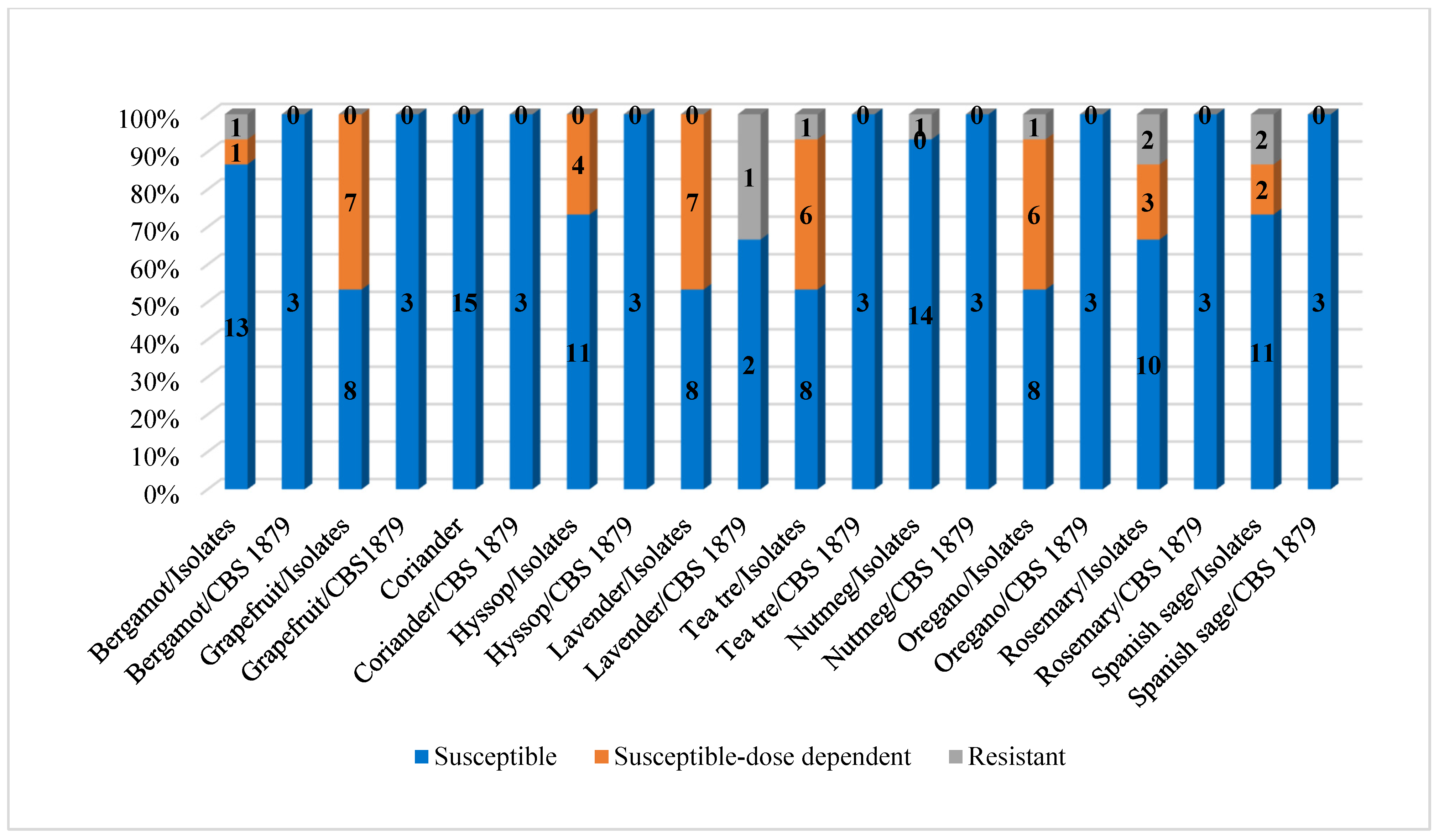
Figure 1. Susceptibility of M. pachydermatis strains to the tested plant EOs.
Microorganisms 13 02675 g001
Table 1. Characteristics of essential oils used in the experiment *.
Essential OilBotanical NameFamilyPlant PartMajor Components
CorianderCoriandrum sativum L.ApiaceaeFruitLinalool (64 ± 2%)
HyssopHyssopus officinalis L.LamiaceaeAerial part of the plantPinocamphone (50.0 ± 2%) Isopinocamphone (28.0 ± 1%)
α-pinene (11.0 ± 1%)
LavenderLavandula angustifolia MILLERFlowerLinalool (48 ± 2%)
RosemaryRosmarinus officinalis L.Leaf1,8-cineole (25.0 ± 1%)
α-pinene (19.0 ± 1%)
Camphor (19.0 ± 1%)
Spanish sageSalvia lavandulifolia VahlAerial part of the plantCamphor (27 ± 1)
OreganoOriganum vulgare L.Aerial part of the plantCarvacrol (57 ± 3%)
Tea treeMelaleuca alternifolia CheefMyrtaceaeLeafTerpinen-4-ol (33 ± 2)
NutmegMyristica fragrans HouttMyristicaceaeCoreα-pinene (18.0 ± 1%)
Sabinene (14.0 ± 1%)
β-pinene (13.0 ± 1%)
Myristicin (5 ± 0.2%)
BergamotCitrus aurantium L. subsp. bergamiaRutaceaePericarpLimonene (36 ± 1%)
Linalyl acetate
(23 ± 1%)
Linalool (15 ± 1%)
GrapefruitCitrus paradisi MacfadPericarpLimonene (87 ± 3%)
* Note—The table was prepared based on certificates obtained from the company Calendula a. s. (Nová Ľubovňa, SR). The major components were identified using gas chromatography analyses.
Table 2. Evaluation of plant essential oil MIC (µg/mL).
ParameterBergamotGrapefruitCorianderHyssopLavenderTea TreeNutmegOreganoRosemarySage
Clinical isolates
Minimum4008004004004004001600400400400
Maximum3125100,000400160080025,00012,50012,50031253125
x ¯ 821.67 a15,015 a,b,c,d,e,f400 b666.67 c586.67 d23605838.332430741.67 e816.67 f
SD715.0734,516.950493.77206.566281.132528.853057.90736.53947.44
Mode400312540040040040062503125400400
Median800160040040040040062501600400400
MIC50800160040040040040062501600400400
MIC901600100,0004001600800160062503125800800
Malassezia pachydermatis CBS 1879
Minimum80016004004004004001600400400400
Maximum8001600400400160040031251600400400
x ¯ 800 h1600400 g,400 i800 l400 j2616.67 g,h,i,j,k,l,m,n1200 k400 m400 n
SD0000692.820880.46692.8200
Mode800160040040040040031251600400400
Median800160040040040040031251600400400
x ¯ —average of MIC; MIC50 and MIC90—minimal inhibitory concentration at which 50% or 90% of the strains were inhibited; SD—standard deviation, a–n—MIC values with the same superscript letter are statistically significantly different (p ˂ 0.05).
Table 3. Susceptibility of Malassezia pachydermatis strains to tested plant essential oils.
ParameterBergamotGrapefruitCorianderHyssopLavenderTea TreeNutmegOreganoRosemarySage
Clinical isolates
S
(n/%)
13/86.668/53.3315/10011/73.338/53.338/53.3314/93.338/53.3310/66.6711/73.34
S-DD
(n/%)
1/6.677/46.6704/26.677/46.676/4006/403/202/13.33
R
(n/%)
1/6.6700001/6.671/6.671/6.672/13.332/13.33
Malassezia pachydermatis CBS 1879
S-DD
(n*/%)
3/1003/1003/1003/1002/66.673/1003/1003/1003/1003/100
Resistant
(n*/%)
00001/33.3300000
S—susceptible, S-DD—susceptible dose-dependent, R—resistant, n—number of strains, n*—the reference strain was tested in triplicate.
Disclaimer/Publisher’s Note: The statements, opinions and data contained in all publications are solely those of the individual author(s) and contributor(s) and not of MDPI and/or the editor(s). MDPI and/or the editor(s) disclaim responsibility for any injury to people or property resulting from any ideas, methods, instructions or products referred to in the content.