Article

Impact of Swabbing Location, Self-Swabbing, and Food Intake on SARS-CoV-2 RNA Detection

1
Division of Internal Medicine, University Hospital Basel, 4031 Basel, Switzerland
2
Department of Clinical Research, University of Basel, 4031 Basel, Switzerland
3
Laboratory of Biosensors and Bioelectronics, Institute for Biomedical Engineering, ETH Zurich, 8092 Zurich, Switzerland
4
Clinical Virology, University Hospital Basel, 4031 Basel, Switzerland
5
Transplantation & Clinical Virology, Department Biomedicine, University of Basel, 4031 Basel, Switzerland
6
Infectious Diseases & Hospital Epidemiology, University Hospital Basel, 4031 Basel, Switzerland
7
Botnar Research Centre for Child Health, 4051 Basel, Switzerland
*
Author to whom correspondence should be addressed.
Microorganisms 2024, 12(3), 591; https://doi.org/10.3390/microorganisms12030591
Submission received: 27 February 2024 / Revised: 13 March 2024 / Accepted: 13 March 2024 / Published: 15 March 2024
(This article belongs to the Section Medical Microbiology)

Abstract

:
This study compared SARS-CoV-2 RNA loads at different anatomical sites, and the impact of self-swabbing and food intake. Adult symptomatic patients with SARS-CoV-2 or non-SARS-CoV-2 respiratory tract infection were included between 2021 and 2022. Patients performed a nasal and buccal swab before a professionally collected nasopharyngeal/oropharyngeal swab (NOPS). Buccal swabs were collected fasting and after breakfast in a subgroup of patients. SARS-CoV-2 RNA loads were determined by nucleic acid testing. Swabbing convenience was evaluated using a survey. The median age of 199 patients was 54 years (interquartile range 38–68); 42% were female and 52% tested positive for SARS-CoV-2. The majority of patients (70%) were hospitalized. The mean SARS-CoV-2 RNA load was 6.6 log10 copies/mL (standard deviation (SD), ±1.5), 5.6 log10 copies/mL (SD ± 1.9), and 3.4 log10 copies/mL (SD ± 1.9) in the professionally collected NOPS, and self-collected nasal and buccal swabs, respectively (p < 0.0001). Sensitivity was 96.1% (95% CI 90.4–98.9) and 75.3% (95% CI 63.9–81.8) for the nasal and buccal swabs, respectively. After food intake, SARS-CoV-2 RNA load decreased (p = 0.0006). Buccal swabbing was the preferred sampling procedure for the patients. In conclusion, NOPS yielded the highest SARS-CoV-2 RNA loads. Nasal self-swabbing emerged as a reliable alternative in contrast to buccal swabs. If buccal swabs are used, they should be performed before food intake.

1. Introduction

Severe acute respiratory syndrome coronavirus 2 (SARS-CoV-2) causing coronavirus disease 2019 (COVID-19), is responsible for a major health crisis still generating repercussions in the fourth year after its emergence. Testing for SARS-CoV-2 remained one of the cornerstones to identify infected individuals early. Diagnostic testing allowed the breaking of chains of infection and enabled timely management of risk-stratified treatment, infection control measures, and contact tracing [1,2]. The gold standard testing technique defined by the World Health Organization (WHO) [3,4] is nucleic acid testing, preferably in a quantitative format (QNAT). This molecular assay is characterized by a high sensitivity and specificity [4]. However, the reliable performance of diagnostic QNATs is a resource-consuming and costly process, requiring laboratory set-up and professionally trained persons to process and collect the specimens. Although the burden on laboratories and resources was partly alleviated by rapid antigen testing and appropriate self-testing, QNAT remained the reference test for critical decision making in the clinics [5,6].
The performance of a nasopharyngeal or the combination of a nasopharyngeal with a pharyngeal swab (NOPS) collected by health care personnel was recommended by the WHO [5]. Nasal or mid-turbinate self-testing has been increasingly performed during the pandemic, and has been shown to give reliable results compared to professional collection of NOPS [7,8]. However, nasopharyngeal, nasal, and pharyngeal swabbing frequently cause discomfort or pain and may result in unwillingness to test, nose bleeding, and a higher risk of virus transmission to the health care personnel due to provoked sneezing and coughing [9,10]. Subsequently, saliva was increasingly used for large-scale testing, although it showed a lower sensitivity (approx. 85%) than NOPS [4,11] and was more difficult and time-consuming to process compared to swabs [12]. Due to its less invasive and more comfortable collection technique, it was accepted as a testing technique, in particular in children or elderly people, and in the setting of repetitive routine testing. Nevertheless, the easiest swabbing method might be buccal swabbing. It causes the least uncomfortable sensations, is easy to learn, and may be performed at home [13,14]. However, the SARS-CoV-2 RNA loads of buccal swabbing procedures may be influenced by additional factors such as enzymatic degradation, hygiene procedures, or food intake, leading to a lower test sensitivity observed in this specimen [15,16]. In previous studies, SARS-CoV-2 RNA loads have rarely been measured with quantitative test methods, but semi-quantitative QNAT systems (e.g., Roche cobas® system) or rapid antigen testing have been used to assess test performances.
This study aimed to compare SARS-CoV-2 RNA loads between different anatomical sites (nasal, buccal, nasopharyngeal/pharyngeal) and between professionally and self-collected swabs using QNAT. Furthermore, we aimed to assess the impact of food intake on RNA load in self-collected buccal swabs and patients’ satisfaction according to the swabbing method and location.

2. Materials and Methods

2.1. Study Design

This project was a prospective, observational, single-center, cohort study at the University Hospital Basel, a 750-bed tertiary care center in Switzerland. Patients were enrolled between January 2021 and December 2022. The study was approved by the Ethics Committee of Northwest and Central Switzerland (EKNZ Project-ID: 2020-02260) and conducted in accordance with the Declaration of Helsinki and the principles of Good Clinical Practice. All patients provided written informed consent for participation in the study.

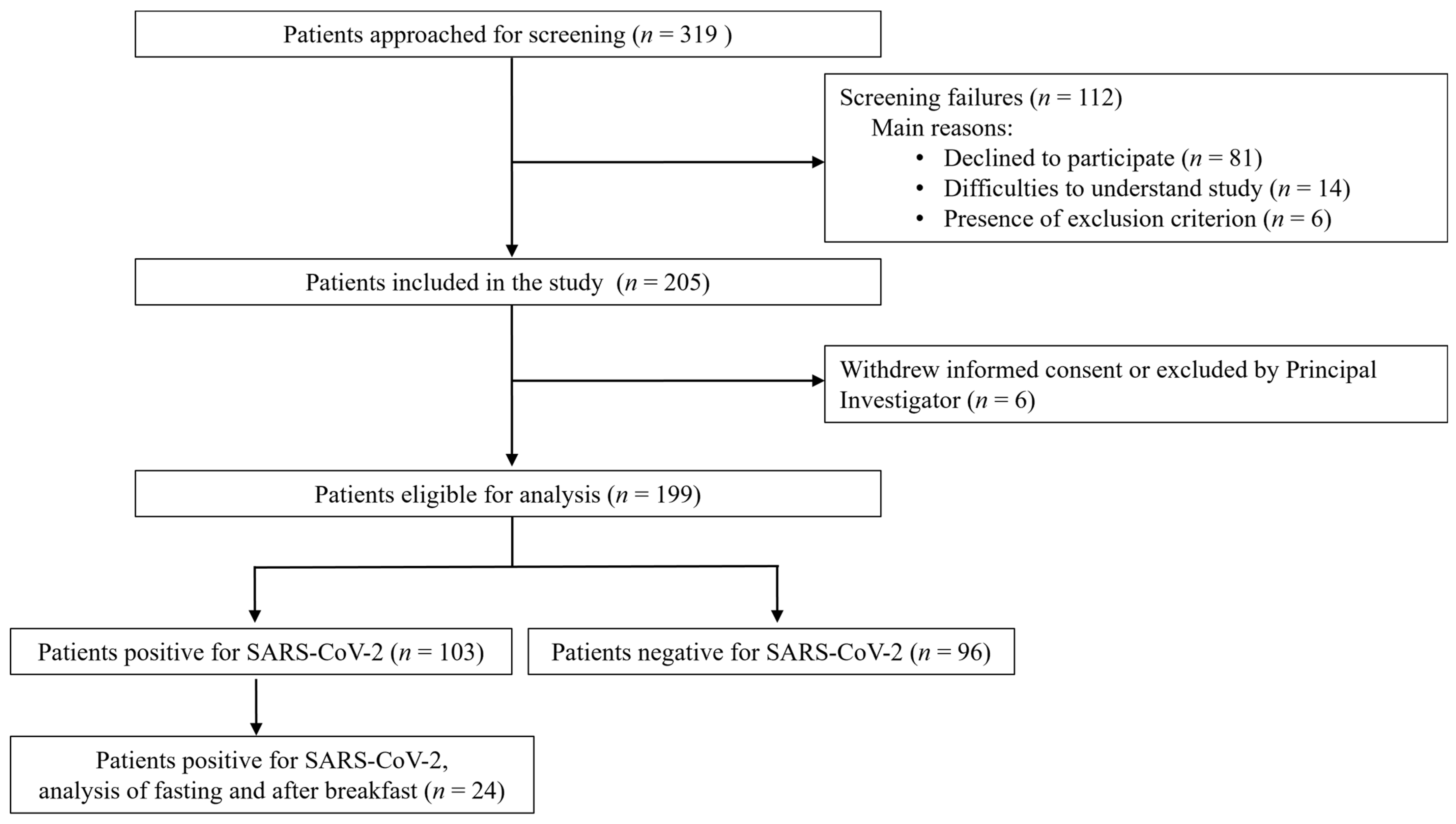
2.2. Study Population

Four different patient groups were enrolled in this study. Inclusion criteria comprised: age ≥ 18 years, and presence of one of the following: (1) at least one of the influenza-like illness (ILI) and/or COVID-19 symptoms and laboratory confirmed SARS-CoV-2 infection (by a positive SARS-CoV-2 QNAT result); (2) at least one of the ILI and/or COVID-19 symptoms, but laboratory confirmed SARS-CoV-2 negative (by a negative SARS-CoV-2 QNAT result) and detection of another virus causing respiratory tract infections; (3) at least one of the ILI and/or COVID-19 symptoms but laboratory confirmed SARS-CoV-2 negative (by a negative SARS-CoV-2 QNAT result) and no detection of another virus causing respiratory tract infections; or (4) outpatients presenting for SARS-CoV-2 testing at the local test center. ILI and/or COVID-19 symptoms comprised: fever, chills, cough, sore throat, rhinitis, earaches, dyspnea, headaches, dizziness, muscle/limb pain, chest pain, abdominal pain, nausea/vomiting, diarrhea, conjunctivitis, exanthema, lymphadenopathy, fatigue/weakness, dysuria, anosmia/ageusia. The onset of symptoms had to be within the previous 10 days. Exclusion criteria for all patients included previous enrolment into the current study, inability to perform a self-collected nasal or buccal swab, nose bleeding within the last 24 h, nasal surgery within the last 2 weeks, and acute facial trauma. For the routine SARS-CoV-2 testing and confirmation of infection, a professional NOPS was collected and analyzed via QNAT on the cobas® platform. A positive QNAT result was defined by a cycle threshold (Ct) value of <40.

2.3. Study Procedures and Sample Collection

After obtaining informed consent, written and visual but no oral instructions for self-collection of nasal (both nostrils) and buccal swabs were given to the patient (Figures S1–S3). The study physician was always present when self-swabbing was performed. A subgroup of patients who tested positive for SARS-CoV-2 (n = 24) was asked to collect two buccal swabs: the first fasting in the morning and before teeth brushing and the second after breakfast.
Nasal self-swabbing was performed by inserting the swab 2–3 cm into one nostril, twisting it five times, and leaving it there for ten seconds before the procedure was repeated with the same swab in the other nostril. Buccal self-swabbing was performed by swabbing five times on the inside of the cheek on both sides. For each swabbing location (nasal and buccal), a separate swab was used. After the swabbing procedure, which was not supported by the study physician, each swab was put into a separate tube containing 1.2 mL of viral culture medium. Subsequently, a NOPS was collected by a trained study physician. Both swabs were pooled into one tube containing 1.2 mL of viral culture medium.
Viral culture media (0.86× Hanks balanced salt solution (Gibco, Thermo Fisher Scientific, Waltham, MA, USA, 14060040), pH 7.4 adjusted by sodium bicarbonate solution) containing the swabs were directly processed to perform the QNAT or stored (max. 4 h) on ice or in the refrigerator (2–8 °C) before processing. After vortexing for 30 s, aliquots of 250 μL were prepared. The RNA loads of SARS-CoV-2 were determined by QNAT targeting the S-gene encoding a surface protein, the spike protein, of the virus. SARS-CoV-2 RNA loads were expressed in log10 copies/mL (c/mL) [17]. A SARS-CoV-2 RNA load of <1000 c/mL was defined as a negative test result. Syndromic panel testing for respiratory infections other than SARS-CoV-2 such as influenza A/B or respiratory syncytial virus was performed with QNAT using the Biofire® respiratory panel (biomerieux, Marcy-l’Étoile, France), GeneXpert® for co-testing of SARS-CoV-2, Influenza A, Influenza B and RSV (Cepheid, Sunnyvale, CA, USA) or the cobas® platform for co-testing of SARS-CoV-2, Influenza A and Influenza B (Roche, Rotkreuz, Switzerland) [17]. After the swabbing procedures, convenience and acceptance of the swabbing methods were assessed using a 15-item questionnaire. Regarding the questionnaire, answers about the swabbing convenience were summarized into four categories: uncomplicated, acceptable, uncomfortable, painful, and not tolerable, which was merged into the fourth category.
Patient data were prospectively collected and managed using Research Electronic Data Capture (REDCap® 14.2.1) hosted at the University Hospital Basel. Routine clinical variables were extracted from the patient’s records of the hospital information system and were entered in an electronic case report form in REDCap® [18,19].

2.4. Statistical Analysis

The Mann–Whitney U-test, the Chi Square test, and Fisher’s exact test were used where appropriate. Data on SARS-CoV-2 RNA load were analyzed by a mixed-effects model with post hoc Tukey adjustment or by paired-sample t-test to compare means. Variable selection for characteristics of study participants was based on biological plausibility and/or demonstrated associations in the literature. For the correlation testing, Spearman correlation was used. Results were considered statistically significant if the p-value was less than 0.05. To calculate the diagnostic accuracy of the swabbing procedure, sensitivity, specificity, positive and negative likelihood ratios, and positive and negative predictive values were calculated with their 95% confidence interval (CI). Furthermore, sensitivity was calculated fasting compared to after breakfast for a sub-group of patients. The reference standard method was the NOPS collected by a trained study physician. All analyses were performed with SPSS Version 28 (IBM SPSS Statistics for Windows. Armonk, NY, USA) and GraphPad Prism 9.0.0 (GraphPad Software, San Diego, CA, USA).

3. Results

3.1. Demographic and Clinical Characteristics

A total of 205 patients was enrolled in this study, and 199 patients completed the study (Figure 1).
Outpatients were most frequently recruited at the test center (60/61, 98%). Four patients withdrew their consent, and two patients were excluded at the investigator’s discretion because of difficulties in performing the self-swabbing, and SARS-CoV-2 replication confined to the lower respiratory tract only. The median age was 54 years (interquartile range (IQR) 38–68), 42% (83/199) were female, and 70% were hospitalized (139/199). Overall, 103/199 patients (52%) were tested positive for SARS-CoV-2. The median time from symptom onset to the swabbing was 4 days (IQR 2–7). The majority of patients had either one (n = 45, 23%) or no comorbidity (n = 86, 43%). In both groups of patients tested negative or positive for SARS-CoV-2, the main comorbidities were arterial hypertension (21.9% and 23.3%, respectively), cardiovascular disease (15.6% and 23.3%, respectively), and diabetes mellitus (14.6% and 19.4%, respectively) (Table 1).
Symptoms present among patients who tested positive for SARS-CoV-2 are presented in Table S1.

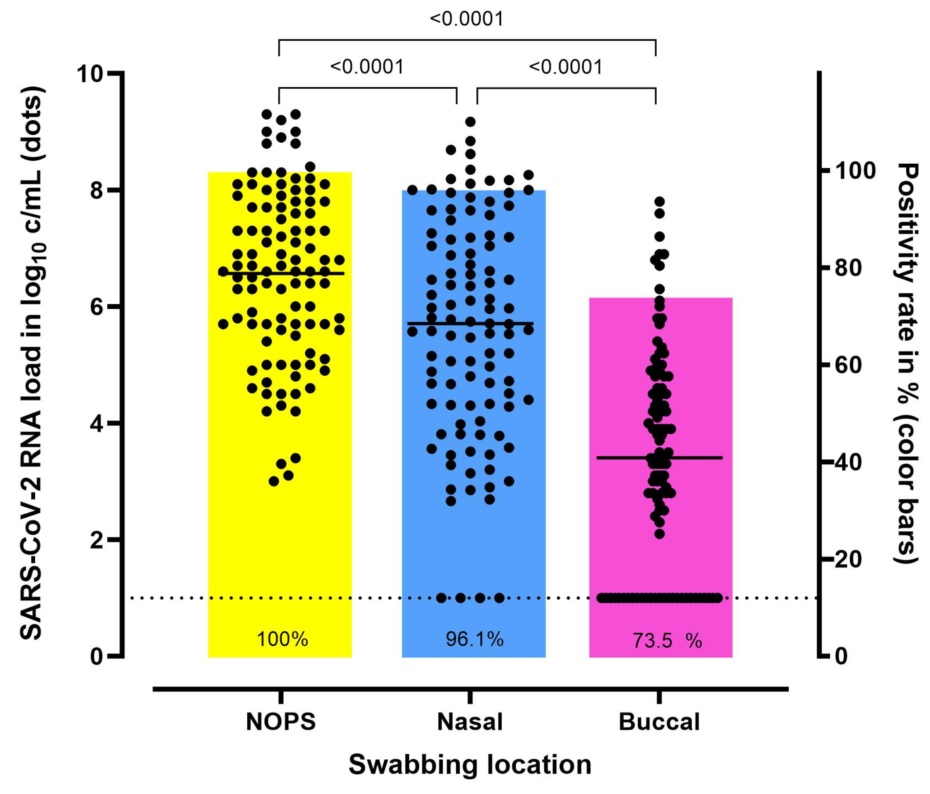
3.2. Comparison of SARS-CoV-2 RNA Load According to the Swabbing Location

In total, 103 SARS-CoV-2 positive-tested patients were included. The RNA load in one of the 103 buccal samples could not be determined. The mean SARS-CoV-2 RNA load was 6.6 log10 c/mL (standard deviation (SD), ±1.5) in the professionally collected NOPS and 5.6 log10 c/mL (SD, ±1.9) and 3.4 log10 c/mL (SD, ±1.9) in the self-collected nasal and buccal swabs, respectively, p < 0.0001 (Figure 2).
In a Bland–Altman plot, the difference between SARS-CoV-2 RNA loads in NOPS and nasal swabs is displayed (Figure S4). Compared to NOPS, 4/103 (4%) and 27/102 (26%) of the nasal and buccal swabs were false negative, respectively. The SARS-CoV-2 RNA load in the nasal and the NOPS correlated negatively with the symptom duration (r = −0.52, p < 0.001). A similar result was observed with regard to buccal swabs (r = −0.335, p < 0.001).

3.3. Diagnostic Accuracy

Table 2 displays the diagnostic value of the different swabbing locations. Nasal and buccal self-swabbing showed a sensitivity of 96.1% (95% CI: 90.4–98.9) and 75.3% (95% CI: 63.9–81.8), respectively. Specificity was high at both anatomical sites (nasal 100% (95% CI 96.2–100, buccal 99.0% (94.3–100))). The majority of false-negative test results was observed in the group of self-collected buccal swabs.

3.4. The Impact of Food Intake on Buccal SARS-CoV-2 RNA Load

In 24/103 patients (23%) who tested positive for SARS-CoV-2, two buccal self-swabs were performed before (fasting) and after breakfast. The mean SARS-CoV-2 RNA load was 3.8 log10 c/mL (SD, ±1.5) before and 2.6 log10 c/mL (SD, ±1.3) after breakfast (p = 0.0006) (Figure 3).
Fasting swabbing demonstrated a sensitivity of 87.5% (95% CI: 67.6–97.3), while a decrease in sensitivity was seen after food intake to 66.7% (95% CI: 44.7–84.4).

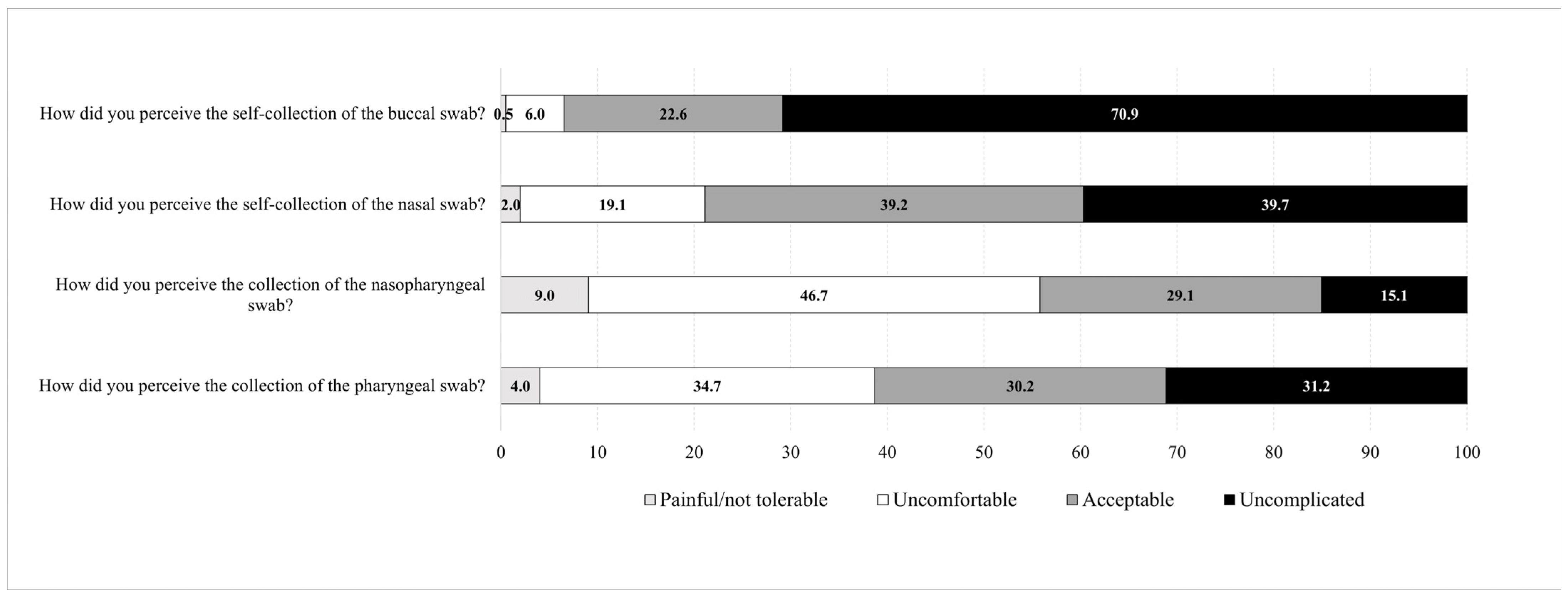
3.5. Patients’ Convenience Related to the Collection Method and Anatomical Site

The questionnaire was completed by all 199 patients. The majority of patients (191/199, 96%) stated that the self-instructions for nasal and buccal self-swabbing were easy to follow. Overall, 64% (127/199) of the patients had never performed a nasal swab, and 89% (178/199) had never performed a buccal swab before participating in the study. The professionally collected NOPS were painful/not tolerable or caused discomfort to 56% (111/199) and 39% (77/199) of the patients, respectively. Self-collected nasal and buccal swabs were stated to cause pain or discomfort in 21% (42/199) and 7% (13/199) of the patients, respectively (Figure 4).
The nasal and buccal self-swabbing were easy to perform for 91% (182/199) and 97% (192/199) of the patients, respectively. Furthermore, the great majority of patients was confident to perform nasal or buccal self-testing at home (191/199, 96%, and 192/199, 96%, respectively). Most patients (131/199, 66%) would prefer self-swabbing over being swabbed by a professional healthcare worker.

4. Discussion

In the present study, we evaluated SARS-CoV-2 RT-PCR testing at different locations in a cohort of 199 patients, and demonstrated that NOPS yielded the highest SARS-CoV-2 RNA loads. Nasal self-swabbing evolved as a reliable alternative to professionally collected NOPS. Buccal swabs had a significantly lower sensitivity compared to NOPS and nasal swabbing, but were the most convenient swabbing procedure for the patient. SARS-CoV-2 RNA loads decreased significantly after food intake.
The SARS-CoV-2 RNA load differed significantly between NOPS, nasal, and buccal swabbing, demonstrating a sensitivity of 96.1% for nasal and 73.5% for buccal self-swabbing for the detection of SARS-CoV-2. Despite a slightly lower SARS-CoV-2 RNA load observed in nasal swabs compared to NOPS, the high sensitivity of 96.1% of self-collected nasal swabs underscores the reliability of this swabbing location, which has already been shown previously [4,20,21,22]. Consequently, nasal swabbing entered international guideline recommendations as a reliable alternative for professionally collected nasopharyngeal swabs [5,23]. Furthermore, self-collected nasal swabs for SARS-CoV-2 are easy to perform, and are highly accepted [8,24,25]. Interestingly, the sensitivity of the nasal swabs was slightly higher in the present study compared to previously published studies, stating a sensitivity of approximately 85% for professionally collected or self-collected nasal swabs [4,26,27,28]. This observed difference may be related to the sensitive quantitative testing method used in the present study compared to semi-quantitative techniques or rapid antigen testing used in previous studies [4,27,29,30]. Although it has been shown that semi-quantitative techniques correlate qualitatively and quantitatively, the sensitivity of the QNAT used in the present study might be slightly higher [17]. In line, Teo et al. demonstrated that not only different testing techniques, but also the use of different RT-PCR kits impacts the sensitivity of the test results [31]. Furthermore, the majority of patients were hospitalized and were included early during the course of the disease, therefore representing a sicker patient population with a possibly higher viral load. Additionally, the introduction of public self-testing strategies may have led to an improved quality of the self-swabbing procedure and a higher sensitivity.
In the present study, the sensitivity of buccal swabs was 73.5% and, therefore, significantly lower than the sensitivity of nasal swabs. In previous studies, their performance was even poorer (sensitivity of oral swabs (rapid antigen tests): 9% [32], 18% [13], and 32% [15]) (sensitivity of buccal swabs (semi-quantitative QNAT): 20.8% [33], 65.4% [9]. 56.7% [34]). The distinctly higher sensitivity in the present study may be associated with the very sensitive testing method used, but also the patient population included, as the majority of patients were symptomatic and the median time between symptom onset and inclusion in the study was only 4 days, resulting in high SARS-CoV-2 RNA loads at the time of testing. Furthermore, the differences in sensitivity of oral or buccal swabbing in previous studies might be related to the instructions patients were given before swabbing, ranging from fasting 30 min before testing [13,15] to additional coughing before sample collection [15] or no instructions at all [33]. Optimal patient instructions may be crucial, especially when collecting buccal or saliva samples. If correctly instructed and performed, saliva samples may even yield a comparable or higher sensitivity compared to NOPS [31]. In line with this, the sensitivity of buccal swabs increased significantly in the present study when the patients were optimally instructed, i.e., to perform the buccal swabbing fasting.
The higher SARS-CoV-2 RNA load in the fasting samples may be related to viral particles accumulating in the oropharynx and mouth during nighttime, resulting in higher oral virus concentrations in the morning [35]. During food intake, more saliva is produced, which leads to a dilution of viral particles in the mouth. Furthermore, a large number of viral particles are probably swallowed [36]. Saliva is rich in RNA degrading enzymes and potential PCR inhibitors, which may break down the viral RNA, and reduce the yield even of the very sensitive QNAT testing. A similar impact on viral load has been presented when saliva samples are collected after mouthwashing [37].
Overall, despite the higher sensitivity of buccal self-swabbing observed in the present study compared to previously published studies, it cannot be recommended as a reliable alternative to the professionally collected NOPS or the self-collected nasal swab due to the high rate of false-negative results, which is in line with previous studies [9,32]. If buccal swabs or oral specimens are used for SARS-CoV-2 testing, it is recommended to collect them fasting in the morning before food intake, drinking, or brushing teeth, to increase the overall sensitivity. Importantly, the positivity rate would further decrease when other, less sensitive test methods such as rapid antigen tests are used [38], and when the patient is not instructed correctly.
In the survey conducted in the present study, the majority of patients (56%) judged professionally collected NOPS as painful/not tolerable or causing discomfort. Conversely, self-collected nasal and buccal swabs were rated in regard to the same inconveniences with only 21% and 7%, respectively. Accordingly, two-thirds of the patients (66%) would prefer self-swabbing over being swabbed by a professional healthcare worker, which is in line with previous studies [26,39,40].
The presented study has several limitations. The COVID-19 patients were all inpatients, representing a more severely ill patient population. There was a selection bias, as patients who participated in the study were more likely to be interested in COVID-19-related testing in general and, hence, the performance of self-swabbing might be better compared to the general population. Additionally, as a member of the study team was always present during the self-swabbing procedure, the participants may have followed the instructions more closely. As the questionnaire was not answered anonymously, but in the presence of the study team, the answers may have been biased, as patients may have felt that a socially accepted answer should be provided.
However, the results of our study may not solely be applicable for SARS-CoV-2 infection, but also for the confirmation of other viral respiratory pathogens, and, hence, may be relevant for testing strategies in future pandemics. In line, Jung et al. demonstrated the superiority of NOPS for detecting non-SARS-CoV-2 respiratory viruses, but concluded that nasal swabs and saliva samples may represent alternatives to NOPS [28], which is in accordance with our findings.
In conclusion, SARS-CoV-2 RNA load differed significantly between professionally collected NOPS and self-collected nasal and buccal swabs. Self-performed nasal swabs proved to be a reliable diagnostic method in symptomatic COVID-19 patients. The practice of nasal swab collection is supported by patients favoring self-swabbing over professional swabbing. Conversely, buccal swabbing cannot be recommended as an alternative swabbing method due to the high rate of false negative results. Food intake impacts significantly on the oral SARS-CoV-2 RNA load. Therefore, if buccal swabbing or other oral specimens are used, they should be collected during fasting in the morning. As convenience, acceptance, and performance of nasal self-swabbing is high, it should remain a cornerstone of any SARS-CoV-2 testing strategy. These results may inform testing strategies in future pandemics.

Supplementary Materials

The following supporting information can be downloaded at: https://www.mdpi.com/article/10.3390/microorganisms12030591/s1, Figure S1: Instruction file 1 for the self-collection of nasal and buccal swabs; Figure S2: Instructions for the self-collection of nasal swabs; Figure S3: Instructions for the self-collection of buccal swabs; Figure S4: Bland–Altman plot showing the differences of SARS-CoV-2 RNA load between professionally collected nasopharyngeal/oropharyngeal swabs and nasal swabs. NOPS, nasopharyngeal/oropharyngeal swab; SARS-CoV-2, severe acute respiratory syndrome coronavirus 2. Table S1: Symptoms and clinical signs of the patients tested positive for SARS-CoV-2 (n = 103). Data are presented as count (percentages) or median (interquartile range).

Author Contributions

Conceptualization, S.D. and M.O.; Formal analysis, S.D., A.H.-H., K.L. and M.O.; Funding acquisition, M.O.; Investigation, S.D., F.B., M.B. and M.O.; Methodology, V.J., A.T., Y.B. and M.O.; Project administration, M.O.; Resources, M.O.; Supervision, M.O.; Validation, A.H.-H.; Visualization, S.D. and M.O.; Writing—original draft, S.D. and M.O.; Writing—review and editing, S.D., F.B., M.B., A.H.-H., V.J., A.T., Y.B., K.L., H.H.H. and M.O. All authors have read and agreed to the published version of the manuscript.

Funding

This research was funded by the Botnar Research Centre for Child Health as part of the Fast Track Call for Acute Global Health Challenges.

Data Availability Statement

The datasets generated are available from the corresponding author on reasonable request (requiring ethics approval).

Conflicts of Interest

The authors declare no conflict of interest.

References

  1. Atzrodt, C.L.; Maknojia, I.; McCarthy, R.D.P.; Oldfield, T.M.; Po, J.; Ta, K.T.L.; Stepp, H.E.; Clements, T.P. A Guide to COVID-19: A global pandemic caused by the novel coronavirus SARS-CoV-2. FEBS J. 2020, 287, 3633–3650. [Google Scholar] [CrossRef] [PubMed]
  2. Kritikos, A.; Caruana, G.; Brouillet, R.; Miroz, J.P.; Abed-Maillard, S.; Stieger, G.; Opota, O.; Croxatto, A.; Vollenweider, P.; Bart, P.A.; et al. Sensitivity of Rapid Antigen Testing and RT-PCR Performed on Nasopharyngeal Swabs versus Saliva Samples in COVID-19 Hospitalized Patients: Results of a Prospective Comparative Trial (RESTART). Microorganisms 2021, 9, 1910. [Google Scholar] [CrossRef] [PubMed]
  3. Rahbari, R.; Moradi, N.; Abdi, M. rRT-PCR for SARS-CoV-2: Analytical considerations. Clin. Chim. Acta 2021, 516, 1–7. [Google Scholar] [CrossRef]
  4. Tsang, N.N.Y.; So, H.C.; Ng, K.Y.; Cowling, B.J.; Leung, G.M.; Ip, D.K.M. Diagnostic performance of different sampling approaches for SARS-CoV-2 RT-PCR testing: A systematic review and meta-analysis. Lancet Infect. Dis. 2021, 21, 1233–1245. [Google Scholar] [CrossRef]
  5. WHO. Diagnostic Testing for SARS-CoV-2 Online: WHO. 2020. Available online: https://iris.who.int/bitstream/handle/10665/334254/WHO-2019-nCoV-laboratory-2020.6-eng.pdf?sequence=1 (accessed on 17 November 2023).
  6. Van der Moeren, N.; Zwart, V.F.; Louise van Leest, M.; Thijssen, M.; Groenewegen, R.; Heer, M.K.; Murk, J.L.; Tjhie, J.T.; Diederen, B.M.W.; Stohr, J. A SARS-CoV-2 and influenza rapid antigen test-based hospital isolation policy awaiting RT-PCR, a prospective observational study. Clin. Microbiol. Infect. 2023, 29, 1595–1599. [Google Scholar] [CrossRef]
  7. Wehrhahn, M.C.; Robson, J.; Brown, S.; Bursle, E.; Byrne, S.; New, D.; Chong, S.; Newcombe, J.P.; Siversten, T.; Hadlow, N. Self-collection: An appropriate alternative during the SARS-CoV-2 pandemic. J. Clin. Virol. 2020, 128, 104417. [Google Scholar] [CrossRef] [PubMed]
  8. Goyal, S.; Prasert, K.; Praphasiri, P.; Chittaganpitch, M.; Waicharoen, S.; Ditsungnoen, D.; Jaichuang, S.; Lindblade, K.A. The acceptability and validity of self-collected nasal swabs for detection of influenza virus infection among older adults in Thailand. Influenza Other Respir. Viruses 2017, 11, 412–417. [Google Scholar] [CrossRef]
  9. Laferl, H.; Seitz, T.; Baier-Grabner, S.; Kelani, H.; Scholz, E.; Heger, F.; Gotzinger, F.; Frischer, P.T.; Wenisch, C.; Allerberger, P.F. Evaluation of RT-qPCR of mouthwash and buccal swabs for detection of SARS-CoV-2 in children and adults. Am. J. Infect. Control 2022, 50, 176–181. [Google Scholar] [CrossRef]
  10. Baker, J.M.; Nakayama, J.Y.; O’Hegarty, M.; McGowan, A.; Teran, R.A.; Bart, S.M.; Mosack, K.; Roberts, N.; Campos, B.; Paegle, A.; et al. SARS-CoV-2 B.1.1.529 (Omicron) Variant Transmission Within Households—Four U.S. Jurisdictions, November 2021–February 2022. MMWR Morb. Mortal. Wkly. Rep. 2022, 71, 341–346. [Google Scholar] [CrossRef]
  11. Jamal, A.J.; Mozafarihashjin, M.; Coomes, E.; Anceva-Sami, S.; Barati, S.; Crowl, G.; Faheem, A.; Farooqi, L.; Kandel, C.E.; Khan, S.; et al. Sensitivity of midturbinate versus nasopharyngeal swabs for the detection of severe acute respiratory syndrome coronavirus 2 (SARS-CoV-2). Infect. Control Hosp. Epidemiol. 2021, 42, 1001–1003. [Google Scholar] [CrossRef]
  12. Landry, M.L.; Criscuolo, J.; Peaper, D.R. Challenges in use of saliva for detection of SARS CoV-2 RNA in symptomatic outpatients. J. Clin. Virol. 2020, 130, 104567. [Google Scholar] [CrossRef] [PubMed]
  13. Wolfl-Duchek, M.; Bergmann, F.; Jorda, A.; Weber, M.; Muller, M.; Seitz, T.; Zoufaly, A.; Strassl, R.; Zeitlinger, M.; Herkner, H.; et al. Sensitivity and Specificity of SARS-CoV-2 Rapid Antigen Detection Tests Using Oral, Anterior Nasal, and Nasopharyngeal Swabs: A Diagnostic Accuracy Study. Microbiol. Spectr. 2022, 10, e0202921. [Google Scholar] [CrossRef] [PubMed]
  14. Kojima, N.; Turner, F.; Slepnev, V.; Bacelar, A.; Deming, L.; Kodeboyina, S.; Klausner, J.D. Self-Collected Oral Fluid and Nasal Swabs Demonstrate Comparable Sensitivity to Clinician Collected Nasopharyngeal Swabs for Coronavirus Disease 2019 Detection. Clin. Infect. Dis. 2021, 73, e3106–e3109. [Google Scholar] [CrossRef] [PubMed]
  15. Blanco, I.; Violan, C.; Suner, C.; Garcia-Prieto, J.; Argerich, M.J.; Rodriguez-Illana, M.; Moreno, N.; Cardona, P.J.; Blanco, A.; Toran-Monserrat, P.; et al. Comparison between mid-nasal swabs and buccal swabs for SARS-CoV-2 detection in mild COVID-19 patients. J. Infect. 2022, 84, e78–e79. [Google Scholar] [CrossRef] [PubMed]
  16. Hanson, K.E.; Barker, A.P.; Hillyard, D.R.; Gilmore, N.; Barrett, J.W.; Orlandi, R.R.; Shakir, S.M. Self-Collected Anterior Nasal and Saliva Specimens versus Health Care Worker-Collected Nasopharyngeal Swabs for the Molecular Detection of SARS-CoV-2. J. Clin. Microbiol. 2020, 58, e01824-20. [Google Scholar] [CrossRef] [PubMed]
  17. Leuzinger, K.; Gosert, R.; Sogaard, K.K.; Naegele, K.; Bielicki, J.; Roloff, T.; Bingisser, R.; Nickel, C.H.; Khanna, N.; Sutter, S.T.; et al. Epidemiology and precision of SARS-CoV-2 detection following lockdown and relaxation measures. J. Med. Virol. 2021, 93, 2374–2384. [Google Scholar] [CrossRef] [PubMed]
  18. Harris, P.A.; Taylor, R.; Thielke, R.; Payne, J.; Gonzalez, N.; Conde, J.G. Research electronic data capture (REDCap)—A metadata-driven methodology and workflow process for providing translational research informatics support. J. Biomed. Inform. 2009, 42, 377–381. [Google Scholar] [CrossRef]
  19. Harris, P.A.; Taylor, R.; Minor, B.L.; Elliott, V.; Fernandez, M.; O’Neal, L.; McLeod, L.; Delacqua, G.; Delacqua, F.; Kirby, J.; et al. The REDCap consortium: Building an international community of software platform partners. J. Biomed. Inform. 2019, 95, 103208. [Google Scholar] [CrossRef]
  20. Altamirano, J.; Govindarajan, P.; Blomkalns, A.L.; Kushner, L.E.; Stevens, B.A.; Pinsky, B.A.; Maldonado, Y. Assessment of Sensitivity and Specificity of Patient-Collected Lower Nasal Specimens for Severe Acute Respiratory Syndrome Coronavirus 2 Testing. JAMA Netw. Open 2020, 3, e2012005. [Google Scholar] [CrossRef]
  21. Kagan, R.M.; Rogers, A.A.; Borillo, G.A.; Clarke, N.J.; Marlowe, E.M. Performance of Unobserved Self-Collected Nasal Swabs for Detection of SARS-CoV-2 by RT-PCR Utilizing a Remote Specimen Collection Strategy. Open Forum. Infect. Dis. 2021, 8, ofab039. [Google Scholar] [CrossRef]
  22. Tu, Y.P.; Jennings, R.; Hart, B.; Cangelosi, G.A.; Wood, R.C.; Wehber, K.; Verma, P.; Vojta, D.; Berke, E.M. Swabs Collected by Patients or Health Care Workers for SARS-CoV-2 Testing. N. Engl. J. Med. 2020, 383, 494–496. [Google Scholar] [CrossRef] [PubMed]
  23. CDC. Interim Guidelines for Clinical Specimens for COVID-19 | CDC, (n.d.): CDC. 2021. Available online: https://www.cdc.gov/coronavirus/2019-ncov/lab/guidelines-clinical-specimens.html (accessed on 17 November 2023).
  24. Dhiman, N.; Miller, R.M.; Finley, J.L.; Sztajnkrycer, M.D.; Nestler, D.M.; Boggust, A.J.; Jenkins, S.M.; Smith, T.F.; Wilson, J.W.; Cockerill, F.R., 3rd; et al. Effectiveness of patient-collected swabs for influenza testing. Mayo Clin. Proc. 2012, 87, 548–554. [Google Scholar] [CrossRef] [PubMed]
  25. Akmatov, M.K.; Gatzemeier, A.; Schughart, K.; Pessler, F. Equivalence of self- and staff-collected nasal swabs for the detection of viral respiratory pathogens. PLoS ONE 2012, 7, e48508. [Google Scholar] [CrossRef] [PubMed]
  26. Marx, G.E.; Biggerstaff, B.J.; Nawrocki, C.C.; Totten, S.E.; Travanty, E.A.; Burakoff, A.W.; Scott, T.; De Hey, J.C.; Carlson, J.J.; Wendel, K.A.; et al. Detection of Severe Acute Respiratory Syndrome Coronavirus 2 on Self-Collected Saliva or Anterior Nasal Specimens Compared With Healthcare Personnel-Collected Nasopharyngeal Specimens. Clin. Infect. Dis. 2021, 73, S65–S73. [Google Scholar] [CrossRef]
  27. Todsen, T.; Jakobsen, K.K.; Gronlund, M.P.; Callesen, R.E.; Folke, F.; Larsen, H.; Ersboll, A.K.; Benfield, T.; Gredal, T.; Klokker, M.; et al. COVID-19 Rapid Antigen Tests With Self-Collected vs. Health Care Worker-Collected Nasal and Throat Swab Specimens: A Randomized Clinical Trial. JAMA Netw. Open 2023, 6, e2344295. [Google Scholar] [CrossRef]
  28. Jung, E.J.; Lee, S.K.; Shin, S.H.; Kim, J.S.; Woo, H.; Cho, E.J.; Hyun, J.; Kim, J.S.; Kim, H.S. Comparison of Nasal Swabs, Nasopharyngeal Swabs, and Saliva Samples for the Detection of SARS-CoV-2 and other Respiratory Virus Infections. Ann. Lab. Med. 2023, 43, 434–442. [Google Scholar] [CrossRef]
  29. Viloria Winnett, A.; Akana, R.; Shelby, N.; Davich, H.; Caldera, S.; Yamada, T.; Reyna, J.R.B.; Romano, A.E.; Carter, A.M.; Kim, M.K.; et al. Daily SARS-CoV-2 Nasal Antigen Tests Miss Infected and Presumably Infectious People Due to Viral Load Differences among Specimen Types. Microbiol. Spectr. 2023, 11, e0129523. [Google Scholar] [CrossRef]
  30. Goodall, B.L.; LeBlanc, J.J.; Hatchette, T.F.; Barrett, L.; Patriquin, G. Investigating the Sensitivity of Nasal or Throat Swabs: Combination of Both Swabs Increases the Sensitivity of SARS-CoV-2 Rapid Antigen Tests. Microbiol. Spectr. 2022, 10, e0021722. [Google Scholar] [CrossRef]
  31. Teo, A.K.J.; Choudhury, Y.; Tan, I.B.; Cher, C.Y.; Chew, S.H.; Wan, Z.Y.; Cheng, L.T.E.; Oon, L.L.E.; Tan, M.H.; Chan, K.S.; et al. Saliva is more sensitive than nasopharyngeal or nasal swabs for diagnosis of asymptomatic and mild COVID-19 infection. Sci. Rep. 2021, 11, 3134. [Google Scholar] [CrossRef] [PubMed]
  32. Schrom, J.; Marquez, C.; Pilarowski, G.; Wang, C.Y.; Mitchell, A.; Puccinelli, R.; Black, D.; Rojas, S.; Ribeiro, S.; Tulier-Laiwa, V.; et al. Comparison of SARS-CoV-2 Reverse Transcriptase Polymerase Chain Reaction and BinaxNOW Rapid Antigen Tests at a Community Site During an Omicron Surge: A Cross-Sectional Study. Ann. Intern. Med. 2022, 175, 682–690. [Google Scholar] [CrossRef] [PubMed]
  33. Connor, M.C.; Copeland, M.; Curran, T. Investigation of saliva, tongue swabs and buccal swabs as alternative specimen types to nasopharyngeal swabs for SARS-CoV-2 testing. J. Clin. Virol. 2022, 146, 105053. [Google Scholar] [CrossRef]
  34. Ku, C.W.; Shivani, D.; Kwan, J.Q.T.; Loy, S.L.; Erwin, C.; Ko, K.K.K.; Ng, X.W.; Oon, L.; Thoon, K.C.; Kalimuddin, S.; et al. Validation of self-collected buccal swab and saliva as a diagnostic tool for COVID-19. Int. J. Infect. Dis. 2021, 104, 255–261. [Google Scholar] [CrossRef] [PubMed]
  35. Hung, D.L.; Li, X.; Chiu, K.H.; Yip, C.C.; To, K.K.; Chan, J.F.; Sridhar, S.; Chung, T.W.; Lung, K.C.; Liu, R.W.; et al. Early-Morning vs. Spot Posterior Oropharyngeal Saliva for Diagnosis of SARS-CoV-2 Infection: Implication of Timing of Specimen Collection for Community-Wide Screening. Open Forum. Infect. Dis. 2020, 7, ofaa210. [Google Scholar] [CrossRef] [PubMed]
  36. Tng, D.J.H.; Yin, B.C.Y.; Cao, J.; Ko, K.K.K.; Goh, K.C.M.; Chua, D.X.W.; Zhang, Y.; Chua, M.L.K.; Low, J.G.H.; Ooi, E.E.; et al. Amplified parallel antigen rapid test for point-of-care salivary detection of SARS-CoV-2 with improved sensitivity. Mikrochim. Acta 2021, 189, 14. [Google Scholar] [CrossRef] [PubMed]
  37. Costa, M.M.; Benoit, N.; Tissot-Dupont, H.; Million, M.; Pradines, B.; Granjeaud, S.; Almeras, L. Mouth Washing Impaired SARS-CoV-2 Detection in Saliva. Diagnostics 2021, 11, 1509. [Google Scholar] [CrossRef]
  38. Puyskens, A.; Michel, J.; Stoliaroff-Pepin, A.; Bayram, F.; Sesver, A.; Wichmann, O.; Harder, T.; Schaade, L.; Nitsche, A.; Peine, C. Direct comparison of clinical diagnostic sensitivity of saliva from buccal swabs versus combined oro-/nasopharyngeal swabs in the detection of SARS-CoV-2 B.1.1.529 Omicron. J. Clin. Virol. 2023, 165, 105496. [Google Scholar] [CrossRef]
  39. Wurstle, S.; Spinner, C.D.; Voit, F.; Hoffmann, D.; Hering, S.; Weidlich, S.; Schneider, J.; Zink, A.; Treiber, M.; Iakoubov, R.; et al. Self-sampling versus health care professional-guided swab collection for SARS-CoV-2 testing. Infection 2021, 49, 927–934. [Google Scholar] [CrossRef]
  40. Valentine-Graves, M.; Hall, E.; Guest, J.L.; Adam, E.; Valencia, R.; Shinn, K.; Hardee, I.; Sanchez, T.; Siegler, A.J.; Sullivan, P.S. At-home self-collection of saliva, oropharyngeal swabs and dried blood spots for SARS-CoV-2 diagnosis and serology: Post-collection acceptability of specimen collection process and patient confidence in specimens. PLoS ONE 2020, 15, e0236775. [Google Scholar] [CrossRef]
Figure 1. Flow chart. Two patients were excluded at the investigator’s discretion due to intolerance of nasopharyngeal swabbing, and due to replication of SARS-CoV-2 only in the lower respiratory tract at time of inclusion. SARS-CoV-2, severe acute respiratory syndrome coronavirus 2.
Microorganisms 12 00591 g001
Figure 2. Comparison of the SARS-CoV-2 RNA load in professionally collected nasopharyngeal/oropharyngeal (NOPS) swabs and in self-collected nasal and buccal swabs (n = 103). In patients who performed two buccal swabs (n = 24), the result of the first (fasting) swab was included. Horizontal lines represent the mean of the SARS-CoV-2 RNA load in log10. The RNA loads of SARS-CoV-2 were determined by a quantitative nucleic acid amplification test targeting the S-gene encoding the spike protein, of the virus. SARS-CoV-2 RNA load was analyzed by a mixed-effects model with post hoc Tukey adjustment. Dashed line: limit of detection. NOPS, nasopharyngeal/oropharyngeal swab; SARS-CoV-2, severe acute respiratory syndrome coronavirus 2.
Microorganisms 12 00591 g002
Figure 3. Impact of food intake on the SARS-CoV-2 RNA load in self-collected buccal swabs before and after breakfast, and according to positivity rate compared to professionally performed nasopharyngeal/pharyngeal test (n = 24). Horizontal lines represent the mean of the SARS-CoV-2 RNA load in log10. Dashed line: limit of detection. SARS-CoV-2 RNA load was analyzed by a paired-sample t-test to compare means. SARS-CoV-2, severe acute respiratory syndrome coronavirus 2.
Microorganisms 12 00591 g003
Figure 4. Convenience and acceptance related to self-collected swabs and the swabbing location as assessed by a questionnaire (n = 199, 100%).
Microorganisms 12 00591 g004
Table 1. Baseline characteristics of the study participants. Data are presented as count (percentages) or median (interquartile range).
VariableCOVID-19 Patients n = 103Non-COVID-19 Patients n = 96All Patients
n = 199
Sex: female40 (38.8)43 (44.8)83 (42)
Age in years, median55 (43–71)51 (34–65)54 (38–68)
Length of hospital stay in days 6 (3–8)8 (5–13)6 (4–9)
Inpatients101 (98.1)37 (38.5)138 (69)
Main diagnosis for admission:
  • COVID-19
92 (89)-92 (46)
  • Congestive heart failure
1 (1)1 (1)2 (1)
  • COPD exacerbation
-8 (8)8 (4)
  • Pneumonia other than COVID-19
-8 (8)8 (4)
  • Other
8 (8)21 (22)29 (15)
  • No admission
2 (2)58 (60)60 (30)
Comorbidities:
  • Arterial hypertension
24 (23.3)21 (21.9)45 (23)
  • Cardiovascular disease
24 (23.3)15 (15.6)39 (20)
  • Diabetes mellitus
20 (19.4)14 (14.6)34 (17)
  • Chronic kidney disease
12 (11.7)10 (10.4)22 (11)
  • Asthma
11 (10.7)11 (11.5)22 (11)
  • Hematological disease/cancer
10 (7.5)20 (20.8)30 (15)
  • Chronic lung disease
9 (9.7)18 (18.8)27 (14)
Pathogen identified (other than SARS-CoV-2):
  • Coronavirus HKU1
-1 (1)1 (1)
  • Coronavirus NL63
-4 (4)4 (2)
  • Coronavirus OC43
-1 (1)1 (1)
  • Human Rhinovirus/Enterovirus
-7 (7)7 (4)
  • Parainfluenza virus 3
-2 (2)2 (1)
  • Respiratory syncytial virus
-1 (1)1 (1)
Vaccinated against SARS-CoV-2:
  • Yes
38 (36.9)19 (19.8)57 (29)
  • No
32 (31.1)16 (16.7)48 (24)
  • Unknown
33 (32.0)61(63.5)94 (47)
Abbreviations: COVID-19: coronavirus disease 2019; COPD: chronic obstructive pulmonary disease; SARS-CoV-2: severe acute respiratory syndrome coronavirus type 2.
Table 2. Diagnostic value of self-collected nasal and buccal swabs for the detection of SARS-CoV-2 RNA.
Swabbing LocationnSensitivity
(95% CI)
Specificity
(95% CI)
PLR
(95% CI)
NLR
(95% CI)
PPV
(95% CI)
NPV
(95% CI)
Nasal 10396.1%
(90.4–98.9)
100%
(96.2–100)
-0.04
(0–0.10)
100%
(96.3–100)
96.0%
(90.2–98.4)
Buccal 102 *75.5%
(63.9–81.8)
99.0%
(94.3–100.0)
70.6
(10.0–497.8)
0.27
(0.2–0.4)
98.7%
(91.4–99.8)
77.9%
(71.8–83.0)
* Analysis of one buccal sample was not possible. CI, confidence interval; PLR, positive likelihood ratio; NLR, negative likelihood ratio; PPV, positive predictive value; NPV, negative predictive value.
Disclaimer/Publisher’s Note: The statements, opinions and data contained in all publications are solely those of the individual author(s) and contributor(s) and not of MDPI and/or the editor(s). MDPI and/or the editor(s) disclaim responsibility for any injury to people or property resulting from any ideas, methods, instructions or products referred to in the content.

