Article

Effects of mscM Gene on Desiccation Resistance in Cronobacter sakazakii

State Key Laboratory of Food Nutrition and Safety, College of Food Science and Engineering, Tianjin University of Science and Technology, Tianjin 300457, China
*
Authors to whom correspondence should be addressed.
These authors contributed equally to this work.
Microorganisms 2024, 12(12), 2464; https://doi.org/10.3390/microorganisms12122464
Submission received: 4 November 2024 / Revised: 28 November 2024 / Accepted: 28 November 2024 / Published: 30 November 2024

Abstract

:
Cronobacter sakazakii, an opportunistic foodborne pathogen, has a strong resistance to osmotic stress and desiccation stress, but the current studies cannot elucidate this resistance mechanism absolutely. A mechanosensitive channel MscM was suspected of involving to desiccation resistance mechanism of C. sakazakii. To investigate the specific molecular mechanism, the mscM mutant strain (ΔmscM) was constructed using the homologous recombination method, and the cpmscM complementary strain was obtained by gene complementation, followed by the analysis of the difference between the wild-type (WT), mutant, and complementary strains. Compared to the wild-type bacteria (WT), the inactivation rate of the ΔmscM strain decreased by 15.83% (p < 0.01) after desiccation stress. The absence of the mscM gene led to an increase in the membrane permeability of mutant strains. Through turbidity assay, it was found that the intracellular content of potassium ion (K+) of the ΔmscM strain increased by 2.2-fold (p < 0.05) compared to the WT strain, while other metal ion contents, including sodium ion (Na+), calcium ion (Ca2+), and magnesium ion (Mg2+), decreased by 48.45% (p < 0.001), 24.29% (p < 0.001), and 26.11% (p < 0.0001), respectively. These findings indicate that the MscM channel primarily regulates cell membrane permeability by controlling K+ efflux to maintain the homeostasis of intracellular osmotic pressure and affect the desiccation tolerance of bacteria. Additionally, the deletion of the mscM gene did not affect bacterial growth and motility but impaired surface hydrophobicity (reduced 20.52% compared to the WT strain, p < 0.001), adhesion/invasion capability (reduced 26.03% compared to the WT strain, p < 0.001), and biofilm formation ability (reduced 30.19% compared to the WT strain, p < 0.05) of the bacteria. This study provides a reference for the role of the mscM gene in the desiccation resistance and biofilm formation of C. sakazakii.

1. Introduction

C. sakazakii is a Gram-negative (G) bacterium that can induce deadly bacteremia, meningitis, and necrotizing small intestine colitis and may lead to serious sequelae [1]. Compared to other enterobacteria, this bacterium has strong desiccation resistance and can survive in low-water-activity environments such as infant formula, flour, and cheese powder, thus posing a serious threat to the target population, especially for infants and the elderly with weakened immunity [2]. According to reports, some factors, including unique yellow pigmentation, biofilm components, and osmolytes, are believed to contribute to the desiccation resistance of C. sakazakii, but the specific mechanism has not been studied in depth [3,4,5].
Desiccation is a kind of extreme form of osmotic stress, and some related models have been established to explore the desiccation resistance mechanism of C. sakazakii. For example, the intracellular accumulation of trehalose or betaine is considered to be an important biological process for restoring osmotic balance and protecting C. sakazakii from desiccation stress [6,7]. Additionally, the rapid intracellular accumulation of some electrolytes is believed to assist bacteria in resisting high osmotic pressure [8]. Potassium glutamate and less potassium acetate are believed to provide temporary protection for cells upon hyperosmotic shock, especially the former [9]. When bacteria are dehydrated in a low-water-activity environment, they immediately absorb potassium from the outside and subsequently produce potassium glutamate, which forms the first line of defense to resist hyperosmotic shock. Furthermore, potassium glutamate controls the synthesis and transport of other more effective osmolytes by inhibiting the binding of RNA polymerase and ribosomal promoters [10].
Mechanosensitive (MS) channels located at the cytoplasmic membrane are gated by mechanical forces caused by turgor pressure. Two major types (classification based on conductance) of MS channels have been found in both prokaryotes and eukaryotes, including the mechanosensitive channel of large conductance (MscL) and the mechanosensitive channel of small conductance (MscS) [11,12]. These channels act as “emergency relief valves” that protect cells from lysis during hypoosmotic shock by exporting osmotically active solutes and ions. Additionally, another mechanosensitive channel of miniconductance (MscM) opens through weaker membrane tension. Compared to MscL and MscS, its structure and function is less known [13]. Michelle D [14] believed that MscM is composed of several different channels, including YjeP (also named as MscM protein directly), YbdG, and other possible homologous proteins. The channel composed of YjeP heptamer was considered to be the major component of MscM, and this channel also contributes to resistance against hypoosmotic shock. In short, these non-specific channels that promote the exportation of osmolytes may be crucial for the desiccation resistance of C. sakazakii.
In this study, we deleted the homologous mscM gene (ESA_00167) of C. sakazakii and focused on the desiccation resistance of the mutant strains. The deletion of the mscM gene led to an increase in intracellular potassium levels and a decrease in bacterial inactivation rate in a dry environment, indicating that MscM negatively regulated the desiccation resistance of C. sakazakii by promoting the efflux of K+. In addition, the deletion of the mscM gene also affected the surface hydrophobicity, adhesion/invasion capability, and biofilm formation of C. sakazakii. This study provides a reference for further studying the mechanism of desiccation resistance and other biological characteristics of C. sakazakii.

2. Materials and Methods

2.1. Bacterial Strains, Plasmids, and Growth Conditions

All the strains and plasmids (stored at Tianjin University of Science and Technology) used in this study are shown in Table S1. C. sakazakii ATCC BAA-894 (ATCC, Manassas, VA, USA) was the target strain. Escherichia coli DH5α (Thermo, Waltham, MA, USA)and DH5α S17-λpir (Thermo, Waltham, MA, USA) were used for gene cloning. All bacterial strains were continuously shaken in a Luria–Bertani (LB) medium at 37 °C for cultivation. The suicide plasmid pCVD442 (Miaolingbio, Wuhan, China) was used for gene editing, and the pACYC184 plasmid (Miaolingbio, Wuhan, China) was used to complement the deletion gene.

2.2. Deletion of the mscM Gene and Gene Complementation

The mutant strains were constructed using a reported method [15], with minor modifications. In brief, two pairs of primers (pCVD442-F, pCVD442-R) were used to amplify the pCVD442 plasmid backbone, and the up and down homologous arms of the mscM gene were amplified from C. sakazakii genome using corresponding primers (mscM-UF, mscM-UR, mscM-DF, and mscM-DR). The fragments of the homologous arm were constructed into the linearized pCVD442 plasmids using a ClonExpress® II One Step Cloning Kit (Vazyme, Nanjing, China), and then the recombinant plasmid was transformed into E. coli DH5α S17-λpir for storage. A two-step screening method was adopted to perform the mscM gene deletion. First, the recombinant plasmids containing the homologous arm fragments were introduced into C. sakazakii (electrotransformation), and the screening of the bacterial strains based on the ampicillin resistance was performed. Second, the above strains were inoculated on an LB solid medium containing 30% sucrose to achieve mscM gene knockout. As for the ΔmscM gene complementation, the amplified mscM gene (cpmscM F, cpmscM R) was constructed into pACYC184 plasmid, followed by transforming into mutant strains to produce the complementary strains harboring the mscM gene (cpmscM strain). Additionally, the primers including mscM 1 F/R, mscM 2 F/R, and mscM 3 F/R were used to verify the strain mentioned above. The primers used in this study are listed in Table S2.

2.3. Bacterial Growth Curves

Bacteria cultured overnight were transferred into fresh LB medium at a ratio of 1:100 for further cultivation. The OD600nm value was measured every 1 h using a Bio-Radometer Plus spectrophotometer (Eppendorf, Hamburg, Germany).

2.4. Desiccation Resistance Evaluation

Logarithmic-phase bacteria were inoculated into a 96-well plate (100 μL/well) and further cultured at 37 °C for 24 h. Briefly, 1 μL liquid cultures were continuously diluted in a 50 mM PBS buffer (pH 7.4) and coated on the LB solid medium for counting the initial number of cells. The 96-well plate was then placed in a sterile desiccator and incubated for an additional 9 days at 37 °C. After drying, the residual cells were resuspended in PBS buffer and coated on the LB solid medium for counting. The inactivation rate of bacteria was calculated using the following formula:
Inactivation rate (%) = (AI − AR)/AI × 100%
Here, AI represents the initial number of bacteria before drying, and AR represents the residual number of bacteria after drying. The higher inactivation rate indicated the weaker desiccation resistance of bacteria.

2.5. Determination of Intracellular K+ Contents

Intracellular K+ contents were analyzed using a commercial kit. Briefly, bacteria were cultured toOD600nm 0.6 and then collected by centrifugation at 4000× g for 5 min at 4 °C. After washing three times, the cells were resuspended in precooled ultrapure water and broken by sonication (300 W, 4 s × 4 s). The sample was further centrifuged at 12,000× g for 15 min at 4 °C to isolate the supernatants. The K+ contents in the supernatants were measured using a potassium (K) turbidimetric assay kit (Elabscience, Wuhan, China) and calculated using the following formula:
K+ contents (mmol/gprot) = (A450 − b)/a × f/Cpr
Here, A450 represents the OD450nm value, a represents the slope of the standard curve, b represents the intercept of the standard curve, f represents the dilution rate, and Cpr represents the protein concentration (gprot/L).

2.6. Determination of Total K+, Na+, Ca2+, and Mg2+ Contents

The total contents of different ions(including K+, Na+, Ca2+, and Mg2+) were determined by atomic absorption spectroscopy. Bacteria were cultured to OD600nm 0.6 and then collected by centrifugation, freeze-dried, and digested with nitric acid in a microwave oven. Subsequently, the total ion contents were carried out using an atomic absorption spectrometer (Thermo, Waltham, MA, USA). The calculation formula is as follows:
Ion (mg/g) = B*d*V/m
Here, B represents the value measured with an atomic absorption spectrometer, d represents the dilution rate, V represents the reaction volume, and m represents the sample weight.

2.7. Permeability Assay

1-N-phenylnaphthylamine (NPN) was used to determine the outer membrane permeability. Logarithmic-phase bacteria were washed three times and resuspended in a PBS buffer, and the OD600nm values were adjusted to 0.5. Subsequently, NPN was added to the bacterial suspension at the final concentration of 40 μΜ in the darkness, and the fluorescence value was measured using a microplate reader (Sunrise-Basic, Vienna, Austria) with excitation and emission wavelength of 350 nm and 420 nm, respectively.

2.8. Bacterial Adhesion/Invasion

HCT-8 cells (ATCC, Manassas, VA, USA) were used to test the bacterial adhesion/invasion ability. Briefly, HCT-8 cells were cultured in RPMI 1640 medium (Gibco, Stockrick, CA, USA) containing a 10% fetal bovine serum (Gibco, Stockrick, CA, USA) until monolayer cells covered the bottom of a 24-well plate. After washing three times with the PBS buffer, the logarithmic-phase bacteria were resuspended in an RPMI 1640 medium (10% fetal bovine serum). Bacteria were added to the 24-well plate in an equal amount (about 0.5 × 10⁷ CFU/well) and incubated for 3 h. Subsequently, the HCT-8 cells were washed three times with the PBS buffer to remove unattached bacteria and further lysed with 0.1% Triton-100. Finally, the lysate was diluted with PBS, coated on the LB solid medium, and incubated at 37 °C overnight for the adhesion/invasion rate assay.

2.9. Hydrophobicity Assay

The surface hydrophobicity of bacteria was determined with xylene. Logarithmic-phase bacteria were washed three times with the PBS buffer, and the OD600nm values were adjusted to 0.5. Then, 2 mL of bacteria suspension was mixed with 800 μL of xylene and placed at room temperature for 3 h. After removing the upper organic phase, the absorbance of the aqueous phase was measured at OD600nm. The formula was calculated as follows:
Hydrophobicity (%) = (0.5 − A600)/0.5 × 100%.
where 0.5 represents the initial absorbance at OD600nm, and A600 represents the absorbance of the aqueous phase.

2.10. Biofilm Formation

The biofilm formation was determined by crystal violet (CV) staining. Bacteria were cultured to OD600nm 0.6 and then transferred to a 96-well plate (LB medium, 100 μL/well) and further cultured at 37 °C for 48 h to establish a biofilm. The biofilm was fixed with 99% methanol and stained with 0.1% CV (Solarbio, Beijing, China) for 30 min. Then, 95% ethanol was used for decolorization. The absorbance was detected with a microplate reader (Sunrise-Basic, Austria) at OD570nm.

2.11. Statistical Analysis

Statistical analysis was carried out with Prism 8.0 software. The significant differences in the results were assessed using Duncan’s test or analysis of variance (ANOVA). A threshold below a p-value of 0.05 was considered statistically significant (* p < 0.05, ** p < 0.01, *** p < 0.001, and **** p < 0.0001). Each experiment was repeated independently at least three times, and the results were expressed as mean ± deviation.

3. Results

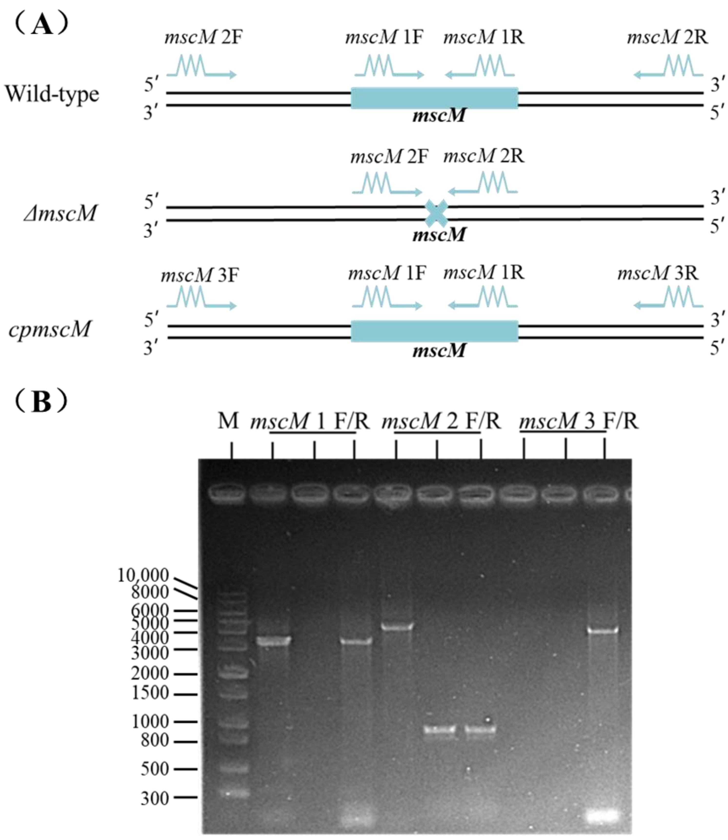
3.1. Construction of the mscM Mutant Strain

The suicide plasmid pCVD442 was used to knock out the mscM gene of C. sakazakii. The amplification location of verified primers in the different C. sakazakii strains is shown in Figure 1A. When using mscM 1 F/R as primers, a 3378 bp band was amplified from the WT and cpmscM strains, and no band was amplified from the ΔmscM strain (Figure 1B). An expected band (4334 bp) was obtained from the WT strain, while a shorter band (1001 bp) was amplified from the ΔmscM or cpmscM strains when using the mscM 2 F/R primer. In addition, the mscM 3 F/R primer could amplify an expected band (4020 bp) on the pACYC184 plasmid in the cpmscM strains (Figure 1B). These results indicated that the mscM gene was deleted in the ΔmscM strain, and this gene was also complemented in the cpmscM strain.

3.2. Effects of MscM on the Bacterial Growth Curves

To determine whether the mscM gene was essential for C. sakazakii, the growth curves of the three groups of bacteria (WT, ΔmscM, and cpmscM) were recorded. As shown in Figure 2, the WT, ΔmscM, and cpmscM strains shared similar growth trends, indicating that the mscM gene is a non-lethal gene, and its absence does not impair the bacterial vitality and growth, thus ruling out the possible growth differences that would interfere with subsequent experiments.

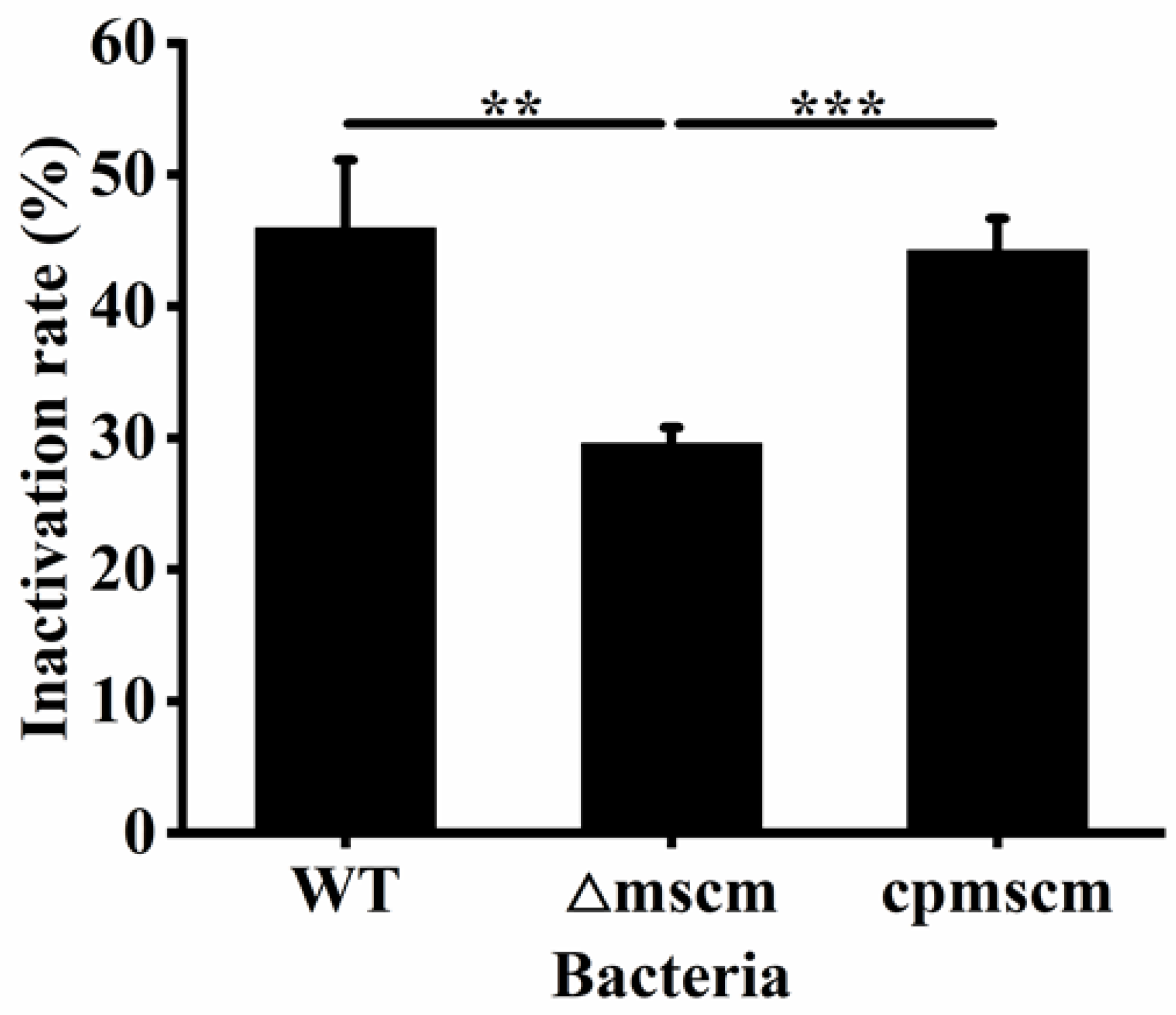
3.3. Effects of MscM on the Desiccation Resistance of C. sakazakii

To investigate the effects of the MscM channel on the desiccation resistance of C. sakazakii, the desiccation resistance ability of the three strains, namely WT, ΔmscM, and cpmscM, was evaluated. As shown in Figure 3, the inactivation rate of the WT strain was 45.94 ± 5.18% after 9 days of drying treatment, while the inactivation rate of the ΔmscM strain was 30.11 ± 1.47%. The inactivation rate of the ΔmscM strain was reduced by 15.83% (p < 0.01) compared with the WT strain. In addition, the inactivation rate of the cpmscM strain was 44.26 ± 2.43%, which was not significantly different from that of the WT strain. Therefore, the MscM protein had a negative effect of the regulation on the desiccation resistance of C. sakazakii.

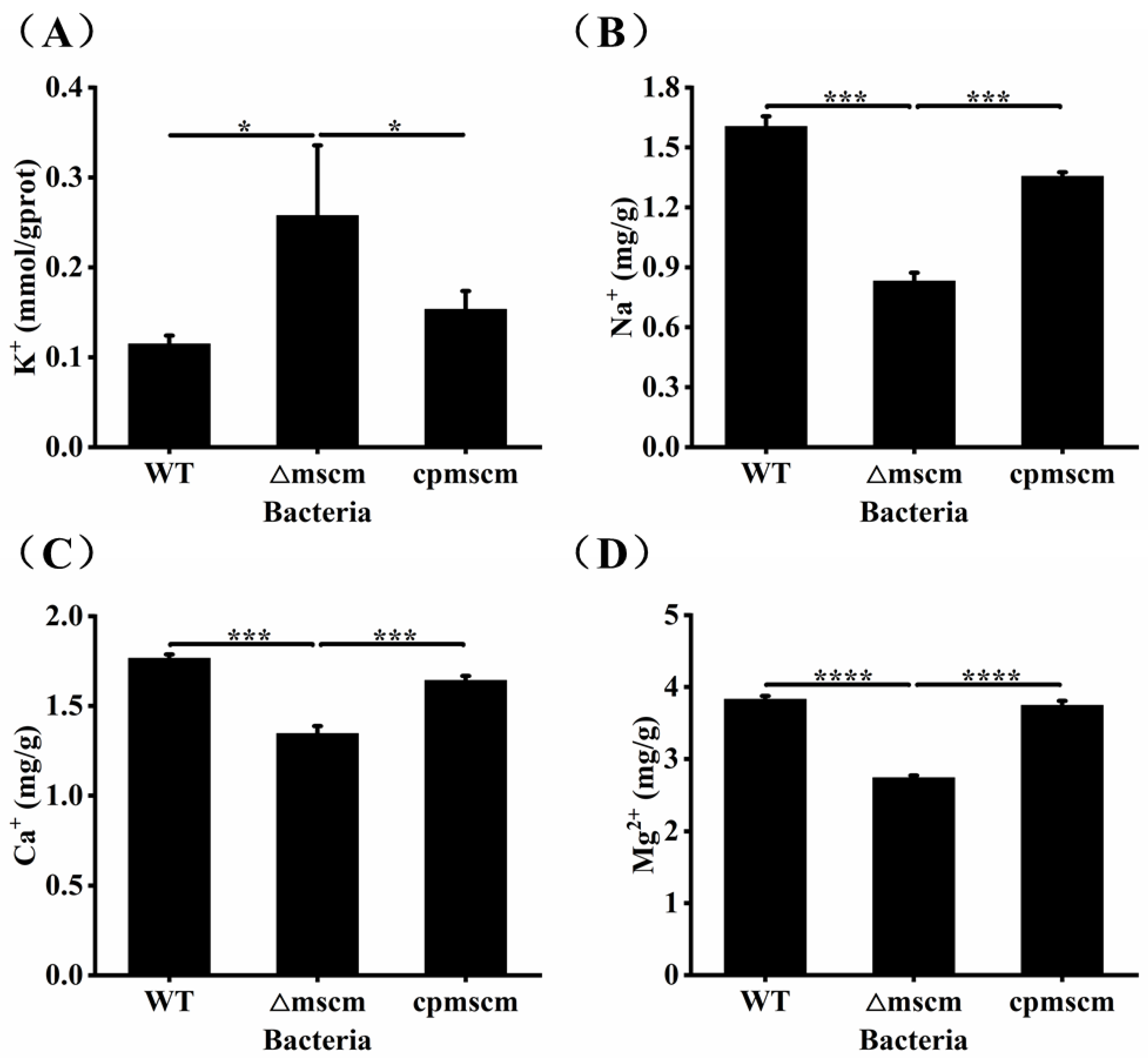
3.4. The Different Ion Contents in WT, ΔmscM and cpmscM Strains

As an ion channel, the reported MS channel has the ability to maintain the osmotic balance of the cells by regulating the efflux of metal ions. In order to study the molecular mechanism of MscM related to the desiccation resistance of C. sakazakii, the levels of intracellular potassium were detected. As shown in Figure 4A, the intracellular potassium ion concentration of the WT strain was 0.11 ± 0.01 mM/gprot, which was significantly higher than that of the ΔmscM strain, at 0.26 ± 0.08 mM/gprot (p < 0.05). As for the cpmscM strain, the complementary mscM gene promoted a decrease in cytoplasmic K+ to 0.15 ± 0.02 mM/gprot. In addition, the total K+ content of the WT, ΔmscM, and cpmscM strains were 5.89 ± 0.14 mg/g, 6.80 ± 0.08 mg/g, and 5.94 ± 0.18 mg/g, respectively (Figure S1). Consistent with the changing trend of intracellular potassium ion concentration, the total K+ content of the ΔmscM strain also increased compared to the WT strain. Other metal ions also contribute to the osmotic balance. To further explore the effects of the mscM gene on the other metal ion contents, we detected the total ion contents of Na+, Ca2+, and Mg2+ ions among the three strains. As shown in Figure 4B–D, the total ion contents of Na+, Ca2+, and Mg2+ were 0.83 ± 0.04 mg/g, 1.34 ± 0.04 mg/g, and 2.83 ± 0.03 mg/g in the ΔmscM strain, which were less than the values in the WT strain 1.61 ± 0.05 mg/g (p < 0.001), 1.77 ± 0.02 mg/g (p < 0.001), and 3.83 ± 0.04 mg/g (p < 0.0001), respectively. The total contents of the three ions were 1.35 ± 0.02 mg/g (Na+), 1.64 ± 0.03 mg/g (Ca2+), and 3.74 ± 0.06 mg/g (Mg2+) in the cpmscM strain, indicating a successful gene complementation. The above results indicate that C. sakazakii can control the K+ efflux through MscM channels, and the deletion of the mscM gene may stimulate the opening of other MS channels, thereby promoting the efflux of Na+, Ca2+, and Mg2+.

3.5. Detection of the Outer Membrane Permeability

The outer membrane permeability is an essential indicator for G bacteria to respond to osmotic stress and regulate osmotic balance. To investigate the effects of the MscM channel on the outer membrane permeability, NPN was used as the fluorescent probe in this study. As shown in Figure 5, the fluorescent value of the ΔmscM strain was 45.09 ± 0.17, which was about 19.05 ± 0.006% (p < 0.0001) and 18.19 ± 0.02% (p < 0.0001) higher than the WT (36.5 ± 0.24) and cpmscM (36.89 ± 0.84) strains, respectively, indicating that the outer membrane permeability was increased in the ΔmscM strain, and the MscM channel had a negative regulatory effect on it.

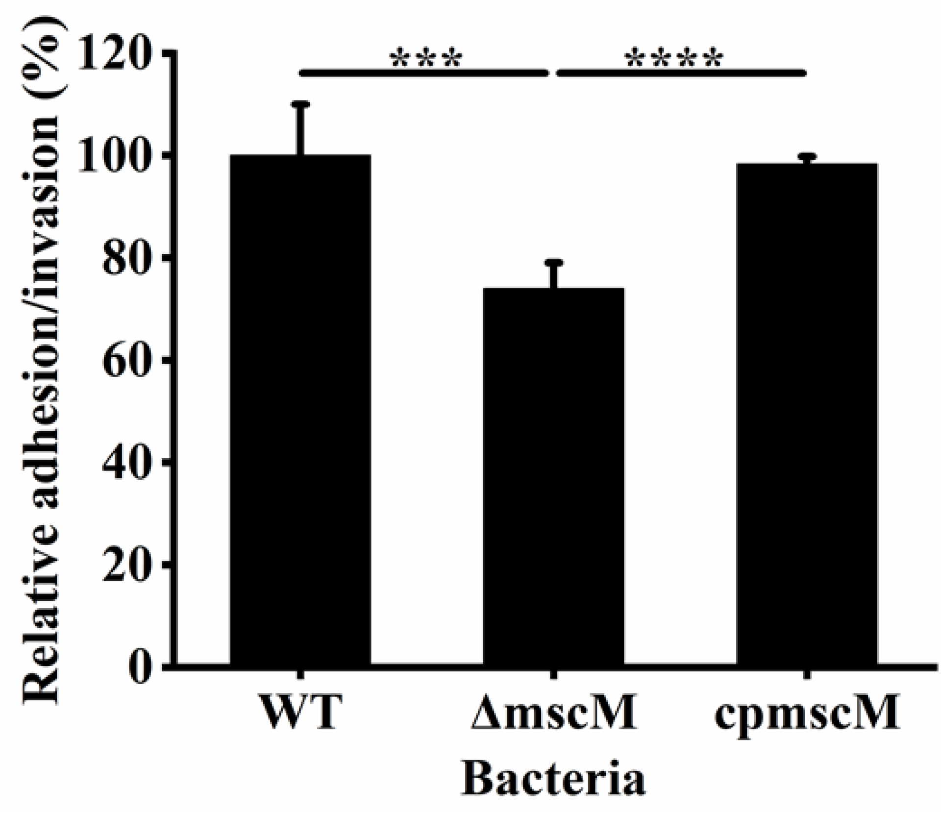
3.6. Effects of MscM on Bacterial Adhesion/Invasion

Bacterial attachment is related not only to pathogenicity but also to growth, aggregation, and colonization. To study the effects of MscM protein on bacterial attachment, HCT-8 cells were used to determine the adhesion/invasion of the different strains. As shown in Figure 6, compared to the WT and cpmscM strains, the adhesive/invasive rate decreased to 73.97 ± 5.07% in the ΔmscM strain, indicating that the deletion of the mscM gene damaged the adhesion/invasion of C. sakazakii.

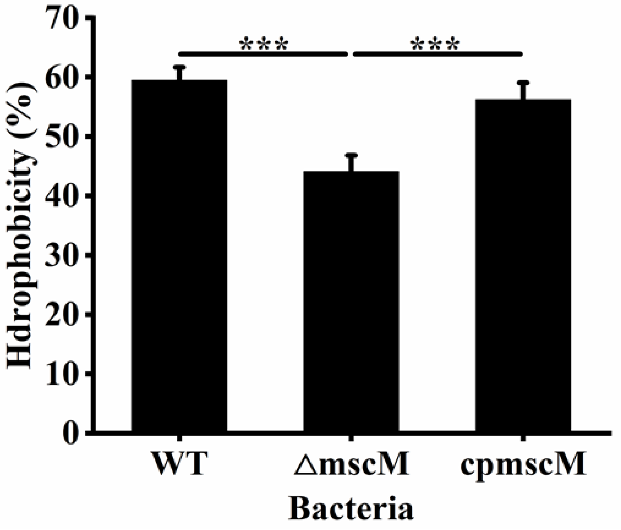
3.7. Effects of MscM on the Surface Hydrophobicity

The surface hydrophobicity is vital for bacterial adhesion and aggregation. To study the surface hydrophobicity of the three strains (WT, ΔmscM, and cpmscM), the bacteria were treated with xylene and the absorbance of the aqueous phase was recorded. As shown in Figure 7, the surface hydrophobicity values were 59.47 ± 2.81%, 44.09 ± 2.74%, and 56.22 ± 2.83% in the WT, ΔmscM, and cpmscM strains, respectively. The surface hydrophobicity of the ΔmscM strain was 20.52% (p < 0.001) lower than that of the WT strain, indicating that the absence of the mscM gene had an adverse effect on bacterial surface hydrophobicity.

3.8. Effects of MscM on the Biofilm Formation

Biofilms have been shown to be associated with most bacteria overcoming environmental barriers and surviving for a long time. To investigate the effect of the MscM channel on biofilm formation, the biofilm formation of the three strains was detected by CV staining. The results are illustrated in Figure 8. The OD570nm value of the WT strain was 0.53 ± 0.04, while that of the ΔmscM strain was 0.37 ± 0.03. The biofilm formation of the ΔmscM strain decreased by 30.19% (p < 0.05) when lacking MscM protein, whereas the complementary MscM protein could restore the biofilm formation to the normal level (0.49 ± 0.003), indicating that the presence of the MscM channel was beneficial for the biofilm formation of C. sakazakii.

4. Discussion

As an opportunistic pathogen, C. sakazakii is famous for its desiccation resistance. Several models have been established to clarify the molecular mechanism of desiccation resistance of this bacterium, but the special mechanism still harbors numerous ambiguities. In this study, the deletion of the mscM gene was used to research the effect of this gene on the desiccation resistance of C. sakazakii. In addition, the changes in some biological characters caused by the absence of the mscM gene were also explored in C. sakazakii. The physiological effect of MscM proteins on C. sakazakii is shown in Figure 9.
The deletion of the mscM gene did not impair bacterial vitality and growth and enhanced the desiccation resistance of C. sakazakii. The opening of MS channels was crucial for bacterial survival when osmolarity changed. For example, it has been found that the MscS and MscL channels contributed to maintaining the integrity of the bacterial envelope during osmotic shock [16]. Solutes flow out through these channels to reduce the water influx rate and avoid cell lysis. In our study, compared with the WT strain, the ΔmscM strain accumulated more intracellular K+ and total K+ contents, indicating that the MscM channel primarily downregulated the desiccation resistance of C. sakazakii by reducing the efflux of K+. The phenomenon that the potassium efflux damaged the capacity of bacterial desiccation resistance was also reported in other studies [17,18]. The high concentration of potassium glutamate was considered to compose the first line of defense to resist the desiccation. However, it was also a less preferred osmolyte due to its ability to disrupt cellular metabolism [9,19]. The accumulation of potassium glutamate was accompanied by the production of other theoretical osmoprotectants. These osmoprotectants might form the second line of defense in resisting the desiccation and did not induce disturbances to cellular metabolism [20,21]. In this study, the total contents of Na+, Ca2+, and Mg2+ decreased in the ΔmscM strain, leading to the dilution of intracellular ion concentrations that promote the water absorption of C. sakazakii. This manner might reduce the effective concentration of K+ to a certain extent and minimize the damage to cells. These ions might export through other MS channels, but we did not detect any changes in related gene expression by using RT-qPCR (Figure S2). In fact, the opening of MS channels was unable to be detected in the body. Theoretically, they would successively open as the membrane tension increased [13]. In addition, the absence of the mscM gene also increased the membrane permeability of the mutant strains. Additionally, the defective membrane could lead to the leakage of other intracellular molecules, such as DNA and proteins, which would cause an adverse effect on bacterial survival in dry environments [17,22]. Bacteria could adapt to extremely dry environments by regulating the expression of the genes related to the desiccation resistance of C. sakazakii. The transcription level of the rpoS gene was increased, while that of the betI gene was decreased in the ΔmscM strain (Figure S2). RpoS is the main signal to respond to the osmotic pressure, and BetI was involved in the inhibition of the biosynthesis of the osmoprotectant glycine betaine [3,23]. Both changes in the above two genes helped the bacteria to cope with the desiccation stress. In short, several direct or indirect cascades of events contribute to the desiccation adaption of bacteria and lead to cellular survival.
Biofilm has been shown to be involved in the survival and infection of C. sakazakii, and the adhesion and colonization of bacteria are crucial for the formation of their biofilms [24,25]. Compared to the WT strain, the ΔmscM strain had defects in biofilm formation and poorer adhesion to HCT-8 cells. The above differences were indeed caused by the lack of MscM protein in the ΔmscM strain rather than the differences in bacterial vitality, as the growth curves of the ΔmscM and WT strains were almost identical. MS channels could stimulate the expression of the related proteins located in the cell membrane and could improve bacterial adhesion and biofilm formation [26]. In Pseudomonas aeruginosa, stimulating MS channels could promote cell growth and biofilm formation [27]. The deletion of the mscM gene also impaired the surface hydrophobicity of the ΔmscM strain. Surface hydrophobicity is based on the interactions of different molecules in the cell membrane and plays an important role in cell adhesion. When a hydrophobic amino acid was replaced in the transmembrane domain of the MscL protein, it reduced the synthesis of the MscL protein and damaged the surface hydrophobicity of E. coli [28]. These complicating factors ultimately led to failure in biofilm formation. The poor biofilm formation may provide little assistance for the environmental persistence of bacteria, especially in a dry environment. However, the inactivation rate of the ΔmscM strain was lower than the WT strain after a short-term drying treatment (drying for 9 days). C. sakazakii could survive in a low-water-activity environment for 1–2 years [29]. We might detect a higher inactivation rate caused by poor biofilm formation in the mutant strains after a longer drying process. Therefore, from another perspective, this also reflects that the accumulation of potassium is crucial for the initial survival of C. sakazakii in a low-water-activity environment.
The absence of the mscM gene did not affect bacterial motility, but the weakening of adhesion ability also indicated that the MscM protein might play an important role in the pathogenicity of C. sakazakii. Bacterial adhesion is key to causing infection. It has been reported that the MS channel protein YnaI of Salmonella is essential for intestinal colonization, and the deletion of the ynaI gene impairs the adhesion of S. typhimurium [30]. In addition, the mechanical forces produced in the adhesion process could pull the bacterial envelope, thus causing conformational changes in MS proteins and activating the MS channel, which was beneficial for the interaction between bacterial integrins (the major adhesive mediators of bacteria) and the host cell membrane and generating adhesion signals [31]. Of course, the effects of the MscM protein on the pathogenicity of C. sakazakii need further investigation.

5. Conclusions

In this study, we mainly reported the effects of the MscM protein on the desiccation resistance of C. sakazakii. The deletion of the mscM gene could reduce K+ efflux and improve the desiccation resistance of C. sakazakii, indicating that the intracellular accumulation of K+ was important for the tolerance of this bacterium in a dry environment. Additionally, the mscM gene mutant strains showed failure in biofilm formation and bacterial adhesion/invasion. This study enhances our understanding of the function of the mscM gene and the desiccation resistance mechanism of C. sakazakii.

Supplementary Materials

The following supporting information can be downloaded at: https://www.mdpi.com/article/10.3390/microorganisms12122464/s1, Table S1. Bacterial strains and plasmids used in this study. Table S2. Primers used in this study. Figure S1. The total K+ contents in WT, ΔmscM and cpmscM strains. Figure S2. The transcription level of some genes in ΔmscM strains. Figure S3. The bacterial motility of WT, ΔmscM and cpmscM strains.

Author Contributions

Validation, D.Z. and Z.Z.; Writing—original draft, D.Z.; Writing—review & editing, P.L.; Supervision, P.L. and X.D.; Project administration, X.D. All authors have read and agreed to the published version of the manuscript.

Funding

This work was supported by the Natural Science Foundation of China (32472322) and the National Key R&D Program of China (2023YFF1103900).

Data Availability Statement

The original contributions presented in this study are included in the article/Supplementary Material. Further inquiries can be directed to the corresponding authors.

Conflicts of Interest

The authors declare no conflict of interest.

References

  1. Jin, T.; Pang, L.; Yue, T.; Niu, L.; Li, T.; Liang, Y.; Zhang, Y.; Yan, C.; Yang, B.; Zhang, C.; et al. The role of DsbA and PepP genes in the environmental tolerance and virulence factors of Cronobacter sakazakii. Food. Res. Int. 2024, 190, 114555. [Google Scholar] [CrossRef] [PubMed]
  2. Ekundayo, T.C.; Ijabadeniyi, O.A. Global and regional prevalence of Cronobacter sakazakii in powdered milk and flour. Sci. Rep. 2024, 14, 6865. [Google Scholar] [CrossRef]
  3. Johler, S.; Stephan, R.; Hartmann, I.; Kuehner, K.A.; Lehner, A. Genes involved in yellow pigmentation of Cronobacter sakazakii ES5 and influence of pigmentation on persistence and growth under environmental stress. Appl. Environ. Microbiol. 2010, 76, 1053–1061. [Google Scholar] [CrossRef] [PubMed]
  4. Du, X.J.; Wang, X.Y.; Dong, X.; Li, P.; Wang, S. Characterization of the desiccation tolerance of Cronobacter sakazakii strains. Front. Microbiol. 2018, 9, 2867. [Google Scholar] [CrossRef]
  5. Wang, L.; Forsythe, S.J.; Yang, X.; Fu, S.; Man, C.; Jiang, Y. Invited review: Stress resistance of Cronobacter spp. affecting control of its growth during food production. J. Dairy. Sci. 2021, 104, 11348–11367. [Google Scholar] [CrossRef]
  6. Hu, S.; Yu, Y.G.; Wu, X.W.; Xia, X.Z.; Xiao, X.L.; Wu, H. Comparative proteomic analysis of Cronobacter sakazakii by iTRAQ provides insights into response to desiccation. Food. Res. Int. 2017, 100 Pt 1, 631–639. [Google Scholar] [CrossRef] [PubMed]
  7. Srikumar, S.; Cao, Y.; Yan, Q.; Van Hoorde, K.; Nguyen, S.; Cooney, S.; Gopinath, G.R.; Tall, B.D.; Sivasankaran, S.K.; Lehner, A.; et al. RNA Sequencing-based transcriptional overview of xerotolerance in Cronobacter sakazakii SP291. Appl. Environ. Microbiol. 2019, 85, e01993-18. [Google Scholar] [CrossRef]
  8. Feeney, A.; Sleator, R.D. An in silico analysis of osmotolerance in the emerging gastrointestinal pathogen Cronobacter sakazakii. Bioeng. Bugs. 2011, 2, 260–270. [Google Scholar] [CrossRef]
  9. Gralla, J.D.; Vargas, D.R. Potassium glutamate as a transcriptional inhibitor during bacterial osmoregulation. EMBO J. 2006, 25, 1515–1521. [Google Scholar] [CrossRef]
  10. Roe, A.J.; O’Byrne, C.; McLaggan, D.; Booth, I.R. Inhibition of Escherichia coli growth by acetic acid: A problem with methionine biosynthesis and homocysteine toxicity. Microbiology 2002, 148, 2215–2222. [Google Scholar] [CrossRef] [PubMed]
  11. Blount, P.; Iscla, I. Life with Bacterial Mechanosensitive Channels, from Discovery to Physiology to Pharmacological Target. Microbiol. Mol. Biol. Rev. 2020, 84, e00055-19. [Google Scholar] [CrossRef]
  12. Ramsey, K.; Britt, M.; Maramba, J.; Ushijima, B.; Moller, E.; Anishkin, A.; Häse, C.; Sukharev, S. The dynamic hypoosmotic response of Vibrio cholerae relies on the mechanosensitive channel MscS. iScience 2024, 27, 110001. [Google Scholar] [CrossRef] [PubMed]
  13. Schumann, U.; Edwards, M.D.; Rasmussen, T.; Bartlett, W.; van West, P.; Booth, I.R. YbdG in Escherichia coli is a threshold-setting mechanosensitive channel with MscM activity. Proc. Natl. Acad. Sci. USA 2010, 107, 12664–12669. [Google Scholar] [CrossRef] [PubMed]
  14. Edwards, M.D.; Black, S.; Rasmussen, T.; Rasmussen, A.; Stokes, N.R.; Stephen, T.-L.; Miller, S.; Booth, I.R. Characterization of three novel mechanosensitive channel activities in Escherichia coli. Channels 2012, 6, 272–281. [Google Scholar] [CrossRef] [PubMed]
  15. Ji, X.; Lu, P.; Xue, J.; Zhao, N.; Zhang, Y.; Dong, L.; Zhang, X.; Li, P.; Hu, Y.; Wang, J.; et al. The lipoprotein NlpD in Cronobacter sakazakii responds to acid stress and regulates macrophage resistance and virulence by maintaining membrane integrity. Virulence 2021, 12, 415–429. [Google Scholar] [CrossRef] [PubMed]
  16. Pivetti, C.D.; Yen, M.-R.; Miller, S.; Busch, W.; Tseng, Y.-H.; Booth, I.R.; Saier, M.H. Two families of mechanosensitive channel proteins. Microbiol. Mol. Biol. R. 2003, 67, 66–85. [Google Scholar] [CrossRef]
  17. Wang, X.Y.; Li, P.; Du, X.J.; Wang, S. Effect of glutathione-transport-related gene gsiD on desiccation tolerance of Cronobacter sakazakii and its related regulatory mechanism. Appl. Environ. Microbiol. 2024, 90, e0156223. [Google Scholar] [CrossRef] [PubMed]
  18. Abdelhamid, A.G.; Yousef, A.E. Carvacrol and thymol combat desiccation resistance mechanisms in Salmonella enterica serovar tennessee. Microorganisms 2021, 10, 44. [Google Scholar] [CrossRef]
  19. Sarah Finn, S.; Condell, O.; McClure, P.; Amézquita, A.; Fanning, S. Mechanisms of survival, responses and sources of Salmonella in low-moisture environments. Front. Microbiol. 2013, 4, 331. [Google Scholar] [CrossRef]
  20. Jovanovich, S.B.; Martinell, M.; Record, M.T.J.; Burgess, R.R. Rapid response to osmotic upshift by osmoregulated genes in Escherichia coli and Salmonella typhimurium. J. Bacteriol. 1988, 170, 534–539. [Google Scholar] [CrossRef]
  21. Kempf, B.; Bremer, E. Uptake and synthesis of compatible solutes as microbial stress responses to high-osmolality environments. Arch. Microbiol. 1998, 170, 319–330. [Google Scholar] [CrossRef] [PubMed]
  22. Scherber, C.M.; Schottel, J.L.; Aksan, A. Membrane phase behavior of Escherichia coli during desiccation, rehydration, and growth recovery. Biochim. Biophys. Acta. 2009, 1788, 2427–2435. [Google Scholar] [CrossRef] [PubMed]
  23. Lamark, T.; Røkenes, T.P.; McDougall, J.; Strøm, A.R. The complex bet promoters of Escherichia coli: Regulation by oxygen (ArcA), choline (BetI), and osmotic stress. J. Bacteriol. 1996, 178, 1655–1662. [Google Scholar] [CrossRef] [PubMed]
  24. Henry, M.; Fouladkhah, A. Outbreak history, biofilm formation, and preventive measures for control of Cronobacter sakazakii in infant formula and infant care settings. Microorganisms 2019, 7, 77. [Google Scholar] [CrossRef]
  25. Carniello, V.; Peterson, B.W.; van der Mei, H.C.; Busscher, H.J. Physico-chemistry from initial bacterial adhesion to surface-programmed biofilm growth. Adv. Colloid. Interface. Sci. 2018, 261, 1–14. [Google Scholar] [CrossRef]
  26. Irene, I.; Paul, B. Sensing and responding to membrane tension: The bacterial MscL channel as a model system. Biophys. J. 2012, 103, 169–174. [Google Scholar] [CrossRef]
  27. Gu, S.; Zhang, Y.; Wu, Y. Effects of sound exposure on the growth and intracellular macromolecular synthesis of E. coli k-12. PeerJ 2016, 4, e1920. [Google Scholar] [CrossRef]
  28. Sukharev, S.I.; Blount, P.; Martinac, B.; Blattner, F.R.; Kung, C. A large-conductance mechanosensitive channel in E. coli encoded by mscL alone. Nature 1994, 368, 265–268. [Google Scholar] [CrossRef]
  29. Barron, J.C.; Forsythe, S.J. Dry stress and survival time of Enterobacter sakazakii and other Enterobacteriaceae in dehydrated powdered infant formula. J. Food. Prot. 2007, 70, 2111–2117. [Google Scholar] [CrossRef] [PubMed]
  30. Chaudhuri, R.R.; Morgan, E.; Peters, S.E.; Pleasance, S.J.; Hudson, D.L.; Davies, H.M.; Wang, J.; Van Diemen, P.M.; Buckley, A.M.; Bowen, A.J.; et al. Comprehensive assignment of roles for Salmonella typhimurium genes in intestinal colonization of food-producing animals. PLoS. Genet. 2013, 9, e1003456. [Google Scholar] [CrossRef]
  31. Glogauer, M.; Arora, P.; Yao, G.; Sokholov, I.; Ferrier, J.; McCulloch, C.A. Calcium ions and tyrosine phosphorylation interact coordinately with actin to regulate cytoprotective responses to stretching. J. Cell. Sci. 1997, 110 Pt 1, 11–21. [Google Scholar] [CrossRef] [PubMed]
Figure 1. Locations of specific primers and PCR amplification: (A) Locations of specific primers used for PCR validation; (B) PCR amplification result from different bacterial strains using different primers pairs, M. Maker; msccM 1 F/R, the WT strain; msccM 2 F/R, the ΔmscM strain; msccM 3 F/R, the cpmscM strain.
Microorganisms 12 02464 g001
Figure 2. The growth curves of WT, ΔmscM, and cpmscM strains.
Microorganisms 12 02464 g002
Figure 3. The drying inactivation rate of WT, ΔmscM, and cpmscM strains. ** p < 0.01, *** p < 0.001.
Microorganisms 12 02464 g003
Figure 4. The different ion contents in WT, ΔmscM, and cpmscM strains: (A) intracellular potassium ion concentration; (BD) total Na+, Ca2+, and Mg2+ ion contents. * p < 0.05, *** p < 0.001, **** p < 0.0001.
Microorganisms 12 02464 g004
Figure 5. Outer membrane permeability. **** p < 0.0001.
Microorganisms 12 02464 g005
Figure 6. Relative adhesion/infestation rate of the different strains.*** p < 0.001, **** p < 0.0001.
Microorganisms 12 02464 g006
Figure 7. The surface hydrophobicity of bacteria. *** p < 0.001.
Microorganisms 12 02464 g007
Figure 8. Biofilm formation. * p < 0.05, ** p < 0.01.
Microorganisms 12 02464 g008
Figure 9. The illustration of the effects of MscM proteins on C. sakazakii.
Microorganisms 12 02464 g009
Disclaimer/Publisher’s Note: The statements, opinions and data contained in all publications are solely those of the individual author(s) and contributor(s) and not of MDPI and/or the editor(s). MDPI and/or the editor(s) disclaim responsibility for any injury to people or property resulting from any ideas, methods, instructions or products referred to in the content.

