Article

Impact of Mycobacterium tuberculosis H37Rv Infection on Extracellular Vesicle Cargo in Macrophages: Implications for Host–Pathogen Interaction

by
Manuel G. Salgado-Cantú
1,†,
Luis Horacio Gutiérrez-González
2,†,
Silvia Guzmán-Beltrán
1,
María Teresa Herrera
1,
Carmen Sarabia
1 and
Yolanda González
1,*
1
Department of Microbiology, Instituto Nacional de Enfermedades Respiratorias Ismael Cosío Villegas, Mexico City 14080, Mexico
2
Laboratory of Transcriptomics and Molecular Immunology, Instituto Nacional de Enfermedades Respiratorias Ismael Cosío Villegas, Mexico City 14080, Mexico
*
Author to whom correspondence should be addressed.
These authors contributed equally to this work.
Microorganisms 2024, 12(12), 2405; https://doi.org/10.3390/microorganisms12122405
Submission received: 29 September 2024 / Revised: 13 November 2024 / Accepted: 20 November 2024 / Published: 23 November 2024
(This article belongs to the Special Issue Bacterial Pathogenesis and Host Immune Responses)

Abstract

:
Tuberculosis (TB) is one of the most common respiratory infections worldwide, and it is caused by Mycobacterium tuberculosis (Mtb). Mtb employs immune evasion mechanisms that allow the disease to become chronic. Despite extensive research, the host–pathogen interaction remains incompletely understood. Extracellular vesicles (EVs) are small membrane particles that play a regulatory role in infectious diseases. Host-derived EVs have been identified as carriers of proteins, messenger RNA, and lipids from both the host cells and the pathogens. In this study, we assessed the cargo of EVs in human macrophages infected with the virulent strain H37Rv of Mtb at 1 and 24 h post-infection (hpi). The results showed that 1 hpi, infected macrophages secreted EVs containing Mtb proteins (15 to 37 kDa) and Ag85 kDa, as well as RNA transcripts (ESAT-6, 5KST, Ag85, IS6110, 30 kDa, 19 kDa, and MPT64). However, these decreased at 24 hpi. The infection of macrophages with Mtb was observed to result in the release of EVs containing Ag85 protein and RNA transcripts of Mtb; this process appeared to diminish after 24 hpi, suggesting the existence of an evasion mechanism. Both Ag85 and the RNA transcripts could be potential biomarkers for the diagnosis of TB patients.

1. Introduction

Although tuberculosis (TB) is a preventable and generally curable disease, an estimated 10.8 million people worldwide were infected with TB in 2023, and 1.25 million people died from the disease. TB is the leading cause of death due to a single infectious agent and the second leading cause of death due to an infectious agent after COVID-19. In 2023, TB probably returned to being the world’s leading cause of death from a single infectious agent, following 3 years in which it was replaced by coronavirus disease. The causative agent, Mycobacterium tuberculosis (Mtb), is a pathogen with multiple mechanisms to evade the immune system and cause active disease in susceptible individuals but can remain latent in non-susceptible individuals [1].
The H37Rv strain of Mtb, which was isolated in 1905, has served as the neotype for the species and has played an important role in understanding the pathogenicity and drug resistance mechanisms of mycobacteria. Despite extensive research, the evasion mechanisms of Mtb remain incompletely understood. The possible involvement of extracellular vesicles (EVs) has been investigated under pathological conditions. EVs are small membrane-derived particles (50–100 nm) that are critical mediators of intercellular communication and play an important role in both physiological and pathological conditions. These EVs are characterized by their heterogeneous lipid bilayer membrane containing various biological cargoes, such as cytosol, proteins, lipids, and nucleic acids [2,3], and are present in many body fluids [3]. EVs are resistant to enzymatic degradation during transit. This occurs through the formation of intraluminal vesicles and late endosomes. Macrophage-derivated EVs have the ability to encapsulate a variety of biological molecules, including proteins, messenger RNAs (mRNAs), microRNAs (miRNAs), and DNA, as well as lipids derived from both the parent cell and the pathogen. EVs are then released from cells into the extracellular space, where they modulate the biological function of recipient cells [4].
Host EVs have been identified as carriers of Mtb lipids and proteins, further expanding the range of molecules that can be transported between Mtb and host cells [5,6]. In latent and active TB patients, it has been reported that Mtb resides intracellularly, and its secreted mycobacterial and host RNAs are packaged into host EVs and released into the bloodstream [7,8,9]. In in vitro experiments with Mtb-infected RAW264.7 murine macrophages, which is a distinct repertoire of miRNAs, mRNAs were identified; pathogen-derived mycobacterial RNA, Rv0740, Rv0288, Rv1344, Rv0968, Rv1942c, Rv0664, Rv0190, Rv1757c, and Rv1369c proteins were also vesicle-encapsulated and transferred to recipient cells, where they promoted both innate and acquired immune responses [6]. Most studies of EVs and Mtb have involved immortalized human monocyte and macrophage or murine cell lines and Mtb strains, such as M. bovis, M. avium, or non-virulent mycobacteria [10,11,12,13]. However, the precise role of EVs in the regulation of the immune system regulation and the persistence of Mtb in the host remains poorly understood.
Conversely, it has been documented that macrophage-derived EVs (Mφ-EVs) deliver abundant proteins, lipids, RNA, and DNA that may vary with different macrophage phenotypes or microenvironments, indicating different processes or pathological events [4]. This poses a challenge in elucidating the immunological mechanisms involved when utilizing different experimental models. Here, we used an in vitro model with macrophages and a virulent strain of live Mtb (H37Rv). We evaluated some abundant molecules packaged in EVs secreted by the infected macrophages at 1 or 24 h post-infection (hpi) to identify the proteins and RNA of Mtb carried in these EVs. The results show that infected macrophages secrete EVs containing Mtb proteins and RNA after one hour of infection. However, at 24 hpi, while the number of vesicles remains constant, the composition of the proteins and RNA within them is reduced.

2. Materials and Methods

2.1. Ethics Statement

The protocol was reviewed and approved by the Institutional Ethics and Research Committee (protocol number: C57-17). All participants were recruited at the National Institute of Respiratory Diseases (INER), Mexico City, and provided written informed consent to participate in the protocol.

2.2. Biological Samples

2.2.1. Study Population

Six healthy volunteers (three men and three women) participated in this study. They were vaccinated with BCG at birth and had a positive tuberculin skin test (TST). Five of them were from Mexico City, and one was from Puebla. Mexico is considered a moderate-incidence country for TB.

2.2.2. Peripheral Blood Mononuclear Cell Isolation

Heparinized whole blood (120 mL) was collected from six healthy volunteers, and human peripheral blood mononuclear cells (PBMCs) were gradient purified. Briefly, blood was diluted 1:2 with RPMI 1640 (Lonza, Walkersville, MD, USA), lymphoprep solution (Axis Shield Diagnostics, Oslo, Norway) was added, and then the mixture was centrifuged at 1200 rpm for 45 min at room temperature to obtain PBMCs.

2.2.3. Monocyte Isolation, Culture, and Differentiation

Monocytes were isolated from PBMCs by positive selection using the MACS separation system (Miltenyi Biotec, Auburn, CA, USA) according to the manufacturer’s recommendations. Briefly, PBMCs were mixed with ant-CD14 monoclonal antibody conjugated to magnetic beads (Miltenyi Biotec) and incubated at 4 °C for 15 min. Monocytes were then washed with wash solution (2 mM EDTA, 0.5% bovine serum albumin in PBS 0.01 M, pH = 7.2) and resuspended in wash solution after centrifugation. Monocyte suspension was added to the column under a magnetic field and washed three times. Finally, the column was removed from the magnetic field, and CD14positive cells (monocytes) were eluted. The pure monocyte population was centrifuged and resuspended in an RPMI medium containing 10% heat-inactivated human serum (Valley Biomedical, Winchester, VA, USA). The viability was assessed using trypan blue exclusion (>95%). In addition, the purity of the monocyte population was determined through staining with anti-human CD14-PeTxRed (Invitrogen-Thermo Fisher Scientific, Carlsbad, CA, USA), acquired in a BD FACS Aria flow cytometer, and analyzed using FlowJo software (FlowJo, v10.4.2, Ashland, OR, USA). The number of CD14-positive cells was >90%. Monocytes were resuspended in RPMI medium with 10% heat-inactivated human serum at 1 × 10⁶ MN/mL. A total of 1 mL of the cell suspension was added to 24-well culture plates and incubated at 37 °C in 5% CO2 for 7 days to obtain human monocyte-derived macrophages (MDM, also referred to as macrophages).

2.2.4. Human Acute Monocytic Leukemia Cells (THP-1)

Human acute monocytic leukemia cells, THP-1, were obtained from the American Type Culture Collection (ATCC, Manassas, VA, USA). THP-1 cells were grown in RPMI 1640 supplemented with 10% fetal bovine serum (Lonza), 0.05 µM β-mercaptoethanol (Bio-Rad, Hercules, CA, USA), and 4 mM L-glutamine (Thermo Fisher Scientific).

2.3. Strains and Culture Conditions

The virulent Mtb H37Rv strain from the American Type Culture Collection (ATCC 25618, Rockville, MD, USA) was cultivated in Middlebrook 7H9 broth medium (Becton Dickinson, NJ, USA) and stored at −70 °C. Prior to macrophage infection, the bacteria were thawed and disaggregated to obtain a single-cell suspension. The bacteria were centrifuged at 9000× g for 10 min, after which the supernatant was discarded and 1 mL RPMI-1640 with glutamine and 5% of exosome-depleted fetal bovine serum (FBS) (SBI, Palo Alto, CA, USA) were added. Then, 3 mm sterile glass beads were added and vortexed for 5 min. The bacterial suspension was then centrifuged at 900× g for two minutes. The suspension was then transferred to a new tube for infection assays.

2.4. Human Monocyte-Derived Macrophage (MDM/Macrophage) Infection

The medium of the MDM culture was replaced with fresh medium containing 5% of exosome-depleted FBS (SBI, USA) and infected with Mtb at a multiplicity of infection (MOI) of 10; uninfected MDM were included as negative controls. Plates were incubated at 37 °C and 5% CO2. After 1 hpi or 24 hpi, culture supernatants from infected or uninfected MDM were collected and filtered with sterile 0.22 μm syringe filters (Corning, NY, USA) and stored at 20 °C until EVs isolation.

2.5. Isolation of Extracellular Vesicles (EVs)

EVs were isolated from filtered supernatants of uninfected or Mtb-infected MDM. Briefly, supernatants were thawed and centrifuged at 2000× g for 30 min at 4 °C, and the supernatant was then transferred to a new tube. EVs were then purified from cell supernatants using Total Exosome Isolation Reagent (Invitrogen, Carlsbad, CA, USA) according to the manufacturer’s instructions.

2.6. Quantitation of EV Proteins

Individual or pooled EV samples were treated with RIPA lysis solution (Thermo Fisher Scientific) supplemented with EDTA (5 mM) and halt protease inhibitor cocktail (Thermo Fisher Scientific); the mixture was then shaken at 4 °C for 15 min and sonicated for additional 15 s. The samples were centrifuged at 14,000× g for 15 min at 4 °C, and the supernatant was collected. The extracted proteins were diluted 1:10 with double-distilled water, and samples of bovine serum albumin standard (Bio-Rad) and the Quick Start Bradford Protein Assay (Bio-Rad) were added to a 96-well flat-bottom plate (Thermo Fisher Scientific). The plate was shaken at 21 °C. The absorbance was measured at 595 nm using an Epoch™ microplate reader (Biotek, Santa Clara, CA, USA).

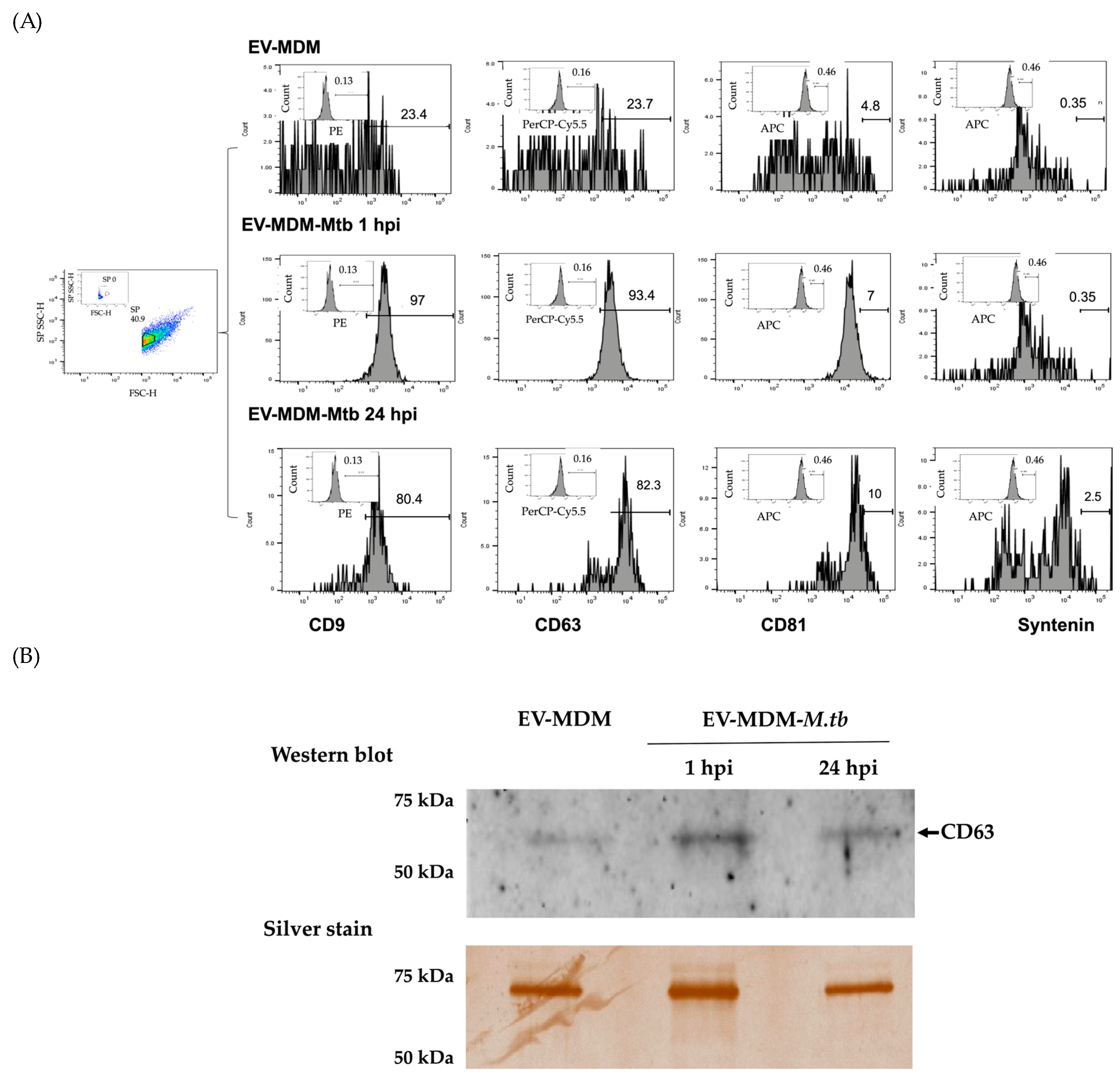
2.7. Expression of CD9, CD63, CD81, and Syntenin in EVs Using Flow Cytometry

Pooled EVs from uninfected MDM (EV-MDM) or infected MDM (EV-MDM-Mtb) were incubated with antibodies anti-CD9-PE, anti-CD63-PerCP-Cy5.5, anti-CD81-APC (Biolegend, San Diego, CA, USA), or anti-syntenin-APC (Assaypro, St Charles, MO, USA) for 20 min at room temperature. EVs were then isolated using the ExoQuick kit according to the manufacturer’s instructions and resuspended in filtered PBS containing 1% paraformaldehyde. A total of 200,000 small-particle or diluent events were acquired using a BD FACSymphony A1 (Cell Analyzer, San Jose, CA, USA) and a BD® Small Particle Detector with a High Sensitivity Side Scatter Channel Detector (SP SSC-H) that detects light scattering from small particles, such as EVs. A mixture of Megamix-Plus SSC, fluorescent beads 0.1 to 1 μm (Thermo Fisher Scientific), and SSCs, as a size-dependent parameter (BioCyteX, Marseille, FR), were used for gating. The purity of EVs was analyzed using the FlowJo software.

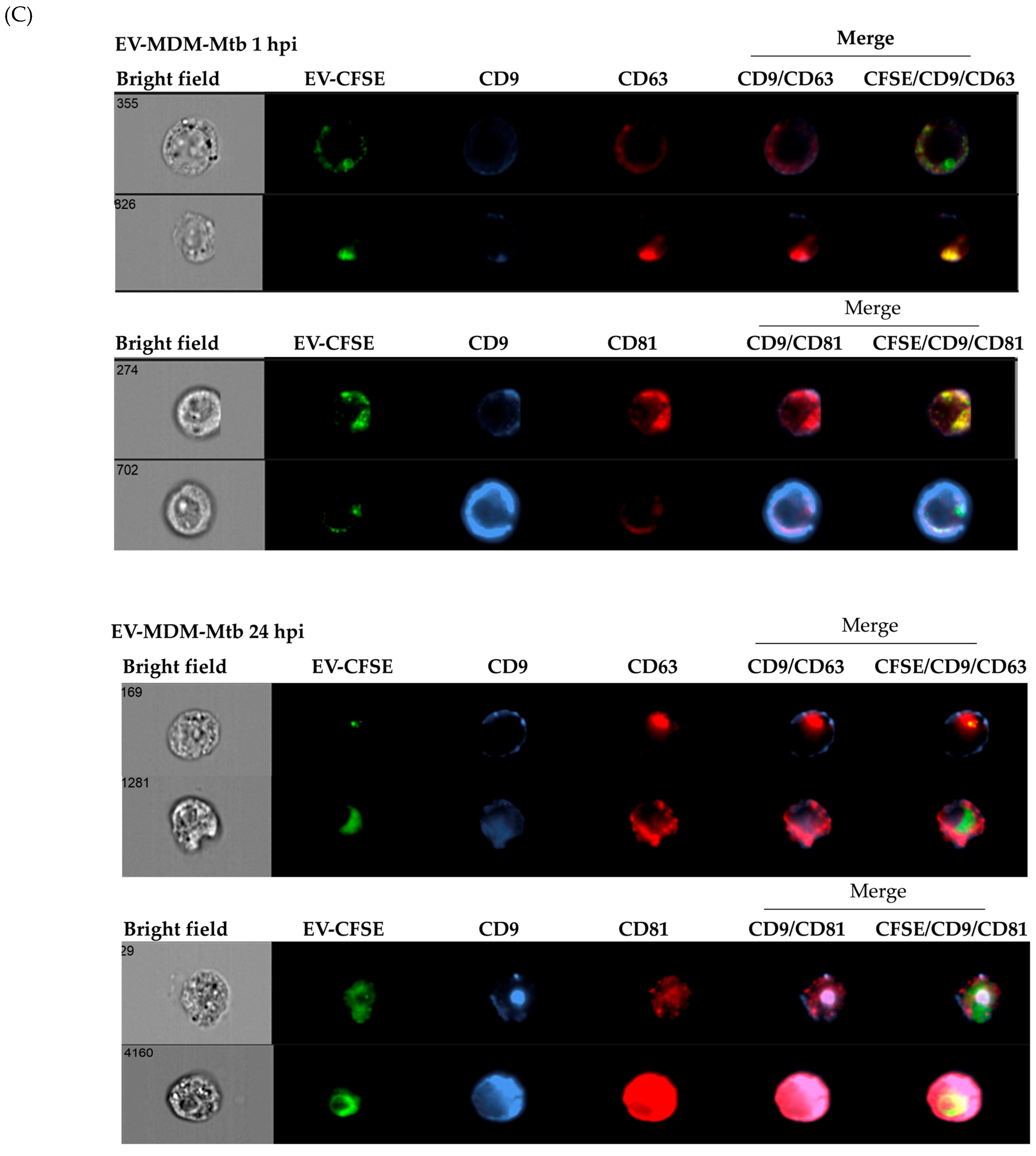
2.8. Internalization of EVs by THP1 Cells

Pooled EV-MDM-Mtb were stained with 5(6)-Carboxyfluorescein diacetate N-succinimidyl ester (CFSE) (Sigma Aldrich, Darmstadt, Germany) according to the manufacturer’s instructions. Briefly, CFSE was added to the EVs to a final concentration of 100 μM and incubated for 10 min at 37 °C. The reaction was stopped by adding 3 volumes of cold PBS and incubating the mixture for 5 min on ice; EVs were then precipitated by adding ExoQuickTM reagent (SBI) at 4 °C for 30 min and then centrifuging the mixture at 1500× g for 30 min at 4 °C. The supernatant was removed, and the pellet was resuspended in 100 µL of RPMI with 200 mM of L-glutamine and 5% of SBF exosome depleted (Gibco-Thermo Fisher Scientific); 1 × 10⁶ THP1 cells were incubated with 50 µL of the pool of EV-MDM-Mtb-CFSE for one (1 hpi) or twenty-four hours (24 hpi) at 37 °C in 5% CO2. The cells were subsequently harvested and centrifuged at 1500 rpm for 5 min at 4 °C. The supernatant was collected and stored at −20 °C, the cells were resuspended in 1x Perm/Wash™ Buffer (BD), anti-human CD9-PE, CD63-PerCpCy5.5, and CD81-APC (Biolegend) were added, and the mixture was incubated for 30 min at room temperature protected from light. Then, 1 mL of washing solution (5% FBS in PBS) was added, the mixture was centrifuged at 1500× g for 5 min at 4 °C, and the cells were resuspended in 400 µL of 1% paraformaldehyde in PBS. Cells were acquired on a six-channel ImageStreamX MKII system (Amnis, Fremont, CA, USA) using INSPIRE ver.2 software with brightfield illumination and 488 nm and 642 nm lasers. For compensation, a total of 12,000 events were acquired with the brightfield illuminator and SSC turned off. Cells were selected from the histogram of “Gradient RMS of Channel 1 (brightfield)”, and a plot of “Area of Channel 1” vs. “Aspect Ratio Intensity of Channel 1 (brightfield)” was created from the region in focus. The EVs were selected as fluorescent green spots with a plot of “Intensity of channel 2 (CFSE)” vs. “Max Pixel of channel 2”. From the region of cells with EVs, the EVs inside the cell were selected using the internalization score. From the cells with EVs inside, the cells positive for CD9 (PE) or CD81 (APC) tags were selected using the “Max Pixel of Channel 3 (PE)” vs. “Max Pixel of Channel (APC)” graph.

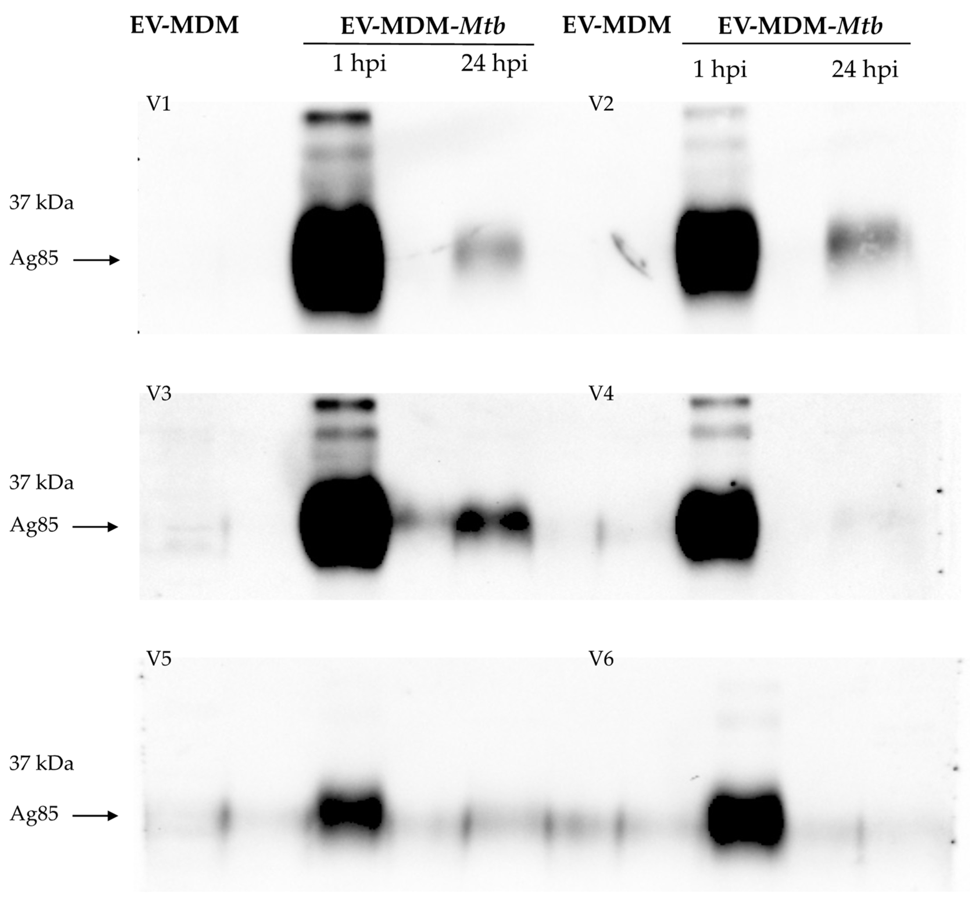
2.9. Identification of Mtb Protein Cargo in EVs Using Western Blotting

A total of 10 μg of either EV-MDM or EV-MDM-Mtb protein was combined with Laemmli buffer and β-mercaptoethanol. The proteins were then separated using 4–15% SDS-PAGE and transferred to PVDF membranes. The membranes were probed with polyclonal anti-Mtb (donated by Dr. Iris Estrada-García), anti-CD63, or anti-Ag85B (Abcam, Cambridge, UK) antibodies, followed by secondary antibody IgG-HRP (Thermo Fisher Scientific). Immune complexes were identified using the chemiluminescent substrate Clarity (Bio-Rad) and the ChemiDocMP imaging system (Bio-Rad). Total proteins were stained using a silver staining kit (Sigma Aldrich) after SDS-PAGE.

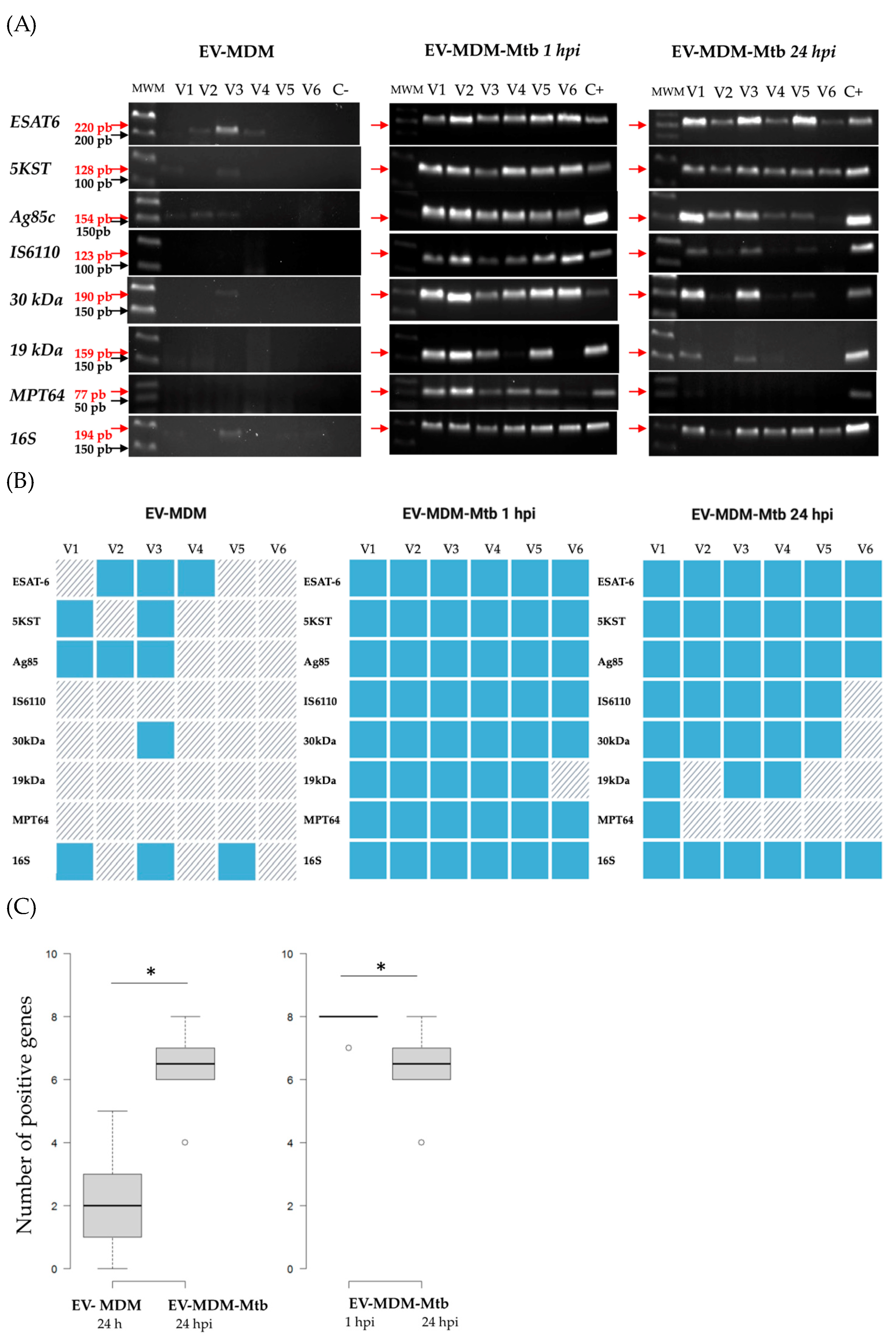
2.10. Identification of Mtb RNA Cargo in EVs Using PCR Amplification

According to the manufacturer’s instructions, the total exosomal RNA and protein isolation kit (Invitrogen-Thermo Fisher Scientific) was used to extract RNA from EV-MDM or EV-MDM-Mtb. Next, using the RNA as a template, cDNA was synthesized and prepared using the TaqMan MicroRNA Reverse Transcription Kit (Applied Biosystems-Thermo Fisher Scientific) with hexamers (Invitrogen, Thermo Fisher Scientific). A total of 70 ng of cDNA and forward and reverse specific primers (Table 1) for seven mycobacterial gene fragments (ESAT−6, 5KST, Ag85, IS6110, 30 kDa, 19 kDa, and MPT64) and rRNA16S as the endogenous gene were amplified in a Thermal Cycle (Applied Biosystems-Verity Thermo Fisher Scientific). The amplified fragments were then separated on an agarose gel, stained with Gel Red (Biotum, Fremont, CA, USA), and photographed under UV light using a Chemi-DocMP imaging system (Bio-Rad).

2.11. Statistical Analyses
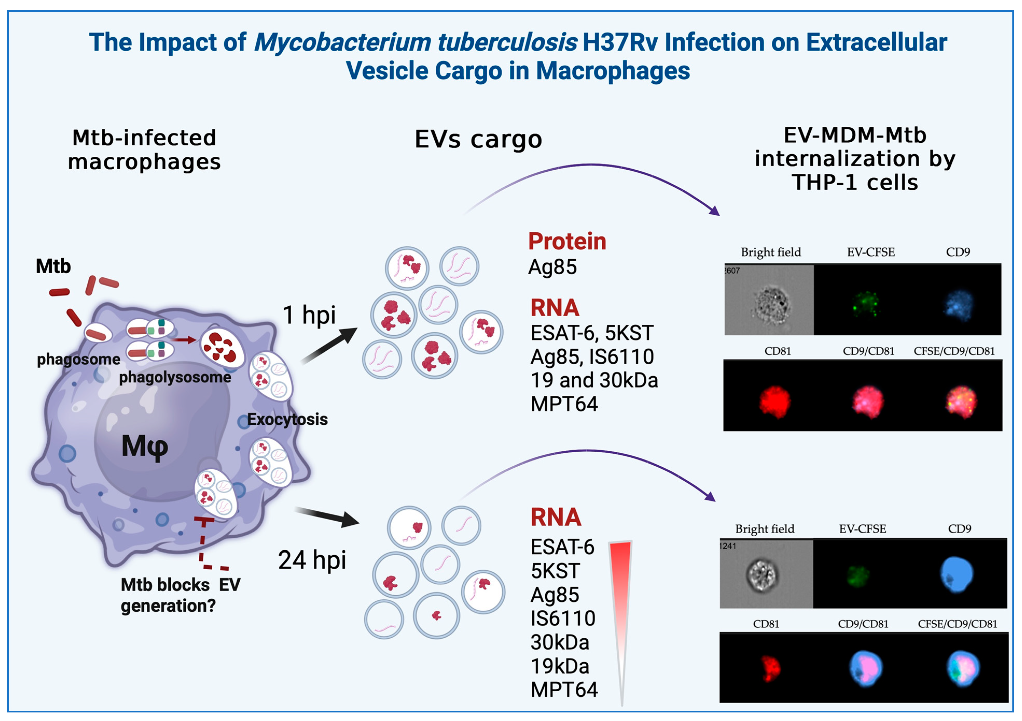
Numbers and percentages are used to represent categorical data without statistical tests. Comparisons between groups were performed by the Kruskal–Wallis test using GraphPad Prism software 10.4.0 version (Boston, MA, USA); p ≤ 0.05 was considered significant. The number of positive genes per individual in each group was compared using the Wilcoxon rank sum test (p = 0.003) using R-project software version 4.1.2 (2021) [17]. More details can be found in the figure legends.

3. Results

3.1. Characterization of EVs

We assessed tetraspanin expression on the surface of EVs using flow cytometry and Western blotting. Specific proteins of EVs have been described previously, including CD63, CD9, CD81, and syntenin-1 [18]. Pooled EVs from uninfected MDM expressed CD63, CD9, CD81, and syntenin at rates of 23.7%, 23.4%, 4.8%, and 0.35%, respectively. EVs from MDM infected with Mtb after 1 hpi showed high expressions of CD9 (97%) and CD63 (93%) and low expressions of CD81 (7%) and syntenin (0.35%), while EVs from MDM, after 24 hpi, expressed CD9 (80%) and CD63 (82%) and exhibited low expressions of CD81 (10%) and syntenin (2.5%) (Figure 1A). In addition, the expression of CD63 was examined using Western blotting of pooled EVs derived from both uninfected and infected MDM. The CD63 protein was observed in all samples using Western blotting, and the total protein was measured for each sample (Figure 1B). Additionally, pooled EV-MDM-Mtb (1 and 24 hpi) were characterized by the expression of CD9, CD63, and CD81 inside THP1 cells (Figure 1C).

3.2. Detection of Mycobacterial Proteins in EVs

We found that Mtb infection greatly reduces the total protein concentration in EVs at both 1 and 24 hpi (Figure 2A); however, we observed distinct mycobacterial proteins in EVs from uninfected or at 1 or 24 hpi cells. Rabbit serum polyclonal antibodies identified mycobacterial proteins, with molecular weights ranging from 15 to 37 kDa in EV-MDM-Mtb. At 1 hpi, these proteins were highly expressed, but their expression decreased by 24 hpi. EV-MDM-Mtb from volunteers V1, V2, V3, and V6 contained the most proteins (Figure 2B). We then examined CFP-10, ESAT-6, and Ag85 (specific antigens for Mtb) in EVs. Ag85 antigen was observed in infected MDM at 1 hpi and was significantly reduced at 24 hpi (Figure 3). CFP-10 and ESAT-6 proteins were not detected in EVs from any subject.

3.3. Detection of Mycobacterial RNAs in EVs

Mycobacterial hypothetical protein was present in exosomes from infected RAW264.7 murine macrophages [6]. In addition, we detected RNA from 30 kDa and 5KST of Mtb in the serum-EVs of TB-MDR patients [8]. Here we evaluate the RNA of the mycobacterial genes ESAT-6, 5KST, Ag85, IS6110, 30 kDa, 19 kDa, MPT64, and rRNA16S in EVs from infected MDM.
The RNA expression of six healthy volunteers (Figure 4A) and the overall analysis of all volunteers is shown as a heatmap (Figure 4B). ESAT-6, 5KST, Ag85, 30 kDa, and rRNA16S were detected in EVs from the uninfected or infected MDM of five healthy volunteers. The mycobacterial ESAT-6, 5KST, Ag85, IS6110, 30 kDa, 19 kDa, MPT64, and rRNA16S gene fragments were present in EVs at 1 hpi, except V6 for the 19 kDa fragment. In contrast, at 24 hpi, EVs’ RNA cargo decreased significantly. Specifically, mycobacterial transcripts for IS6110 and 30 kDa were not detected in V6; 19 kDa was not detected in V2, V5, or V6; and MPT64 was not detected in V2–V6 (Figure 4C). Figure 5 provides a graphical summary of the results of this study.

4. Discussion

The role of EVs in regulating or exacerbating non-communicable and infectious diseases has opened new avenues for understanding the progression of diseases and developing new treatment options. In TB, the role of EVs in pathogenesis and biomarker identification has been reported. However, due to the heterogeneity of EV cargo, further studies are required to understand their role in TB.
In this study, EV cargo was examined in human MDM at 1 and 24 hpi with the virulent H37Rv strain of Mtb. The EVs obtained from MDM culture supernatants exhibited expression of tetraspanins, with CD9 and CD63 present at approximately 20%. This expression of CD9 and CD63 increased to over 90% and 80% in EVs from MDM after 1 and 24 hpi, whereas CD81 and syntenin increased up to 10 and 2.5% in EVs at 24 hpi. Although the increased EV secretion has been shown in response to a variety of stimuli, including electric stimulation, drug administration, X-ray and UV radiation, sound waves, mitochondrial metabolism, and viruses [19], it is possible that the increased expression of CD9, CD63, CD81, and syntenin may be associated with Mtb infection. The role of tetraspanins in TB disease has been studied before. CD81 binds to the M. abscessus antioxidant enzyme alkyl hydroperoxidase C (AhpC) to facilitate early macrophage interactions. Given that Mtb AhpC shares ∼83 % similarity with M. abscessus AhpC, it is possible that CD81 is a receptor for the latter pathogenic mycobacteria and may also facilitate macrophage interactions [20]. The C-terminus of ESAT-6 also binds to the PDZ-domains of syntenin-1, suggesting that the host protein syntenin-1 is a possible cellular receptor for the mycobacterial protein ESAT-6 [21]. Further studies are required to demonstrate whether EVs in conjunction with Mtb antigens may be involved in modulating the immune response.
EVs containing Mtb proteins or RNA do not necessarily express tetraspanins. It has been reported that Mtb-derived EVs are not necessarily packaged in host EVs [22]. This opens an important field for future studies. Here we find that that Mtb infection increased the expression of CD9 and CD63 tetraspanins in EVs. Syntenin and tetraspanins are involved in many cellular processes, such as cell adhesion, the regulation of cell motility and/or morphology, fusion, signaling, and other functions, and increasing evidence suggests that they play a central role in the pathogenesis of viral infections [23]. Additionally, it has been reported that during Mtb infection, the lysosomal glycoprotein CD63 is recruited to Mtb phagosomes [24]. Based on this, it is possible to speculate that Mtb could evade immune responses by modulating the expression of syntenin and tetraspanins.
The total protein in EVs from infected MDM was not associated with a decrease in the abundance of Mtb proteins, as our results showed that some mycobacterial proteins were present in EVs at 1 hpi, whereas these proteins were significantly reduced at 24 hpi. Previous studies have reported more than 41 mycobacterial proteins in EVs released from Mtb-infected J774 macrophages using highly sensitive techniques, such as proteomics [25], but the transport of mycobacterial proteins in EVs at different infection stages remains to be evaluated. In another study using non-virulent M. bovis BCG–infected J774 macrophages, it was reported that lipoarabinomannan and the 19 kDa lipoprotein took between 72 and 120 h to appear [26]. In contrast, in virulent Mtb strains, we only detected the RNA of these proteins at an early stage of infection, followed by a decrease in 19 kDA lipoprotein RNA. Furthermore, our results showed a decrease in Ag85 kDa after 24 hpi. We investigated the presence of Mtb Ag85 kDa in EVs, as Ag85A kDa and Ag85B kDa account for approximately 60% of the total protein secreted by Mtb [27]. Previous studies have reported that Ag85B is downregulated during chronic infection, inducing CD4+ T cells to recognize the Ag85B epitope [28]. This suggests that this decrease may be associated with an immune evasion mechanism of Mtb. However, this is an issue that requires further investigation.
RNA transcripts have been reported to originate from vesicles released by mycobacteria during infection. Both host and mycobacterial small RNA fragments have been identified in Mtb-infected macrophages and patients with TB [6,8]. Mycobacterial transcripts have been identified in exosomes/EVs using the RAW264.7 murine macrophage [6]. Here, we evaluated transcripts previously studied in TB patients and found that ESAT-6, 5KST, Ag85, IS6110, 30 kDa, 19 kDa, and MPT64 were expressed in EVs. As with Mtb proteins, RNA transcripts decreased after 24 hpi, with a decrease in MPT64 observed in most patients, followed by the 19 kDa transcript and, in some individuals, the 30 kDa transcript and IS6110. The function of mycobacterial RNAs as pro-inflammatory mediators and in recruiting cells to sites of infection is well-established [6,29]. However, further studies are needed.
This was an unexpected demonstration that RNA transcripts of ESAT-6, 5KST, Ag85, and 30 kDa were present in EVs from uninfected healthy subjects. This finding may indicate the possibility of latent infection or exposure to Mtb, as all subjects were from TB-endemic areas and were TST-positive. Previously, it has been reported that Mtb occurs intracellularly; its secreted mycobacterial antigens and host RNAs are packaged into host EVs and released into the bloodstream. In addition, Mtb proteins have been detected in healthy individuals, possibly with prior infections [30,31]. RNA transcripts of Mtb have also been detected in both healthy and latently TB-infected individuals [32]. It is difficult to determine whether the healthy subjects tested were exposed to Mtb or latently infected. Further analysis may provide more insight into this issue.
One of the limitations in studying EVs from healthy human subjects or patients is the number of EVs that can be obtained using assays. While the use of cell lines can provide a distinct advantage for their analysis, the response of primary cells and the inherent variability in response can only be elucidated by studying primary cells from healthy subjects.

5. Conclusions

Mtb infection resulted in the increased expression of the CD9 and CD63 tetraspanins in EVs released by macrophages. EVs carry Ag85-Mtb and various RNA sequences from Mtb in the early stages of infection; however, the EV cargo appears to diminish by 24 hpi. Given that Mtb is known to evade phagosome–lysosome fusion, the reduction in EV cargo may be linked to this evasion mechanism. Both Ag85 and the RNA transcripts could therefore serve as potential biomarkers for diagnosing Mtb infection or latency.

Author Contributions

M.G.S.-C., formal analysis, methodology, data curation; L.H.G.-G., writing—original draft preparation, review, formal analysis, data curation and editing; S.G.-B., software, formal analysis, writing—original draft preparation; M.T.H., software, formal analysis, writing—original draft preparation; C.S., software, methodology, formal analysis; Y.G., conceptualization, writing—review and editing, funding acquisition and project administration. All authors have read and agreed to the published version of the manuscript.

Funding

This study was funded by institutional protocol number C57-20 and the Fondo Sectorial de Investigación en Salud y Seguridad Social (FOSISS), Proyecto No. 00000000290282, Consejo Nacional de Humanidades, Ciencias y Tecnologías (CONAHCyT), Mexico.

Data Availability Statement

The original contributions presented in the study are included in the article, further inquiries can be directed to the corresponding author.

Acknowledgments

We thank Luz del Carmen Balderas and Andrea Araceli González Zavala for valuable technical assistance and Iris Citlali Estrada García for the antibodies anti-Mtb. We thank the Laboratorio Nacional Conahcyt de Investigación y Diagnóstico por Inmunocitofluorometría (LANCIDI) for technical assistance.

Conflicts of Interest

The authors declare no conflicts of interest.

References

  1. World Health Organization. Global Tuberculosis Report 2024; World Health Organization: Geneva, Switzerland, 2024. Available online: https://www.who.int/teams/global-tuberculosis-programme/tb-reports/global-tuberculosis-report-2024 (accessed on 31 October 2024).
  2. Ng, C.Y.; Kee, L.T.; Al-Masawa, M.E.; Lee, Q.H.; Subramaniam, T.; Kok, D.; Ng, M.H.; Law, J.X. Scalable Production of Extracellular Vesicles and Its Therapeutic Values: A Review. Int. J. Mol. Sci. 2022, 23, 7986. [Google Scholar] [CrossRef] [PubMed]
  3. Gurunathan, S.; Kang, M.H.; Qasim, M.; Khan, K.; Kim, J.H. Biogenesis, Membrane Trafficking, Functions, and next Generation Nanotherapeutics Medicine of Extracellular Vesicles. Int. J. Nanomed. 2021, 16, 3357–3383. [Google Scholar] [CrossRef] [PubMed]
  4. Wang, Y.; Zhao, M.; Liu, S.; Guo, J.; Lu, Y.; Cheng, J.; Liu, J. Macrophage-Derived Extracellular Vesicles: Diverse Mediators of Pathology and Therapeutics in Multiple Diseases. Cell Death Dis. 2020, 11, 924. [Google Scholar] [CrossRef] [PubMed]
  5. Prados-Rosales, R.; Baena, A.; Martinez, L.R.; Luque-Garcia, J.; Kalscheuer, R.; Veeraraghavan, U.; Camara, C.; Nosanchuk, J.D.; Besra, G.S.; Chen, B.; et al. Mycobacteria Release Active Membrane Vesicles That Modulate Immune Responses in a TLR2-Dependent Manner in Mice. J. Clin. Investig. 2011, 121, 1471–1483. [Google Scholar] [CrossRef]
  6. Singh, P.P.; Li, L.; Schorey, J.S. Exosomal RNA from Mycobacterium Tuberculosis-Infected Cells Is Functional in Recipient Macrophages. Traffic 2015, 16, 555–571. [Google Scholar] [CrossRef]
  7. Lyu, L.; Zhang, X.; Li, C.; Yang, T.; Wang, J.; Pan, L.; Jia, H.; Li, Z.; Sun, Q.; Yue, L.; et al. Small RNA Profiles of Serum Exosomes Derived from Individuals with Latent and Active Tuberculosis. Front. Microbiol. 2019, 10, 1174. [Google Scholar] [CrossRef]
  8. Carranza, C.; Herrera, M.T.; Guzmán-Beltrán, S.; Salgado-Cantú, M.G.; Salido-Guadarrama, I.; Santiago, E.; Chávez-Galán, L.; Gutiérrez-González, L.H.; González, Y. A Dual Marker for Monitoring MDR-TB Treatment: Host-Derived MiRNAs and M. Tuberculosis-Derived RNA Sequences in Serum. Front. Immunol. 2021, 12, 760468. [Google Scholar] [CrossRef]
  9. Zheng, W.; LaCourse, S.M.; Song, B.; Singh, D.K.; Khanna, M.; Olivo, J.; Stern, J.; Escudero, J.N.; Vergara, C.; Zhang, F.; et al. Diagnosis of Paediatric Tuberculosis by Optically Detecting Two Virulence Factors on Extracellular Vesicles in Blood Samples. Nat. Biomed. Eng. 2022, 6, 979–991. [Google Scholar] [CrossRef]
  10. Mehaffy, C.; Dobos, K.M.; Nahid, P.; Kruh-Garcia, N.A. Second Generation Multiple Reaction Monitoring Assays for Enhanced Detection of Ultra-Low Abundance Mycobacterium Tuberculosis Peptides in Human Serum. Clin. Proteom. 2017, 14, 21. [Google Scholar] [CrossRef]
  11. Ramachandra, L.; Qu, Y.; Wang, Y.; Lewis, C.J.; Cobb, B.A.; Takatsu, K.; Boom, W.H.; Dubyak, G.R.; Harding, C.V. Mycobacterium Tuberculosis Synergizes with ATP to Induce Release of Microvesicles and Exosomes Containing Major Histocompatibility Complex Class II Molecules Capable of Antigen Presentation. Infect. Immun. 2010, 78, 5116–5125. [Google Scholar] [CrossRef]
  12. Bhatnagar, S.; Schorey, J.S. Exosomes Released from Infected Macrophages Contain Mycobacterium Avium Glycopeptidolipids and Are Proinflammatory. J. Biol. Chem. 2007, 282, 25779–25789. [Google Scholar] [CrossRef] [PubMed]
  13. García-Martínez, M.; Vázquez-Flores, L.; Álvarez-Jiménez, V.D.; Castañeda-Casimiro, J.; Ibáñez-Hernández, M.; Sánchez-Torres, L.E.; Barrios-Payán, J.; Mata-Espinosa, D.; Estrada-Parra, S.; Chacón-Salinas, R.; et al. Extracellular Vesicles Released by J774A.1 Macrophages Reduce the Bacterial Load in Macrophages and in an Experimental Mouse Model of Tuberculosis. Int. J. Nanomed. 2019, 14, 6707–6719. [Google Scholar] [CrossRef] [PubMed]
  14. Raj, A.; Singh, N.; Gupta, K.B.; Chaudhary, D.; Yadav, A.; Chaudhary, A.; Agarwal, K.; Varma-Basil, M.; Prasad, R.; Khuller, G.K.; et al. Comparative Evaluation of Several Gene Targets for Designing a Multiplex-PCR for an Early Diagnosis of Extrapulmonary Tuberculosis. Yonsei Med. J. 2016, 57, 88–96. [Google Scholar] [CrossRef] [PubMed]
  15. Neshani, A.; Kakhki, R.K.; Sankian, M.; Zare, H.; Chichaklu, A.H.; Sayyadi, M.; Ghazvini, K. Modified Genome Comparison Method: A New Approach for Identification of Specific Targets in Molecular Diagnostic Tests Using Mycobacterium Tuberculosis Complex as an Example. BMC Infect. Dis. 2018, 18, 517. [Google Scholar] [CrossRef]
  16. Parandaman, V.; Narayanan, P.R.; Venkatesan, P.; Girish, C.; Mahadevan, S.; Rajajee, S. Evaluation of PCR Using TRC(4) and IS6110 Primers in Detection of Tuberculous Meningitis. J. Clin. Microbiol. 2001, 39, 2006–2008. [Google Scholar] [CrossRef]
  17. Team, R.C. R Core Team 2023 R: A Language and Environment for Statistical Computing; R Foundation for Statistical Computing: Vienna, Austria, 2023. [Google Scholar]
  18. Théry, C.; Witwer, K.W.; Aikawa, E.; Alcaraz, M.J.; Anderson, J.D.; Andriantsitohaina, R.; Antoniou, A.; Arab, T.; Archer, F.; Atkin-Smith, G.K.; et al. Minimal Information for Studies of Extracellular Vesicles 2018 (MISEV2018): A Position Statement of the International Society for Extracellular Vesicles and Update of the MISEV2014 Guidelines. J. Extracell Vesicles 2018, 7, 1535750. [Google Scholar] [CrossRef]
  19. Erwin, N.; Serafim, M.F.; He, M. Enhancing the Cellular Production of Extracellular Vesicles for Developing Therapeutic Applications. Pharm. Res. 2023, 40, 833–853. [Google Scholar] [CrossRef]
  20. Karam, J.; Blanchet, F.P.; Vivès, É.; Boisguérin, P.; Boudehen, Y.M.; Kremer, L.; Daher, W. Mycobacterium Abscessus Alkyl Hydroperoxide Reductase C Promotes Cell Invasion by Binding to Tetraspanin CD81. iScience 2023, 26, 106042. [Google Scholar] [CrossRef]
  21. Schumann, G.; Schleier, S.; Rosenkrands, I.; Nehmann, N.; Hälbich, S.; Zipfel, P.F.; De Jonge, M.I.; Cole, S.T.; Munder, T.; Möllmann, U. Mycobacterium Tuberculosis Secreted Protein ESAT-6 Interacts with the Human Protein Syntenin-1. Cent. Eur. J. Biol. 2006, 1, 183–202. [Google Scholar] [CrossRef]
  22. Palacios, A.; Gupta, S.; Rodriguez, G.M.; Prados-Rosales, R. Extracellular Vesicles in the Context of Mycobacterium Tuberculosis Infection. Mol. Immunol. 2021, 133, 175–181. [Google Scholar] [CrossRef]
  23. Jankovičová, J.; Sečová, P.; Michalková, K.; Antalíková, J. Tetraspanins, More than Markers of Extracellular Vesicles in Reproduction. Int. J. Mol. Sci. 2020, 21, 7568. [Google Scholar] [CrossRef] [PubMed]
  24. Seto, S.; Matsumoto, S.; Tsujimura, K.; Koide, Y. Differential Recruitment of CD63 and Rab7-Interacting-Lysosomal-Protein to Phagosomes Containing Mycobacterium Tuberculosis in Macrophages. Microbiol. Immunol. 2009, 54, 170–174. [Google Scholar] [CrossRef]
  25. Giri, P.K.; Kruh, N.A.; Dobos, K.M.; Schorey, J.S. Proteomic Analysis Identifies Highly Antigenic Proteins in Exosomes from M. Tuberculosis-Infected and Culture Filtrate Protein-Treated Macrophages. Proteomics 2010, 10, 3190–3202. [Google Scholar] [CrossRef] [PubMed]
  26. Bhatnagar, S.; Shinagawa, K.; Castellino, F.J.; Schorey, J.S. Exosomes Released from Macrophages Infected with Intracellular Pathogens Stimulate a Proinflammatory Response in Vitro and in Vivo. Blood 2007, 110, 3234–3244. [Google Scholar] [CrossRef]
  27. Wiker, H.G.; Harboe, M. The Antigen 85 Complex: A Major Secretion Product of Mycobacterium Tuberculosis. Microbiol. Rev. 1992, 56, 648–661. [Google Scholar] [CrossRef]
  28. Chandra, P.; Grigsby, S.J.; Philips, J.A. Immune Evasion and Provocation by Mycobacterium Tuberculosis. Nat. Rev. Microbiol. 2022, 20, 750–766. [Google Scholar] [CrossRef]
  29. Cheng, Y.; Schorey, J.S. Extracellular Vesicles Deliver Mycobacterium RNA to Promote Host Immunity and Bacterial Killing. EMBO Rep. 2019, 20, e46613. [Google Scholar] [CrossRef]
  30. Mehaffy, C.; Kruh-Garcia, N.A.; Graham, B.; Jarlsberg, L.G.; Willyerd, C.E.; Borisov, A.; Sterling, T.R.; Nahid, P.; Dobos, K.M. Identification of Mycobacterium Tuberculosis Peptides in Serum Extracellular Vesicles from Persons with Latent Tuberculosis Infection. J. Clin. Microbiol. 2020, 58, e00393-20. [Google Scholar] [CrossRef]
  31. Kruh-Garcia, N.A.; Wolfe, L.M.; Chaisson, L.H.; Worodria, W.O.; Nahid, P.; Schorey, J.S.; Davis, J.L.; Dobos, K.M. Detection of Mycobacterium Tuberculosis Peptides in the Exosomes of Patients with Active and Latent M. Tuberculosis Infection Using MRM-MS. PLoS ONE 2014, 9, e103811. [Google Scholar] [CrossRef]
  32. Lv, L.; Li, C.; Zhang, X.; Ding, N.; Cao, T.; Jia, X.; Wang, J.; Pan, L.; Jia, H.; Li, Z.; et al. RNA Profiling Analysis of the Serum Exosomes Derived from Patients with Active and Latent Mycobacterium Tuberculosis Infection. Front. Microbiol. 2017, 8, 1051. [Google Scholar] [CrossRef]
Figure 1. Characterization of pooled EVs from infected MDM. The surface markers CD9, CD63, CD81, and syntenin were detected using flow cytometry (A). EV proteins were separated using SDS-PAGE, and CD63 was detected using Western blotting; the lower membrane shows silver-stained total protein as a loading control (B). Characterization of EVs inside THP1 cells. EVs-CFSE from Mtb-infected MDM (1 or 24 hpi) were added to THP-1 cells, and intracellular vesicles were stained with antibodies against CD9-PE, CD63-PerCP-Cy5.5, and CD81-APC; the expression was detected using an Amnis® imaging flow cytometer. This figure shows a representative image of CD9, CD63, and CD81 expression in EVs (C).
Microorganisms 12 02405 g001aMicroorganisms 12 02405 g001b
Figure 2. Mycobacterial proteins in EVs from MDM. EVs from six volunteers were isolated from supernatants of MDM at 24 hpi or MDM infected with Mtb at 1 or 24 hpi. EVs were lysed and proteins were quantified by Bradford (A,B), comparisons between groups were performed by the Kruskal–Wallis test, * p < 0.05. Proteins were separated using SDS-PAGE and transferred to a PVDF membrane. The membrane was incubated with rabbit polyclonal antibodies against Mtb (C).
Microorganisms 12 02405 g002
Figure 3. Detection of the mycobacterial antigen Ag85 in EVs. EVs from six subjects were isolated from supernatants of MDM- or MDM-Mtb-infected cells (1 or 24 hpi). EVs were lysed and proteins were separated using SDS-PAGE and transferred to a PVDF membrane. The membrane was incubated with an antibody against Ag85 from Mtb.
Microorganisms 12 02405 g003
Figure 4. EVs contain RNA sequences of mycobacterial antigens. RNA was extracted from EVs, and cDNA was synthesized, followed by PCR detection of the following mycobacterial genes: ESAT-6, 5KST, Ag85, IS6110, 30 kDa, 19 kDa, MPT64, and rRNA16S. Fragments were separated using agarose gel electrophoresis, stained, and visualized under UV light. Mycobacterial amplified fragments in EVs from uninfected or Mtb-infected MDM (1 or 24 hpi) (A). Heatmap of EVs extracted from six healthy volunteers; blue represents positive amplification, and diagonal gray lines represent negative amplification (B). Box plot of the number of positive genes per individual for each group (Biorender). It shows a significant difference between groups, as follows: * Wilcoxon rank sum test for “EV-MDM” vs. “EV-MDM-Mtb 24 hpi” showed p = 0.007796 (left graphic), and Wilcoxon rank sum test for “EV-MDM-Mtb 1 hpi” vs. “EV-MDM-Mtb 24 hpi” showed p = 0.0248 (right graphic) (C).
Microorganisms 12 02405 g004
Figure 5. EVs released from Mtb-infected cells with the H37Rv Mtb strain at 1 hpi carry mycobacterial proteins, as well as RNA transcripts of ESAT-6, 5KST, Ag85, IS6110, 19 kDa, 30 kDa, and MPT64 proteins. These EVs can be inserted into other cells and express tetraspanins, such as CD9, CD63, and CD81. At 24 hpi, Mtb arrests phagosome–lysosome fusion and probably prevents the release of EVs carrying Mtb antigens and genetic material, as well as tetraspanin expression.
Microorganisms 12 02405 g005
Table 1. Mycobacterial gene primers.
TargetPrimer SequencesSize (bp)Reference
ESAT-6F: GTCCATTCATTCCCTCCT
R. CTATGCGAACATCCCAGT
220[14]
5KSTF: TTGCTGAACTTGACCTGCCCGTA
R: GCGTCTCTGCCTTCCTCCGAT
128[15]
Ag85F: AAGGTCCAGTTCCAGGGCG
R: ATTGGCCGCCCACGGGCATGAT
154[8]
IS6110F: CCTGCGAGCGTAGGCGTCGG
R: CTCGTCCAGCGCCGCTFCGG
123[16]
30 kDaF: TGTACCAGTCGCTGTAGAAG
R: GACATCAAGGTTCAGTTCC
190[14]
19 kDaF: GAGACCACGACCGCGGCAGG
R: AATGCCGGTCGCCGCCCCGCCGAT
159[8]
MPT64F: GTGAACTGAGCAAGCAGACCG
R: GTTCTGATAATTCACCGGGTCC
77[8]
rRNA16SF: GCCGTAAACGGTGGGTACTA
R: TGCATGTCAAACCCAGGTAA
194[8]
Disclaimer/Publisher’s Note: The statements, opinions and data contained in all publications are solely those of the individual author(s) and contributor(s) and not of MDPI and/or the editor(s). MDPI and/or the editor(s) disclaim responsibility for any injury to people or property resulting from any ideas, methods, instructions or products referred to in the content.

