Article

Diversity of Bacteria with Quorum Sensing and Quenching Activities from Hydrothermal Vents in the Okinawa Trough

1
Frontiers Science Center for Deep Ocean Multispheres and Earth System, College of Marine Life Sciences, Ocean University of China, 5 Yushan Road, Qingdao 266003, China
2
Laboratory for Marine Ecology and Environmental Science, Laoshan Laboratory, Qingdao 266237, China
3
Institute of Evolution & Marine Biodiversity, Ocean University of China, Qingdao 266003, China
*
Author to whom correspondence should be addressed.
Microorganisms 2023, 11(3), 748; https://doi.org/10.3390/microorganisms11030748
Submission received: 16 January 2023 / Revised: 9 March 2023 / Accepted: 10 March 2023 / Published: 14 March 2023
(This article belongs to the Special Issue Microbial Quorum Sensing: Advances and Challenges)

Abstract

:
Quorum sensing (QS) is a chemical communication system by which bacteria coordinate gene expression and social behaviors. Quorum quenching (QQ) refers to processes of inhibiting the QS pathway. Deep-sea hydrothermal vents are extreme marine environments, where abundant and diverse microbial communities live. However, the nature of chemical communication in bacteria inhabiting the hydrothermal vent is poorly understood. In this study, the QS and QQ activities with N-acyl homoserine lactones (AHLs) as the autoinducer were detected in bacteria isolated from hydrothermal vents in the Okinawa Trough. A total of 18 and 108 isolates possessed AHL-producing and AHL-degrading abilities, respectively. Bacteria mainly affiliated with Rhodobacterales, Hyphomicrobiales, Enterobacterales and Sphingomonadales showed QS activities; QQ was mainly associated with Bacillales, Rhodospirillales and Sphingomonadales. The results showed that the bacterial QS and QQ processes are prevalent in hydrothermal environments in the Okinawa Trough. Furthermore, QS significantly affected the activities of extracellular enzymes represented by β-glucosidase, aminopeptidase and phosphatase in the four isolates with higher QS activities. Our results increase the current knowledge of the diversity of QS and QQ bacteria in extreme marine environments and shed light on the interspecific relationships to better investigate their dynamics and ecological roles in biogeochemical cycling.

1. Introduction

Quorum sensing (QS) is a bacterial communication system that enables bacterial populations to regulate their group behaviors according to density [1]. QS relies on the production, diffusion, accumulation and group-wide detection of extracellular signaling molecules termed autoinducers [2]. QS coordinates various intra- and inter-bacterial behaviors, such as extracellular enzyme production [3], DNA uptake [4] and biofilm formation [5]. It has been estimated that a significant portion of the bacterial genome (4–10%) and proteome (≥20%) is influenced by QS, suggesting that QS is important for the adaptation of bacteria to various environments [6]. A range of autoinducers has been discovered and identified. Thus, QS mediated by N-acyl homoserine lactones (AHLs) has been widely studied in a diverse range of Gram-negative bacteria. Moreover, the AHL-producing genes have been identified in many bacteria, including luxI which leads to the formation of 3OC6-HSL in Vibrio spp., and lasI which is involved in 3OC10-HSL in Pseudomonas aeruginosa [7].
Marine bacteria with AHL-mediated QS (AHL-QS) activities have been isolated from multiple marine habitats, such as marine snow [8], corals [9,10], sponges [11], dinoflagellates [12], Trichoderma colonies [13] and sea anemones [14]. These bacteria belong mainly to Alphaproteobacteria and Gammaproteobacteria [15]. In addition, previous studies have shown that Actinobacteria [3,16], Firmicutes [16,17], archaea [18] and cyanobacteria [19] could produce AHLs. Doberva et al. [20] identified a large number of luxI genes in the global ocean metagenomes, indicating that AHL-QS may be of great significance for communication among microorganisms, and the regulation of biological behaviors in marine habitats. Additionally, AHL-QS was associated with large-scale bioluminescence events caused by algal blooms [21] and was important in maintaining the health of the coral reef ecosystem [22].
AHL-QS exerts an important role in regulating the elements cycling in the global ocean by affecting the activity of extracellular hydrolase and biofilm formation. For example, Jatt et al. [8] determined that QS could regulate bacteria to attach to marine snow particles and produce exoenzymes, thereby affecting the degradation of marine particulate organic carbon. Moreover, the biofilm formation and lipase production of Ruegeria mobilis Rm01, which was isolated from marine particles, could be regulated by QS [3]. In addition, adding AHL molecules to the Trichodesmium consortia could double the activity of phosphatase in the epibiotic bacteria, thereby promoting phosphorus circulation [13].
Quorum quenching (QQ) inhibits QS pathways by interfering with the production, release, recognition, and degradation of signaling molecules, thereby affecting various physiological functions dependent on bacterial QS [23,24]. Indeed, bacterial signaling molecules in the microenvironment could be removed by QQ activity [25], which was a crucial process for bacteria to maintain environmental homeostasis [2]. Bacteria with QQ activities have been isolated from various marine habitats, including biofilms [26], estuaries [23], corals [9] and sediment [27]. In addition, it was reported that QQ enzymes could cause decreases in autoinducer release and the activities of extracellular pectolytic enzymes to attenuate pathogenicity [28]. Despite their important ecological implications, the function of QS and QQ, in deep-sea hydrothermal vents have been poorly characterized [2].
In deep-sea hydrothermal vents, the dramatic changes in temperature (approximately 4–301 °C) and pH (ca. 5.0–7.6) vary in different marine environments [29]. Compared with other hydrothermal vents, the vent fluids from hydrothermal systems of the Okinawa Trough, influenced by the sediment from Chinese marginal seas, have lower pH and higher concentrations of CO2, CH4, NH4+, iodine and potassium [29]. In addition, the unique physicochemical conditions associated with hydrothermal vent systems give rise to abundant microbial communities. For example, there are abundant magnetotactic bacteria, sulfur-oxidizing bacteria (Sulfurimonas and Sulfurovum), sulfate-reducing bacteria (Thermosulfidibacter and Desulfothermus) and methanogens (Methanococcales and Methanosarcinales) living in the hydrothermal vents [30,31,32,33,34,35,36]. However, the interspecies relationship of microbial communities and their ecological function in hydrothermal vents from Okinawa Trough has been rarely studied.
In this study, we explored the diversity of QS and QQ isolates, and the regulatory function of QS on extracellular enzymatic activities in bacteria isolated from hydrothermal fields of the Okinawa Trough. The AHL-based QS and QQ activities of culturable bacteria were screened, and the biofilm formation of QS and partial-QQ strains was detected to reveal the relationship between QS/QQ and biofilm production. In addition, the effects of QS on the extracellular enzymatic activities of QS bacteria were explored.

2. Materials and Methods

2.1. Isolation and Identification of Bacterial Strains from the Okinawa Trough

Bacteria used in this study were isolated from seawater, sediment and macroorganisms (mussel, crab and shrimp) from the hydrothermal fields of the Okinawa Trough during the HOBAB2 voyage in 2014 and the HOBAB4 voyage in 2016 and 2018. The samples were collected by a TV-grab sampler or the remotely operated vehicle (ROV) Faxian during the cruises conducted by the R/V Kexue. The information about samples, including longitude and latitude and depth, were listed in Table S1).
The seawater samples were serially diluted from 10−1 to 10−3, and the sediment samples were soaked in 0.85 % (w/v) saline serially diluted from 10−1 to 10−3, and 100 μL of each dilution was spread onto the surface of marine agar 2216E (MA) plates [37] and R2A plates [38]. The macroorganisms were dissected aseptically, and the tissues, including gill and intestine, were washed with 100 mL of 0.22 µm filtered seawater, which was then diluted and spread onto MA and R2A plates. The plates were cultured at 28 °C, and colonies were purified by streaking and re-streaking three to four times on MA or R2A plates. Further identification of the isolates relied on 16S rRNA gene amplification using the primers B8F (5′-AGAGTTTGATCCTGGCTCAG-3′) and B1510R (5′-GGTTACCTTGTTACGACTT-3′) followed by sequencing at Sangon Biotech (Qingdao, China). Sequence similarities between isolates and their most closely related bacteria were calculated using the EzBiocloud server (accessed on 20 July 2022. https://www.ezbiocloud.net). The phylogenetic tree of 16S rRNA genes from QS strains, their closest type strains and the reported AHL-producing strains was conducted using MEGA v11 based on the neighbor-joining algorithm [39]. The phylogenetic tree of QQ strains was based on their 16S rRNA gene sequences using MEGA v11 based on the neighbor-joining algorithm [39].

2.2. Screening for AHL-Producing and AHL-Degrading Bacteria

Screening for AHL-producing and AHL-degrading bacteria was conducted using the bacterial biosensor Agrobacterium tumefaciens (pCF218) (pCF372) A136 [40,41,42]. AHL-producing strains were identified by the co-feeding method according to Chu et al. [43]. Briefly, the bacterial cultures were inoculated into marine broth 2216E (MB) and cultured with shaking (170 rpm) at 28 °C for 24 h. A. tumefaciens A136 was added into the LB medium with 1% agar and X-gal (250 μg/L), and then the Oxford cups were placed on the plate to form wells. Meanwhile, fresh cultures were added into the wells, and the C6-HSL and MB were used as positive and negative controls, respectively. After co-incubation at 28 °C for 24 h, positivity was indicated by the presence of indigo spots. In addition, the high-throughput method described by Su et al. [3] was used to confirm the QS activities of these cultures. The fresh culture and an A136 X-gal assay solution [an overnight broth culture of A136 inoculated in AT minimal glucose medium [44] and mixed with X-Gal (250 μg/L)] were mixed and added into the 96-well microtiter plates, and the results were observed after cultivation at 28 °C for 24 h. C6-HSL and MB were used as positive and negative controls, respectively. All the experiments were performed in quadruplicate.
AHL-degrading activity was detected using the high-throughput method described by Tang et al. [45]. C6-HSL (representative for AHLs with short acyl chains) and C12-HSL (representative for AHLs with long acyl chains) were used as the substrates. Briefly, C6-HSL or C12-HSL was mixed with the bacterial culture and MB medium (negative control), and maintained at 28 °C for 24 h. After incubation, the supernatant was obtained after centrifugation at 4 °C and 6000 rpm for 10 min, and filtered through a 0.22 μm filter (JINTENG® PES syringe filter). The supernatant was then mixed with an A136 X-gal assay solution in 96-well microtiter plates, and incubated at 28 °C for another 24 h. All the experiments were performed in triplicate. Positive results were indicated by reduced indigo color compared with the negative control.

2.3. Identification of QQ Activities in QS Strains

The QQ activities of QS strains were further identified using the high-throughput method. In short, QS strains were inoculated into MB with shaking (170 rpm) at 28 °C for 24 h. The cells were harvested after centrifugation at 4 °C and 6000 rpm for 10 min and re-suspended in HEPES buffer (20 mM Na-HEPES with 0.5 M NaCl, 10% glycerol, and 0.1% Triton X-100, pH 8.5) for sonication. Additional centrifugation for the lysed cells was conducted at 4 °C and 12,000 rpm to obtain the crude enzyme supernatant. The crude enzyme supernatant, C6-HSL or C12-HSL, HEPES buffer, and A136 X-gal assay solution were mixed and added into a 96-well microtiter plate, and incubated at 28 °C for 24 h. All the experiments were performed in triplicate. Positive results were indicated by a reduced indigo color compared with the negative control.

2.4. Biofilm Production in QS and QQ Strains

Biofilm production of all the bacteria with QS activities, and 23 isolates with C6- and C12-HSL-degrading abilities were quantified using a crystal violet (CV) assay, as described previously [25] with slight modifications. Briefly, the bacterial cultures were grown in MB, and 1% of the suspension was inoculated in MB medium contained in 96-well microtiter plates with incubation at 28 °C for 18 h. Each subsequent step was performed gently to preserve the biofilm. Thus, the supernatant was removed, and non-adherent cells were washed with sterile double distilled water. The 96-well microtiter plates were dried, and methanol was added to fix the cells; biofilms were stained with 0.2% (w/v) CV. Then, the CV in the biofilm was dissolved with 150 μL of 95% acetic acid, and the resulting OD was measured at 570 nm (OD570). MB was used as a negative control, and all the experiments were performed in triplicate. The significant difference between the experimental groups and the controls was calculated by the t-test (*** p < 0.001, ** p < 0.01, and * p < 0.05).

2.5. Extracellular Enzyme Activities in QS Strains

The extracellular enzyme (EE) activities were quantified following the methods described by Hmelo [46]. Five EE activities were tested using fluorescent substrates, including MUF-α-glucopyranoside (for α-glucosidase activity), MUF-β-glucopyranoside (for β-glucosidase activity), MUF-butyrate (for lipase activity), MCA-leucine (for aminopeptidase activity) and MUF-phosphate (for phosphatase activity) (all fluorescent substrates were purchased from Sigma-Aldrich). In brief, bacterial isolates were grown in MB at 28 °C, and the supernatants were retained after centrifugation at 4 °C and 12,000 rpm for 10 min and filtering through 0.22 μm filters. The supernatants and fluorescent substrates were mixed and added into a fluorescent multi-well plate with incubation at 28 °C for 12 h. Released fluorescent signals were detected with a Fluoroskan Ascent FL multi-well plate reader. The excitation and emission characteristics of the fluorophores were previously programmed into the instrument. In addition, the effects of QS on the EE activities in the isolates with higher QS activities were detected. The AHL lactonase MomL, which could degrade both short- and long-chain AHLs with or without substitution of oxo-group at the C-3 position [47], was used to test the effects of QS on the EE activities. Isolates were grown with purified MomL (1 U mL−1) [47] or inactive MomL (with boiling to inactive) in MB at 28 °C, 170 rpm for 48 h, and the supernatants were obtained after centrifugation at 4 °C and 12,000 rpm for 10 min at intervals of 12 h. MB with MomL and inactive MomL were used as controls. Released fluorescent signals were detected, as described above, and the EE activities were tested after the supernatants and fluorescent substrates were mixed and incubated at 28 °C for 3 h. All the experiments were performed in triplicate. The variations of EE activities were calculated by subtracting the EE activities of blank controls from that of the corresponding treatments. The significance of the difference between the treatments with and without MomL was calculated by the t-test (*** p < 0.001, ** p < 0.01, and * p < 0.05).

3. Results

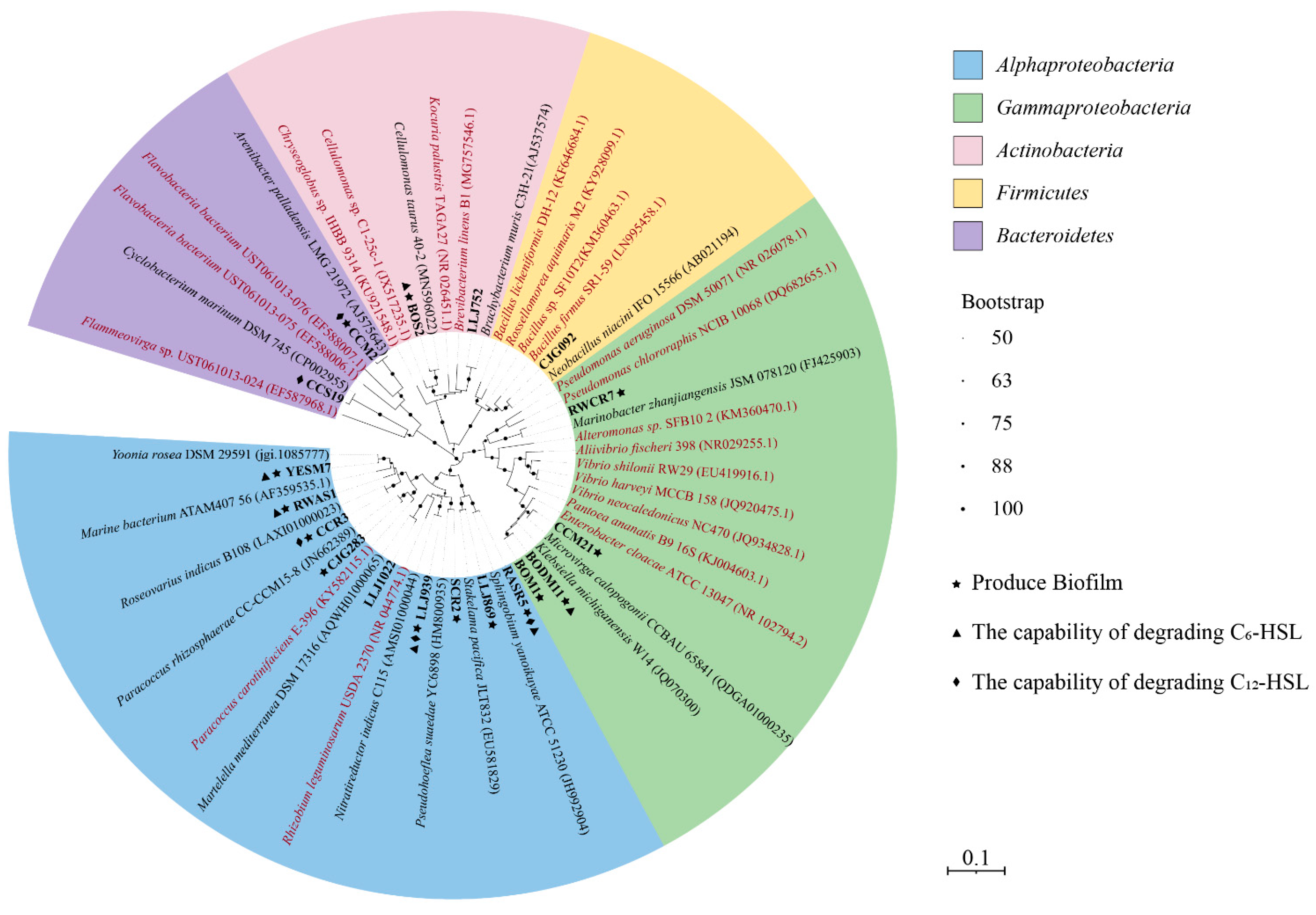
3.1. AHL-Producing Bacteria Isolated from the Hydrothermal Fields of the Okinawa Trough

To reveal the diversity of QS bacteria isolated from deep-sea hydrothermal vents, the AHL-producing activities of bacteria isolated from hydrothermal fields in the Okinawa Trough were detected. A total of 305 isolates belonging to 178 species have been recovered from seawater, sediment and macroorganisms in hydrothermal fields of the Okinawa Trough in a previous study. After the preliminary selection, a total of 159 isolates belonging to 158 different species were chosen to examine their QS activities. There were 56, 55 and 48 strains isolated from sediment, seawater and macroorganisms, respectively (Table S1). According to the 16S rRNA gene sequence analyses, these isolates belonged to Proteobacteria, Bacteroidetes, Actinobacteria and Firmicutes (Table S1). Among them, 18 cultures showed AHL-producing activities, including eight isolates from macroorganisms (16.67%), seven from seawater (12.73%) and three from sediment (5.35%) (Table 1). These AHL-producing bacteria belonged to the orders Rhodobacterales (4), Hyphomicrobiales (3), Enterobacterales (3), Sphingomonadales (2), Pseudomonadales (1), Cellulomonadales (1), Dermabacterales (1), Cytophagales (1), Flavobacteriales (1) and Bacillales (1) (Table 1 and Figure 1). Four isolates exhibited relatively higher QS activities, and were equated with Nitratireductor indicus LLJ939, Thalassococcus profundi RWAS1, Stakelama pacifica LLJ869 and Pseudohoeflea suaedae SCR2 (Figure 2 and Figure S1).

3.2. Identification of Species Capable of AHL Degradation

The AHL-degrading activities of the 159 isolates were further examined, and C6-HSL and C12-HSL were used as the representative AHLs with short and long acyl chains, respectively. Among them, 60 (38.37%) and 89 cultures (55.98%) showed C6-HSL degradation and C12-HSL degradation, respectively. A total of 41 (25.78%) isolates were capable of degrading both C6-HSL and C12-HSL (Table S1 and Figure 3). Consequently, there were 108 isolates (67.92%) having the ability of degrading AHLs.
The 108 bacterial isolates with AHLs degrading activities belong to Alphaproteobacteria (35), Gammaproteobacteria (26), Betaprotebacteria (3), Firmicutes (32), Actinobacteria (8) and Bacteroidetes (4). At the order level, it was shown that the QQ strains were mainly affiliated with Bacillales (33), Rhodospirillales (19), and Sphingomonadales (9). Furthermore, bacteria showing both C6-HSL- and C12-HSL-degrading activities were mainly affiliated with Bacillales (10) and Rhodobacterales (9) (Table 2 and Figure 3).
For bacteria isolated from seawater samples, 20 (41.07% of isolates from seawater) and 29 (52.73%) cultures exhibited the C6- and C12-HSL-degrading activities, respectively. Among the isolates from sediment samples, 23 (41.07%) and 33 (58.93%) showed C6- and C12-HSL-degrading activities, respectively. As for cultures from macroorganism, 17 (35.41%) and 33 (56.25%) of the isolates were capable of degrading C6- and C12-HSL, respectively. The QQ cultures isolated from macroorganism sand seawater samples belonged mainly to Alphaproteobacteria and Gammaproteobacteria. QQ strains isolated from sediment were affiliated mainly with Firmicutes. It is noteworthy that more bacteria from sediment samples were found to exhibit QQ activities (Table 3 and Table S1).
Among bacteria with QS activities, six cultures showed C6-HSL-degrading activities, including Klebsiella michiganensis BODM11, Cellulomonas taurus BOS2, N. indicus LLJ939, Sphingobium yanoikuyae RASR5, Thalassococcus profundi RWAS1 and Yoonia rosea YESM7. Meanwhile, five cultures exhibited C12-HSL-degrading activities, including Arenibacter palladensis CCM2, Roseovarius indicus CCR3, Cyclobacterium marinum CCS19, N. indicus LLJ939 and S. yanoikuyae RASR5. QS strains N. indicus LLJ939 and S. yanoikuyae RASR5 exhibited both C6-HSL- and C12-HSL-degrading activities (Figure 3).

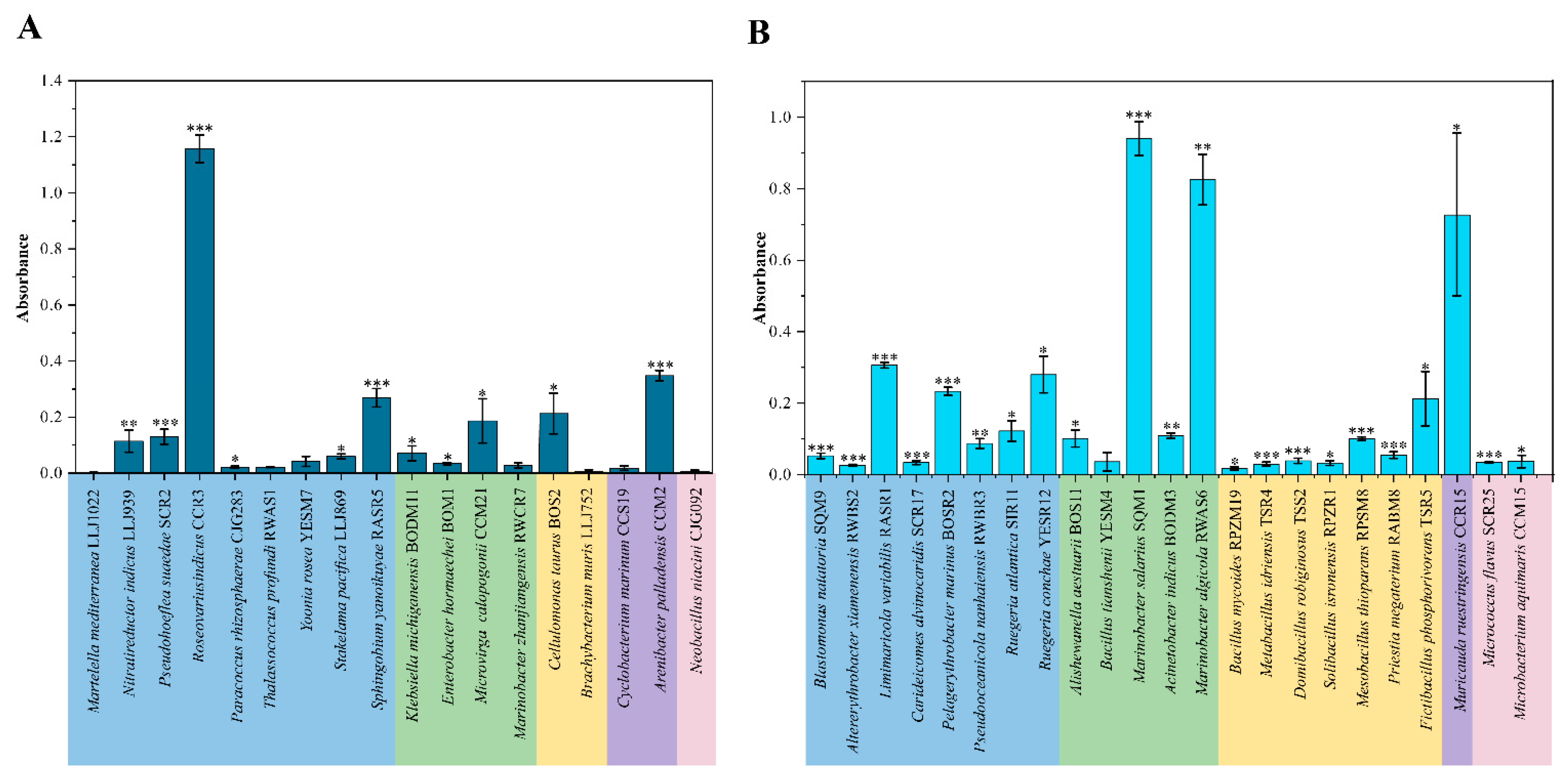
3.3. Biofilm Production in QS and QQ Cultures

In order to preliminarily reveal the relationship between QS/QQ and biofilm production, the CV assay was used to detect the biofilm production of 18 and 23 isolates with QS activities and QQ activities, respectively. Among them, 11 QS cultures demonstrated the ability to produce biofilm, with significantly stronger capabilities found in A. palladensis CCM2, S. yanoikuyae RASR5 and R. indicus CCR3 (Figure 4A). A total of 22 QQ isolates exhibited the abilities of biofilm production with higher production in Limimaricola variabilis RASR1, Pelagerythrobacter marinus BOSR2, Ruegeria conchae YESR12, Marinobacter salarius SQM1, Marinobacter algicola RWAS6 and Muricauda ruestringensis CCR15 (Figure 4B).

3.4. Extracellular Enzyme Activities in QS Isolates

To reveal how the QS regulated the extracellular enzymes in QS isolates, MUF-α-glucopyranoside, MUF-β-glucopyranoside, MUF-butyrate, MCA-leucine and MUF-phosphate were selected as fluorescent substrates for the activities of α-glucosidase, β-glucosidase, lipase, aminopeptidase and phosphatase, respectively. The EE activities of the four cultures with higher QS activities, including N. indicus LLJ939, T. profundi RWAS1, S. pacifica LLJ869 and P. suaedae SCR2, were detected. It was found that neither of the four QS isolates showed α-glucosidase or lipase activity. However, T. profundi WRAS1 and S. pacifica LLJ869 exhibited β-glucosidase activities, S. pacifica LLJ869 and P. suaedae SCR2 revealed phosphatase activities, and N. indicus LLJ939 and S. pacifica LLJ869 had aminopeptidase activities. After AHL lactonase MomL was added into the cultures to degrade the AHLs, the activities of β-glucosidase in T. profundi WRAS1 (at 12, 24, 36 and 48 h; Figure 5A), the activities of aminopeptidase in N. indicus LLJ939 (at 48 h; Figure 5C) and S. pacifica LLJ869 (at 24, 36 and 48 h; Figure 5D), and the activities of phosphatase in P. suaedae SCR2 (at 48 h; Figure 5E) and S. pacifica LLJ869 (at 12, 24, 36 and 48 h; Figure 5F) were significantly lower than that in these strains with inactive MomL. Moreover, it was shown that the activities of β-glucosidase in S. pacifica LLJ869 at 12 and 24 h were lower than that in strain LLJ869 with inactive MomL, but higher at 36 h (Figure 5B). It was suggested that QS showed significant effects on the EE activities in these QS strains.

4. Discussion

Although the functions of QS and QQ have been extensively studied in marine environments, much less work has been conducted in the extreme environments, such as hydrothermal vents. Our study expanded the current knowledge of the diversity of QS and QQ isolates in hydrothermal fields, and the regulation of QS on bacterial behaviors. In this study, a total of 159 bacterial isolates, which were recovered from hydrothermal fields in the Okinawa Trough and belong to 158 different species, were selected to screen their QS and QQ activities. The diversity of QS and QQ bacteria in hydrothermal vents was revealed, and the relationship between QS and the EE activities was explored. Our results showed that abundant QS regulation existed in hydrothermal fields, which might play important roles in regulating bacterial metabolic pathways and even biogeochemical cycles in hydrothermal vents.

4.1. The Diversity of QS Strains in Hydrothermal Fields

Compared with marine snow, corals, sponges and marine particles, the hydrothermal vents have unique and complex environments, where abundant and various microbial populations live. However, the diversity of QS and QQ bacteria in hydrothermal fields has never been revealed before. In this study, bacterial isolates, which were recovered from hydrothermal fields, were selected to evaluate their AHL-based QS and QQ abilities using bacterial biosensor A. tumefaciens A136.
AHL synthase sequences belonging to Alphaproteobacteria have been reported in several environmental metagenomics datasets. Doberva et al. [20] reported that all sequences encoding for AHL synthases luxI in the Global Ocean Sampling dataset were derived from Alphaproteobacteria with 19 (65%), 3 (14%), 2 (7%) belonging to Rhodobacterales, Rhizobiales and Sphingomonadales, respectively. In addition, Su et al. [48] determined that luxI belonging to Alphaproteobacteria existed in particulate organic matter (POM) samples collected from coastal water and sediment in the Yellow Sea of China. In this study, 9 out of 55 isolates belonging to Alphaproteobacteria produced AHLs, with affiliation to Rhodobacterales (4), Hyphomicrobiales (3) and Sphingomonadales (2) (Figure 1); they were first reported to have QS activities at the species level. Many bacteria in the order Rhodobacterales, especially the Roseobacter Clade, have been found to have QS activities, which may play important roles in their association with marine invertebrates and nutrient-rich marine snows or organic particles [49]. For example, Ruegeria sp. KLH11 was isolated from a marine sponge, and could produce 3-OH-C14-HSL, 3-OH-C12-HSL and 3-OH-C14-HSL, which were engaged in temporal regulation of tropodithietic acid (TDA) production [50,51]. In our study, R. indicus CCR3, Y. rosea YESM7, P. rhizosphaerae CJG 283 and T. profundi WRAS1 were affiliated with the Roseobacter Clade and showed QS activities (Figure 1). Indeed, they may produce various AHLs and regulate their metabolic activities. Strains of Sphingomonadales from Yellow Sea particles [3] and Aulne estuary [52] have been found to possess QS activities. Similarly, S. pacifica LLJ869 and S. yanoikuyae RASR5 belong to Sphingomonadales and S. pacifica LLJ869 exhibited relatively higher QS activity, and the signaling molecules will be detected in the following study (Figure 1).
Gammaproteobacteria are among the most common AHL producers isolated from marine habitats [3,8]. However, in our study, only 4 out of 41 Gammaproteobacteria isolates were AHL producers. They were affiliated with Enterobacterales (3) and Pseudomonadales (1). Enterobacterales are usually found in marine aquatic environments and have emerged as major players in antimicrobial resistance worldwide. Jatt et al. [8] found that Pantoea ananatis B9, which belongs to Enterobacterales and was isolated from natural marine snow particles, could produce six AHLs, and its extracellular alkaline phosphatase activity was enhanced when adding exogenous AHLs. In our study, three isolates belonging to Enterobacterales showed QS activities, and they were all isolated from marine organisms inhabiting hydrothermal fields (Figure 1). It was suggested that there might be close relationships between bacteria and their hosts mediated by QS in hydrothermal vent environments [53].
Actinobacteria are widespread in marine environments and reached high abundance in deep-sea sediments, organic aggregates and macroalgae [54]. In terms of QS, several bacteria belonging to Actinobacteria, which were isolated from marine snow [3] and microbial mats in Shark Bay Australia [16] or reef macroalgae [55], have been identified to show AHL-producing activities. In the hydrothermal fields of the Okinawa Trough, we found that C. taurus BOS2 and B. muris LLJ752 affiliated with Actinobacteria showed AHL-QS activities (Figure 1). Although the QS activities of closely related bacteria in Actinobacteria were reported, our study provided the first report of AHL-producing bacteria isolated from hydrothermal vents in the Okinawa Trough. At present, a few studies have reported some AHL-producers belonging to Firmicutes, but the ecological importance and QS-regulated function is rarely studied [16,17]. In our study, N. niacin CJG092 belongs to Firmicutes, but it could not form biofilms. Therefore, the bacterial behaviors affected by QS in Firmicutes should be explored. In addition, few bacteria belonging to Bacteroidetes have been well studied in terms of QS [26]. In our study, two isolates belonging to Bacteroidetes showed QS activities, which may provide bacterial resources to find novel QS regulatory pathways in the future.

4.2. The Diversity of QQ Isolates in Hydrothermal Fields

At present, the metagenomic survey shows that genes encoding for both AHL acylase and lactonase showed relatively high abundance in marine environments [23]. Moreover, Romero et al. found that 14% and 18% of cultivable bacteria had been experimentally verified to harbor QQ activities in microbial biofilms and pelagic communities, respectively [23,56]. However, the diversity of QQ bacteria in hydrothermal fields has been rarely studied. In our study, 108 isolates (67.92%) showed QQ activities, and were mainly distributed in Alphaproteobacteria, Gammaproteobacteria and Firmicutes, which agreed with previous studies [2].
Many marine QQ bacteria belong to both Alphaproteobacteria [56], Gammaproteobacteria [57] and Firmicutes [58]. In our study, Alphaproteobacteria (64.81%), Gammaproteobacteria (63.41%) and Firmicutes (69.56%) showed a relatively higher proportion of QQ activities. Sequentially, Su et al. [3] found that R. mobilis Rm01 exhibited the degrading activities of C10-HSL, C12-HSL and C14-HSL because of AHL lactonase. Additionally, we found that both Ruegeria atlantica SIR11 and Ruegeria arenilitorisi CCM11, isolated from shrimp gut and crab gill, respectively, degraded C12-HSL, but only R. atlantica SIR11 attacked C6-HSL (Figure 3). Moreover, it was reported that Pseudomonas sihuiensis M4-84, which was isolated from the microbiota of sea anemones, had the capacity of degrading C4-HSL, C6-HSL, C10-HSL and C12-HS [59]. We found that P. sihuiensis CJG117 isolated from seawater exhibited QQ activity by degrading both C6-HSL and C12-HSL (Figure 3). Additionally, P. sihuiensis M4-84 was demonstrated to decrease the virulence of pathogens, and improve the survival rate of Artemia salina [59]. Similarly, we found that B. cheonanensis CJG088, B. thioparans RPSM8, B. mycoides RPZM19, B. idriensis TSR4, and B. tianshenii YESM4 could degrade both C6- and C12-HSL (Figure 3). Additionally, compared with short-chain AHLs, these QQ strains isolated from hydrothermal fields in the Okinawa Trough were more likely to degrade long-chain AHLs. It was reported that AHL-acylases preferred to degrade AHLs with longer carbon chain and could not degrade C4-HSL and C6-HSL in some bacteria [60,61]. Further, AHL acylases in cultivable marine bacteria [3,27,59,62] and in marine metagenomic collections [23,24] seems to be more abundant than AHL lactonases. In our study, more bacteria from sediment samples were found to exhibit QQ activities. It is possible that the abundance of bacteria was higher in sediments, and QQ might play more important roles in coordinating the relationships of various microorganisms.
In our study, 9 out of 18 QS strains exhibited degrading activities of AHLs; N. indicus LLJ939 and S. yanoikuyae RASR5 showed both C6-HSL- and C12-HSL-degrading activities (Figure 1). The reports of bacteria that simultaneously showed QS and QQ activities had been demonstrated before. For example, R. mobilis Rm01 not only produced 3OC10-HSL, C10-HSL and C12-HSL, but also had the capacity of degrading C10-HSL, C12-HSL and C14-HSL [3]. Strains belonging to Marinobacterium and Shewanella had the abilities of QS and QQ [63]. Furthermore, Urvoy et al. [52] found that ten QS strains showed QQ abilities to interfere with C14-HSL in the estuarine environment. QS and QQ pathways that co-existed were beneficial to the competition of microbial populations by limiting the growth and the coordination of bacteria taking part in QS communication [64,65], which could help maintain the homeostasis of the microbial community.

4.3. Biofilm Formation and the Regulation of EE Activities in QS Isolates

Biofilms may provide a relatively stable environment for bacteria to access resources, maintain the growth and reproduction of colonies, and protect the bacteria from detrimental conditions. It was reported that biofilm development processes were regulated by QS [66]. In our study, 11 QS isolates (61.11% of QS cultures) and 22 QQ isolates were able to produce biofilms (Figure 4 and Table 1). The capabilities of biofilm formation of QQ strains may be regulated by QS systems, other than AHL-QS, and the presence of biofilm had significant effects on whether bacteria could survive stably in extreme marine environments.
The QS pathways presented among marine bacteria may directly or indirectly influence biogeochemical cycles through the regulation of genes and EE activities, and the degradation of marine particles and marine snows [54]. Hmelo et al. [46] demonstrated that the addition of 3OC6-HSL or 3OC8-HSL improved the extracellular aminopeptidase and phosphatase activities of bacterial communities colonizing marine snow. Moreover, Krupke [67] showed that the addition of 3OC8-HSL significantly stimulated or inhibited hydrolytic phosphatase and aminopeptidase activities in sunken particle samples collected from the Atlantic and Pacific Oceans. Su et al. [3] reported that the addition of 3OC8-HSL to marine snow reduced the activities of β-glucosidase. In our study, we found that when the AHL-QS pathway was disrupted using MomL, the β-glucosidase, aminopeptidase and phosphatase activities were significantly decreased (Figure 5). The activities of β-glucosidase in S. pacifica LLJ869 with MomL were higher than the treatment with inactive MomL at 36 h, and it could be attributed to the drastic decrease in the β-glucosidase activities after the treatment of MomL after 24 h. It was shown that AHL-QS could affect the β-glucosidase, aminopeptidase and phosphatase activities in bacteria isolated from hydrothermal environments. Moreover, it is suggested that QS and QQ were essential, and complicated mechanisms engaged in microbial interactions and had important ecological implications in hydrothermal environments.

5. Conclusions

This study revealed the diversity of QS and QQ strains isolated from hydrothermal vents in the Okinawa Trough. The QS and QQ bacteria belong mainly to Alphaproteobacteria, Gammaproteobacteria and Firmicutes. The QS strains were mainly affiliated with Rhodobacterales (4), Hyphomicrobiales (3), Enterobacterales (3) and Sphingomonadales (2), and QQ strains mainly belong to Bacillales (33), Rhodospirillales (19) and Sphingomonadales (9). It implied the abundant QS and QQ microbial resources in the extreme marine environment. In addition, the diversity of QQ strains was higher than that of QS strains, and they were more likely to degrade long-chain AHLs compared with short-chain AHLs. The biofilm formation might help the QS and QQ strains survive in the hydrothermal environments. Moreover, the β-glucosidase, aminopeptidase and phosphatase activities in bacteria isolated from hydrothermal environments could be regulated by QS. In conclusion, our study revealed the diversity of culturable QS and QQ bacteria and explored the QS regulation of extracellular enzyme in deep-sea hydrothermal environments. In the future, the QS mechanisms and QQ enzymes will be further studied to explore the important roles of QS and QQ in regulating biogeochemical cycles in hydrothermal vents.

Supplementary Materials

The following supporting information can be downloaded at: https://www.mdpi.com/article/10.3390/microorganisms11030748/s1, Table S1: The information of strains from hydrothermal vents in the Okinawa Trough; Figure S1: The AHL-producing abilities of strains are tested by the high-throughput method.

Author Contributions

Conceptualization, M.Y. and X.-H.Z.; methodology, M.Y. and F.Y.; formal analysis, F.Y. and L.Y.; investigation, F.Y. and D.G.; validation, F.Y.; writing—original draft preparation, F.Y.; writing—review and editing, F.Y., M.Y., Y.Z., J.L. and X.-H.Z.; supervision, M.Y.; project administration, M.Y.; funding acquisition, M.Y. and X.-H.Z. All authors have read and agreed to the published version of the manuscript.

Funding

This work was supported by projects from the National Natural Science Foundation of China (41976137 and U1706208), the Fundamental Research Funds for the Central Universities (202172002), and the National Key R&D Program of China (2018YFC0310701).

Data Availability Statement

The data presented in this study are fully available in the main text and Supplementary Materials of this article.

Acknowledgments

We would like to thank Austin for checking and modifying our manuscript. We are grateful to Xi Li, Jingguang Cheng, Yanhong Wu and Lijun Liu for isolating and identifying the strains from hydrothermal vents in the Okinawa Trough.

Conflicts of Interest

The authors declare no conflict of interest.

References

  1. Decho, A.W.; Norman, R.S.; Visscher, P.T. Quorum Sensing in Natural Environments: Emerging Views from Microbial Mats. Trends Microbiol. 2010, 18, 73–80. [Google Scholar] [CrossRef] [PubMed]
  2. Hmelo, L.R. Quorum Sensing in Marine Microbial Environments. Annu. Rev. Mar. Sci. 2017, 9, 257–281. [Google Scholar] [CrossRef] [PubMed]
  3. Su, Y.; Tang, K.; Liu, J.; Wang, Y.; Zheng, Y.; Zhang, X.-H. Quorum Sensing System of Ruegeria Mobilis Rm01 Controls Lipase and Biofilm Formation. Front. Microbiol. 2019, 9, 3304. [Google Scholar] [CrossRef]
  4. Kleerebezem, M.; Quadri, L.E.N.; Kuipers, O.P.; De Vos, W.M. Quorum Sensing by Peptide Pheromones and Two-component Signal-transduction Systems in Gram-positive Bacteria. Mol. Microbiol. 1997, 24, 895–904. [Google Scholar] [CrossRef] [PubMed] [Green Version]
  5. Wang, J.; Liu, Q.; Dong, D.; Hu, H.; Wu, B.; Ren, H. AHLs-Mediated Quorum Sensing Threshold and Its Response towards Initial Adhesion of Wastewater Biofilms. Water Res. 2021, 194, 116925. [Google Scholar] [CrossRef]
  6. Prazdnova, E.V.; Gorovtsov, A.V.; Vasilchenko, N.G.; Kulikov, M.P.; Statsenko, V.N.; Bogdanova, A.A.; Refeld, A.G.; Brislavskiy, Y.A.; Chistyakov, V.A.; Chikindas, M.L. Quorum-Sensing Inhibition by Gram-Positive Bacteria. Microorganisms 2022, 10, 350. [Google Scholar] [CrossRef]
  7. Pearson, J.P.; Gray, K.M.; Passador, L.; Tucker, K.D.; Eberhard, A.; Iglewski, B.H.; Greenberg, E.P. Structure of the Autoinducer Required for Expression of Pseudomonas Aeruginosa Virulence Genes. Proc. Natl. Acad. Sci. USA 1994, 91, 197–201. [Google Scholar] [CrossRef] [Green Version]
  8. Jatt, A.N.; Tang, K.; Liu, J.; Zhang, Z.; Zhang, X.-H. Quorum Sensing in Marine Snow and Its Possible Influence on Production of Extracellular Hydrolytic Enzymes in Marine Snow Bacterium Pantoea Ananatis B9. FEMS Microbiol. Ecol. 2015, 91, 1–13. [Google Scholar] [CrossRef]
  9. Tait, K.; Hutchison, Z.; Thompson, F.L.; Munn, C.B. Quorum Sensing Signal Production and Inhibition by Coral-Associated Vibrios: Quorum Sensing and Coral-Associated Vibrios. Environ. Microbiol. Rep. 2010, 2, 145–150. [Google Scholar] [CrossRef]
  10. Golberg, K.; Eltzov, E.; Shnit-Orland, M.; Marks, R.S.; Kushmaro, A. Characterization of Quorum Sensing Signals in Coral-Associated Bacteria. Microb. Ecol. 2011, 61, 783–792. [Google Scholar] [CrossRef]
  11. Gardères, J.; Taupin, L.; Saïdin, J.B.; Dufour, A.; Le Pennec, G. N-Acyl Homoserine Lactone Production by Bacteria within the Sponge Suberites Domuncula (Olivi, 1792) (Porifera, Demospongiae). Mar. Biol. 2012, 159, 1685–1692. [Google Scholar] [CrossRef]
  12. Wagner-Döbler, I.; Thiel, V.; Eberl, L.; Allgaier, M.; Bodor, A.; Meyer, S.; Ebner, S.; Hennig, A.; Pukall, R.; Schulz, S. Discovery of Complex Mixtures of Novel Long-Chain Quorum Sensing Signals in Free-Living and Host-Associated Marine Alphaproteobacteria. ChemBioChem 2005, 6, 2195–2206. [Google Scholar] [CrossRef] [PubMed]
  13. Van Mooy, B.A.S.; Hmelo, L.R.; Sofen, L.E.; Campagna, S.R.; May, A.L.; Dyhrman, S.T.; Heithoff, A.; Webb, E.A.; Momper, L.; Mincer, T.J. Quorum Sensing Control of Phosphorus Acquisition in Trichodesmium Consortia. ISME J. 2012, 6, 422–429. [Google Scholar] [CrossRef]
  14. Ransome, E.; Munn, C.B.; Halliday, N.; Cámara, M.; Tait, K. Diverse Profiles of N-Acyl-Homoserine Lactone Molecules Found in Cnidarians. FEMS Microbiol. Ecol. 2014, 87, 315–329. [Google Scholar] [CrossRef] [PubMed] [Green Version]
  15. Zhang, J.; Feng, T.; Wang, J.; Wang, Y.; Zhang, X.-H. The Mechanisms and Applications of Quorum Sensing (QS) and Quorum Quenching (QQ). J. Ocean Univ. China 2019, 18, 1427–1442. [Google Scholar] [CrossRef]
  16. Charlesworth, J.C.; Watters, C.; Wong, H.L.; Visscher, P.T.; Burns, B.P. Isolation of Novel Quorum-Sensing Active Bacteria from Microbial Mats in Shark Bay Australia. FEMS Microbiol. Ecol. 2019, 95, fiz035. [Google Scholar] [CrossRef]
  17. Freckelton, M.L.; Høj, L.; Bowden, B.F. Quorum Sensing Interference and Structural Variation of Quorum Sensing Mimics in Australian Soft Coral. Front. Mar. Sci. 2018, 5, 198. [Google Scholar] [CrossRef]
  18. Zhang, G.; Zhang, F.; Ding, G.; Li, J.; Guo, X.; Zhu, J.; Zhou, L.; Cai, S.; Liu, X.; Luo, Y.; et al. Acyl Homoserine Lactone-Based Quorum Sensing in a Methanogenic Archaeon. ISME J. 2012, 6, 1336–1344. [Google Scholar] [CrossRef] [Green Version]
  19. Sharif, D.I.; Gallon, J.; Smith, C.J.; Dudley, E. Quorum Sensing in Cyanobacteria: N-Octanoyl-Homoserine Lactone Release and Response, by the Epilithic Colonial Cyanobacterium Gloeothece PCC6909. ISME J. 2008, 2, 1171–1182. [Google Scholar] [CrossRef] [Green Version]
  20. Doberva, M.; Sanchez-Ferandin, S.; Toulza, E.; Lebaron, P.; Lami, R. Diversity of Quorum Sensing Autoinducer Synthases in the Global Ocean Sampling Metagenomic Database. Aquat. Microb. Ecol. 2015, 74, 107–119. [Google Scholar] [CrossRef] [Green Version]
  21. Nealson, K.H.; Hastings, J.W. Quorum Sensing on a Global Scale: Massive Numbers of Bioluminescent Bacteria Make Milky Seas. Appl. Environ. Microbiol. 2006, 72, 2295–2297. [Google Scholar] [CrossRef] [PubMed] [Green Version]
  22. Joint, I.; Tait, K.; Callow, M.E.; Callow, J.A.; Milton, D.; Williams, P.; Cámara, M. Cell-to-Cell Communication Across the Prokaryote-Eukaryote Boundary. Science 2002, 298, 1207. [Google Scholar] [CrossRef] [Green Version]
  23. Romero, M.; Martin-Cuadrado, A.-B.; Otero, A. Determination of Whether Quorum Quenching Is a Common Activity in Marine Bacteria by Analysis of Cultivable Bacteria and Metagenomic Sequences. Appl. Environ. Microbiol. 2012, 78, 6345–6348. [Google Scholar] [CrossRef] [PubMed] [Green Version]
  24. Muras, A.; López-Pérez, M.; Mayer, C.; Parga, A.; Amaro-Blanco, J.; Otero, A. High Prevalence of Quorum-Sensing and Quorum-Quenching Activity among Cultivable Bacteria and Metagenomic Sequences in the Mediterranean Sea. Genes 2018, 9, 100. [Google Scholar] [CrossRef] [Green Version]
  25. Blanchet, E.; Prado, S.; Stien, D.; Oliveira da Silva, J.; Ferandin, Y.; Batailler, N.; Intertaglia, L.; Escargueil, A.; Lami, R. Quorum Sensing and Quorum Quenching in the Mediterranean Seagrass Posidonia Oceanica Microbiota. Front. Mar. Sci. 2017, 4, 218. [Google Scholar] [CrossRef] [Green Version]
  26. Huang, Y.; Ki, J.; Case, R.; Qian, P. Diversity and Acyl-Homoserine Lactone Production among Subtidal Biofilm-Forming Bacteria. Aquat. Microb. Ecol. 2008, 52, 185–193. [Google Scholar] [CrossRef] [Green Version]
  27. Rehman, Z.U.; Leiknes, T. Quorum-Quenching Bacteria Isolated From Red Sea Sediments Reduce Biofilm Formation by Pseudomonas Aeruginosa. Front. Microbiol. 2018, 9, 1354. [Google Scholar] [CrossRef] [PubMed]
  28. Dong, Y.-H.; Xu, J.-L.; Li, X.-Z.; Zhang, L.-H. AiiA, an Enzyme That Inactivates the Acylhomoserine Lactone Quorum-Sensing Signal and Attenuates the Virulence of Erwinia carotovora. Proc. Natl. Acad. Sci. USA 2000, 97, 3526–3531. [Google Scholar] [CrossRef]
  29. Toki, T.; Itoh, M.; Iwata, D.; Ohshima, S.; Shinjo, R.; Ishibashi, J.; Tsunogai, U.; Takahata, N.; Sano, Y.; Yamanaka, T.; et al. Geochemical Characteristics of Hydrothermal Fluids at Hatoma Knoll in the Southern Okinawa Trough. Geochem. J. 2016, 50, 493–525. [Google Scholar] [CrossRef] [Green Version]
  30. Chen, S.; Yu, M.; Zhang, W.; He, K.; Pan, H.; Cui, K.; Zhao, Y.; Zhang, X.-H.; Xiao, T.; Zhang, W.; et al. Metagenomic and Microscopic Analysis of Magnetotactic Bacteria in Tangyin Hydrothermal Field of Okinawa Trough. Front. Microbiol. 2022, 13, 887136. [Google Scholar] [CrossRef] [PubMed]
  31. Inagaki, F. Sulfurimonas autotrophica Gen. Nov., Sp. Nov., a Novel Sulfur-Oxidizing -Proteobacterium Isolated from Hydrothermal Sediments in the Mid-Okinawa Trough. Int. J. Syst. Evol. Microbiol. 2003, 53, 1801–1805. [Google Scholar] [CrossRef] [Green Version]
  32. Inagaki, F.; Takai, K.; Nealson, K.H.; Horikoshi, K. Sulfurovum lithotrophicum Gen. Nov., Sp. Nov., a Novel Sulfur-Oxidizing Chemolithoautotroph within the ε-Proteobacteria Isolated from Okinawa Trough Hydrothermal Sediments. Int. J. Syst. Evol. Microbiol. 2004, 54, 1477–1482. [Google Scholar] [CrossRef] [Green Version]
  33. Nunoura, T.; Oida, H.; Miyazaki, M.; Suzuki, Y. Thermosulfidibacter takaii Gen. Nov., Sp. Nov., a Thermophilic, Hydrogen-Oxidizing, Sulfur-Reducing Chemolithoautotroph Isolated from a Deep-Sea Hydrothermal Field in the Southern Okinawa Trough. Int. J. Syst. Evol. Microbiol. 2008, 58, 659–665. [Google Scholar] [CrossRef]
  34. Nunoura, T.; Oida, H.; Miyazaki, M.; Suzuki, Y.; Takai, K.; Horikoshi, K. Desulfothermus okinawensis sp. Nov., a Thermophilic and Heterotrophic Sulfate-Reducing Bacterium Isolated from a Deep-Sea Hydrothermal Field. Int. J. Syst. Evol. Microbiol. 2007, 57, 2360–2364. [Google Scholar] [CrossRef] [PubMed]
  35. Miyazaki, J.; Kawagucci, S.; Makabe, A.; Takahashi, A.; Kitada, K.; Torimoto, J.; Matsui, Y.; Tasumi, E.; Shibuya, T.; Nakamura, K.; et al. Deepest and Hottest Hydrothermal Activity in the Okinawa Trough: The Yokosuka Site at Yaeyama Knoll. R. Soc. Open Sci. 2017, 4, 171570. [Google Scholar] [CrossRef] [PubMed] [Green Version]
  36. McGonigle, J.M.; Lang, S.Q.; Brazelton, W.J. Genomic Evidence for Formate Metabolism by Chloroflexi as the Key to Unlocking Deep Carbon in Lost City Microbial Ecosystems. Appl. Environ. Microbiol. 2020, 86, e02583-19. [Google Scholar] [CrossRef] [Green Version]
  37. Patrick, F.M. The Use of Membrane Filtration and Marine Agar 2216E to Enumerate Marine Heterotrophic Bacteria. Aquaculture 1978, 13, 369–372. [Google Scholar] [CrossRef]
  38. Massa, S.; Caruso, M.; Trovatelli, F.; Tosques, M. Comparison of Plate Count Agar and R2A Medium for Enumeration of Heterotrophic Bacteria in Natural Mineral Water. World J. Microbiol. Biotechnol. 1998, 14, 727–730. [Google Scholar] [CrossRef]
  39. Tamura, K.; Stecher, G.; Kumar, S. MEGA11: Molecular Evolutionary Genetics Analysis Version 11. Mol. Biol. Evol. 2021, 38, 3022–3027. [Google Scholar] [CrossRef]
  40. Fuqua, C.; Winans, S.C. Conserved Cis-Acting Promoter Elements Are Required for Density-Dependent Transcription of Agrobacterium Tumefaciens Conjugal Transfer Genes. J. Bacteriol. 1996, 178, 435–440. [Google Scholar] [CrossRef] [Green Version]
  41. Bernier, S.P.; Beeston, A.L.; Sokol, P.A. Detection of N-Acyl Homoserine Lactones Using a TraI-LuxCDABE-Based Biosensor as a High-Throughput Screening Tool. BMC Biotechnol. 2008, 8, 59. [Google Scholar] [CrossRef] [Green Version]
  42. Zhu, J.; Beaber, J.W.; Moré, M.I.; Fuqua, C.; Eberhard, A.; Winans, S.C. Analogs of the Autoinducer 3-Oxooctanoyl-Homoserine Lactone Strongly Inhibit Activity of the TraR Protein of Agrobacterium Tumefaciens. J. Bacteriol. 1998, 180, 5398–5405. [Google Scholar] [CrossRef] [PubMed] [Green Version]
  43. Chu, W.; Vattem, D.A.; Maitin, V.; Barnes, M.B.; McLean, R.J.C. Bioassays of Quorum Sensing Compounds Using Agrobacterium Tumefaciens and Chromobacterium Violaceum. In Quorum Sensing; Rumbaugh, K.P., Ed.; Methods in Molecular Biology; Humana Press: Totowa, NJ, USA, 2011; Volume 692, pp. 3–19. ISBN 978-1-60761-970-3. [Google Scholar]
  44. Tempé, J.; Petit, A.; Holsters, M.; van Montagu, M.; Schell, J. Thermosensitive Step Associated with Transfer of the Ti Plasmid during Conjugation: Possible Relation to Transformation in Crown Gall. Proc. Natl. Acad. Sci. USA 1977, 74, 2848–2849. [Google Scholar] [CrossRef] [PubMed] [Green Version]
  45. Tang, K.; Zhang, Y.; Yu, M.; Shi, X.; Coenye, T.; Bossier, P.; Zhang, X.-H. Evaluation of a New High-Throughput Method for Identifying Quorum Quenching Bacteria. Sci. Rep. 2013, 3, 2935. [Google Scholar] [CrossRef] [Green Version]
  46. Hmelo, L.R.; Mincer, T.J.; Van Mooy, B.A.S. Possible Influence of Bacterial Quorum Sensing on the Hydrolysis of Sinking Particulate Organic Carbon in Marine Environments: Quorum Sensing on Sinking Particles. Environ. Microbiol. Rep. 2011, 3, 682–688. [Google Scholar] [CrossRef]
  47. Tang, K.; Su, Y.; Brackman, G.; Cui, F.; Zhang, Y.; Shi, X.; Coenye, T.; Zhang, X.-H. MomL, a Novel Marine-Derived N-Acyl Homoserine Lactonase from Muricauda Olearia. Appl. Environ. Microbiol. 2015, 81, 774–782. [Google Scholar] [CrossRef] [Green Version]
  48. Su, Y.; Yang, Y.; Zhu, X.-Y.; Zhang, X.-H.; Yu, M. Metagenomic Insights Into the Microbial Assemblage Capable of Quorum Sensing and Quorum Quenching in Particulate Organic Matter in the Yellow Sea. Front. Microbiol. 2021, 11, 602010. [Google Scholar] [CrossRef] [PubMed]
  49. Zan, J.; Liu, Y.; Fuqua, C.; Hill, R. Acyl-Homoserine Lactone Quorum Sensing in the Roseobacter Clade. Int. J. Mol. Sci. 2014, 15, 654–669. [Google Scholar] [CrossRef]
  50. Berger, M.; Brock, N.L.; Liesegang, H.; Dogs, M.; Preuth, I.; Simon, M.; Dickschat, J.S.; Brinkhoff, T. Genetic Analysis of the Upper Phenylacetate Catabolic Pathway in the Production of Tropodithietic Acid by Phaeobacter Gallaeciensis. Appl. Environ. Microbiol. 2012, 78, 3539–3551. [Google Scholar] [CrossRef] [Green Version]
  51. Prol García, M.J.; D’Alvise, P.W.; Gram, L. Disruption of Cell-to-Cell Signaling Does Not Abolish the Antagonism of Phaeobacter Gallaeciensis toward the Fish Pathogen Vibrio Anguillarum in Algal Systems. Appl. Environ. Microbiol. 2013, 79, 5414–5417. [Google Scholar] [CrossRef] [PubMed] [Green Version]
  52. Urvoy, M.; Lami, R.; Dreanno, C.; Daudé, D.; Rodrigues, A.M.S.; Gourmelon, M.; L’Helguen, S.; Labry, C. Quorum Sensing Disruption Regulates Hydrolytic Enzyme and Biofilm Production in Estuarine Bacteria. Environ. Microbiol. 2021, 23, 7183–7200. [Google Scholar] [CrossRef]
  53. Deng, Z.; Luo, X.M.; Liu, J.; Wang, H. Quorum Sensing, Biofilm, and Intestinal Mucosal Barrier: Involvement the Role of Probiotic. Front. Cell. Infect. Microbiol. 2020, 10, 538077. [Google Scholar] [CrossRef] [PubMed]
  54. Urvoy, M.; Labry, C.; L’Helguen, S.; Lami, R. Quorum Sensing Regulates Bacterial Processes That Play a Major Role in Marine Biogeochemical Cycles. Front. Mar. Sci. 2022, 9, 834337. [Google Scholar] [CrossRef]
  55. Bose, U.; Ortori, C.A.; Sarmad, S.; Barrett, D.A.; Hewavitharana, A.K.; Hodson, M.P.; Fuerst, J.A.; Shaw, P.N. Production of N -Acyl Homoserine Lactones by the Sponge-Associated Marine Actinobacteria Salinispora Arenicola and Salinispora Pacifica. FEMS Microbiol. Lett. 2017, 364, fnx002. [Google Scholar] [CrossRef] [PubMed] [Green Version]
  56. Romero, M.; Martin-Cuadrado, A.-B.; Roca-Rivada, A.; Cabello, A.M.; Otero, A. Quorum Quenching in Cultivable Bacteria from Dense Marine Coastal Microbial Communities: Quorum Quenching in Marine Bacteria. FEMS Microbiol. Ecol. 2011, 75, 205–217. [Google Scholar] [CrossRef] [PubMed]
  57. Zhao, J.; Li, X.; Hou, X.; Quan, C.; Chen, M. Widespread Existence of Quorum Sensing Inhibitors in Marine Bacteria: Potential Drugs to Combat Pathogens with Novel Strategies. Mar. Drugs 2019, 17, 275. [Google Scholar] [CrossRef] [Green Version]
  58. Teasdale, M.E.; Donovan, K.A.; Forschner-Dancause, S.R.; Rowley, D.C. Gram-Positive Marine Bacteria as a Potential Resource for the Discovery of Quorum Sensing Inhibitors. Mar. Biotechnol. 2011, 13, 722–732. [Google Scholar] [CrossRef]
  59. Reina, J.C.; Pérez, P.; Llamas, I. Quorum Quenching Strains Isolated from the Microbiota of Sea Anemones and Holothurians Attenuate Vibriocorallilyticus Virulence Factors and Reduce Mortality in Artemiasalina. Microorganisms 2022, 10, 631. [Google Scholar] [CrossRef] [PubMed]
  60. Shepherd, R.W.; Lindow, S.E. Two Dissimilar N -Acyl-Homoserine Lactone Acylases of Pseudomonas Syringae Influence Colony and Biofilm Morphology. Appl. Environ. Microbiol. 2009, 75, 45–53. [Google Scholar] [CrossRef] [Green Version]
  61. Czajkowski, R.; Krzyżanowska, D.; Karczewska, J.; Atkinson, S.; Przysowa, J.; Lojkowska, E.; Williams, P.; Jafra, S. Inactivation of AHLs by Ochrobactrum Sp. A44 Depends on the Activity of a Novel Class of AHL Acylase: An Ochrobactrum sp. A44 AHL Acylase. Environ. Microbiol. Rep. 2011, 3, 59–68. [Google Scholar] [CrossRef]
  62. Torres, M.; Rubio-Portillo, E.; Antón, J.; Ramos-Esplá, A.A.; Quesada, E.; Llamas, I. Selection of the N-Acylhomoserine Lactone-Degrading Bacterium Alteromonas stellipolaris PQQ-42 and of Its Potential for Biocontrol in Aquaculture. Front. Microbiol. 2016, 7, 646. [Google Scholar] [CrossRef] [PubMed] [Green Version]
  63. Tait, K.; Williamson, H.; Atkinson, S.; Williams, P.; Cámara, M.; Joint, I. Turnover of Quorum Sensing Signal Molecules Modulates Cross-Kingdom Signalling. Environ. Microbiol. 2009, 11, 1792–1802. [Google Scholar] [CrossRef] [PubMed]
  64. Defoirdt, T.; Boon, N.; Bossier, P.; Verstraete, W. Disruption of Bacterial Quorum Sensing: An Unexplored Strategy to Fight Infections in Aquaculture. Aquaculture 2004, 240, 69–88. [Google Scholar] [CrossRef]
  65. Rasmussen, T.B.; Givskov, M. Quorum Sensing Inhibitors: A Bargain of Effects. Microbiology 2006, 152, 895–904. [Google Scholar] [CrossRef] [PubMed] [Green Version]
  66. Whiteley, M.; Diggle, S.P.; Greenberg, E.P. Progress in and Promise of Bacterial Quorum Sensing Research. Nature 2017, 551, 313–320. [Google Scholar] [CrossRef] [PubMed] [Green Version]
  67. Krupke, A. Quorum Sensing Plays a Complex Role in Regulating the Enzyme Hydrolysis Activity of Microbes Associated with Sinking Particles in the Ocean. Front. Mar. Sci. 2016, 3, 55. [Google Scholar] [CrossRef] [Green Version]
Figure 1. Phylogenetic tree of QS bacteria isolated from hydrothermal fields in the Okinawa Trough. The black blot at each branch point indicates the bootstrap values (>50%) based on a Neighbor-Joining analysis of 1000 resampled datasets. GenBank accession numbers of 16S rRNA gene sequences are given in parentheses. The characters in red indicate the QS strains found in the previous study, the characters in black and bold indicate the QS strains, the characters in black indicate the most similar strains of QS strains in this study.
Microorganisms 11 00748 g001
Figure 2. AHL-producing abilities of isolates recovered from hydrothermal fields in the Okinawa Trough. The results are shown via the co-feeding method supplemented with the AHL reporter strain Agrobacterium tumefaciens A136. (A,B), the positive control (C6-HSL, 10 nM) and the negative control (MB). (CT) represent the following test strains, in order: Nitratireductor indicus LLJ939, Thalassococcus profundi RWAS1, Stakelama pacifica LLJ869, Pseudohoeflea suaedae SCR2, Arenibacter palladensis CCM2, Yoonia rosea YESM7, Neobacillus niacini CJG092, Enterobacter hormaechei BOM1, Microvirga calopogonii CCM21, Cellulomonas taurus BOS2, Klebsiella michiganensis BODM11, Marinobacter zhanjiangensis RWCR7, Paracoccus rhizosphaerae CJG283, Cyclobacterium marinum CCS19, Brachybacterium muris LLJ752, Roseovarius indicus CCR3, Sphingobium yanoikuyae RASR5, Martelella mediterranea LLJ1022.
Microorganisms 11 00748 g002
Figure 3. Phylogenetic tree of QQ bacteria isolated from hydrothermal fields in the Okinawa Trough. The black blot at each branch point indicates the bootstrap values (>50%) based on a Neighbor-Joining analysis of 1000 resampled datasets.
Microorganisms 11 00748 g003
Figure 4. Biofilm production of bacterial isolates from the hydrothermal vent in the Okinawa Trough. (A), Biofilm production of QS strains. (B), Biofilm production of partial QQ strains. The data are shown as the mean ± SD, and the differences between the amended groups (the QS strains) and the control groups (the MB medium) are calculated by the t-test (*** p < 0.001, ** p < 0.01, and * p < 0.05).
Microorganisms 11 00748 g004
Figure 5. The β-glucosidase (A,B), aminopeptidase (C,D) and phosphatase activities (E,F) influenced by MomL. The data are shown as the mean ± standard deviation (SD). The differences between the amended groups (MomL) and the untreated groups (inactive MomL) are shown in different colors and calculated by the t-test (*** p < 0.001 and * p < 0.05).
Microorganisms 11 00748 g005
Table 1. Information of QS strains isolated from hydrothermal fields in the Okinawa Trough.
StrainPhylumClassOrderSpeciesSource *
LLJ1022ProteobacteriaAlphaproteobacteriaHyphomicrobialesMartelella mediterraneaSD
LLJ939ProteobacteriaAlphaproteobacteriaHyphomicrobialesNitratireductor indicusSW
SCR2ProteobacteriaAlphaproteobacteriaHyphomicrobialesPseudohoeflea suaedaeSG
CCR3ProteobacteriaAlphaproteobacteriaRhodobacteralesRoseovarius indicusCG
CJG283ProteobacteriaAlphaproteobacteriaRhodobacteralesParacoccus rhizosphaeraeSW
RWAS1ProteobacteriaAlphaproteobacteriaRhodobacteralesThalassococcus profundiSW
YESM7ProteobacteriaAlphaproteobacteriaRhodobacteralesYoonia roseaSD
LLJ869ProteobacteriaAlphaproteobacteriaSphingomonadalesStakelama pacificaSW
RASR5ProteobacteriaAlphaproteobacteriaSphingomonadalesSphingobium yanoikuyaeSD
BODM11ProteobacteriaGammaproteobacteriaEnterobacteralesKlebsiella michiganensisM
BOM1ProteobacteriaGammaproteobacteriaEnterobacteralesEnterobacter hormaecheiM
CCM21ProteobacteriaGammaproteobacteriaEnterobacteralesMicrovirga calopogoniiCG
RWCR7ProteobacteriaGammaproteobacteriaPseudomonadalesMarinobacter zhanjiangensisSW
BOS2ActinobacteriaActinomycetiaCellulomonadalesCellulomonas taurusM
LLJ752ActinobacteriaActinomycetiaDermabacteralesBrachybacterium murisSW
CCS19BacteroidetesCytophagiaCytophagalesCyclobacterium marinumCG
CCM2BacteroidetesFlavobacteriiaFlavobacterialesArenibacter palladensisCG
CJG092FirmicutesBacilliBacillalesNeobacillus niaciniSW
* SD, sediment; SW, seawater; SG, shrimp gill; CG, crab gill; M, mussel.
Table 2. The information of QQ strains isolated from hydrothermal fields in the Okinawa Trough (at the order level).
PhylumClassOrderThe Number of Test StrainsThe Number of Strains Degrading C6-HSLThe Number of Strains Degrading C12-HSL
ProteobacteriaAlphaproteobacteriaRhizobiales1326
Rhodobacterales291217
Sphingomonadales1295
GammaproteobacteriaAlteromonadales1167
Pseudomonadales1058
Enterobacterales400
Oceanospirillales905
Xanthomonadales101
Vibrionales722
BetaproteobacteriaBurkholderiales312
FirmicutesBacillalesBacillaceae461528
ActinobacteriaActinomycetiaCorynebacteriales221
Micrococcales744
BacteroidetesFlavobacterialesFlavobacteriales423
CytophagiaCytophagales100
Total number15960 (37.73%)89 (55.97%)
Table 3. The numbers of QQ cultures isolated from hydrothermal fields in the Okinawa Trough.
SourceTotal NumberThe Number of Isolates Degrading C6-HSL (%)The Number of Isolates Degrading C12-HSL (%)
Seawater5520 (36.36%)29 (52.73%)
Sediment5623 (41.07%)33 (58.93%)
Organism4817 (35.41%)27 (56.25%)
Total number15960 (38.37%)89 (55.98%)
Disclaimer/Publisher’s Note: The statements, opinions and data contained in all publications are solely those of the individual author(s) and contributor(s) and not of MDPI and/or the editor(s). MDPI and/or the editor(s) disclaim responsibility for any injury to people or property resulting from any ideas, methods, instructions or products referred to in the content.

