Article

The Association between Mycoplasma pneumoniae Genotype and Cutaneous Disease

1
Department of Paediatric Pulmonology, University Children’s Hospital, University Medical Centre Ljubljana, 1000 Ljubljana, Slovenia
2
Department of Infectious Diseases, University Medical Centre Ljubljana, 1000 Ljubljana, Slovenia
3
University Clinic of Respiratory and Allergic Diseases Golnik, 4204 Golnik, Slovenia
4
Faculty of Medicine, University of Ljubljana, 1000 Ljubljana, Slovenia
5
Institute of Microbiology and Immunology, Faculty of Medicine, University of Ljubljana, 1000 Ljubljana, Slovenia
*
Author to whom correspondence should be addressed.
Microorganisms 2023, 11(1), 205; https://doi.org/10.3390/microorganisms11010205
Submission received: 5 December 2022 / Revised: 9 January 2023 / Accepted: 10 January 2023 / Published: 13 January 2023
(This article belongs to the Special Issue 10th Anniversary of Microorganisms: Past, Present and Future)

Abstract

:
Mycoplasma pneumoniae (Mp) can cause several extrapulmonary manifestations, most frequently dermatological ones. It is largely unknown whether Mp genotype determines Mp-induced cutaneous disease. The aim of our study was to assess the association between Mp genotype and this clinical outcome. We performed a retrospective study of children referred with signs of acute Mp infection from 1 January 2014 to 31 December 2014. We compared the characteristics of children presenting as cutaneous disease, upper (URTI) and lower respiratory tract infection (LRTI). In addition, we separately analyzed the data of patients presenting with Mp-induced cutaneous disease. We evaluated data from 435 patients (mean age 7.3 years, SD 3.4 years; 52.0% boys) who had Mp PCR-positive pharyngeal swab, P1 genotype and/or multilocus variable-number tandem-repeat analysis (MLVA) genotype defined and no viral co-detection, presenting as cutaneous disease (38/435), URTI (46/435) or LRTI (351/435). The majority of patients had urticarial (55%, 21/38) or maculopapular eruptions (37%, 14/38). We found no association between Mp genotype and clinical outcome of cutaneous disease, nor any specific dermatological presentation. In the group with cutaneous disease, 18% (7/38) required hospital admission because of rash. We found that infection with MLVA-3,6,6,2 strains was more common in admitted patients than in outpatients (40% vs. 4%, p = 0.017) and significantly affected the likelihood of hospital admission in a logistic regression model. The results of our cohort study suggest that Mp genotype does not determine Mp-induced cutaneous disease or a specific dermatological presentation. Nevertheless, infections with certain MLVA strains could induce more severe cutaneous disease requiring hospitalization.

1. Introduction

Mycoplasma pneumoniae (Mp) is a common cause of respiratory tract infections (RTI) in children and adults worldwide [1,2,3]. Nevertheless, it can also cause several extrapulmonary manifestations involving all the major organ systems [1,4]. The most prevalent form of extrapulmonary manifestations are the dermatological ones. It is estimated that up to one-third of patients with Mp infection have some cutaneous form of disease [5,6,7,8,9,10].
Mp strains can be classified into different genotypes by using several typing methods, most frequently P1 typing, multilocus variable-number tandem-repeat analysis (MLVA) and multilocus sequence typing (MLST) [11,12]. P1 typing separates the isolates into two major subtypes, P1 type 1 and P1 type 2, according to nucleotide differences in two repetitive elements (RepMP2/3 and RepMP4) in the MPN141 gene that codes for the P1 adhesion protein [13,14,15]. It was the most frequently used genotyping method until newer methods were developed. In comparison to P1 typing, MLVA and MLST offer a more discriminative categorization of isolates. MLVA typing separates the isolates based on the variable copy numbers of tandem repeat sequence at specific loci [11,16], while MLST offers an even higher discriminative power with the aid of whole-genome sequencing analysis [17].
We recently described an association between P1 and MLVA genotype and severity of lower respiratory tract infections (LRTI) in children [18,19]. However, it is largely unknown whether a specific Mp genotype is associated with a specific clinical outcome, such as cutaneous disease. Moreover, it is unknown whether a specific Mp genotype is associated with specific dermatological presentation.
The aim of this study is to present our experience with dermatological manifestations of Mp infection during a recent Mp epidemic in 2014 [20], with special attention to the association between Mp genotype and Mp-induced cutaneous disease. The hypothesis of our study is that a specific Mp genotype is associated with clinical outcome, specific dermatological presentation and severity of Mp-induced cutaneous disease.

2. Materials and Methods
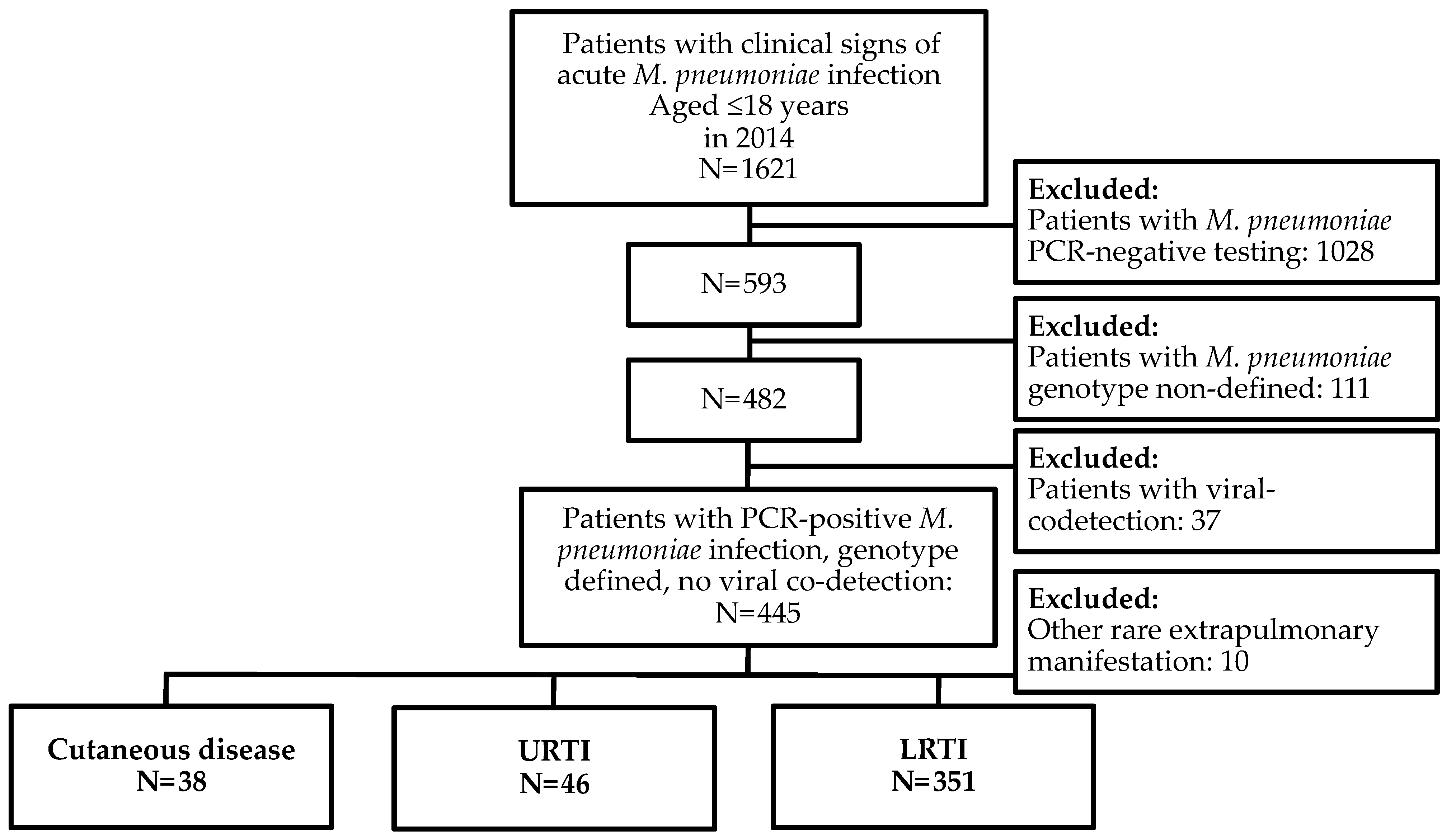
2.1. Study Subjects

Children younger than 18 years, referred to University Children’s Hospital and Department of Infectious Diseases Ljubljana, Slovenia, with clinical signs of acute Mp infection from 1 January 2014 to 31 December 2014, were tested for Mp.
All patients who were PCR positive for Mp in pharyngeal swabs and in whom the P1 and/or MLVA genotype was successfully defined were identified from a laboratory database and included in the study. We excluded cases with a viral co-detection.
The National Medical Ethics Committee of the Republic of Slovenia approved the protocol for this study (No 0120-8/2018/4 and No 0120-244/2021/3).

2.2. Study Design

We performed an observational retrospective study to determine whether Mp genotype is associated with Mp-induced cutaneous disease in children.
The data on age, gender, disease presentation, dermatological presentation, Mp genotype, macrolide susceptibility, interval between onset of disease and initiation of antibiotic therapy, laboratory biomarkers of inflammation, hospital admission, duration of hospital stay and data related to complications and treatment were collected for all patients.
The patients were further divided into groups according to the disease presentation; patients with cutaneous disease, patients with upper (URTI) or lower respiratory tract infection (LRTI), and their epidemiological characteristics and Mp genotype distribution were compared. Patients with other rare extrapulmonary manifestations were excluded from the study. Cutaneous disease was defined as an eruptive lesion, which involved skin. We present Mp-induced dermatological presentations reported previously in the literature [5,6]. LRTI diagnosis was made based on physical examination revealing pathological lung auscultation and radiographic appearance consistent with a diagnosis of LRTI, if an X-ray was performed. Patients were diagnosed with URTI if they had respiratory symptoms and did not fulfill LRTI criteria.
In addition, the group of patients with cutaneous disease was divided according to a specific dermatological presentation, and the characteristics of each group were evaluated and compared.
To better assess the possible factors influencing the severity of the disease, we compared the characteristics of patients hospitalized because of rash with those of outpatients.

2.3. Methods

Pharyngeal swabs were subjected to DNA isolation using MagNA Pure Compact (Roche Diagnostics, Mannheim, Germany) and later tested by Mp real-time PCR (ArgeneBioMerieux diagnostics, Marcy l’Etoile, France). The remainder of each PCR-positive sample was cultivated as described previously [20]. MLVA subtyping was performed by amplification of four variable-number tandem-repeat (VNTR) loci (Mpn13, Mpn14, Mpn15 and Mpn16) according to a standardized MLVA protocol [16]. P1 subtyping was performed using pyrosequencing, which targets the Mp MPN141 and MPN528a genes, while macrolide resistance was recognized by pyrosequencing two parts of domain V in the 23S rRNA gene [20].
Multiplex PCR was performed on the nasopharyngeal aspirate specimens to assess viral co-detection, including respiratory syncytial virus, influenza virus, parainfluenza virus, human bocavirus, adenovirus, metapneumovirus, rhinovirus, enterovirus and coronavirus.

2.4. Analysis

Continuous variables were presented as mean (SD) or median (IQR), where appropriate. Categorical variables were described with counts and percentages.
Continuous variables in two independent groups were compared using the independent samples T-test or the Mann–Whitney U-test, where appropriate, whereas continuous variables in multiple independent groups were compared using the one-way ANOVA or Kruskal–Wallis test, where appropriate [21]. Categorical variables were compared by using the Pearson chi-square test. We performed a multivariable logistic regression analysis to assess whether Mp genotype is associated with hospital admission because of cutaneous disease [22]. The differences were considered statistically significant when the p value was <0.05. Statistical computing was conducted in IBM SPSS Statistics (Version 28.0).

3. Results

During the study period, 1621 children were referred to our hospitals with signs of acute Mp infection. After applying the inclusion and exclusion study criteria, we evaluated data from 435 patients (mean age 7.3 yrs., SD 3.4 yrs.; 52.0% boys) with PCR-positive acute Mp infection, P1 and/or MLVA genotype defined and no viral co-detection (Figure 1), presenting as cutaneous disease (38/435), URTI (46/435) or LRTI (351/435).
The two main P1 genotypes, P1 type 1 and P1 type 2, accounted for 74 and 26% of total isolates, respectively. MLVA typing revealed seven distinct MLVA types: MLVA-3,5,6,2 (10.6%, 34/322), MLVA-3,5,6,3 (0.3%, 1/322), MLVA-3,6,6,2 (12.7%, 41/322), MLVA-4,5,6,2 (0.3%, 1/322), MLVA-4,5,7,2 (72.4%, 233/322), MLVA-4,5,7,3 (3.4%, 11/322) and MLVA-5,5,7,2 (0.3%, 1/322). The majority of isolates (99.3%, 432/435) were macrolide susceptible, and three (0.7%) were found to be macrolide resistant (MRMp). Of the MRMp, infection with one MRMp strain presented as cutaneous disease (3%, 1/38).
We compared P1 and MLVA Mp genotype distribution in children with cutaneous disease, URTI and LRTI (Table 1). We found no association between a specific Mp genotype and a specific disease presentation (Table 1). However, infections with only three MLVA strains presented with cutaneous disease, including MLVA-3,6,6,2, MLVA-4,5,7,2 and MLVA-4,5,7,3.
The two most common skin manifestations during the study period were urticarial (55%, 21/38) and maculopapular eruptions (37%, 14/38), accounting for 92% of all cutaneous disease (Table 2).
When comparing the two groups, we found that patients with urticarial rash were more often treated with antihistamines and were hospitalized more often because of skin disease (Table 3). We found no association between a specific Mp genotype and a specific dermatological presentation. Nevertheless, only infections with MLVA-4,5,7,2 strains occurred as maculopapular eruptions, while infections with all three MLVA strains could present as urticarial rash.
The characteristics of patients with rare skin manifestations (8%, 3/38) are listed in Table 4. No mucocutaneous eruptions, such as toxic epidermal necrolysis or Stevens–Johnson syndrome associated with Mp infection, were observed in our patients.
In the group with Mp-induced cutaneous disease, 18% (7/38) required hospital admission because of rash. When comparing the characteristics of patients admitted to the hospital to outpatients’ ones, we found an important difference in their age, treatment and distribution of MLVA-3,6,6,2 genotype in both groups (Table 5). Children who were hospitalized were younger and more frequently treated with antihistamines and systemic steroids. In addition, infection with MLVA-3,6,6,2 strains was more common in admitted patients.
The observed difference in hospital admission remained significant even after adjusting for age in a logistic regression model for the MLVA-3,6,6,2 genotype (Table 6).

4. Discussion

Cutaneous disease is the most common extrapulmonary presentation of Mp infection [1,5,6,7,8,9,10]. The factors determining disease presentation and severity of Mp-induced cutaneous disease are only partly understood, with undiscovered roles for both pathogen- and host-related factors. The aim of our study was to determine whether Mp genotype is associated with this specific clinical outcome in children. In the study period, 8.5% of all patients presented with Mp-induced cutaneous disease. The two most common dermatological presentations were maculopapular and urticarial eruptions, accounting for 92% of all cutaneous disease. Patients requiring hospitalization because of rash were those with rare Mp-induced cutaneous disease, such as Henoch–Schönlein purpura and erythema multiforme, as well as those with urticarial rash, mimicking allergic reaction, and were therefore frequently treated with antihistamines and steroids.
When comparing Mp P1 and MLVA genotype distribution in patients with cutaneous disease, URTI and LRTI, we found no association between a specific P1 and MLVA genotype and Mp-induced cutaneous disease. Moreover, we found no association between Mp genotype and a specific dermatological presentation. Although seven different MLVA types co-circulated during the study period, infections with only three MLVA strains presented with cutaneous disease, including MLVA-3,6,6,2, MLVA-4,5,7,2 and MLVA-4,5,7,3. Moreover, when assessing the factors associated with severe cutaneous disease requiring hospital admission, we found that infection with MLVA-3,6,6,2 strains was more common in admitted patients. In addition, after adjusting for age, MLVA-3,6,6,2 genotype significantly affected the likelihood of hospital admission because of cutaneous disease in a logistic regression model.
Several possible mechanisms have been proposed to play a role in Mp-induced cutaneous disease [4,5,23]. It is suggested that an indirect mechanism, in which the bacterium is not present at the site of inflammation, but immune modulation, such as autoimmunity or formation of immune complexes, is thought to play an important role [4,24,25].
Microbiological studies have shown that Mp genotypes may have different characteristics, which could potentially impact their virulence and inflammation [26,27,28]. Results from clinical studies have also shown that P1 and MLVA genotypes could influence the severity of LRTI [18,19,29,30]. However, limited clinical studies to date have investigated the influence of the Mp genotype on Mp-induced cutaneous disease. To our knowledge, our sample size is the largest cohort of patients with Mp-induced cutaneous disease addressing this question either in children or adults. In addition, to better assess the impact on disease severity of different genotypes, we included outpatients as well as hospitalized patients in our study. Similar to our study, results from recent research found no association between Mp genotype and clinical outcomes, such as cutaneous disease [31]. In comparison to their study, our cohort of patients is larger, also including patients younger than 3 years of age, with no viral co-detection, to better assess the role of Mp in disease presentation. Macrolide-resistant Mp has been reported to be more frequently associated with mucocutaneous disease, possibly as a result of stronger and more persistent inflammatory stimulation by MRMp because of treatment failure [32]. In our study, we detected only a single MRMp strain in a patient with cutaneous disease.
Our study has several limitations. First, even though the sample of patients is larger compared to that in previous studies, a relatively small number of patients presented with extrapulmonary manifestations, such as Mp-induced cutaneous disease and specific dermatological manifestations. In addition, a relatively small number of patients were infected with specific MLVA genotypes. Second, all of our patients were recruited from tertiary centers, which may have resulted in a disproportionate number of cases with more severe Mp infection. Nevertheless, no mucocutaneous eruptions, such as toxic epidermal necrolysis or Stevens–Johnson syndrome associated with Mp infection, were observed in our patients. Third, the retrospective design of our study limited data collection to the most commonly used clinical variables. However, our study focused mainly on the association between Mp genotype and cutaneous disease, which was not influenced by the study design. Although it may be difficult to draw any definite conclusions, this study provides valuable information on the association between Mp genotype and cutaneous disease. Future studies should investigate other inflammatory parameters, disease severity and host-related factors to better understand the pathogenic role of Mp genotype in this clinical outcome.

5. Conclusions

The results of our cohort study suggest that no specific Mp genotype predisposes children to Mp-induced cutaneous disease and a specific dermatological presentation. Interestingly, infections with few MLVA strains were associated with this clinical outcome. In addition, the results suggest that infection with certain MLVA strains could induce more severe cutaneous disease requiring hospital admission. Future studies are needed to better understand the role of Mp genotype in Mp-induced cutaneous disease.

Author Contributions

Conceptualization, J.R.B. and D.K.; methodology, J.R.B.; software, J.R.B.; validation, J.R.B., T.M., M.K. and D.K.; formal analysis, J.R.B.; investigation, J.R.B.; data curation, J.R.B.; writing—original draft preparation, J.R.B.; writing—review and editing, J.R.B., T.M., M.K. and D.K.; visualization, J.R.B.; supervision, D.K. All authors have read and agreed to the published version of the manuscript.

Funding

This research received no external funding.

Data Availability Statement

The data presented in this study are available on request from the corresponding author. The data are not publicly available due to privacy.

Conflicts of Interest

The authors declare no conflict of interest.

References

  1. Atkinson, T.P.; Waites, K.B. Mycoplasma pneumoniae infections in childhood. Pediatr. Infect. Dis. J. 2014, 33, 92–94. [Google Scholar] [CrossRef] [Green Version]
  2. Kutty, P.K.; Jain, S.; Taylor, T.H.; Bramley, A.M.; Diaz, M.H.; Ampofo, K.; Arnold, S.; Williams, D.; Edwards, K.; McCullers, J.; et al. Mycoplasma pneumoniae among children hospitalized with community-acquired pneumonia. Clin. Infect. Dis. 2019, 68, 5–12. [Google Scholar] [CrossRef] [Green Version]
  3. Blasi, F. Atypical pathogens and respiratory tract infections. Eur. Respir. J. 2004, 24, 171–181. [Google Scholar] [CrossRef] [Green Version]
  4. Hu, J.; Ye, Y.; Chen, X.; Xiong, L.; Xie, W.; Liu, P. Insight into the Pathogenic Mechanism of Mycoplasma pneumoniae. Curr. Microbiol. 2022, 80, 14. [Google Scholar] [CrossRef]
  5. Schalock, P.C.; Dinulos, J.G. Mycoplasma pneumoniae-induced cutaneous disease. Int. J. Dermatol. 2009, 48, 673–680. [Google Scholar] [CrossRef] [PubMed]
  6. Terraneo, L.; Lava, S.A.; Camozzi, P.; Zgraggen, L.; Simonetti, G.D.; Bianchetti, M.G.; Milani, G.P. Unusual eruptions associated with Mycoplasma pneumoniae respiratory infections: Review of the literature. Dermatology 2015, 231, 152–157. [Google Scholar] [CrossRef] [PubMed] [Green Version]
  7. Canavan, T.N.; Mathes, E.F.; Frieden, I. Mycoplasma pneumoniae-induced rash and mucositis as a syndrome distinct from Stevens-Johnson syndrome and erythema multiforme: A systematic review. J. Am. Acad. Dermatol. 2015, 72, 239–245. [Google Scholar] [CrossRef] [PubMed]
  8. Liakos, W.; Xu, A.; Finelt, N. Clinical features of recurrent Mycoplasma pneumoniae-induced rash and mucositis. Pediatr. Dermatol. 2021, 38, 154–158. [Google Scholar] [CrossRef]
  9. Chen, N.; Li, M. Case Report and Literature Review: Clinical Characteristics of 10 Children With Mycoplasma pneumoniae-Induced Rash and Mucositis. Front. Pediatr. 2022, 10, 823376. [Google Scholar] [CrossRef]
  10. Fan, X.; Luo, Y.; Lu, J.; Xu, J.; Chen, Q.; Guo, H.; Jin, P. Erythema Multiforme Major Associated With Community-Acquired Pneumonia: Lessons From a Case Report. Front. Pediatr. 2021, 9, 698261. [Google Scholar] [CrossRef]
  11. Dégrange, S.; Cazanave, C.; Charron, A.; Renaudin, H.; Bébéar, C.; Bébéar, C.M. Development of multiple-locus variable-number tandem-repeat analysis for molecular typing of Mycoplasma pneumoniae. J. Clin. Microbiol. 2009, 47, 914–923. [Google Scholar] [CrossRef] [Green Version]
  12. Dumke, R.; Rodriguez, N. Use of different approaches for the culture-independent typing of Mycoplasma pneumoniae from two geographically distinct regions. J. Microbiol. Methods 2021, 186, 106239. [Google Scholar] [CrossRef]
  13. Xiao, J.; Liu, Y.; Wang, M.; Jiang, C.; You, X.; Zhu, C. Detection of Mycoplasma pneumoniae P1 subtype variations by denaturing gradient gel electrophoresis. Diagn Microbiol. Infect. Dis. 2014, 78, 24–28. [Google Scholar] [CrossRef] [PubMed]
  14. Zhao, F.; Cao, B.; Li, J.; Song, S.; Tao, X.; Yin, Y.; He, L.; Zhang, J. Sequence analysis of the p1 adhesin gene of Mycoplasma pneumoniae in clinical isolates collected in Beijing in 2008 to 2009. J. Clin. Microbiol. 2011, 49, 3000–3003. [Google Scholar] [CrossRef] [Green Version]
  15. Dorigo-Zetsma, J.W.; Dankert, J.; Zaat, S.A. Genotyping of Mycoplasma pneumoniae clinical isolates reveals eight P1 subtypes within two genomic groups. J. Clin. Microbiol. 2000, 38, 965–970. [Google Scholar] [CrossRef] [PubMed] [Green Version]
  16. Sun, H.; Xue, G.; Yan, C.; Li, S.; Cao, L.; Yuan, Y.; Zhao, H.; Feng, Y.; Wang, L.; Fan, Z. Multiple-locus variable-number tandem-repeat analysis of Mycoplasma pneumoniae clinical specimens and proposal for amendment of MLVA nomenclature. PLoS ONE 2013, 8, e64607. [Google Scholar] [CrossRef] [PubMed] [Green Version]
  17. Brown, R.J.; Holden, M.T.; Spiller, O.B.; Chalker, V.J. Development of a Multilocus Sequence Typing Scheme for Molecular Typing of Mycoplasma pneumoniae. J. Clin. Microbiol. 2015, 53, 3195–3203. [Google Scholar] [CrossRef] [Green Version]
  18. Rodman Berlot, J.; Krivec, U.; Mrvič, T.; Kogoj, R.; Keše, D. Mycoplasma pneumoniae P1 genotype indicates severity of lower respiratory tract infections in children. J. Clin. Microbiol. 2021, 59, e0022021. [Google Scholar] [CrossRef]
  19. Rodman Berlot, J.; Mrvič, T.; Keše, D. Mycoplasma pneumoniae multilocus variable-number tandem-repeat analysis genotypes are associated with inflammatory biomarker levels in children with lower respiratory tract infections. Eur. J. Clin. Microbiol. Infect. Dis. 2022, 41, 1099–1105. [Google Scholar] [CrossRef]
  20. Kogoj, R.; Praprotnik, M.; Mrvič, T.; Korva, M.; Keše, D. Genetic diversity and macrolide resistance of Mycoplasma pneumoniae isolates from two consecutive epidemics in Slovenia. Eur. J. Clin. Microbiol. Infect. Dis. 2018, 37, 99–107. [Google Scholar] [CrossRef]
  21. Lee, S.W. Methods for testing statistical differences between groups in medical research: Statistical standard and guideline of Life Cycle Committee. Life Cycle 2022, 2, e1. [Google Scholar] [CrossRef]
  22. Lee, S.W. Regression analysis for continuous independent variables in medical research: Statistical standard and guideline of Life Cycle Committee. Life Cycle 2022, 2, e3. [Google Scholar] [CrossRef]
  23. Meyer Sauteur, P.M.; Theiler, M.; Buettcher, M. Frequency and clinical presentation of mucocutaneous disease due to Mycoplasma pneumoniae infection in children with community-acquired pneumonia. JAMA Dermatol. 2020, 156, 144–150. [Google Scholar] [CrossRef]
  24. Narita, M. Classification of extrapulmonary manifestations due to Mycoplasma pneumoniae infection on the basis of possible pathogenesis. Front. Microbiol. 2016, 7, 23. [Google Scholar] [CrossRef]
  25. Sánchez-Vargas, F.M.; Gómez-Duarte, O.G. Mycoplasma pneumoniae-an emerging extra-pulmonary pathogen. Clin. Microbiol. Infect. 2008, 14, 105–117. [Google Scholar] [CrossRef] [PubMed] [Green Version]
  26. Simmons, W.L.; Daubenspeck, J.M.; Osborne, J.D.; Balish, M.F.; Waites, K.B.; Dybvig, K. Type 1 and type 2 strains of Mycoplasma pneumoniae form different biofilms. Microbiology 2013, 159, 737–747. [Google Scholar] [CrossRef] [Green Version]
  27. Lluch-Senar, M.; Cozzuto, L.; Cano, J.; Delgado, J.; Llórens-Rico, V.; Pereyre, S.; Bebear, C.; Serrano, L. Comparative “-omics” in Mycoplasma pneumoniae clinical isolates reveals key virulence factors . PLoS ONE 2015, 10, e0137354. [Google Scholar] [CrossRef] [PubMed]
  28. Techasaensiri, C.; Tagliabue, C.; Cagle, M.; Iranpour, P.; Katz, K.; Kannan, T.R.; Coalson, J.J.; Baseman, J.B.; Hardy, R.D. Variation in colonization, ADP-ribosylating and vacuolating cytotoxin, and pulmonary disease severity among Mycoplasma pneumoniae strains. Am. J. Respir. Crit. Care Med. 2010, 182, 797–804. [Google Scholar] [CrossRef] [PubMed] [Green Version]
  29. Qu, J.; Yu, X.; Liu, Y.; Yin, Y.; Gu, L.; Cao, B.; Wang, C. Specific multilocus variable-number tandem-repeat analysis genotypes of Mycoplasma pneumoniae are associated with diseases severity and macrolide susceptibility. PLoS ONE 2013, 8, e82174. [Google Scholar] [CrossRef] [PubMed] [Green Version]
  30. Yan, C.; Xue, G.; Zhao, H.; Feng, Y.; Li, S.; Cui, J.; Ni, S.; Sun, H. Molecular and clinical characteristics of severe Mycoplasma pneumoniae pneumonia in children. Pediatr. Pulmonol. 2019, 54, 1012–1021. [Google Scholar] [CrossRef] [PubMed]
  31. Meyer Sauteur, P.M.; Pánisová, E.; Seiler, M.; Theiler, M.; Berger, C.; Dumke, R. Mycoplasma pneumoniae genotypes and clinical outcome in children. J. Clin. Microbiol. 2021, 18, e0074821. [Google Scholar] [CrossRef] [PubMed]
  32. Zhou, Y.; Zhang, Y.; Sheng, Y.; Zhang, L.; Shen, Z.; Chen, Z. More complications occur in macrolide-resistant than in macrolide-sensitive Mycoplasma pneumoniae pneumonia. Antimicrob. Agents Chemother. 2014, 58, 1034–1038. [Google Scholar] [CrossRef] [PubMed]
Figure 1. Study flowchart. Abbreviations: LRTI, Lower respiratory tract infection; MLVA, Multilocus variable-number tandem-repeat analysis; URTI, Upper respiratory tract infection.
Microorganisms 11 00205 g001
Table 1. Epidemiologic characteristics and Mycoplasma pneumoniae genotype distribution in patients with cutaneous disease, upper and lower respiratory tract infection. Data are presented as median (IQR) or as percentage (proportion of subjects).
Cutaneous DiseaseURTILRTITest Statistic, p
Subjects N3846351
Boys/girls (%)50%/50%63%/37%51%/49%χ2(2) = 2.54, 0.281
Age (years)6.6 (IQR 4.6–9.8)5.9 (IQR 4.1–9.4)7.0 (IQR 4.9–9.9)H(2) = 1.34, 0.511
<5 years
5–18 years
32% (12/38)
68% (26/38)
33% (15/46)
67% (31/46)
26% (93/351)
74% (258/351)
χ2(2) = 1.09, 0.579
Mp Genotype
P1-1/P1-2 (%)
MLVA type
82%/18%70%/30%74%/26%χ2(2) = 1.63, 0.442
MLVA-3,6,6,2
MLVA-4,5,7,2
MLVA-4,5,7,3
10% (3/29)
86% (25/29)
4% (1/29)
18% (7/38)
66% (25/38)
3% (1/38)
12% (31/255)
72% (183/255)
4% (9/255)
χ2(2) = 1.33, 0.514
χ2(2) = 3.65, 0.162
χ2(2) = 0.08, 0.960
Abbreviations: IQR, Interquartile range; LRTI, Lower respiratory tract infection; MLVA, Multilocus variable-number tandem-repeat analysis; URTI, Upper respiratory tract infection. Continuous variables were compared using the Kruskal–Wallis test, whereas categorical variables were compared using the Pearson chi-square test.
Table 2. Frequency of dermatological presentations of Mycoplasma-pneumoniae-induced cutaneous disease.
Dermatological PresentationFrequency
Urticarial eruptions55.3% (21/38)
Maculopapular eruptions36.8% (14/38)
Erythema multiforme2.6% (1/38)
Henoch–Schönlein purpura2.6% (1/38)
Pityriasis rosea2.6% (1/38)
Table 3. Characteristics of patients with maculopapular and urticarial M. pneumoniae-induced cutaneous disease. Data are presented as median (IQR) or as percentage (proportion of subjects). Significant differences (p < 0.05) are highlighted in bold.
Maculopapular EruptionsUrticarial EruptionsTest Statistic, p
Subjects N1421
Boys/girls (%)43%/57%48%/52%χ2(1) = 0.08, 0.782
Age (years)6.5 (IQR 4.6–10.0)6.5 (IQR 4.4–8.1)U(1) = 129.0, 0.544
<5 years
5–18 years
29% (4/14)
71% (10/14)
38% (8/21)
62% (13/21)
χ2(1) = 0.34, 0.561
CRP (mg/L)14.0 (IQR 4.0–32.3)19.5 (IQR 4.5–47.0)U(1) = 92.5, 0.413
WBC (×109/L)9.1 (IQR 6.4–12.3)10.0 (IQR 7.5–14.2)U(1) = 96.5, 0.519
Hospitalization
because of rash
0% (0/14)24% (5/21)χ2(1) = 3.89, 0.049
Treatment
Macrolide antibiotic
Antihistamines
Systemic steroids

93% (13/14)
0% (0/14)
0% (0/14)

100% (21/21)
24% (5/21)
10% (2/21)

χ2(1) = 1.54, 0.214
χ2(1) = 3.89, 0.049
χ2(1) = 1.41, 0.234
Mp Genotype
P1-1/P1-2 (%)
MLVA type
93%/7%
 
76%/24%
 
χ2(1) = 1.64, 0.200
 
MLVA-3,6,6,2
MLVA-4,5,7,2
MLVA-4,5,7,3
0% (0/12)
100% (12/12)
0% (0/12)
14% (2/14)
79% (11/14)
7% (1/14)
χ2(1) = 1.86, 0.173
χ2(1) = 2.91, 0.088
χ2(1) = 0.89, 0.345
Abbreviations: CRP, C-reactive protein; IQR, Interquartile range; MLVA, Multilocus variable-number tandem-repeat analysis; WBC, White blood cell count. Continuous variables were compared using the Mann–Whitney U-test, whereas categorical variables were compared using the Pearson chi-square test.
Table 4. Characteristics of patients with rare dermatological presentations of Mycoplasma-pneumoniae-induced cutaneous disease.
Age (yrs.)/
Gender
Dermatological PresentationP1 TypeMLVA TypeSymptomsTreatmentOutcome
11/MErythema multiforme 14,5,7,2Target lesions on limbs, dehydrationazithromycin
parenteral hydration
Complete recovery
6/MHenoch–Schönlein purpura with GIT involvement14,5,7,2Abdominal pain, petechiae on lower limbsazithromycin
methylprednisolone
Complete recovery
12/MPityriasis rosea23,6,6,2RashazithromycinComplete recovery
Abbreviations: GIT—gastrointestinal; M—male; yrs—years.
Table 5. Comparison of characteristics of patients admitted to the hospital because of cutaneous disease to the outpatients’ ones. Data are presented as median (IQR) or as percentage (proportion of subjects). Significant differences (p < 0.05) are highlighted in bold.
InpatientsOutpatientsTest Statistic, p
Subjects N632
Boys/girls (%)50%/50%50%/50%χ2(1) = 0.00, 1.000
Age (years)4.9 (IQR 3.8–7.9)6.9 (IQR 5.0–10.0)U(1) = 64.0, 0.200
<5 years
5–18 years
67% (4/6)
33% (2/6)
25% (8/32)
75% (24/32)
χ2(1) = 4.06, 0.044
CRP (mg/L)12.5 (IQR 4.0–29.0)12.0 (IQR 4.0–39.0)U(1) = 71.5, 0.654
WBC (× 109/L) 9.8 (IQR 6.3–11.5)9.7 (IQR 7.4–12.4)U(1) = 72.0, 0.674
Treatment
Macrolide antibiotic
Antihistamines
Systemic steroids

100% (6/6)
67% (4/6)
50% (3/6)

97% (31/32)
3% (1/32)
0% (0/32)

χ2(1) = 0.19, 0.661
χ2(1) = 17.85, <0.001
χ2(1) = 17.37, <0.001
Mp Genotype
P1-1/P1-2 (%)
MLVA type
67%/33%
 
84%/16%
 
χ2(1) = 1.05, 0.305
 
MLVA-3,6,6,2
MLVA-4,5,7,2
MLVA-4,5,7,3
40% (2/5)
60% (3/5)
0% (0/5)
4% (1/24)
92% (22/24)
4% (1/24)
χ2(1) = 5.73, 0.017
χ2(1) = 3.49, 0.062
χ2(1) = 0.22, 0.642
Abbreviations: CRP, C-reactive protein; IQR, Interquartile range; MLVA, Multilocus variable-number tandem-repeat analysis; WBC, White blood cell count. Continuous variables were compared using the Mann–Whitney U-test, whereas categorical variables were compared using the Pearson chi-square test.
Table 6. Logistic regression analysis of hospital admission of Mycoplasma-pneumoniae-infected patients because of cutaneous disease. Significant differences (p < 0.05) are highlighted in bold.
Crude OR (95%-CI)Adjusted a OR (95%-CI)p Values (Crude/Adjusted)
P1 type 1
P1 type 2
0.37 (0.05–2.60)
2.70 (0.39–18.93)
0.33 (0.04–2.48)
3.02 (0.40–22.60)
0.317/0.281
0.317/0.281
MLVA-3,6,6,2
MLVA-4,5,7,2
15.33 (1.05–224.78)
0.14 (0.01–1.36)
232.43 (1.18–45,972.69)
0.10 (0.01–1.32)
0.046/0.043
0.090/0.081
MLVA-4,5,7,30.00 (0.00)0.00 (0.00)1.000/1.000
a Adjusted for age. Abbreviations: CI, Confidence interval; OR, Odds ratio.
Disclaimer/Publisher’s Note: The statements, opinions and data contained in all publications are solely those of the individual author(s) and contributor(s) and not of MDPI and/or the editor(s). MDPI and/or the editor(s) disclaim responsibility for any injury to people or property resulting from any ideas, methods, instructions or products referred to in the content.

